Pectobacterium carotovorum
Pathogenesis
Maria del Pilar Marquez-Villavicencio1, Brooke Weber1, R. Andrews Witherell1, David K. Willis1,2, Amy O. Charkowski1*
1Department of Plant Pathology, University of Wisconsin-Madison, Madison, Wisconsin, United States of America,2Vegetable Crops Research Unit, Agricultural Research Service, United States Department of Agriculture, Madison, Wisconsin, United States of America
Abstract
Pectobacteriumspecies are necrotrophic bacterial pathogens that cause soft rot diseases in potatoes and several other crops worldwide. Gene expression data identifiedPectobacterium carotovorumsubsp.carotovorum budB, which encodes thea -acetolactate synthase enzyme in the 2,3-butanediol pathway, as more highly expressed in potato tubers than potato stems. This pathway is of interest because volatiles produced by the 2,3-butanediol pathway have been shown to act as plant growth promoting molecules, insect attractants, and, in other bacterial species, affect virulence and fitness. Disruption of the 2,3-butanediol pathway reduced virulence of P. c. subsp. carotovorum WPP14 on potato tubers and impaired alkalinization of growth medium and potato tubers under anaerobic conditions. Alkalinization of the milieu via this pathway may aid in plant cell maceration sincePectobacteriumpectate lyases are most active at alkaline pH.
Citation:Marquez-Villavicencio MdP, Weber B, Witherell RA, Willis DK, Charkowski AO (2011) The 3-Hydroxy-2-Butanone Pathway Is Required forPectobacterium carotovorumPathogenesis. PLoS ONE 6(8): e22974. doi:10.1371/journal.pone.0022974
Editor:Ching-Hong Yang, University of Wisconsin-Milwaukee, United States of America
ReceivedMay 27, 2011;AcceptedJuly 5, 2011;PublishedAugust 18, 2011
This is an open-access article, free of all copyright, and may be freely reproduced, distributed, transmitted, modified, built upon, or otherwise used by anyone for any lawful purpose. The work is made available under the Creative Commons CC0 public domain dedication.
Funding:This work was funded by the National Science Foundation Genomics in the Environment Program award number 0412599, the United States Department of Agriculture NRI program grant 2006-398 35319-17396, and the Wisconsin Potato and Vegetable Growers Association. The funders had no role in study design, data collection and analysis, decision to publish, or preparation of the manuscript.
Competing Interests:The authors have declared that no competing interests exist.
* E-mail: [email protected]
Introduction
Plant associatedEnterobacteriaceaecommonly produce acetoin (3-hydroxy-2-butanone; 3H2B) from pyruvate, which is an end product of fermentation of glucose or other carbon sources through the Embden-Meyerhof (EM) pathway [1,2], whereas related animal pathogens produce acid and sometimes gas through mixed acid fermentation. The Voges-Prokauer reaction, which measures production of 3H2B, has been used in microbiology lab classes and taxonomic schemes for decades to differentiate environmentalEnterobacteriaceaefrom coliform bacteria, which are used as indicators of fecal contamination [3]. In the 3H2B pathway, a-acetolactate synthase condenses two molecules of pyruvate intoa-acetolactate, which is then converted into 3H2B (reviewed in [4]). Nonenzymatic oxidative decarboxylation can also convert a-acetolactate into diacetyl. The enzyme 2,3-butanediol dehydrogenase transforms 3H2B into 2,3-2,3-butanediol (23B) and also transforms diacetyl into 23B. The genes encoding the 3H2B pathway enzymes are commonly referred to as bud
genes. In addition to the plant-associatedEnterobacteriaceae, such as
Pectobacterium, Dickeya, Erwinia, Enterobacter, Serratia, and Klebsiella, many other genera of Gram-positive and Gram-negative bacteria, such as Pseudomonas, Streptomyces, Staphylococcus, Bacillus and
Clostridium species, use this pathway to produce 3H2B and its reductive state analog 23B.
The 3H2B pathway alkalinizes growth media during fermentation, thereby promoting growth of bacteria unable to cope with acidic conditions. For example, acidic conditions are lethal to Vibrio cholerae and acquisition of the 3H2B
pathway by the El Tor biotype appears to have made it a better intestinal and environmental colonist [5]. Since 3H2B and 23B can be metabolized by some bacterial species after glucose and other readily fermented carbon sources are used, it also serves as an energy storing strategy [6,7,8]. The 3H2B pathway may also reduce fitness in some environments; a
Serratia plymuthica budBmutant is more virulent on carrot slices than wild type, even though it does not grow as well in rich medium as wild type [9].
In Arabidopsis plants, 3H2B and 23B promote plant growth, drought tolerance, and defense against pathogens. Experiments with plant growth promotingBacillusspecies showed that volatiles produced by Bacillus were responsible for increased growth of
Arabidopsisplants and these volatiles were later identified as 3H2B and 23B [22]. The plant growth promotingBacilluswas unable to affect Arabidopsis ein2 and cre1 mutants, which are impaired in ethylene and cytokinen sensing, respectively, suggesting that 3H2B and 23B act on plants through these pathways. Bacterial 3H2B and 23B also appear to act as microbe-associated molecular patterns (MAMP). These volatiles induce systemic resistance in Arabidopsis [23] and also cause Arabidopsis stomata to close [24]. The 3H2B pathway is present in the Enterobacteriaceae plant pathogens Pectobacterium and Dickeya[25]. These genera are found worldwide in water, soil, and plants and cause wilt, necrosis and maceration symptoms in potato, as well as numerous other vegetable and ornamental plants [26]. Both genera produce acetoin in culture and volatile products of the HB pathway have been detected when assaying carrots and potatoes inoculated withPectobacterium carotovorum
subsp.carotovorum(P. c.subsp.carotovorum) [27,28]. Volatile products may also play a role in vectoring soft rot pathogens since insects appear to play important roles in bothPectobacteriumandDickeyalife cycles [29,30,31,32,33,34,35,36,37,38,39,40,41].
These soft rot pathogens secrete multiple plant cell wall degrading enzymes during pathogenesis, including pectate and pectin lyases, polygalacturonases, cellulases and proteases [42,43]. The pectate lyses are crucial for disease development. Bacterial pectate lyases tend to have a pH optimum of over 8 (for example, see [44,45]), while the plant host apoplast pH is typically 5 to 6.5 [46]. Thus, raising the local pH is a necessary step in soft rot pathogenesis [47] and it is possible that the 3H2B pathway plays a role in this step of pathogenesis. We recently found that the genes encoding this pathway, includingbudA,budB, andbudC, are highly expressed during P. c. subsp. carotovorum-potato stem and tuber interactions (Marquez-Villavicencio et al., in preparation) and RT-qPCR confirmed thatbudBis differentially expressed in potato stems compared to potato tubers. Here we describe the effects of a
budBmutation onP. c.subsp.carotovorumvirulence in potato tubers.
Materials and Methods
Bacterial strains and growth conditions
Bacterial strains used in this study are listed in Table 1. P. c.
subsp. carotovorum strains were grown in Luria-Bertani medium (LB) at 37uC. When required, antibiotics were used at the following concentration: ampicillin (100mg/ml), spectinomycin (50mg/ml), and kanamycin (50mg/ml).
Plant growth conditions and inoculation methods for plants used for bacterial mRNA isolation
Certified seed potatoes (Solanum tuberosum subsp. tuberosum var. Superior) were used for experiments where bacterial RNA was isolated from plant samples. Potato plants were grown in a growth chamber at 26uC with a 12 hour photoperiod and were not fertilized since the tuber provides enough nutrition for young potato plants. Stems of 4-week old plants were inoculated by stabbing a needle and injecting 10ml of a bacterial suspension of WPP14 (0.3 OD600, equivalent to 1.56108CFU/ml). To prepare bacterial suspensions, cells from 24 hour-old bacterial cultures grown on LB agar medium incubated at 37uC were suspended in water. Each potato stem was inoculated in five different sites approximately 3 cm apart. After inoculation, plants were placed into plastic bags and incubated at 26uC for 13.5 hours. Based on growth curve analysis, the bacteria reached log phase growth between 10 and 15 hours after inoculation into stems (data not shown).
For experiments where bacterial RNA was isolated from inoculated tubers, the potato tubers (S. tuberosum subsp.tuberosum
var. Superior) were surface sanitized by soaking them in 0.5% bleach for 10 minutes. The tubers were rinsed with distilled water and allowed to dry at room temperature for 24 hours. The tubers were inoculated in at least 15 sites per tuber with 10ml of a bacterial suspension of WPP14 (1.56108CFU/ml). Tubers were then placed into plastic bags and incubated at 26uC for 10 hours. Based on growth curve analysis, the bacteria reach log phase growth between 10 and 15 hours after inoculation into stems (data not shown).
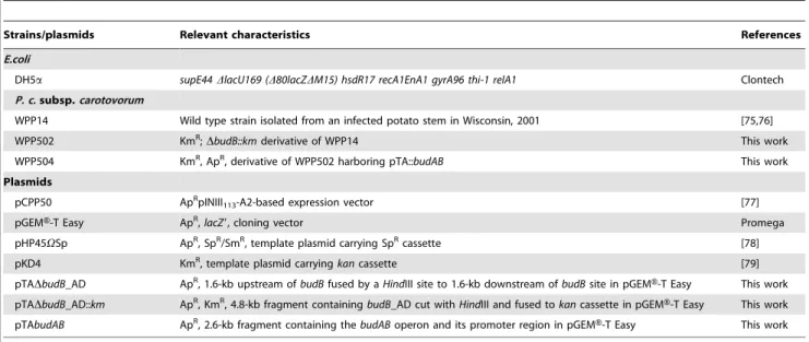
Table 1.Bacterial strains and plasmids used in this study.
Strains/plasmids Relevant characteristics References
E.coli
DH5a supE44DlacU169 (D80lacZDM15) hsdR17 recA1EnA1 gyrA96 thi-1 relA1 Clontech
P. c.subsp.carotovorum
WPP14 Wild type strain isolated from an infected potato stem in Wisconsin, 2001 [75,76]
WPP502 KmR;DbudB::kmderivative of WPP14 This work
WPP504 KmR, ApR, derivative of WPP502 harboring pTA::budAB This work
Plasmids
pCPP50 ApRpINIII113-A2-based expression vector [77]
pGEMH-T Easy ApR,
lacZ9, cloning vector Promega
pHP45VSp ApR, SpR/SmR, template plasmid carrying SpRcassette [78]
pKD4 KmR, template plasmid carryingkancassette [79]
pTADbudB_AD ApR, 1.6-kb upstream of
budBfused by aHindIII site to 1.6-kb downstream ofbudBsite in pGEMH-T Easy This work
pTADbudB_AD::km ApR, KmR, 4.8-kb fragment containingbudB_AD cut withHindIII and fused tokancassette in pGEM
H-T Easy This work
pTAbudAB ApR, 2.6-kb fragment containing thebudABoperon and its promoter region in pGEMH-T Easy This work
Bacterial RNA isolation
Approximately 90 stem and 75 tuber inoculations sites, respectively, were sampled for bacterial RNA isolations. Thirty stem sections of approximately 1 cm each or 25 1-cm cubes of tuber were placed into a cooled sterile RNAse-free mortar that was pre-filled with 17.5 ml of DEPC water and 2.5 ml of ice cold EtOH/ phenol stop solution (5% water-saturated phenol pH,7.0) to stabilize cellular RNA and stop degradation [48,49]. Plant samples were gently ground with a sterile RNAse-free pestle. Supernatants were collected in two 15 ml falcon tubes and RNA was isolated with a hot SDS/hot phenol protocol [48,49]. Briefly, bacterial supernatants were harvested by centrifugation 8,200 g for 2 min-utes at 4uC. The supernatant was aspirated and the pellet was frozen in a dry ice-alcohol bath. The pellets were resuspended in 1.6 ml of lysis buffer (TE: 10 mM Tris - 1 mM EDTA, pH 8.0 and 0.5 mg/ ml Ready-LyseTM
Lysozyme Solution (Epicentre Biotechnologies, Madison, WI) and 160ml of 10% SDS solution, and then mixed and incubated at 65uC for 2 minutes. Then, 176ml of 1 M NaOAc (pH 5.2) was mixed with the lysate. Equal volumes of lysate were transferred to RNase-free 2 ml microfuge tubes and mixed by inversion with 1 ml of water saturated phenol. The tubes were incubated in 65uC water bath for 6 minutes. Then the tubes were placed on ice for 2 minutes and centrifuged at 21,000 g for 10 min at 4uC. The aqueous layer was then transferred to a new 2 ml microfuge tube containing an equal volume of chloroform. Tubes were mixed by inversion, and then spun down at 21,000 g for 10 minutes at 4uC. The aqueous layer was split into equal volumes in 1.5 ml Eppendorf tubes and ethanol precipitated by adding 1/10 volume of 3 M NaOAc (pH 5.2), 1 mM EDTA (final concentra-tion) and 2 volumes of cold 100% EtOH to each sample. Samples were mixed and placed at280uC overnight. Bacterial RNA was pelleted by centrifugation at 21,000 g for 30 minutes at 4uC. The pellets were washed with ice cold 80% EtOH and spun down at 21,000 g for 10 minutes. The supernatant was removed and the pellets were air dried for 30 minutes in the fume hood. The pellets were resuspended in 10ml of DEPC-treated water. All resuspended samples were pooled together (100ml) and DNA contamination was removed by treatment with Turbo DNA-free(Ambion, Inc.) with the rigorous protocol and with 5ml of enzyme. Additional sample purification was performed with RNeasy Mini Kits (Qiagen, Valencia, CA). Purity and concentration of the RNA was verified with a spectrophotometric analysis (NanoDrop ND-1000; Nano-Drop technologies, Wilmington, DE). RNA integrity was measured with an Agilent 2100 Bioanalyzer (Agilent Technologies, Inc.) and the RNA was stored at280uC until use.
RT-qPCR
Quantitative real-time reverse transcriptase PCR (RT-qPCR) was used to validate the transcriptome profiling experiment on selected candidate genes. We followed the recently published MIQE guidelines for publication quality RT-qPCR [50]. Bacterial RNA was isolated as described above. Primers were designed based on the draft genome sequence of WPP14 with Beacon Designer software (Premier Biosoft International, Palo Alto, CA, U.S.A) and primer sequences are shown in Table 2. Primer efficiency was determined with a dilution series of P. c. subsp.
carotovorumWPP14 chromosomal DNA. RNA samples were tested for residual DNA contamination with RT-qPCR and primers specific for theproCtranscript inP. c. subsp.carotovorumWPP14. RNA samples that showed quantification cycle (Cq) values greater than 30 cycles were considered to be sufficiently free of chromosomal DNA contamination for analysis. The synthesis of cDNA was performed with an iScript cDNA synthesis kit according to the manufacturer’s instructions (Bio-Rad
Laborato-ries, Hercules, CA, U.S.A.). Briefly, cDNA synthesis was performed with 250 ng of total RNA in 15ml of DEPC-water plus 4ml of 56iScript reaction mix, which contains a blend of
oligo dT and random hexamer primers and 1ml of iScript reverse transcriptase. The cDNA synthesis mixture was incubated at 25uC for 5 min. followed by 42uC for 30 min., then 85uC for 5 min. Three independent biological replicate samples per experimental condition were completed for the cDNA synthesis. Two iScript reactions were performed for each RNA sample. For each cDNA sample, two 25ml reactions were used as technical replicates. The amount of mRNA specific cDNA was determined with qPCR with the MyiQ detection system (Bio-Rad Laboratories). The PCR conditions were 95uC for 3 min; 40 cycles of 95uC for 10 s and 50uC for 45 s; and 1 cycle of 95uC for 1 min and 55uC for 1 min; followed by a dissociation curve with 80 cycles of 55uC for 10 s with a 0.5uC increase per cycle. Primer dimers and the presence of a single product per reaction were evaluated with the dissociation (melt) curve analysis in the MyiQ detection system software.
For absolute quantification, the target transcript amount in each sample was determined with a standard curve of P. c. subsp.
carotovorumWPP14 DNA. The mean of the starting quantity (SQ) ratio for each target mRNA was internally normalized with the absolute expression mean values of four reference transcripts:ffh,
dspE, pelB, andhrcC. The SQ value of each reference RNA was determined with a standard curve ofP. c.subsp.carotovorumWPP14 DNA. These four RNA were validated as stable reference transcripts under our experimental conditions with the Excel-based software program BestKeeper [49,50]. We determined the mean relative expression ratio (RER) of thebudBtranscript in tuber tissue relative to stem by first normalizing the target transcript in each sample to each of the four reference transcripts and then determining the ratio of expression in each of the three stem and tuber samples relative to the mean of expression in stem tissue. The means of the stem and tuber ratios were analyzed via a two-tailed unpaired t-test with a 95% confidence interval with Prism 5.0a software (GraphPad Software, Inc. La Jolla, CA, U.S.A).
Mutants and plasmid constructions
To construct plasmids for allelic exchange mutagenesis ofbudB, 1.6-kb flanking regions ofbudBwere PCR-amplified with primers
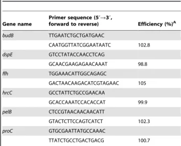
Table 2.RT-qPCR primer sequences and efficiency.
Gene name
Primer sequence (59R39,
forward to reverse) Efficiency (%)A
budB TTGAATCTGCTGATGAAC
CAATGGTTATCGGAATAATC 102.8
dspE GTCCTATACCAACCTCAG
GCAACGAAGAGAACAAAT 98.8
ffh TGGAAACATTGGCAGAGC
GACTAACAAGACATCGTAGAAC 105
hrcC GCCTATTCTGCCGAACAA
GCACCAAATCCACACCAT 99.9
pelB CTCCGTAACAACAACATT
GTACTCTTCCAGTCATCT 102.3
proC GTGCGAATTATGCCAAAC
TTATCTGCCTGACTGACG 100.7
A: Primer efficiencies were calculated from dilution curve with the MyiQ Cycler
software.
sets (University of Wisconsin Biotechnology Center DNA Synthesis Facility, Madison, WI) listed in Table 3. Primers were used to amplify a 3.2-kb fusion of flanking regions amplicons. A uniqueHindIII enzyme site was added into the primers, allowing cleavage and ligation of the antibiotic resistance genekanbetween the flanking regions. The fusion PCR product was cloned into pGEMH-T Easy vector (Promega, Madison, WI), producing pTADbudB_AD::km vector. This plasmid was electrotransformed into wild-type WPP14 for allelic exchange mutagenesis following the methods described by Ried and Collmer [51]. The mutation was confirmed by PCR-amplifying the mutated region and sequencing the PCR fragment. To complement the DbudB mutation, the promoter region and coding regions of budAand
budB were PCR amplified and cloned into the pGEMH-T Easy vector and the resulting plasmid was electrotransformed into DbudB. Transformation, restriction endonuclease digestion and other DNA manipulations were performed essentially as described in Sambrook and Russell [52].
Voges-Proskauer test
To verify a disruption in the acetoin pathway, we used the Voges-Proskauer test, which determines whether 3H2B is present [3]. A single colony of WPP14 wild- type, DbudB, and the complemented strain ofDbudBgrown in LB agar medium, were transferred to 5 ml of methyl red-Voges Proskauer buffered glucose broth (BD DifcoTM, NJ) amended with appropriate antibiotics and the culture were incubated at 37uC. The presence of 3H2B, was monitored at 24 and 48 hours of incubation. For this, 1 ml of each bacterial culture was transferred to a sterile glass tube and 15 drops of BBL Voges-Proskauer reagent A (5% wt/vol alpha-naphthol in absolute alcohol) (BD, Franklin Lakes, NJ) and 5 drops of BBL Voges-Proskauer reagent B (40% wt/vol potassium hydroxide in distilled water) were added. The mixture was stirred vigorously and the development of a red color, which indicates a positive test, was monitored for 30 minutes at room temperature.
Measurements of pH underin vitroandin vivoconditions To evaluate differences in pH between WPP14 wild-type and DbudB after growing in a glucose-rich medium, single colonies grown in LB agar medium, were transferred to 3 ml of SOC (glucose-containing rich medium) and incubated at 30uC for 20 hours. The pH was then measured with EMD colorpHast pH strips (4 to 7 and 6.5 to 10 pH range) (Merck KGaA, Darmstadt, Germany). Measurements were done in duplicate and the experiment was repeated twice. To evaluate differences in pH between WPP14 wild-type and WPP14DbudB during disease in potato tubers, slices of surfaced-sanitized tubers (cv. Yukon Gold) of 1.5 cm thickness were inoculated with 106CFU bacteria in two sites on each slice. For incubation under aerobic conditions, individual potato slices were placed into sterile plastic petri dishes, sealed with parafilm and incubated at room temperature. For incubation under anaerobic conditions, potatoes slices in petri dishes or whole potato tubers, were placed into a GasPak 100 polycarbonate jar (BD, Franklin Lakes, NJ). GasPakTM Plus envelopes (BD, Franklin Lakes, NJ), which are disposable hydrogen and carbon dioxide generators, were used to create an anaerobic atmosphere. The pH in potato slices after inoculation was monitored with the following pH indicator solutions: bromocresol purple (0.1%), which is yellow when the pH is below 5.2 and purple when the pH is above 6.8, and phenol red (0.1%), which is yellow when the pH is below 6.4 and red violet when the pH is above 8.2 [53]. Measurements were done in duplicate and the experiment was repeated twice.
Virulence assays and bacterial growth curves in potatoes Differences in macerated tissue produced by wild type and mutants strains were evaluated with a potato tuber test. For this, potato tubers (cv. Yukon Gold) were surface-sanitized for 10 min with 10% bleach, rinsed thoroughly and allowed to air dry. For inoculation, bacterial strains were grown overnight on LB agar medium and suspended in sterile water. For each biological replicate of the experiment, 10 tubers were stabbed approximately 1.5 cm deep with a pipette tip and 10ml of a 108CFU/ml bacterial suspension was placed into the wound. Negative controls were inoculated with sterile water. The potato tubers were placed into plastic bags and the bags were sealed to maintain moisture and create a low oxygen environment. They were then randomized in plastic trays and incubated at 28uC for 3 days. Diseased tubers were cut open and macerated tissue was scooped out and weighed. The experiment was repeated twice. Statistical analysis was performed with a two-tailed t-test with 95% confidence interval with Prism version 5.0a (GraphPad Software, Inc., La Jolla, CA, U.S.A).
To determine if mutations affected bacterial growth in potato stems and tubers, bacterial populations were monitored. Surface sanitized potato tubers (cv. Superior) were stab-inoculated by placing 10ml of a 107CFU/ml suspension of WPP14 wild-type or DbudBinto 1-cm-deep wounds made with a pipette tip. Negative controls were inoculated with sterile water. Tubers were placed individually into plastic bags, the bags were sealed, and the tubers were incubated at 28uC. Bacterial growth was monitored at 0, 24, 48 and 72 hours after inoculation. Approximately 1 g of potato tissue was recovered from the inoculation site with a number 5 cork borer, weighed, and homogenized in sterile water in sterile polystyrene tubes (12675 mm) with a sterile plastic pestle.
Bacterial populations in the homogenates were determined by dilution plating on LB agar medium, incubating the plates at 37uC for 24 hours, and counting the resulting colonies. Five replicates were used to calculate means and standard errors. Statistical analysis in each time point was performed with a two-tailedt-test Table 3.Oligonucleotides used in this study for cloning and
mutagenesis.
Sequence (59R39) Restriction sites
Amplified region
TTAATACAGGCGAGAGATTCC — 1.6-kb
upstream
CGTACTCTGCGAAGCTTCCGACATG-ACTCCGATGAAATGACTAGCTC
HindIII budB
GGAAGCTTCGCAGAGTACGTATATC-AGGTGCTTATCCGCC
HindIII 1.6-kb
downstream
AAACAT TAATCACGGACCAATATC — budB
ATGGAAAAGTCCACCGAACAGCA — 0.716-kb
CAGATCAGCACGCTAATATTCAGATTC — budBgene
AGATAACGTAGCTCCATTTACGTGA — 2.6-kb
budA::budB
TCAGATCAGCACGCTAATATTCAGATTC — with native
promoter
with 95% confidence interval using Prism version 5.0a (GraphPad Software, Inc., La Jolla, CA, U.S.A).
Assaying acetoin and 2, 3-butanediol for effects on potato plantlet growth
Tissue culture plantlets (cv. Atlantic) obtained from the Wisconsin Seed Potato Certification Laboratory were used to examine twenty-six treatments consisting of 3H2B and 23B separately and in combination (Table 4). 3H2B (Acetoin A17951) and 23B (2, 3-butanediol B84904) were obtained from Sigma-Aldrich. Different concentrations of each compound were made with the solvent dichloromethane (D37-500, Fisher) and sterilized with a 0.2mM nylon filter into sterile centrifuge tubes. Lanolin (S80047-1, Fisher), which has been reported to reduce the initial burst of 3H2B and 23B [23] was added at a rate of 80 mg/ ml to a subset of the treatments to determine if the addition of lanolin affected shoot growth.
Nine single nodal cuttings were spaced evenly in a culture vessel (Magenta GA7 cubes, Magenta Corp.) containing 75 ml of MS propagation medium (pH 5.85) for each treatment [54]. Under sterile conditions, a 1 cm filter paper was randomly placed in each cube and 10ml or 20ml of each compound (individually or in combination) were dispensed as specified per treatment. Two days after initiating the plantlets, shoot height was measured (in cm) every
two days for 10 days. For the duration of the experiment, the plantlets were grown in a walk-in growth room set to a 16-h photoperiod under cool white fluorescent lamps that provided an average photosynthetically active radiation (PAR) of 200mmol m22s21. The temperature was maintained at 19uC with a relative humidity of 25%. The experiment was replicated three times with the culture vessels sealed with micropore tape, which allows volatiles, such as ethylene to disperse, and three times with tightly sealed culture vessels.
An analysis of variance (ANOVA) was performed with PROC MIXED in SASH9.2 (SAS Institute, Cary, NC). Models were fit both for reps combined and separately. PROC GLM was also performed to compare shoot height means per treatment with the Tukey multiple comparison correction. Means were given a letter code to determine significance at aP= 0.05 level.
Results
P. c.subsp.carotovorum budBis expressed at higher
levels during bacterial growth in tubers than during growth in stems
The budAB gene cluster first caught our attention when we noted that it was highly expressed in tubers in our microarray experiments. Details on the data obtained in our microarray
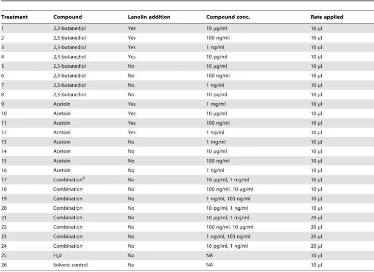
Table 4.Twenty-six treatments used to examine 3H2B (acetoin) and 23B (2, 3-butanediol) separately and in combination at various concentrations, in the presence or absence of lanolin, and at two application rates.
Treatment Compound Lanolin addition Compound conc. Rate applied
1 2,3-butanediol Yes 10mg/ml 10ml
2 2,3-butanediol Yes 100 ng/ml 10ml
3 2,3-butanediol Yes 1 ng/ml 10ml
4 2,3-butanediol Yes 10 pg/ml 10ml
5 2,3-butanediol No 10mg/ml 10ml
6 2,3-butanediol No 100 ng/ml 10ml
7 2,3-butanediol No 1 ng/ml 10ml
8 2,3-butanediol No 10 pg/ml 10ml
9 Acetoin Yes 1 mg/ml 10ml
10 Acetoin Yes 10mg/ml 10ml
11 Acetoin Yes 100 ng/ml 10ml
12 Acetoin Yes 1 ng/ml 10ml
13 Acetoin No 1 mg/ml 10ml
14 Acetoin No 10mg/ml 10ml
15 Acetoin No 100 ng/ml 10ml
16 Acetoin No 1 ng/ml 10ml
17 CombinationA No 10
mg/ml, 1 mg/ml 10ml
18 Combination No 100 ng/ml, 10mg/ml 10ml
19 Combination No 1 ng/ml, 100 ng/ml 10ml
20 Combination No 10 pg/ml, 1 ng/ml 10ml
21 Combination No 10mg/ml, 1 mg/ml 20ml
22 Combination No 100 ng/ml, 10mg/ml 20ml
23 Combination No 1 ng/ml, 100 ng/ml 20ml
24 Combination No 10 pg/ml, 1 ng/ml 20ml
25 H20 No NA 10ml
26 Solvent control No NA 10ml
AIn combination treatments, the applied rate was added per compound with the 23B concentration listed first.
experiment will be reported elsewhere (Marquez-Villavicencio et al., in preparation). We used absolute quantification of mRNA relative to four reference transcripts to confirm that one of the genes in this cluster,budB, was highly expressed during bacterial infection of tubers relative to stems. We determined the relative expression ratio (RER) with the mean values of the absolute amounts of four reference transcripts: ffh,dspE,pelB, andhrcCas previously described [48,55]. The target RNA was normalized to each reference RNA by dividing the absolute amount of target RNA by the amount of reference RNA. To determine the relative expression ratio of the target gene in tubers relative to stems, the normalized target RNA ratio was divided by the average of the normalized ratios of three stem samples. The average of the three stem normalized ratios is designated the ‘‘calibrator’’ since the variation of all samples, including the individual stem samples, is determined relative to this value. Using four reference transcripts resulted in four mean RER values for each RNA sample relative to the calibrator. The four mean RER values for each of the stem and tuber samples were treated as replicates and statistically analyzed byt-test. The Prism software (GraphPad Software, Inc.) averages the four RER values for each of the three stem or tuber RNA samples before performing the t-test analysis. Our RT-qPCR results showed thatbudBexpression was 8.5- fold higher in tubers than in stems (Fig. 1).
TheP. c. subsp. carotovorumWPP14budBmutant does
not produce detectable 3H2B in culture medium P. c. subsp. carotovorum WPP14 encodes genes homologous to
budAB,budCandbudR, which in other enteric bacteria are involved in the production of acetoin and 2,3-butanediol [4,7]. The catabolica-acetolactate synthase (budB) anda-acetolactate decar-boxylase (budA), are found in the same locus and a third gene, butanediol dehydrogenase (budC) is located elsewhere in the chromosome, as described in Bacillus subtilis [56] (Fig. 2). This differs from otherEnterobacteriaceaesuchKlebsiellaandEnterobacterin which the three enzymes are found in one operon [57,58]. In the
Enterobacteriaceae, the transcriptional regulatorbudRis located in the same locus as thebudAB operon, but is divergently transcribed. Other soft rot pathogens, includingP. atrosepticum SCRI1043,P. carotovorumPC1, P. carotovorumsubsp. brasiliensis 1692, P. wasabiae
WPP163,Dickeya dadantii3937 have the same operon structure as
P. c. carotovorum WPP14. Members of the Erwinia genus, such
Erwinia amylovoraCFBP1430 and ATCC 49946,Erwinia pyrifoliae
DSM 12163 and Ep1/96 andErwinia tasmaniensisET1/99, have the same budRAB operon structure as P. c. subsp. carotovorum
WPP14, but all of theseErwiniaspecies lackbudC, suggesting that they produce 3H2B, but not 23B. In the plant associated
Enterobacteriaceae, the budRABlocus does not appear to be located in a conserved region of the chromosome since the genes flanking this locus vary among strains and species.
To test whether the budAB operon is required for 3H2B production, abudBdeletion mutant was constructed. To verify a disruption in the butanediol pathway, a qualitative Voges-Proskauer test was used to detect the presence of acetoin through the formation of a red-colored compound in the medium [3]. While wild type WPP14 and the complemented DbudB mutant strains turned red after adding alpha-naphthol and potassium hydroxide to the medium in which they were grown, theDbudB culture did not. This result was observed at both 24 and 48 hours after incubation.
P. c.subsp.carotovorumWPP14DbudBis unable to
alkalize culture medium or inoculated potato tubers Activity of the 3H2B pathway is known to result in medium alkalization in other species [5]. Wild type WPP14 cultures grown in SOC medium reached pH 7.7 after 20 hours of incubation, whereas the WPP14DbudB culture remained at pH 5.0. The complementedDbudBmutant strain was partially restored, with a culture pH of 7.1. Uninoculated SOC medium remained at pH 7.0.
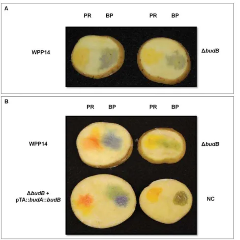
The pH of diseased tuber slices was measured after incubation in aerobic and anaerobic conditions to determine if budB also affected the pH of the milieu during plant-microbe interactions. After 6 or 20 hours of aerobic incubation, no differences in pH around the site of inoculation were observed between wild type and the DbudB mutant; both remained below 6.4 (Fig. 3). However, when potato slices were incubated in an anaerobic chamber, a difference in pH between wild type andDbudBwere seen at both 6 and 20 hours after incubation. While the tissue inoculated with wild type WPP14 reached a pH above 6.8, the
budBmutant remained below pH 6.4. Expression ofbudABfrom a plasmid in the mutant strain restored the wild type phenotype (Fig. 3).
TheP. c.subsp.carotovorumWPP14budBmutant was
reduced in virulence in potato tubers
Because a functionalbudBgene was required for alkalinization of both SOC medium and potato tissue, we hypothesized that this gene was also important for virulence in potato tubers. In two independent experiments, thebudBmutant produced significantly less macerated tissue than wild type (Fig. 4, two-tailed t-test,
P,0.001). Expression of budAB from a plasmid in the mutant strain partially restored the wild type phenotype (Fig. 4). To determine if the reduction in virulence was due to reduced ability of the bacteria to multiply in lesions, the bacterial population in the symptomatic part of the potato tuber was monitored. The mutant and the wild type strains were inoculated in potato tubers both grew to ,109CFU/g of tissue after 3 days of incubation (Fig. 5). No statistically significant difference in bacterial growth Figure 1.P. c.sp.carotovorum budBtranscript is differentially
expressed in potato tubers and stems.Relative expression of the
budBtranscript in potato stems and tubers was determined with four reference mRNA (ffh,dspE,pelB, andhrcC- Table 2). The mean relative expression ratio (RER) of the budB transcript in tubers was 8.5-fold elevated compared to stem tissue, which was used as the calibrator. The difference was found to be significant at a 95% confidence interval byt-test using Prism software (Mac version 5.0d, GraphPad Software, Inc.) with aP-value = 0.0425. Error bars represent standard error of the mean.
was detected at any time point of bacterial growth curve by a two-tailed t-test. Thus, the mutant is not defective in growth in the sense that it was able to grow in potato tuber at the inoculation site to the same density as wild type. However, the reduced ability of the mutant to macerate potato tissue and spread beyond the inoculation site results in an overall reduction in bacterial growth in tubers.
Acetoin (3H2B) and 2,3-butanediol (23B) do not affect potato growth in tissue culture
3H2B suppresses the ethylene response in Arabidopsis [22,23] and insensitivity to ethylene in both tobacco and Arabidopsis increases susceptibility P. c. subsp. carotovorum [59]. Ethylene accumulation in tissue culture vessels is a well-known cause of poor vigor and the reason that culture vessels are sealed with micropore tape to allow for ventilation, while still maintaining a sterile environment for tissue culture plantlets. To determine if 3H2B or 23B affected potato growth, we treated nodal cuttings of potato with these compounds in tightly sealed vessels or in vessels sealed with micropore tape. We were unable to detect any significant effect of 3H2B or 23B on growth of potato plantlets (data not shown).
Discussion
Relatively little is known about global gene expression of bacterial plant pathogens during pathogenesis. We became interested in the budAB operon because it was initially among the most highly expressedP. c.subsp.carotovorumgenes identified in bacterial tuber interactions through our microarray assay (Marquez-Villavicencio et al., in preparation). We used real-time RT-qPCR to confirm thatbudBis expressed during pathogenesis and also found it to be differentially expressed during infections of stems and tubers. In the plant pathogensPectobacterium atrosepticum
and S. plymuthica, the budAB operon is controlled by acyl-homoserine lactone quorum sensing [60,61], and it is also controlled by pH inS. plymuthica[61]. InV. cholerae, this operon is controlled by multiple regulators and is induced by quorum sensing, acetate [62] as well as acidic pH and low oxygen levels [63].
We hypothesize that less oxygen is available in potato tubers than stems and that this alternate fermentation pathway could be responding to low oxygen levels, but since this operon is likely to be controlled by multiple stimuli, there are many other possibilities that could account for this difference. ThebudABoperon does play Figure 2. Operon structure ofbudgenes inKlebsiella,Pectobacterium,Dickeya, andErwiniaspecies.Sequences were retrieved from ASAP [66]. Genes are indicated by arrows, with the direction of the arrowhead indicating the direction of the gene. Genes shaded with the same color are homologous and unlabeled genes have no known function.budAis an alpha-acetolactate decarboxylase,budBis an acetolactate synthase,budCis an acetoin reductase andbudRis a LysR-family transcriptional regulator. InKlebsiellaandEnterobacter(not shown),budABCare in one operon. The locus of thebudCgene is found elsewhere in the chromosome inPectobacteriumandDickeya. TheErwiniaspecies lackbudC.
Figure 3. The P. c.subsp. carotovorum budAB operon affects bacterial modification of pH in tubers slices that are incubated anaerobically.Potato slices were inoculated with 106CFU and incubated under aerobic (A) and anaerobic conditions (B) for 20 hours at room
temperature (,22uC).PR: pH indicator phenol red (yellow at pH below 6.4 and red violet at pH above 8.2),BP: pH indicator bromocresol purple
(yellow at pH below 5.2 and purple at a pH above 6.8). doi:10.1371/journal.pone.0022974.g003
Figure 4. WPP14DbudBis reduced in virulence in potato tubers.Potato tubers (n = 10) were inoculated with 106CFU, placed into plastic bags and incubated at 28uC for 3 days. After incubation, macerated tissue was scooped out and weighed. The graph shown is from one experiment. This experiment was repeated twice with similar results. Error bars indicate standard error.
a role inP. c.subsp.carotovorumpathogenesis in tubers, perhaps in part due to the role of this gene cluster in alkalizing the local environment.
The Voges-Proskauer assay, which detects a product of the EM pathway, 3H2B (acetoin), has been used for decades to differentiate coliform bacteria, such asEscherichia coliandSalmonella enterica from other enterobacteria [3]. Although many plant pathogenic enterobacteria, including soft rot pathogens ( Pectobac-teriumandDickeya), necrotic vegetable pathogens (Enterobacter), and tree pathogens (Erwinia amylovora) encode this pathway, its role in plant pathogenesis has been little explored. We found thatbudB, which encodesa-acetolactate synthase, an essential enzyme in this pathway, is required byP. c.subsp. carotovorumfor full virulence. Although others have reported that volatiles produced via this pathway promote plant growth, we were unable to demonstrate any effect of these compounds on potato plantlets grown in tissue culture.
ThebudBgene may contribute toP. c.subsp.carotovorumgrowth in plants directly by playing a role in nutrient and energy acquisition through fermentation. This fermentation pathway is not absolutely required for growth in potato, however, since the
budB mutant still reached high cell densities in the area it was inoculated into potato. This pathway may contribute to virulence by alkalinizing the local environment, which favors pectate lyase activity. A related plant pathogen,Serratia plymuthica, grows well in carrot slices when thebudBgene is deleted, thus, related pathogens also do not require this pathway for growth on plants [9]. Unlike
P. c.subsp.carotovorum,S. plymuthicadoes not produce pectate lyases and it is a more aggressive plant pathogen whenbudBis deleted. Fungal pathogens, such asColletotrichum gloeosporioides, also alkalin-ize their environment, which favors both expression and activity of fungal pectate lyases [64]. This convergence among bacterial and fungal pathogens that produce pectate lyases suggests that microbial ability to buffer the local environment is required for efficacy, and perhaps therefore selection and maintenance in the genome, of pectate lyases.
Some bacteria are able to utilize 3H2B and 23B as energy sources, but a search of the availablePectobacteriumgenomes in the ASAP database [65,66] show that they lack the required genes, which were initially described inAlcaligenes eutrophus [67], as does the related soft rot pathogenDickeyaand the tree pathogenErwinia amylovora. However, BLASTp searches show that these genes are widespread in other plant associated bacteria, including Pseudomo-nas putida, Pseudomonas fluorescens, Erwinia tasmaniensis, Bacillus
species, andPhytoplasmaspecies. It is also present in opportunistic human pathogens commonly found on plants, such asPseudomonas aeruginosaand Burkholderia cepacia, although it is not inPseudomonas syringae. It is possible that metabolism of 3H2B pathway products, which may buffer the environment for soft rot pathogens, would be counterproductive for these pathogens. There have been a few studies of co-colonization of soft rot bacterial pathogens withS. entericaorE. colion plant leaves [68,69,70], which are bacteria that acidify their environment, andClostridium, which can metabolize 3H2B are which are commonly found with Pectobacterium in decaying tubers [71]. The parameters that determine local pH in mixed infections of these types and whether it affects disease progress remain unknown.
The stability of a key P. carotovorum signal molecule, acyl-homoserine lactone (acyl-HSL), is also affected by pH and it is hydrolyzed into an inactive form above pH 6.8 [72]. As in many bacteria, acyl-HSL acts as a quorum sensing molecule in
Pectobacterium and affects the regulation of approximately one quarter of Pectobacterium genes, including induction of both the
budABoperon and plant cell wall degrading enzymes [60]. Since acyl-HSL hydrolyzes in alkaline pH, it should be less available once fermentation has started and the local pH has increased. Whether local concentrations of acyl-HSL are sufficient or other mechanisms, such as induction by plant cell wall fragments become more important in enzyme expression remains unknown. The butanediol pathway has been examined in plant growth promoting bacteria (PGPB), where it is thought to be responsible for the growth promoting effects of some PGPB [22,23]. A wide Figure 5. WPP14DbudBis not impaired in growth in potato tubers.Bacterial suspensions containing 105CFU were stab-inoculated into potato tubers (cv. ‘‘Superior’’) (n= 5). Tubers were placed into plastic bags and incubated at 28uC. Bacterial population was determined every 24 hrs after inoculation. Error bars indicate standard error. The graph shows data from one of two experiments, neither of which showed significant difference in bacterial growth.
range of bacterial species commonly found in decaying plants, from Pectobacterium to Bacillus subtilis produce 3H2B and 23B through fermentation. If the growth promoting effects of 3H2B and 23B do occur, this suggests that plants may grow in response to this compound because it is a signal of bacterial decay of plants, and hence, a signal that a rich source of nutrients is available from the composting plants. To determine if 3H2B and 23B affect growth of potato, we added these compounds to tissue culture medium used to grow potato plantlets, but were unable to demonstrate that these compounds promote potato growth. Use of 3H2B and 23B as priming molecules for plant resistance and drought resistance has been suggested [23,24], but this may be ineffective in practice since plants are likely already exposed to these molecules, which are produced by a wide range of soil microbes, on a daily basis.
The volatile products of the 3H2B pathway have long been known to attract a wide range of insects and it is common knowledge that a variety of insects are attracted to decaying plant material. For example, pest control recommendations for seed corn maggot (Delia platura) include avoiding plowing weeds, green manure or other cover crops in the spring since the adult female flies are attracted to decaying plants. A similar attraction to decaying onions has been seen with female, but not male, onion flies (Delia antiqua) [73]. Recently, Turner and Ray [19] found that 3H2B pathway volatiles suppress CO2 avoidance in Drosophila, which causes the flies to be attracted to ripening fruit. However, the connection that bacteria may attract insects specifically through the butanediol pathway has not yet been made. Insects are known vectors of soft rot bacteria [29,30,31,32,33,34,35,36,37,38,39,41] and are also vectors of other closely related pathogens that also encode this pathway, such as Erwinia amylovora [74], providing
support for a role of the 3H2B pathway in bacterial pathogen-insect symbiosis. Insects may benefit from this if the volatile compounds produced by the 3H2B pathway indicate a source of food (decaying plants) or a source of beneficial microbes that may aid them in invasion of plant hosts.
The Voges-Proskauer reaction is commonly used in introduc-tory bacteriology laboratories to demonstrate biochemical meth-ods used to identify bacteria, but then essentially ignored thereafter by many microbiologists. However, the butanediol pathway appears to play important roles in plant pathogenesis, insect behavior, and possible plant growth promotion. It may explain, in part, phenomena familiar to any gardener: insects are attracted to decaying plants and plants grow well in compost. It may also be an important step in evolution of soft rot pathogens, since acquisition of a pathway that alkalinizes the local environment may be required for maintenance of pectate lyases in pathogen genomes. Thus, this common diagnostic test is tied to important aspects of bacterial plant pathogenesis and evolution, and insect behavior.
Acknowledgments
We thank Russell Groves for providing information about insect kairomones and also the reviewers of this manuscript for their helpful comments.
Author Contributions
Conceived and designed the experiments: MMV BW RAW DKW AOC. Performed the experiments: MMV BW RAW DKW AOC. Analyzed the data: MMV BW RAW DKW AOC. Contributed reagents/materials/ analysis tools: MMV BW RAW DKW AOC. Wrote the paper: MMV BW RAW DKW AOC.
References
1. Huang M, Oppermann-Sanio FB, Steinbu¨chel A (1999) Biochemical and molecular characterization of the Bacillus subtilis acetoin catabolic pathway. J Bacteriol 181: 3837–3841.
2. Lopez JM, Thoms B, Rehbein H (1975) Acetoin degradation inBacillus subtilisby direct oxidative cleavage. Eur J Biochem 57: 425–430.
3. Levine M (1916) On the significance of the Voges-Proskauer reaction. J Bacteriol 1: 153–164.
4. Xiao Z, Xu P (2007) Acetoin metabolism in bacteria. Crit Rev Micobiol 33: 127–140.
5. Yoon SS, Mekalanos JJ (2006) 2,3-butanediol synthesis and the emergence of the
Vibrio choleraeEl Tor biotype. Infect Immun 74: 6574–6556.
6. Johansen L, Bryn K, Stormer FC (1975) Physiological and biochemical role of the butanediol pathway inAerobacter (Enterobacter) aerogenes. J Bacteriol 123: 1124–1130.
7. Mayer D, Schlensog V, Bock A (1995) Identification of the transcriptional activator controlling the butanediol fermentation pathway inKlebsiella terrigena. J Bacteriol 177: 5261–5269.
8. Grundy FJ, Waters DA, Takova TY, Henkin TM (1993) Identification of genes involved in utilization of acetate and acetoin inBacillus subtilis. Mol Microbiol 10: 259–271.
9. Wevers E, Moons P, Van Houdt R, Lurquin I, Aertsen A, et al. (2009) Quorum sensing and butanediol fermentation affect colonization and spoilage of carrot slices bySerratia plymuthica. Int J Food Microbiol 134: 63–69.
10. Hsieh S-C, Lu C-C, Horng Y-T, Soo P-C, Chang Y-L, et al. (2007) The bacterial metabolite 2,3-butanediol ameliorates endotoxin-induced acute lung injury in rats. Microbes Infect 9: 1402–1409.
11. van Rooy FGBGJ, Rooyackers JM, Prokop M, Houba R, Smit LAM, et al. (2007) Bronchiolitis obliterans syndrome in chemical workers producing diacetyl for food flavorings. Amer J Respiratory Crit Care Med 176: 498–504. 12. Nout MJR, Bartelt RJ (1998) Attraction of a flying nitidulid (Carpophilus humeralis)
to volatiles produced by yeasts grown on sweet corn and a corn-based medium. J Chem Ecol 24: 1217–1239.
13. Buttery RG, Kamm JA, Ling LC (1984) Volatile components of red clover leaves, flowers, and seed pods: possible insect attractants. J Agric Food Chem 32: 254–256.
14. Moore AJ, Moore PJ (1999) Balancing sexual selection through opposing mate choice and male competition. Proc R Soc Lond B Biol Sci 266: 711–716. 15. Moore AJ, Haynes KF, Preziosi RF, Moore PJ (2002) The evolution of
interacting phenotypes: genetics and evolution of social dominance. Am Nat 160: 5186–5197.
16. Rochat D, Morin JP, Kakul T, Beaudoin-Ollivier L, Prior R, et al. (2002) Activity of male pheromone of Melanesian rhinoceros beetleScapanes australis. J Chem Ecol 28: 479–500.
17. Bengtsson JM, Wolde-Hawariat Y, Khbaish H, Negash M, Jembere B, et al. (2009) Field attractants forPachnoda interruptaselected by means of GC-EAD and single sensillum screening. J Chem Ecol 35: 1063–1076.
18. Robacker DC, Lauzon CR (2002) Purine metabolizing capability ofEnterobacter agglomeransaffects volatiles production and attractiveness to Mexican fruit fly. J Chem Ecol 28: 1549–1563.
19. Turner SL, Ray A (2009) Modification of CO2 avoidance behaviour in
Drosophilaby inhibitory odorants. Nature 461: 277–281.
20. Moore AJ, Gowaty PA, Moore PJ (2003) Females avoid manipulative males and live longer. J Evol Biol 16: 523–530.
21. Moore AJ, Gowaty PA, Moore PJ (2001) Sexual conflict and the evolution of female mate choice and male social dominance. Proc R Soc Lond B Biol Sci 268: 517–523.
22. Ryu C-M, Farag MA, Hu C-H, Reddy MS, Wei H-X, et al. (2003) Bacterial volatiles promote growth in Arabidopsis. Proc Natl Acad Sci U S A 100: 4927–4932.
23. Ryu CM, Farag AF, Hu C, Reddy MS, Kloepper JW, et al. (2004) Bacterial volatiles induce systemic resistance in Arabidopsis. Plant Physiol 134: 1017–1026.
24. Cho SM, Kang BR, Han SH, Anderson AJ, Park J-Y, et al. (2008) 2R,3R-butandiol, a bacterial volatile produced byPseudomonas chlororaphis06, is involved in induction of systemic tolerance to drought inArabidopsis thaliana. Mol Plant-Microbe Interact 21: 1067–1075.
25. Brenner DJ, Krieg NR, Staley JT, Garrity GM (2004) The Proteobacteria. Berlin, New York, London: Springer. 1256 p.
26. Ma B, Hibbing ME, Kim H-S, Reedy RM, Yedidia I, et al. (2007) The host range and molecular phylogenies of the soft rot enterobacterial genera
PectobacteriumandDickeya. Phytopathology 97: 1150–1163.
27. Vikram A, Lui LH, Hossain A, Kushalappa AC (2006) Metabolic fingerprinting to discriminate disease of stored carrots. Ann Appl Biol 148: 17–26. 28. Lui LH, Vikram A, Abu-Nada Y, Kushalappa AC, Raghavan GSV, et al. (2005)
Volatile metabolic profiling for discrimination of potato tubers inoculated with dry and soft rot pathogens. Amer J Potato Res 82: 1–8.
30. Howard CM, Leach JG (1963) Relation of the iris borer to bacterial soft rot of iris. Phytopathology 53: 1190–1193.
31. Dalmacio SC, Lugod TR, Serrano EM, Munkvold GP (2007) Reduced incidence of bacterial rot on transgenic insect-resistant maize in the Philippines. Plant Dis 91: 346–351.
32. Thind BS, Singh N (1976) Maize borer [Chilo partellus(Swinhoe)] as carrier of
Erwinia carotovoravar.zeae, tha causal agent of bacterial stalk rot of maize. Curr Sci 45: 117–118.
33. Leach JG (1931) Further studies on the seed-corn maggot and bacteria with special reference to potato blackleg. Phytopathol 21: 387–406.
34. Leach JG (1933) The method of survival of bacteria in the puparia of the seed-corn maggot (Hylemyia cilicrura Rond.). Zeitschr fur angewondte Entomologie 20: 150–161.
35. Harrison MD, Quinn CE, Sells A, Graham DC (1977) Waste potato dumps as sources of insects contaminated with soft rot coliform bacteria in relation to re-contamination of pathogen-free potato stocks. Potato Res 20: 37–52. 36. Kloepper JW, Brewer JW, Harrison MD (1981) Insect transmission ofErwinia
carotovoravar.carotovoraandErwinia carotovoravar.atrosepticato potato plants in the field. Am Potato J 58: 165–175.
37. Kloepper JW, Harrison MD, Brewer JW (1979) The association of Erwinia carotovora var. atroseptica and Erwinia carotovora var. carotovora with insects in Colorado. Am Potato J 56: 351–361.
38. Kloepper JW, Harrison MD, Brewer JW (1981) Effect of temperature on differential persistence and insect transmission of Erwinia carotovora var. Atroseptica and Erwinia carotovora var. Carotovora. The American Potato Journal 58: 585–592.
39. Phillips JA, Kelman A (1982) Direct fluorescent antibody stain procedure applied to insect transmission ofErwinia carotovora. Phytopathology 72: 898–901. 40. Grenier AM, Duport G, Pages S, Condemine G, Rahbe Y (2006) The phytopathogenDickeya dadantii(Erwinia chrysanthemi3937) is a pathogen of the pea aphid. Appl Environ Microbiol 72: 1956–1965.
41. Molina JM, Harrison D, Brewer JW (1974) Transmission ofErwinia carotovora
var.atrosepticabyDrosophila melanogasterMeig. I. Acquisition and transmission of the bacterium. Am Potato J 51: 245–250.
42. Toth IK, Bell KS, Holeva MC, Birch PRJ (2003) Soft rot erwiniae: from genes to genomes. Mol Plant Pathol 4: 17–30.
43. Barras F, Van Gijsegem F, Chatterjee AK (1994) Extracellular enzymes and pathogenesis of soft rotErwinia. Annu Rev Phytopathol 32: 210–234. 44. Lei S-P, Lin H-C, Wang S-S, Callaway J, Wilcox G (1987) Characterization of
theErwinia carotovora pelBgene and its product pectate lyase. J Bacteriol 169: 4379–4383.
45. Lei S-P, Lin H-C, Wang S-S, Wilcox G (1988) Characterization of theErwinia carotovora pelAgene and its product pectate lyase A. Gene 62: 159–164. 46. Grignon C, Sentenac H (1991) pH and ionic conditions in the apoplast. Ann
Rev Plant Physiol 42: 103–128.
47. Nachin L, Barras F (2000) External pH: an environmental signal that helps to rationalize pel gene duplication in Erwinia chrysanthemi. Mol Plant Microbe Interact 13: 882–886.
48. Jahn CE, Charkowski AO, Willis DK (2008) Evaluation of isolation methods and RNA integrity for bacterial RNA quantitation. Jounal of Microbiological Methods 75: 318–324.
49. Linchao S, Bremer H (1986) Effect of the bacterial-growth rate on replication control of plasmid pBR322 inEscherichia coli. Mol Gen Genet 203: 143–149. 50. Bustin SA, Benes V, Garson JA, Hellemans J, Huggett J, et al. (2009) The MIQE
guidelines: minimum information for publication of quantitative real-time PCR experiments. Clin Chem 55: 611–622.
51. Ried JL, Collmer A (1986) Comparison of pectic enzymes produced byErwinia chrysanthemi, Erwinia carotovora subsp. carotovora, and Erwinia carotovora subsp.
atroseptica. Appl Environ Microbiol 52: 305–310.
52. Sambrook J, Russell DW (2001) Molecular Cloning. A Laboratory Manual. Cold Spring Harbor: Cold Spring Harbor Laboratory Press.
53. Llama-Palacios A, Lopez-Solanilla E, Rodriguez-Palenzuela P (2002) TheybiT
gene ofErwinia chrysanthemicodes for a putative ABC transporter and is involved in competitiveness against endophytic bacteria during infection. Applied and Environmental Microbiology 68: 1624–1630.
54. Haberlach GT, Cohen BA, Reichert NA, Baer MA, Towill LE, et al. (1985) Isolation, culture and regeneration of protoplasts from potato and several related
Solanumspecies. Plant Sci 39: 67–74.
55. Rotenberg DT, Thompson TS, German TL, Willis DK (2006) Methods for effective real-time RT-PCR analysis of virus-induced gene silencing. Virological Methods. pp 1–11.
56. Renna MC, Najimudin N, Winik LR, Zahler SA (1993) Regulation of the
Bacillus subtilis alsS,alsD, and alsRgenes involved in post-exponential-phase production of acetoin. J Bacteriol 175: 3863–3875.
57. Blomqvist K, Nikkola M, Lehtovaara P, Suihko ML, Airaksinen U, et al. (1993) Characterization of genes of the 2,3-butanediol operons fromKlebsiella terrigena
andEnterobacter aerogenes. J Bacteriol 175: 1392–1404.
58. Mayer D, Schlensog V, Bo¨ck A (1995) Identification of the transcriptional activator controlling the butanediol fermentation pathway inKlebsiella terrigena. J Bacteriol 177: 5261–5269.
59. Geraats BP, Bakker PA, Lawrence CB, Achuo EA, Ho¨fte M, et al. (2003) Ethylene-insensitive tobacco shows differentially altered susceptibility to different pathogens. Phytopathology 93: 813–821.
60. Liu H, Coulthurst SJ, Pritchard L, Hedley PE, Ravensdale M, et al. (2008) Quorum sensing coordinates brute force and stealth modes of infection in the plant pathogen Pectobacterium atrosepticum. PloS Pathogens 4: e1000093. doi:1000010.1001371/journal.ppat.1000093.
61. Moons P, Van Houdt R, Vivijs B, Michiels CM, Aertsen A (2011) Integrated regulation of acetoin fermentation by quorum sensing and pH in Serratia plymuthicaRVH1. Appl Environ Microbiol.
62. Kovacikova G, Lin W, Skorupski K (2005) Dual regulation of genes involved in acetoin biosynthesis and motility/biofilm formation by the virulence activator AphA and the acetate-responsive LysR-type regulator AlsR inVibrio cholerae. Mol Microbiol 57: 420–433.
63. Kovacikova G, Lin W, Skorupsk iK (2010) The LysR-type virulence activator AphB regulates the expression of genes inVibrio choleraein response to low pH and anaerobiosis. J Bacteriol 192: 4181–4191.
64. Kramer-Haimovich H, Servi E, Katan T, Rollins J, Okon Y, et al. (2006) Effect of ammonia production byColletotrichum gloeosporioidesonpelBactivation, pectate lyase secretion, and fruit pathogenicity. Appl Environ Microbiol 72: 1034–1039. 65. Glasner JD, Liss P, Plunkett G, Darling A, Prasad T, et al. (2003) ASAP, a systematic annotation package for community analysis of genomes. Nucl Acids Res 31: 147–151.
66. Glasner JD, Rusch M, Liss P, Plunkett G, 3rd, Cabot EL, et al. (2006) ASAP: a resource for annotating, curating, comparing, and disseminating genomic data. Nucl Acids Res 34: D41–45.
67. Fru¨nd C, Priefert H, Steinbu¨chel A, Schlegel HG (1989) Biochemical and genetic analyses of acetoin catabolism inAlcaligenes eutrophus. J Bacteriol 171: 6539–6548.
68. Wells JM, Butterfield JE (1997) Salmonella contamination associated with bacterial soft rot of fresh fruits and vegetables in the marketplace. Plant Dis 81: 867–872.
69. Brandl MT (2008) Plant lesions promote the rapid multiplication ofEscherichia coliO157:H7 on postharvest lettuce. Appl Environ Microbiol 74: 5285–5289. 70. Noel JT, Joy J, Smith JN, Fatica M, Schneider KR, et al. (2010)SalmonellaSdiA
recognizes N-acyl homoserine lactone signals fromPectobacterium carotovorum in vitro, but not in a bacterial soft rot. Mol Plant-Microbe Interact 23: 273–282. 71. Pe´rombelon MCM, Gullings-Handley J, Kelman A (1979) Population dynamics
ofErwinia carotovoraand pectolyticClostridiumspp. in relation to decay of potatoes. Phytopathol 69: 167–173.
72. Byers JT, Lucas C, Salmond GPC, Welch M (2002) Nonenzymatic turnover of an Erwinia carotovora quorum-sensing signaling molecule. J Bacteriol 184: 1163–1171.
73. Dindonis LL, Miller JR (1980) Host-finding responses of onion and seedcorn flies to healthy and decomposing onions and several synthetic constituents of onion. Environ Entomol 9: 467–472.
74. Purcell AH (1982) Insect vector relationships with procaryotic plant pathogens. Ann Rev Phytopathol 20: 397–417.
75. Yap M-N, Barak JD, Charkowski AO (2004) Genomic diversity ofErwinia carotovora subsp. carotovora and its correlation with virulence. Appl Environ Microbiol 70: 3013–3023.
76. Glasner JD, Marquez-Villavicencio M, Kim H-S, Jahn CE, Ma B, et al. (2008) Niche-specificity and the variable fraction of thePectobacteriumpan-genome. Mol Plant-Microbe Interact 21: 1549–1560.
77. Bogdanove AJ, Bauer DW, Beer SV (1998)Erwinia amylovorasecretes DspE, a pathogenicity factor and functional AvrE homolog, through the Hrp (type III secretion) pathway. J Bacteriol 180: 2244–2247.
78. Fellay R, Frey J, Krisch H (1987) Interposon mutagenesis of soil and water bacteria: a family of DNA fragments designed forin vitroinsertional mutagenesis of gram-negative bacteria. Gene 52: 147–154.