Global Transcriptome Analysis Reveals
Distinct Aluminum-Tolerance Pathways in the
Al-Accumulating Species
Hydrangea
macrophylla
and Marker Identification
Haixia Chen*, Changping Lu, Hui Jiang, Jinhui Peng
College of Horticulture and landscape, Hunan Agriculture University, Changsha, 410128, People republic of China
Abstract
Hydrangea (Hydrangea macrophylla) is a well known Al-accumulating plant, showing a high level of aluminum (Al) tolerance and accumulation. Although the physiological mechanisms for detoxification of Al and the roles of Al in blue hydrangea sepals have been reported, the molecular mechanisms of Al tolerance and accumulation are poorly understood in hydran-gea. In this study, we conducted a genome-wide transcriptome analysis of Al-response genes in the roots and leaves of hydrangea by RNA sequencing (RNA-seq). The assembly of hydrangea transcriptome provides a rich source for gene identification and mining molec-ular markers, including single nucleotide polymorphism (SNP) and simple sequence repeat (SSR). A total of 401,215 transcripts with an average length of 810.77bp were assembled, generating 256,127 unigenes. After annotation, 4,287 genes in the roots and 730 genes in the leaves were up-regulated by Al exposure, while 236 genes in the roots and 719 genes in the leaves were down-regulated, respectively. Many transporters, including MATE and ABC families, were involved in the process of Al-citrate complex transporting from the roots in hydrangea. A plasma membrane Al uptake transporter, Nramp aluminum transporter was up-regulated in roots and leaves under Al stress, indicating it may play an important role in Al tolerance by reducing the level of toxic Al. Although the exact roles of these candidate genes remain to be examined, these results provide a platform for further functional analysis of the process of detoxification of Al in hydrangea.
Introduction
Aluminum (Al) is the most abundant metal in the earth’s crust and is a toxic element for plants. All over the world, up to 40% ~50% of cultivable land is acidic and Al3+toxicity is a major limiting factor for crop production [1]. In acidic soil, high concentrations of Al are existed and inhibit the root growth [2]. It also increases the levels of reactive oxygen species (ROS), which targets the plasma membrane and interacts with lipid components to initiate
OPEN ACCESS
Citation:Chen H, Lu C, Jiang H, Peng J (2015) Global Transcriptome Analysis Reveals Distinct Aluminum-Tolerance Pathways in the Al-Accumulating SpeciesHydrangea macrophyllaand Marker Identification. PLoS ONE 10(12): e0144927. doi:10.1371/journal.pone.0144927
Editor:Debasis Chakrabarty, CSIR-National Botanical Research Institute, INDIA
Received:July 22, 2015
Accepted:November 25, 2015
Published:December 14, 2015
Copyright:© 2015 Chen et al. This is an open access article distributed under the terms of the Creative Commons Attribution License, which permits unrestricted use, distribution, and reproduction in any medium, provided the original author and source are credited.
Data Availability Statement:The raw sequence data obtained have been deposited at the NCBI in the Short Read Archive (SRA) database under the accession number: SRP061814.
lipid peroxidation [3]. Therefore, enhancing Al tolerance of crops is a key to increase crop pro-ductivity on acidic soils. Elucidation of the detoxify strategies of some Al-accumulating plants will help us to increase the Al tolerance for crops. The Al-tolerance plant species can detoxify Al internally and externally [3,4]. The root, and more specifically, the root tip, is the primary site of Al3+toxicity, and the majority (~90%) of the Al3+resides in the root cell wall [5]. To date, Al-activated secretion of organic acid from root apex is a well-documented mechanism of Al internal detoxification [6]. Different plants secrete different organic acids such as malate, citrate and oxalate to chelate Al and thereby attenuate Al3+toxicity [7]. However, the process leading to the gene expression in the secretion and genes of Al response and tolerance remains not fully understood.
Hydrangea (Hydrangea macrophylla) is a well known Al-accumulating plant, and can become blue sepal color when it’s cultivated in acidic soil. Hydrangea plants can accumulate 5 mg Al g-1dry weight in the leaves within several months [8]. Aluminum, as Al3+, is not available to the hydrangea roots in basic or neutral soils, because it forms aluminum hydroxide and other insoluble precipitates. The main Al species in the cell sap of hydrangea leaves is the 1:1 complex of Al and citrate [9]. In acidic soils, Al3+becomes available to roots, stimulating the hydrangea roots to exude citrates. The citrates form complexes with Al3+and then the alu-minum citrate complexes enter the roots and transport throughout the shrub [8].
Previous studies on aluminum in hydrangea were mainly about the sepals color changes [10,11]. Few Al tolerance genes have been cloned and the corresponding molecular mecha-nism remains unclear in hydrangea, although the gene expression for Al tolerance and accu-mulation in several plants, including rice bean (vigna umbellata) [12], common buckwheat (Fagopyrum esculentum) [13] and tartary buckwheat (Fagopyrum tataricum) [7], were investi-gated. Therefore, identification of Al responsive genes associated with Al stress would provide valuable information for the detoxification mechanism of Al in hydrangea. However, as hydrangea is a non-model plant without the genome information, it will be a challenge to uncover the molecular mechanism of how the hydrangea receives the signal of Al stress and the expression of downstream genes, which finally lead to the physiological response.
Recently, RNA sequencing (RNA-Seq), a high-throughput sequencing method, is developed to analysis the transcriptome for both having or without genome information. It’s an efficient tool to promise simultaneous estimation of abundance and new transcript discovery [14]. RNA-Seq has been applied for transciptome analysis in many plant species, includingYoungia japonica[15], sugarcane [16] and buckwheat [7,13]. In this study, we used the RNA-Seq tech-nique to analyze the transcriptome of roots and leaves of hydrangea exposed or not to Al, for identifying the differentially expressed genes. The aim is to identify Al responsive genes and new insight of molecular mechanisms of Al3+toxicity and tolerance in hydrangea.
Materials and Methods
Plant materials
TheHydrangea macrophyllacultivars were cultivated in the garden of department of ornamen-tal horticulture, Hunan Agricultural University. Cuttings ofHydrangea macrophyllacultivar
“Lavbla”were subjected to hydroponic culture for growing. The solution contained KNO3
(1.0mM), Ca (NO3)2(4mM), MgSO4(1mM), KH2PO4(1mM), NaFeEDTA (10μM), H3BO3
(50μM), MnSO4(0.5μM), ZnSO4(0.4μM), CuSO40.5μM, (NH4)6Mo7O24(1μM). The solution
was changed every day. The Al stress was treated as previous described [7,13]. When the new roots grew, the cuttings were exposed to a 0.5mM CaCl2solution (PH 4.5) containing 50μM
AlCl3. At the same time, the control cuttings groups grew in the same solution (0.5mM CaCl2, Competing Interests:The authors have declared
PH 4.5) only without AlCl3. After 4h, the roots (3cm from the root tip) and the leaves were
har-vested and frozen in liquid nitrogen and stored at -80°C for further use.
RNA extraction, library construction and sequencing
Total RNA were extracted from the roots and leaves using an RNeasy Plant Mini Kit (QIA-GEN) according to the manufacturer’s protocol. Messenger RNAs (mRNAs) from the total RNA were isolated using Oligo (dT) and were randomly cleaved into short fragments. Then the first strand cDNAs were synthesized with random hexamer primers and followed by sec-ond strand cDNAs synthesis using DNA polymerase I (New England BioLabs) and RNase H (Invitrogen). After end repair, adaptor ligation, and index codes adding for each sample, PCR amplification was performed. The quality and quantity of the libraries were detected using an Agilent 2100 Bioanalyzer and an ABI real time RT-PCR system. The qualified cDNA libraries were carried out for sequencing by an Illumina HiSeq 2500 platform with PE100. The raw sequence data obtained have been deposited at the NCBI in the Short Read Archive (SRA) database under the accession number: SRP061814.
Sequence data analysis and De novo assembly
Raw reads were quality-checked with FastQC package, and adaptor sequences and low quality reads were removed. We carried out a stringent filtering criterion to minimize the effects of sequencing errors during the assembly. Firstly, bases with phred quality score lower than 20 and reads length short than 50bp would be discarded. Secondly, reads of 70% bases in a read having high phred quality scores (20) will be used for assembly. Thirdly, only the paired-end reads were used for further assembly. The obtained clean reads of all four samples were assem-bled by Trinity (Release 2013-07-08) using a paired-end model [17]. To annotate the assembled transcripts, BLASTx searches (E-value<1e-5) were performed against the protein databases,
including NCBI non-redundant protein (NR) database, Swiss-Prot, Kyoto Encyclopedia of Genes and Genomes (KEGG) pathway database and Clusters of eukaryotic Orthologous Groups of proteins (KOG) database. The transcripts abundance was normalized by the reads per kilobase of transcript per million mapped reads (RPKM) value using the RSEM (RNASeq by Expectation Maximization) package [18]. And those transcripts with RPKM value equal or larger than 0.1 were defined as expressed. All the unigenes were translated into potential pro-teins according to ORF prediction by Getorf (http://emboss.sourceforge.net/apps/cvs/emboss/ apps/getorf).
Differential expression analysis and GO and KEGG enrichment analysis
Validation of RNA-seq by quantitative Real time RT-PCR
For the quantitative real time RT-PCR (qPCR) of the mRNAs, 1μg of total RNA was used to
synthesize the cDNA using the RevertAid First Strand cDNA Synthesis Kit (Fermentas). Quan-titiative PCR was performed using the FastStart Universal SYBR Green Master (Roche) accord-ing to the manufacturer’s instruction on the StepOne plus Real time PCR Platform (Applied Biosystems). The qPCRs were carried out with the following protocol: 95°C for 10min, fol-lowed by 40 cycles of 95°C for 15s, and at 60°C for 60s. The 18S rRNA which is one of the refer-ence genes in Hydrangea was used as the internal control [11]. After the amplification, the melting curve was determined for specific product. Three independent biological replicates for each sample and three technical replicates for each biological replicate were analyzed. All the primers used are listed inS1 Table. Significant differences of the expression level between roots (-Al) and roots (+Al) or leaves (-Al) and leaves (+Al) were evaluated using Student’sttest.
Mining of SNP and Simple Sequence Repeat (SSR)
SNPs of the transcripts between different samples were detected using SOAPsnp [20]. The filter criteria were as follow: (1) the total coverage and the number of reads to cover a candidate SNP (>8 reads); (2) low phred quality (<25) reads from the coverage were removed; (3) frequency
of mutated bases among all reads covering the position was higher than 30%.
SSR markers were identified by the MISA (MIcroSAtellite identification tool) Perl script (http://pgrc.ipk-gatersleben.de/misa). The settings for minimum number of repeats were mono-, di-, tri-, tetra-, penta-, and hexa-nucleotide motifs with numbers of uninterrupted repeat units more than 10, 6, 5, 5, 5, and 5, respectively. For compound SSRs, the maximum distance between the two SSR was 50bp.
Results
De novo assembly of the transcripts and annotation
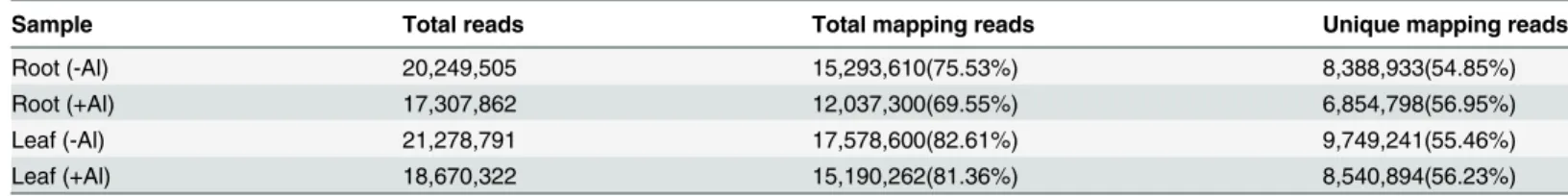
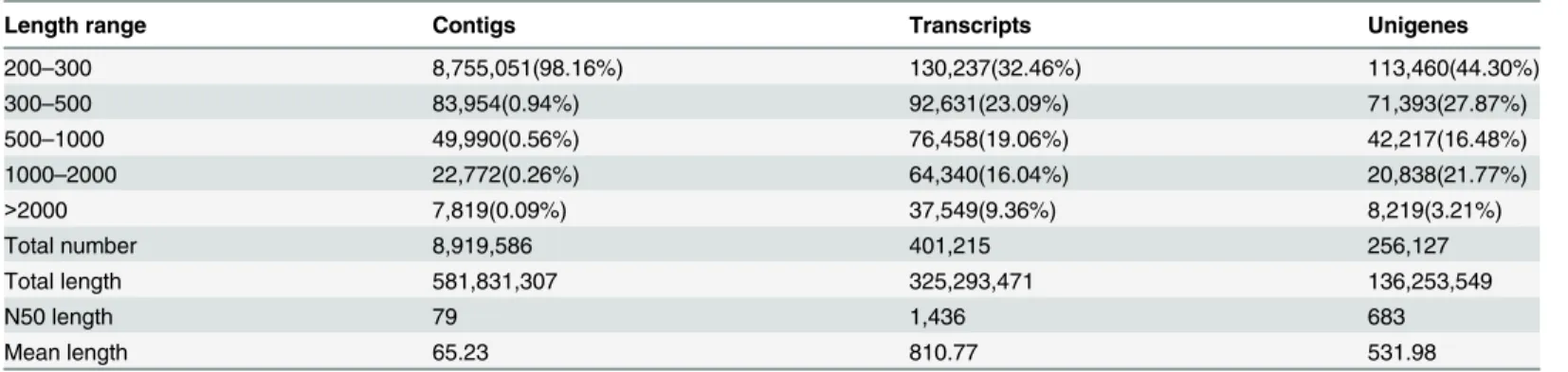
The total reads of hydrangea roots and leaves were ranging from 17~21 million, depending on the tissues and treatments (Table 1). Using the de novo assembly program Trinity, the consen-sus sequences were constructed. As a result, a total of 8,919,586 contigs from the four samples were assembled (Table 2). Of them, a total of 401,215 transcripts and 256,127 unigenes were found. The assembled unigenes had a length distribution from 201 to 15,907 with an average length of 531.98bp (Fig 1andTable 2). Recently, transcriptome sequencing of buckwheat (F.
tataricum) assembled 148,734 contigs and 84,516 unigenes [13]. Compared with their results, we produced more contigs (8,919,586) and more unigenes (256,127). Therefore, the assembled contigs in this study can provide a useful resource for further research of hydrangea.
For annotation, the 256,127 unigenes were subjected to BLASTx searches against the sequences in the NR, Swiss-Prot and KOG databases (E-value1e-5). As a result, a total of 74,167 sequences were annotated, of them, 73,853(77.16%), 57,278 (59.84%) and 28,596 (29.88%) unigenes were aligned against the three protein databases, respectively. GO and KEGG analysis were also performed, having 47,670 and 18,270 unigenes, respectively (Table 3).
Table 1. Summary of short-read data from hydrangea produced by Illumina sequencing.
Sample Total reads Total mapping reads Unique mapping reads
Root (-Al) 20,249,505 15,293,610(75.53%) 8,388,933(54.85%)
Root (+Al) 17,307,862 12,037,300(69.55%) 6,854,798(56.95%)
Leaf (-Al) 21,278,791 17,578,600(82.61%) 9,749,241(55.46%)
Leaf (+Al) 18,670,322 15,190,262(81.36%) 8,540,894(56.23%)
Global effect of Al stress on gene expression
To get insight into the gene expression patterns in roots and leaves of hydrangea under Al stress, RPKMs were calculated for each sample and all the unigenes were annotated (Fig 2,S2 Tableand
S3 Table). The results showed that the roots had more differentially expressed genes (DEGs) than that in leaves (Fig 2A, 2B and 2C) and many genes were up regulated in roots or leaves under Al stress (Fig 2D). The unigene abundance was quantified using Bowtie and RSEM pack-age. The significant DEGs were judged by |log2 (fold change)|2 and FDR value less than 0.01. In the roots, compared with control (-Al), 4,287 and 236 annotated genes were up-regulated and down-regulated in roots (+Al), respectively (S4 Table). In the leaves, the number of genes up-and down- regulated by Al was 730 up-and 719, respectively (S4 Table). Although the down- regu-lated genes in roots and DEGs in leaves were lower than buckwheat [13], the results in this study were similar with previous reports, having more up-regulated genes in roots under Al stress.
Functional classification of the Al stress genes
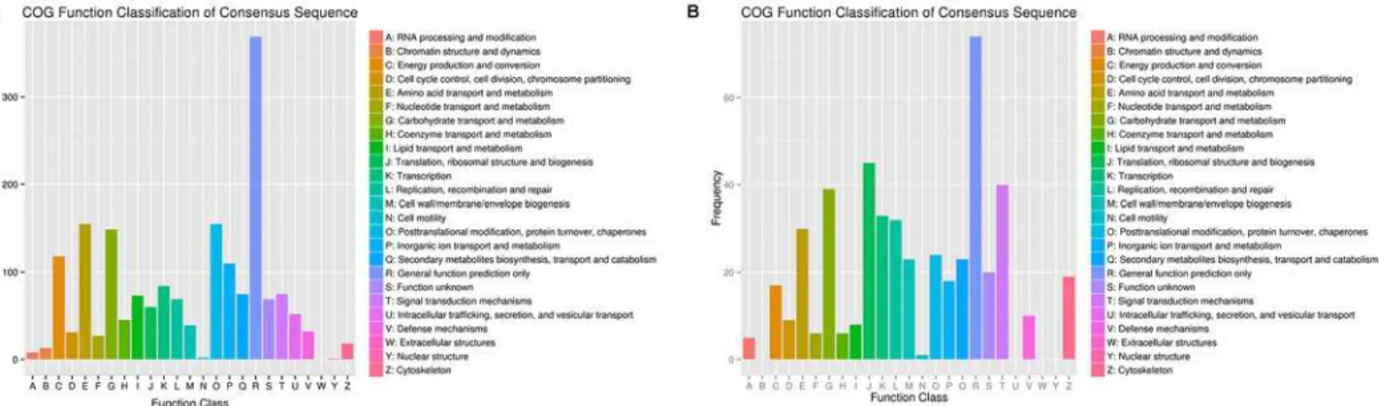
To identify the genes that are differentially expressed under Al stress, a functional categoriza-tion was carried out by GO and KEGG funccategoriza-tional annotacategoriza-tion, respectively (Table 3). The anno-tated unigenes were also compared with the eukaryotic orthologous groups (KOG) database for functional prediction and classification. In total, 28,596 annotated putative proteins were classified into 25 KOG groups (Fig 3). Among the 25 categories,“Extracellular structures”was not represented in both hydrangea roots and leaves under Al stress. In roots, the larger groups included“Posttranslational modification, protein turnover, chaperones”,“Amino acid
Table 2. The length distribution of assembled contigs, transcripts and unigenes.
Length range Contigs Transcripts Unigenes
200–300 8,755,051(98.16%) 130,237(32.46%) 113,460(44.30%)
300–500 83,954(0.94%) 92,631(23.09%) 71,393(27.87%)
500–1000 49,990(0.56%) 76,458(19.06%) 42,217(16.48%)
1000–2000 22,772(0.26%) 64,340(16.04%) 20,838(21.77%)
>2000 7,819(0.09%) 37,549(9.36%) 8,219(3.21%)
Total number 8,919,586 401,215 256,127
Total length 581,831,307 325,293,471 136,253,549
N50 length 79 1,436 683
Mean length 65.23 810.77 531.98
doi:10.1371/journal.pone.0144927.t002
Fig 1. Distribution of the length of transcript assembly unigenes.
Table 3. Annotation of Unigenes were searched against from NR, SwissProt, GO, KOG, KEGG by BLAST.
Annotated databases All sequence >= 300bp >= 1000bp
KOG 28,596 17,029 11,567
GO 47,670 28,697 18,973
KEGG 18,270 11,429 6,841
Swiss-Prot 57,278 34,059 23,219
NR 73,853 47,143 26,710
All 74,167 47,410 26,757
doi:10.1371/journal.pone.0144927.t003
Fig 2. The overview of differential expression between no Al and Al stress in roots and leaves in hydrangea.(A) Boxplots of the DEGs in the four samples; (B) Sequencing saturation level analysis of DEGs in the four samples; (C) Density distribution of the differentially expressed genes (DEGs) in the four samples; (D) heat map of the DEGs in roots and leaves between Al(-) and Al (+).
transport and metabolism”and“Carbohydrate transport and metabolism”. Unlike the roots, three groups“Translation, ribosomal structure and biogenesis”,“Signal transduction mecha-nisms”and“Carbohydrate transport and metabolism”were represented as the major number groups in leaves of hydrangea.
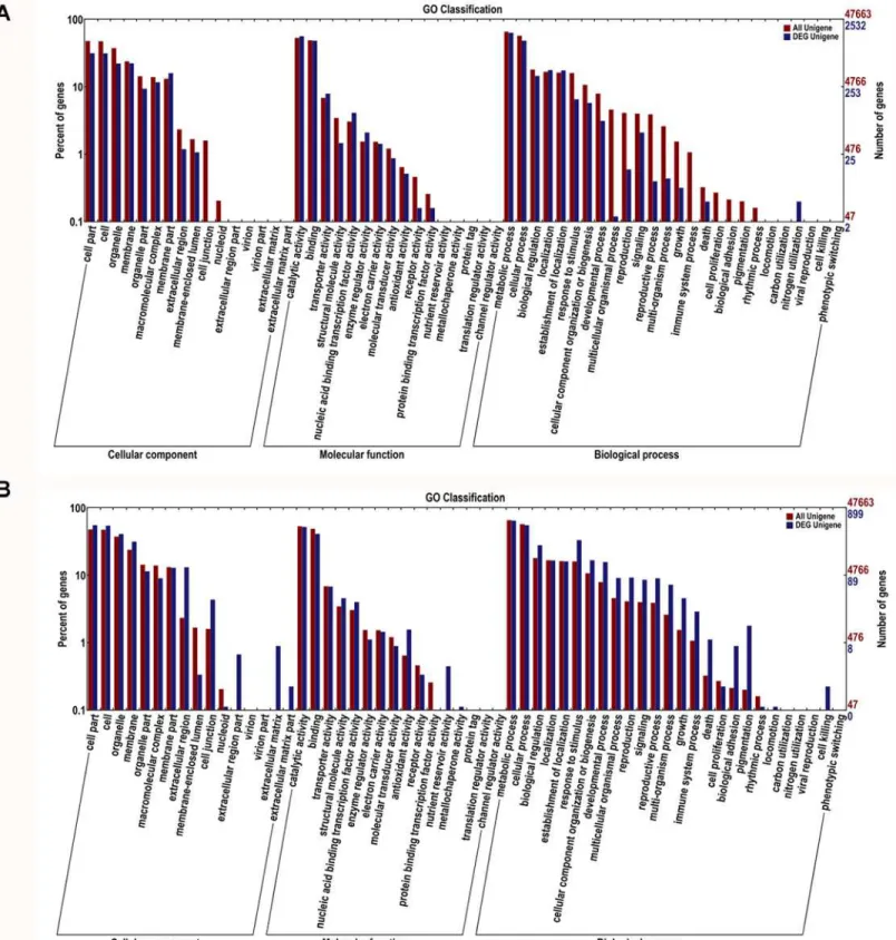
GO analysis showed that the total of 47,670 predicted proteins were categorized into 57 functional groups under three main divisions (Cellular Components, Molecular Functions and Biological Processes) (Fig 4). Among DEGs by Al in the roots, the“membrane part”in cellular component and“transporter activity”or“enzyme regulator activity”in molecular functions was overrepresented. Particularly,“nitrogen utilization”in biological processes was only repre-sented in Al stress roots (S5 TableandFig 4A). In the leaves, among the Al-induced up- and down-regulated genes,“extracellular matrix”,“extracellular region”and“membrane”in cellu-lar component were the predominant groups. In the molecucellu-lar functions category,“nutrient reservoir activity”and“antioxidant activity”were the most representative ones. With regard to biological processes, the predominant categories were“cell killing”, followed by“death”,
“immune system process”and“response to stimulus”(S6 TableandFig 4B).
To determine whether the genes involved in specific metabolic or signal transduction path-ways, the DEGs were search against the KEGG pathway database. By comparing the control without Al stress, all DEGs were assigned to 107 and 79 pathways in roots and leaves under Al stress, respectively (S7 Table). The top 20 obviously enriched pathways were shown inFig 5. These DEGs were enriched in“ABC transporters”,“signal transduction”, and“lipid metabo-lism”, including“Ether lipid metabolism”,“Glycerophospholipid metabolism”,“Sphingolipid metabolism”(Fig 5). The significant pathways in roots were“Protein processing in endoplas-mic reticulum”(gene number [N] = 72), followed by“Proteasome”(N = 37) and“ glyceropho-spholipid metabolism”(N = 26) (Fig 5AandS7Table). In Al stress leaves, more DEGs were enriched in the pathways“plant hormone signal transduction”(N = 16),“ribosome”(N = 34) and“pentose and glucuronate interconversions”(N = 9) (Fig 5BandS8Table).
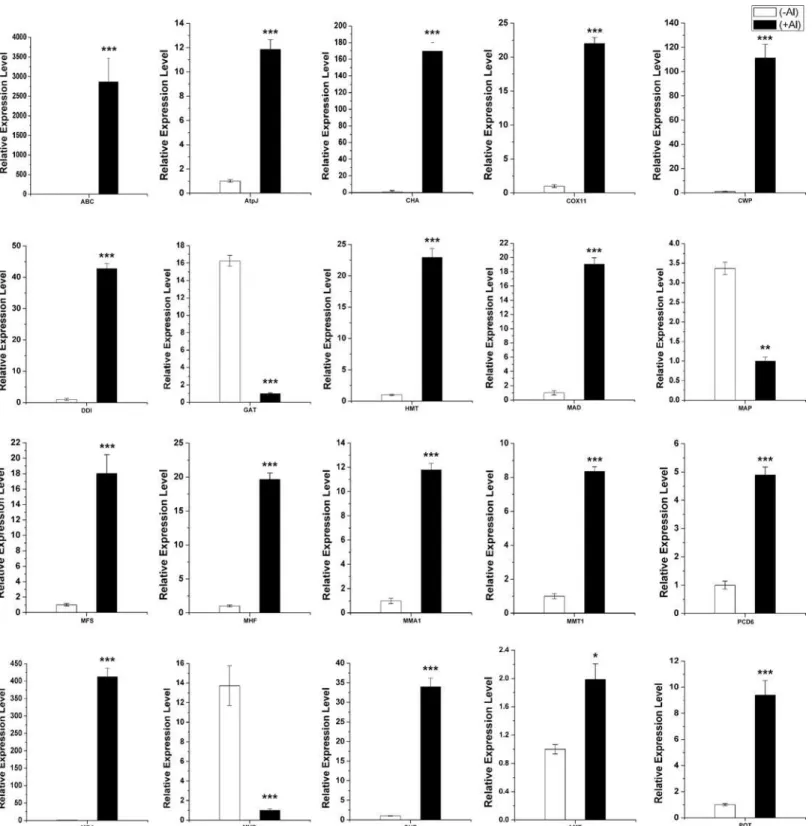
Validation of RNA-seq data by quantitative real-time RT-PCR
The expression of each gene from the RNA-seq data was calculated by RPKM. To verify the RNA-seq expression data, we selected 20 genes displaying diverse expression profiles in the roots and leaves for quantitative real-time RT-PCR analysis. A good correlation (R2= 0.89) was observed between RNA-seq data and qPCR data (Fig 6). These results confirmed the high reliability of the RNA-seq data obtained in the present study.
Fig 3. Histogram of KOG classification of differential expressed unigenes in roots (A) and leaves (B) under Al stress in hydrangea.
Fig 4. Gene Ontology classifications of differential expressed unigenes in roots (A) and leaves (B) under Al stress in hydrangea.
Expression profile of STOP/ART1-regulated genes and organic acid
metabolism
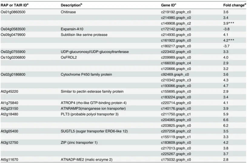
STOP1 (Sensitive to Proton Rhizotoxicity1), a C2H2 zinc finger-type transcription factor for Al response gene expression, was firstly identified in Arabidopsis [21]. And the AtSTOP1 homolog in rice (Oryza sativa) is ART1 (Al Resistance Transcription Factor1) [22]. It has been reported that STOP1/ART1 regulates the expression of three major Al tolerance genes: Alumi-num activated Malate Transporter1 (ALMT1), ALUMINUMSENSITIVE3 (AtALS3) and MATE (Multidrug and Toxic Compound Extrusion). In Arabidopsis, 43 genes are regulated by STOP1 [23,24]. Among these regulated genes, 7 homologous genes were up-regulated in hydrangea roots (Table 4). Genes homologous to ATROP4 (rho-like GTP-binding protein 4) (c220714.graph_c0), PLT3 (probable polyol transporter 3) (c211750.graph_c1, c204065. graph_c0, c203825.graph_c0) and ATNADP-ME2 (malic enzyme 2) (c175032.graph_c0) were also up-regulated by Al (Table 4). However, their roles in Al tolerance were unknown.
Previous reports showed that ART1 regulated 32 genes in rice roots [22,25]. Five homolo-gous genes in hydrangea roots were also up-regulated by Al (fold change>2) (Table 4).
Fig 5. KEGG enrichment analysis of differential expressed unigenes in roots (A) and leaves (B) under Al stress in hydrangea.
doi:10.1371/journal.pone.0144927.g005
Fig 6. Validation of the expression data from RNA-seq analysis via quantitative real-time RT-PCR (qPCR) analysis.Twenty genes exhibiting diverse expression profiles in the RNA-seq data were chosen for qPCR analysis. Average value of each RNA-seq expression data was plotted against that from qPCR and fit into a linear regression. Both x- and y-axes were shown in log2scale.
Especially, three MATE genes (c209989.graph_c0, c166030.graph_c4, c120666.graph_c0) homologous to OsFRDL2 (FERRIC REDUCTASE DEFECTIVE LIKE2), were up-regulated more than 3-fold by Al (Table 4). The MATE genes from the citrate transporter AtFRD3 have been shown to be involved in transporting citrate [26]. Furthermore, MDH (malate dehydroge-nase) (c86593.graph_c0), CS (citrate synthase) (c171733.graph_c0, c182066.graph_c1), key enzymes in the tricarboxylic acid (TCA) cycle, were up-regulated in roots (Fig 7). In the glyco-late/glyoxylate pathway, genes encoding isocitrate lyase (c171402.graph_c1) and serine hydro-xymethyltransferase (c191330.graph_c0) were up-regulated in roots. However, in the leaves, gene encoding malate dehydrogenase (c205150.graph_c2) was down-regulated. All the other genes involved in TCA cycle were not induced by Al in the present study. These results were consisted with other Al-accumulating plant, such as buckwheat [13]. In addition, we found
Table 4. Comparison of expression profiling between STOP/ART-regulated genes in rice or Arabidopsis and corresponding genes in hydrangea roots.
RAP or TAIR IDa Descriptionb Gene IDc Fold changed
Os01g0860500 Chitinase c219192.graph_c0 3.6
c214980.graph_c0 3.4
c149908.graph_c2 3.9***
Os04g0583500 Expansin-A10 c172142.graph_c0 -3.8
Os09g0479900 Subtilisin like serine protease c214930.graph_c0 4.1
c161922.graph_c0 4.2***
c180217.graph_c0 -3.7
Os02g0755900 UDP-glucuronosyl/UDP-glucosyltranferase c223402.graph_c0 3.3
Os10g0206800 OsFRDL2 c209989.graph_c0 4.0
c166030.graph_c4 2.9
c120666.graph_c0 3.2
Os02g0186800 Cytochrome P450 family protein c92469.graph_c0 3.6
c210342.graph_c3 4.3
c193066.graph_c0 4.7
At2g45220 Similar to pectin esterase family protein c155695.graph_c0 2.9
c183224.graph_c0 3.4
At1g75840 ATROP4 (rho-like GTP-binding protein 4) c220714.graph_c0 4.1
At2g23150 ATNRAMP3(manganese ion transporter) c140176.graph_c0 3.9
At2g18480 PLT3 (probable polyol transporter 3) c211750.graph_c1 5.9
c204065.graph_c0 6.6
c203825.graph_c0 6.2
At3g05400 SUGTL5 (sugar transporter ERD6-like 12) c207258.graph_c2 3.5
c155119.graph_c1 3.3
At3g12750 ZIP (zinc transporter 1) c183609.graph_c0 4.2
c217013.graph_c0 3.8
c225267.graph_c0 3.7
At5g11670 ATNADP-ME2 (malic enzyme 2) c175032.graph_c0 2.8
a, TAIR (The Arabidopsis Information Resourse) ID. b, Description based on the TAIR-DB.
c, Hydrangea assembly unigenes ID.
d, Fold change of expression ratio of +Al treatment and–Al treatment with log2 normalization.
***The expression level was validated by qPCR. The asterisks indicate significant differences between roots (+Al) and roots (-Al), as determined by Student’sttest (***P<0.001).
three genes belonging to the MATE family were induced in roots and the OsFRDL4 homolo-gous gene (c202686.graph_c0) was also up-regulated in leaves. Although Al-activated citrate secretion is not the Al-tolerance mechanism, citrate might be transported into the xylem for Al translocation [7,27].
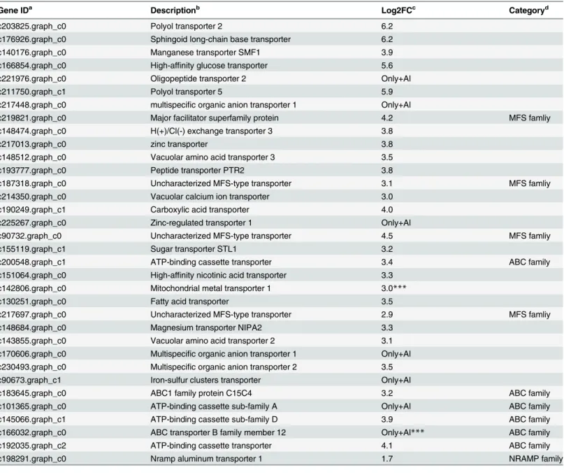
Al-up-regulated transporter genes in roots and leaves
Transporters are required for Al-induced secretion of citrate and for Al uptake, sequestration to vacuoles [28]. In this study, 57 and 37 transporter genes were Al-induced in roots and leaves, respectively (Tables5and6andS9 Table). Many of these genes in roots (6) and leaves (6) belong to the ABC (ATP-binging cassette) transporter family (Table 5). Some other transporter genes induced by Al belonged to different types of transporter families (Tables5and6andS9 Table). For example, three genes similar to MFS (Major Facilitator superfamily protein) family have been reported to be involved in vacuolar sequestration of Al [13]. Genes showing high similarity to nitatre transporter, magnesium transporter, potassium transporter and sulfate transporter were also up-regulated by Al (Tables5and6). In addition, vacuolar amino acid transporter genes, multispecific organic anion and peptide transporter genes were also up-reg-ulated (Tables5and6). Similar results were reported in buckwheat, implied that they might play important roles in Al tolerance. Although these transporters have not been reported to be associated with Al tolerance, investigation of them in the next step will help for further under-standing the mechanism of Al tolerance in hydrangea.
In rice, uptake of Al3+is mediated by NRAT1 (NRAMP ALUMINUM TRANSPORTER1), a member of the NRAMP family. In the study, we also found a homolog gene of NRAT1 in our RNA-seq data of the roots and leaves, indicating its role in Al uptake (Table 5). Some members of MATE family can transport citrate to participate in Al tolerance. A gene similar to citrate efflux MATE transporter was also found to be up-regulated in leaves in this study (Table 6). Two up-regulated transporter genes mitochondrial metal transporter 1 (c142806.graph_c0), ABC transporter (c166032.graph_c0) in roots, Aluminum-activated malate transporter (c208330.graph_c1), and potassium transporter 1 (c86493.graph_c0) in leaves were further verified by qPCR analysis (Fig 7).
Genes related to stress/defence, energy and cell wall organization in
roots
A number of genes related to stress and energy metabolism were identified to be affected by Al (S10 Table). For example, stress response protein nst1 (c141215.graph_c1, c87075.graph_c0 and c173987.graph_c1), ATP synthase subunit 5 (c195438.graph_c0), AtpJ (c176296. graph_c0) and Atpα(c172514.graph_c0) were up-regulated in roots. Many genes related to
oxidative stress, such as Cu/Zn superoxide dismutase (c145636.graph_c0, c199996.graph_c1) and L-ascorbate oxidase (c179819.graph_c0) were induced by Al. In the study, genes involved in cell wall organization were identified, in which three were cell wall integrity and stress response genes (c166600.graph_c0, c209786.graph_c0, c92982.graph_c0). Many cell wall pro-teins and vegetative cell wall propro-teins were also up-regulated in roots under Al stress (S10 Table). Some of these genes including cell wall protein DAN4 (c193058.graph_c1), cox11
proteinase Spm1. Two genes in leaves: AMT (c208330. graph_ c1), Aluminum-activated malate transporter; POT (c86493.graph_c0), Potassium transporter 1. The asterisks indicate significant differences between root (+Al) and root (-Al), or leaves (+Al) and leaves (-Al), as determined by Student’sttest (*P<0.05, **P<0.01,***P<0.001).
(c220486.graph_c0) and AtpJ (c176296.graph_c0) were selected for qPCR analysis and con-firmed their high expression level in roots (+Al) (Fig 7).
Genes related to other biological processes, including signal transduction, transcription, DNA damage and metal homeostasis, were induced by Al in hydrangea roots. For instance, many mitogen-activated protein kinases and Zinc finger transcription factors were
up-Table 5. Al-induced transporter genes in hydrangea roots.
Gene IDa Descriptionb Log2FCc Categoryd
c203825.graph_c0 Polyol transporter 2 6.2
c176926.graph_c0 Sphingoid long-chain base transporter 6.2
c140176.graph_c0 Manganese transporter SMF1 3.9
c166854.graph_c0 High-affinity glucose transporter 5.6
c221976.graph_c0 Oligopeptide transporter 2 Only+Al
c211750.graph_c1 Polyol transporter 5 5.9
c217448.graph_c0 multispecific organic anion transporter 1 Only+Al
c219821.graph_c0 Major facilitator superfamily protein 4.2 MFS famliy
c148474.graph_c0 H(+)/Cl(-) exchange transporter 3 3.8
c217013.graph_c0 zinc transporter 3.8
c148512.graph_c0 Vacuolar amino acid transporter 3 3.5
c193777.graph_c0 Peptide transporter PTR2 3.8
c187318.graph_c0 Uncharacterized MFS-type transporter 3.1 MFS famliy
c214350.graph_c0 Vacuolar calcium ion transporter 3.0
c190249.graph_c1 Carboxylic acid transporter 4.0
c225267.graph_c0 Zinc-regulated transporter 1 Only+Al
c90732.graph_c0 Uncharacterized MFS-type transporter 4.5 MFS famliy
c155119.graph_c1 Sugar transporter STL1 3.2
c200548.graph_c1 ATP-binding cassette transporter 3.4 ABC family
c151064.graph_c0 High-affinity nicotinic acid transporter 3.3
c142806.graph_c0 Mitochondrial metal transporter 1 3.0***
c130251.graph_c0 Fatty acid transporter 3.5
c217697.graph_c0 Uncharacterized MFS-type transporter 2.9 MFS famliy
c148684.graph_c0 Magnesium transporter NIPA2 3.3
c143855.graph_c0 Vacuolar amino acid transporter 2 3.1
c170606.graph_c0 Multispecific organic anion transporter 1 Only+Al c230493.graph_c0 Multispecific organic anion transporter 2 3.5
c90673.graph_c1 Iron-sulfur clusters transporter Only+Al
c183645.graph_c0 ABC1 family protein C15C4 3.2 ABC family
c101365.graph_c0 ATP-binding cassette sub-family A Only+Al ABC family
c145066.graph_c1 ATP-binding cassette sub-family D 3.9 ABC family
c166032.graph_c0 ABC transporter B family member 12 Only+Al*** ABC family
c192035.graph_c2 ATP-binding cassette transporter 4.1 ABC family
c198291.graph_c0 Nramp aluminum transporter 1 1.7 NRAMP family
a, Hydrangea assembly unigenes ID.
b, Description based on the BLASTx with NR and Swiss-Prot database.
c, Fold change of expression ratio of +Al treatment and–Al treatment with log2 normalization. d, Transporter category.
***The expression level was validated by qPCR. The asterisks indicate significant differences between roots (+Al) and roots (-Al), as determined by Student’sttest (***P<0.001).
Table 6. Al-induced transporter genes in Hydrangea leaves.
Gene IDa Descriptionb Log2FCc Categoryd
c86493.graph_c0 Potassium transporter 1 2.9***
c211481.graph_c0 ABC transporter 2.2 ABC family
c205928.graph_c0 Potassium transporter 2 1.2
c208330.graph_c1 Aluminum-activated malate transporter 1.1
c199526.graph_c0 Plastidic glucose transporter 2 1.4
c207435.graph_c0 Sulfate transporter 3.2
c181864.graph_c2 ABC transporter 1.6 ABC family
c186746.graph_c0 Sulfate transporter 1.8
c205623.graph_c1 Nitrate transporter 1.0
c198291.graph_c0 Nramp aluminum transporter 1 0.8 NRAMP family
c190578.graph_c0 Peptide transporter 1.2
c156211.graph_c0 Equilibrative nucleotide transporter 5.3
c204623.graph_c0 Sugar transporter 1.2
c208984.graph_c0 MATE efflux protein 0.3 MATE family
c207435.graph_c1 Low affinity sulfate transporter 3 2.8
c122554.graph_c0 Vacuolar iron transporter 2.4
c202686.graph_c0 Citrate efflux MATE transporter 1.3 MATE family
c191802.graph_c0 Auxin transporter-like protein 1.3
c179282.graph_c0 ABC transporter 1.8 ABC family
c204800.graph_c2 Amino acid transporter 6 1.3
c187439.graph_c0 ABC transporter 2.0 ABC family
c205514.graph_c0 Nitrate excretion transporter 1.3
c176091.graph_c0 Phosphate transporter 4.3
a, Hydrangea assembly unigenes ID.
b, Description based on the BLASTx with NR and Swiss-Prot database.
c, Fold change of expression ratio of +Al treatment and–Al treatment with log2 normalization. d, Transporter category.
***The expression level was validated by qPCR. The asterisks indicate significant differences between leaves (+Al) and leaves (-Al), as determined by Student’sttest (***P<0.001).
doi:10.1371/journal.pone.0144927.t006
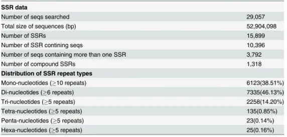
Table 7. Classification of SSR repeats in the hydrangea transcriptome assembly.
SSR data
Number of seqs searched 29,057
Total size of sequences (bp) 52,904,098
Number of SSRs 15,899
Number of SSR contining seqs 10,396
Number of seqs containing more than one SSR 3,792
Number of compound SSRs 1,318
Distribution of SSR repeat types
Mono-nucleotides (10 repeats) 6123(38.51%)
Di-nucleotides (6 repeats) 7335(46.13%)
Tri-nucleotides (5 repeats) 2258(14.20%)
Tetra-nucleotides (5 repeats) 135(0.85%)
Penta-nucleotides (5 repeats) 23(0.14%)
Hexa-nucleotides (5 repeats) 25(0.16%)
regulated in roots (S10 Table). Moreover, DNA damage-inducible protein (c201184.graph_c0), metal homeostasis factor (c185758.graph_c0), metallothionein (c129315.graph_c0) and heavy metal tolerance protein (c171214.graph_c0) were also showed high expression in Al stress roots (S8 Table). Further qRT-PCR analysis verified these results (Fig 7).
SSR and SNP mining
The assembled transcripts provide a rich source for potential genic-SSR markers, which are potentially linked with functional genes. Using MISA Perl script, a total of 15,899 SSRs with 1–6 repeat motifs and 1,318 compound SSR motifs were identified in 29,057 sequences (Table 7andS11 Table). The largest proportion of SSRs consisted of di-nucleotide repeats (46.13%), followed by mono-nucleotide repeats (38.51%) (Table 7). The most abundant di-nucleotide and tri-di-nucleotide motifs were AG/CT (69.29% in di-SSRs) and AAG/CTT (26.62% in tri-SSRs), respectively (S11 Table).
Genic-SNPs are useful for linking to candidate genes or QTLs. In this study, 27,816 SNPs were identified in the four samples. The most abundance of base substitution was A/G (2931, 10.54%), followed by A/T (2826, 10.16%) (Fig 8). Although a stringent criteria were used to identify the SNPs (seeMethods), the frequencies of SNPs in unigenes were detected highly (S12 Table).
Discussion
Using high-throughput RNA sequencing in non-model plant hydrangea, a large amount of sequence data was generated. Afterde novoassembly of the sequences, nearly 130,237 tran-scripts were constructed in hydrangea (Table 1). Since the EST data of hydrangea available in NCBI is few, these results provided a large number of resources for gene discovery, quantifica-tion of transcripts in Al stress and the identificaquantifica-tion of putative SNPs and SSRs.
The differential expression analysis of RNA-seq data in control and Al stress revealed that more genes up-regulated in roots than leaves (S7 TableandS8Table), which suggested that root might be more sensitive to Al stress at the primary time. And further investigation needed to validate the results. GO enrichment analysis showed that the DEGs in roots or leaves were significantly overrepresented in“Response to stimulus”,“Antioxidant activity”,
Fig 8. SNP distribution of different variation types in hydrangea.
“membrane”,”Extracellular region”and“death”. The results suggested that defensive genes and genes encoding extracellular-localized proteins, such as cell wall component, were preferen-tially induced in expression by Al stress. For KEGG pathway analysis,“Lipid metabolism”was significantly enriched in root and leaves. The enrichment of genes in the lipid metabolism pathway supported the observation that Al can interfere with the function of the plasma mem-brane. By contrast, genes in“ABC transporters”,“Proteasome”and“Protein processing in endoplasmic reticulum”pathways were only significantly enriched in roots, while genes in
“Plant hormone signal transduction”were overrepresented in leaves but not in roots under Al stress. The DEGs were also subjected to KOG classification, showing different categories in roots and leaves except for“Carbohydrate transport and metabolism”. Therefore, the roots and leaves may possess different mechanism of Al responsiveness in hydrangea.
Recently, STOP1 or ART1 gene homologs from Eucalyptus, wheat (Triticum aestivum), tea (Camellia sinensis),Lotus japonicas, black poplar (Populus nigra) and moss (Physcomitrella petens) have been isolated [24,29–30]. In this study, the STOP1 or ART1 homologous genes had not been found in hydrangea. Moreover, the majority of AtSTOP1/OsART1-regulated genes homologs were unaffected in hydrangea under Al stress. The results suggested that hydrangea may have distinct Al tolerance mechanisms. Previous reports suggested that a tono-plast and a plasma membrane localized aquaporin (HmVALT1 and HmPALT1, respectively) were involved in Al transport inHydrangea macrophylla[11]. However, in our study, their expression was not up-regulated by Al in roots or leaves. That may be because of different tis-sues (sepals) used in the former study [11].
Organic acids such as oxalate and citrate have been well documented to involve in Al toler-ance and accumulation in some plants [7,13]. Oxalate and citrate play important roles in Al tolerance and oxalate is able to form a complex with Al, detoxifying Al externally and internally [13,28]. Glycolate/glyoxylate is the efficient precursor for oxalate biosynthesis and is produced during photorespiration or glyoxylate cycle reactions [31]. Except for CS and MDH, other genes involved in the TCA cycle were not changed by Al. These results suggested that citrate biosynthesis was not critical for Al tolerance. Previous studies also documented that it is the transporters rather than biosynthesis of organic acids which are more important for Al toler-ance [28].
Al tolerance involves internal mechanisms that allow plants to detoxify Al3+which enters root cells by forming nontoxic organic acid (OA)-Al complexes in the cytosol and/or by sequestering the Al into vacuoles [8]. To facilitate Al hyperaccumulation into the vacuoles, the plasma membrane-localized Al transporter should be present simultaneously. In rice, Nrat1, a member of Nramp family, has been reported to be an Al transporter localized at the plasma membrane [32]. And coordinated plasma membrane/tonoplast Al transport systems are the major contributor to rice’s superior level of Al tolerance compared with other cereal crops [33]. In this study, more than 50 membrane proteins have been up-regulated in the roots. Further-more, an Nramp aluminum transporter 1 (c198291.graph_c0) was found to be up-regulated in both roots and leaves under Al stress (Tables5and6). In rice, the Nramp aluminum trans-porter (NRAT1) was involved in Al tolerance by reducing the level of toxic Al in the root cell wall and transporting Al into the root cell, sequestering in the vacuole. The results in our study suggested that the internal Al tolerance mechanism may be also involved in the Al detoxifica-tion of hydrangea.
mediated by a member of MATE family and was involved in Al tolerance [23,25,36]. In this study, we also found that many ABC family transporters and the MATE transporters were up-regulated in roots and leaves under Al stress (Tables5and6).
It is notable that a number of genes relative to energy metabolism, cell wall organization, membrane, DNA damage and metal homeostasis were up-regulated in roots induced by Al in this study. Transcriptomic analysis ofA.halleriandN.caerulescenswith non-accumulator spe-cies revealed that a gene set encoding metal-transporters and metal-chelator biosynthestic pro-teins were highly and constitutively expressed in the hyperaccumulators. In the
hyperaccumulator plants, many proteins work simultaneously, such as P-type ATPase (Heavy Metal ATPase 4, HMA4) functions in xylem loading/ unloading and metal tolerance protein 1 (MTP1) functions in vacuolar storage [37]. Four metal tolerance proteins were identified to be up-regulated in roots under Al stress (S9 Table).
Plasma membrane ATPase is the most abundant proteins on the plasma membrane and is reported to be involved in citrate exudation under Al3+toxicity conditions [38]. The process might be achieved through a citrate-proton co-transport system [39]. And most compounds in vacuoles are imported or exported using the electrochemical gradient generated by V-type ATPase [40]. The interesting thing is that many V-type protons ATPase were up-regulated in the roots under Al stress in the study (S10 Table). The results suggested that citrate exudation might be important for Al tolerance in hydrangea.
The cell wall is a major target for Al accumulation and Al3+toxicity in higher plants. The root tip is the primary site of Al3+toxicity, and most of the root Al resides in the cell wall [5,
41]. Al accumulation can increase the rigidity of the cell wall by cross-linking pectin residues which inhibits cell wall loosening needed for root growth [41]. In addition, another protein, expansin which loosen the cell wall during the process of cellular expansion and growth, is very sensitive to Al [42]. In this study, the majority of cell wall proteins were up-regulated and an expansin gene (c172142.graph_c0) was down-regulated in roots under Al stress (S4 Tableand
S10 Table). The results indicated that increasing the rigidity of cell wall played important roles for Al tolerance in hydrangea. Further functional analysis will reveal their roles of these homol-ogous genes in Al detoxification and accumulation in hydrangea.
Conclusion
Through RNA-seq data analysis, we constructed about 400,000 high-quality transcripts, which provided information for further investigation of the molecular mechanism of Al accumulation and tolerance in hydrangea and for SNP and SSR markers identifying. The markers will be a rich resource for constructing genetic linkage maps and breeding research in the future. In this study, the results revealed that the roots and leaves may have common and distinct mecha-nisms of Al responsiveness. Citrate synthase, a key enzyme in the tricarboxylic acid (TCA) cycle, was up-regulated in hydrangea roots and many transporters, including MATE and ABC families, were involved in the process, suggesting that citrate secretion may have its role in the Al tolerance in hydrangea. A plasma membrane Al uptake transporter, Nramp aluminum transporter was up-regulated in roots and leaves under Al stress, and may play an important role in hydrangea Al tolerance by reducing the level of toxic Al. Although the exact roles of these candidate genes remain to be examined, the results in the study provide a platform for further functional analysis of these genes.
Supporting Information
S1 Table. Primers used in the study for quantitative RT-PCR.
S2 Table. All unigenes with expression levels (RPKM) in in roots and leaves under Al stress in hydrangea.
(XLSX)
S3 Table. Annotations of all unigenes that assembled in hydrangea in the study.
(XLSX)
S4 Table. Up-regulated and Down-regulated genes in roots and leaves under Al stress in hydrangea.Summary of the up-regulated and down-regulated genes in roots and leaves under Al stress.
(XLSX)
S5 Table. GO analysis and enrichment of roots under Al stress in hydrangea.
(XLSX)
S6 Table. GO analysis and enrichment of leaves under Al stress in hydrangea.
(XLSX)
S7 Table. KEGG analysis and enrichment of roots under Al stress in hydrangea.
(XLSX)
S8 Table. KEGG analysis and enrichment of leaves under Al stress in hydrangea.
(XLSX)
S9 Table. The differential expression transporters in roots and leaves in hydrangea.
(XLSX)
S10 Table. Functional classification of the up-regulated genes in roots in hydrangea.
(XLSX)
S11 Table. Characterization and distribution of SSRs identified in hydrangea.
(XLSX)
S12 Table. All the SNPs detected in the study.
(XLSX)
Acknowledgments
The authors thank the scientific research fund of Hunan co-innovation center for utilization of botanicals functional ingredients. This work was supported by grants from the National Natu-ral Science Foundation of China (31201656) and Scientific Research Fund of Hunan Provincial Education Department (14B093).
Author Contributions
Conceived and designed the experiments: HXC CPL. Performed the experiments: HXC CPL HJ. Analyzed the data: HXC HJ JHP. Wrote the paper: HXC.
References
1. von Uexküll HR, Mutert E. Global extent, development and economic-impact of acid soils. Plant Soil. 1995; 171(1):1–15.
2. Kochan LV. Cellular mechanisms of aluminum toxicity and resistance in plants. Annu Rev Plant Phys. 1995; 46: 237–260.
4. Gould B, McCouch S, Geber M.De Novotranscriptome assembly and identification of gene candidates for rapid evolution of soil Al tolerance inAnthoxanthum odoratumat the long-term park grass experi-ment. PloS ONE. 2015; 10(7): e0124424. doi:10.1371/journal.pone.0124424PMID:26148203
5. Chang YC, Yamamoto Y, Matsumoto H. Accumulation of aluminium in the cell wall pectin in cultured tobacco (Nicotiana tabacumL.) cells treated with a combination of aluminium and iron. Plant Cell Envi-ron. 1999; 22(8):1009–1017.
6. Ryan P, Delhaize E, Jones D. Function and mechanism of organic anion exudation from plant roots. Annu Rev Plant Physiol. 2001; 52:527–60.
7. Zhu HF, Wang H, Zhu YF, Zou JW, Zhao FJ, Huang CF. Genome-wide transcriptomic and phylogenetic analyses reveal distinct aluminum-tolerance mechanisms in the aluminum- accumulating species buck-wheat (Fagopyrum tataricum). BMC Plant Biol. 2015; 15:16. doi:10.1186/s12870-014-0395-zPMID: 25603892
8. Ma JF, Hiradate S, Nomoto K, Iwashita T, Matsumoto H. Internal detoxification mechanism of Al in hydrangea. Plant Physiol. 1997; 113:033–1039.
9. Hotta H, Wang Q, Fukuda M, Aizawa S, Umemura T, Sekizawa K, et al. Identification of Aluminum spe-cies in an Aluminum-accumulating plant, Hydrangea (Hydrangea macrophylla), by Electrospray Ioniza-tion Mass Spectrometry. Anal Sci. 2008; 24:795–798. PMID:18544872
10. Schreiber HD, Jones AH, Lariviere CM, Mayhew KM, Cain JB. Role of aluminum in red-to-blue color changes in Hydrangea macrophylla sepals. Biometals. 2011; 24:1005–1015. doi: 10.1007/s10534-011-9458-xPMID:21584711
11. Negishi T, Oshima K, Hattori M, Kanai M, Mano S, Nishimura M, et al. Tonoplast- and plasma mem-brane-localized aquaporin-Family transporters in blue hydrangea sepals of Aluminum hyperaccumulat-ing plant. PloS ONE. 2012; 7(8):e43189. doi:10.1371/journal.pone.0043189PMID:22952644
12. Fan W, Lou HQ, Gong YL, Liu MY, Wang ZQ, Yang JL, et al. Identification of early Al-resposive genes in rice bean (Vigna umbellata) roots provides new clues to molecular mechanisms to Al toxicity and tol-erance. Plant Cell Environ. 2014; 37: 1586–1597. doi:10.1111/pce.12258PMID:24372448
13. Yokosho K, Yamji N, Ma JF. Global transcriptome analysis of Al-induced genes in an Al-accumulating species, common buckwheat (Fagopyrum esculentumMoench). Plant Cell Physiol. 2014; 55 (12):2077–2091. doi:10.1093/pcp/pcu135PMID:25273892
14. Cloonan N, Forrest AR, Kolle G, Gardiner BB, Faulkner GJ, Brown MK, et al. Stem cell transcriptome profiling via massive-scale mRNA sequencing. Nat Methods. 2008; 5: 613–619. doi:10.1038/nmeth. 1223PMID:18516046
15. Peng Y, Gao X, Li R, Cao G. Transcriptome sequencing and de novo analysis ofYoungia japonica
using the Illumina platform. PloS ONE. 2014; 9: e90636. doi:10.1371/journal.pone.0090636PMID: 24595283
16. Cardoso-Silva CB, Costa EA, Mancini MC, Balsalobre TW, Canesin LE, Pinto LR, et al. De novo assembly and transcriptome analysis of contrasting sugarcane varieties. PloS ONE. 2014; 9:e88462. doi:10.1371/journal.pone.0088462PMID:24523899
17. Grabherr MG, Haas BG, Yassour M, Levin JZ, Thompson DA, Amit I, et al. Full-length transcriptome assembly from RNA-Seq data without a reference genome. Nat Biotechnol. 2011; 29: 644–652. doi: 10.1038/nbt.1883PMID:21572440
18. Li B, Dewey CN. RSEM: accurate transcript quantification from RNA-Seq data with or without a refer-ence genome. BMC Bioinformatics. 2011; 12: 33. doi:10.1186/1471-2105-12-323PMID:21816040
19. Langmead B, Salzberg SL. Fast gapped-read alignment with Bowtie 2. Nat Methods. 2012; 9: 357– 359. doi:10.1038/nmeth.1923PMID:22388286
20. Li R, Li Y, Fang X, Yang H, Wang J, Kristiansen K, et al. SNP detection for massively parallel whole-genome resequencing. Genome Res. 2009; 19:1124–1132. doi:10.1101/gr.088013.108PMID: 19420381
21. Iuchi S, Koyama H, Iuchi A, Kobayashi Y, Kitabayashi S, Kobayashi Y, et al. Zinc finger protein STOP1 is critical for proton tolerance in Arabidopsis and coregulates a key gene in aluminum tolerance. Proc Natl Acad Sci USA. 2007; 104: 9900–9905. PMID:17535918
22. Yamaji N, Huang CF, Nagao S, Yano M, Sato Y, Nagamura Y, et al. A zinc finger transcription factor ART1 regulates multiple genes implicated in aluminum tolerance in rice. Plant Cell. 2009; 21: 3339– 3349. doi:10.1105/tpc.109.070771PMID:19880795
24. Sawaki Y, Iuchi S, Kobayashi Y, Kobayashi Y, Ikka T, Sakurai N, et al. STOP1 regulates multiple genes that protect Arabidopsis from proton and aluminum toxicities. Plant Physiol. 2009; 150: 281–294. doi: 10.1104/pp.108.134700PMID:19321711
25. Yokosho K, Yamaji N, Ma JF. An Al-inducible MATE gene is involved in external detoxification of Al in rice. Plant J. 2011; 69: 1061–1069.
26. Rogers EE, Guerinot ML. FRD3, a member of the multidrug and toxin efflux family, controls iron defi-ciency responses in Arabidopsis. Plant Cell. 2002; 14(8):1787–99. PMID:12172022
27. Ma JF. Role of organic acids in detoxification of aluminum in higher plants. Plant Cell Physiol. 2000; 41: 383–390. PMID:10845450
28. Ma JF, Ryan PR, Delhaize E. Aluminium tolerance in plants and the complexing role of organic acids. Trends Plant Sci. 2001; 6: 273–278. PMID:11378470
29. Garcia-Oliveira AL, Benito C, Prieto P, Menezes RA, Rodrigues-Pousada C, Guedes-Pinto H, et al. Molecular characterization of TaSTOP1 homoeologues and their response to aluminium and proton (H +) toxicity in bread wheat (Triticum aestivumL.). BMC Plant Biol. 2013; 13: 134. doi: 10.1186/1471-2229-13-134PMID:24034075
30. Ohyama Y, Ito H, Kobayashi Y, Ikka T, Morita A, Kobayashi M, et al. Characterization of AtSTOP1 orthologous genes in tobacco and other plant species. Plant Physiol. 2013; 162: 1937–1946. doi:10. 1104/pp.113.218958PMID:23749850
31. Yu L, Jiang J, Zhang C, Jiang L, Ye N, Lu Y. et al. Glyoxylate rather than ascorbate is an efficient pre-cursor for oxalate biosynthesis in rice. J Exp Bot. 2010; 61: 1625–1634. doi:10.1093/jxb/erq028PMID: 20194922
32. Xia J, Yamaji N, Kasai T, Ma JF. Plasma membrane-localized transporter for aluminum in rice. Proc Natl Acad Sci USA. 2010; 107: 18381–18385. doi:10.1073/pnas.1004949107PMID:20937890
33. Li JY, Liu JP, Dong DK, Jia XM, McCouch SR, Kochian LV. Natural variation underlies alterations in Nramp aluminum transporter (NRAT1) expression and function that play a key role in rice aluminum tol-erance. Proc Natl Acad Sci USA. 2014; 111(17): 6503–6508. doi:10.1073/pnas.1318975111PMID: 24728832
34. Frelet-Barrand A, Kolukisaoglu HU, Plaza S, Ruffer M, Azevedo L, Hörtensteiner S, et al. Comparative mutant analysis of Arabidopsis ABCC-type ABC transporters: AtMRP2 contributes to detoxification, vacuolar organic anion transport and chlorophyll degradation. Plant Cell Physiol. 2008; 49: 557–69. doi:10.1093/pcp/pcn034PMID:18325934
35. Huang CF, Yamaji N, Mitani N, Yano M, Nagamura Y, Ma JF. A bacterial-type ABC transporter is involved in aluminum tolerance in rice. Plant Cell. 2009; 21: 655–667. doi:10.1105/tpc.108.064543 PMID:19244140
36. Yang XY, Yang JL, Zhou Y, Piueros MA, Kochian LV, Li GX, et al. A de novo synthesis citrate transpo-ter, Vigna umbellate multidrug and toxic compound extrusion, implicates in Al-activeted citrate efflux in rice bean (Vigna umbellate) root apex. Plant Cell Environ. 2011; 34:2138–2148. doi: 10.1111/j.1365-3040.2011.02410.xPMID:21848845
37. Hanikenne M, Nouet C. Metal hyperaccumulation and hypertolerance: a model for plant evolutionary genomics. Curr Opin Plant Biol. 2011; 14:252–259. doi:10.1016/j.pbi.2011.04.003PMID:21531166
38. Janicka-Russak M, Kabala K, Burzynski M, Klobus G. Response of plasma membrane H+-ATPase to
heavy metal stress inCucumis sativusroots. J Exp Bot. 2008; 59: 3721–3728. doi:10.1093/jxb/ern219 PMID:18820260
39. Ohno T, Koyama H, Hara T. Characterization of citrate transport through the plasma membrane in a carrot mutant cell line with enhanced citrate excretion, Plant Cell Physiol. 2003; 44:156–162. PMID: 12610218
40. Schumacher K, Krebs M. The V-ATPase: small cargo, large effects. Curr Opin Plant Biol. 2010; 13:724–30. doi:10.1016/j.pbi.2010.07.003PMID:20801076
41. Tabuchi A, Matsumoto H. Changes in cell-wall properties of wheat (Triticum aestivum) roots during alu-minum-induced growth inhibition. Physiol Plant. 2001; 112(3):353–358. PMID:11473692