OLÍVIO BRITO MALHEIRO
INDICADORES DE RESERVA OVARIANA EM MULHERES
COM LÚPUS ERITEMATOSO SISTÊMICO
INDICADORES DE RESERVA OVARIANA EM MULHERES COM LÚPUS ERITEMATOSO SISTÊMICO
Dissertação apresentada ao Programa de Pós-Graduação em Saúde da Mulher, da Faculdade de Medicina da Universidade Federal de Minas Gerais como requisito parcial à obtenção do título de Mestre em Medicina: Saúde da Mulher Área de concentração: Reprodução Humana e Patologia Ginecológica e Mamária
Orientador: Prof. Dr. Fernando Marcos dos Reis Co-orientadora: Profa. Dra. Gilda Aparecida Ferreira
Ficha catalográfica elaborada pela Biblioteca J. Baeta Vianna – Campus Saúde UFMG Malheiro, Olívio Brito.
M249i Indicadores de reserva ovariana em mulheres com Lúpus Eritematoso Sistêmico [manuscrito]. / Olívio Brito Malheiros. - - Belo Horizonte: 2011. 79f.: il.
Orientador: Fernando Marcos dos Reis. Co-Orientador: Gilda Aparecida Ferreira.
Área de concentração: Reprodução Humana e Patologia Ginecológica e Mamária.
Dissertação (mestrado): Universidade Federal de Minas Gerais, Faculdade de Medicina.
1. Lúpus Eritematoso Sistêmico. 2. Hormônio Antimülleriano. 3.
Hormônio Foliculoestimulante. 4. Dissertações Acadêmicas. I. Reis, Fernando Marcos dos. II. Ferreira, Gilda Aparecisa. III. Universidade Federal de Minas Gerais, Faculdade de Medicina. IV. Título
Professor Clélio Campolina Diniz
VICE-REITORA
Professora Rocksane de Carvalho Norton
PRÓ-REITOR DE PÓS-GRADUAÇÃO
Professor Ricardo Santiago Gomez
PRÓ-REITOR DE PESQUISA
Professor Renato de Lima dos Santos
DIRETOR DA FACULDADE DE MEDICINA
Professor Francisco José Penna
VICE-DIRETOR DA FACULDADE DE MEDICINA
Professor Tarcizo Afonso Nunes
COORDENADOR DO CENTRO DE PÓS-GRADUAÇÃO
Professor Manoel Otávio da Costa Rocha
SUBCOORDENADORA DO CENTRO DE PÓS-GRADUAÇÃO
Professora Teresa Cristina de Abreu Ferrari
CHEFE DO DEPARTAMENTO DE GINECOLOGIA E OBSTETRÍCIA
Professor Cezar Alencar de Lima Rezende
COORDENADOR DO PROGRAMA DE PÓS-GRADUAÇÃO EM SAÚDE DA MULHER
Professor Antônio Carlos Vieira Cabral
SUBCOORDENADOR DO PROGRAMA DE PÓS-GRADUAÇÃO EM SAÚDE DA MULHER
Professora Alamanda Kfoury Pereira
COLEGIADO DO PROGRAMA DE PÓS-GRADUAÇÃO EM SAÚDE DA MULHER: PATOLOGIA GINECOLÓGICA
Professor Antônio Carlos Vieira Cabral Professora Alamanda Kfoury Perreira
Professor Selmo Geber Professor Victor Hugo de Melo
Nomear pessoas neste momento é muito difícil, mas não posso deixar de mencionar e tornar conhecido de todos a minha gratidão e o reconhecimento àqueles que tornaram este trabalho possível.
A toda minha família, pelo suporte necessário nos momentos de dificuldade. Às pacientes do serviço de Reumatologia do Ambulatório Bias Fortes, que na intimidade de sua doença, me permitiram realizar a coleta de sangue para análise e ultrassonografias transvaginais.
Ao professor Fernando Marcos dos Reis, por ter me aceitado como orientando e pelo brilhantismo como orientador e formador de grupos de pesquisa.
À professora Gilda, por sua paciência na elucidação de dúvidas e confiança na viabilidade deste projeto.
A toda equipe do laboratório Prof. Aroldo Camargos de Reprodução Humana do Hospital das Clínicas da UFMG, pelos ensinamentos e ajuda no projeto. Sinto muita satisfação em conviver com um grupo tão qualificado e solícito.
À Dra Carolina Passos Resende, pela imensa disponibilidade em realizar os exames de ultrassonografia e esclarecer minhas dúvidas no cotidiano do serviço de reprodução humana da UFMG.
A toda equipe de Reumatologia do Ambulatório Bias Fortes, principalmente os professores Marco Antônio Parreira de Carvalho e Cristina Costa Duarte Lanna por permitirem que este estudo fosse realizado no Serviço de Reumatologia do Hospital das Clínicas da UFMG.
Aos meus colegas dos Hospitais Alberto Cavalcanti, Felício Rocho e Life Center, que dividiram comigo as responsabilidades cotidianas, proporcionando assim tempo para que pudesse cursar as disciplinas do curso e me dedicar ao projeto.
À Neusa e Vânia, secretárias do 2° andar do ambulatório Bias Fortes pelo carinho e atenção, assim como a Juliana no laboratório de Reprodução Humana.
"Eu ouço e esqueço. Eu vejo e lembro. Eu faço e entendo".
Introdução: O Lúpus Eritematoso Sistêmico (LES) é uma doença multissistêmica, que compromete principalmente mulheres em idade reprodutiva, e pode promover disfunção ovariana precoce relacionada a fatores relacionados à doença ou ao seu próprio tratamento. A avaliação de indicadores de reserva ovariana pode determinar se há diferenças entre estas pacientes e a população geral, previamente ao estabelecimento do climatério. Objetivos: O objetivo deste estudo foi avaliar se há diferenças nos marcadores tradicionais de reserva ovariana em pacientes com LES comparados a um grupo controle, e avaliar sua relação com características clínicas e terapêuticas de pacientes com LES. Pacientes e métodos: Este foi um estudo transversal controlado, incluindo 27 mulheres com LES e 27 controles. Todas as participantes tinham entre 18 e 40 anos, eram eumenorréicas e não haviam usado terapia hormonal ou contraceptivos hormonais nos últimos 6 meses. Os dados clínicos foram avaliados no acompanhamento regular da doença, e as pacientes foram selecionadas para coletar amostras de sangue para dosagem de hormônio folículo estimulante (FSH) e hormônio anti-mulleriano (AMH), e submetidas a ultrassonografia transvaginal para contagem de folículos antrais (AFC). Todos os procedimentos foram feitos na fase folicular inicial do ciclo menstrual. Resultados: A média de idade e do tempo de duração da doença do grupo LES foi 32 (SD±3.8) anos e 102,7 (SD±66,7) meses, respectivamente. Não encontramos diferença entre o grupo LES e o grupo controle na análise de AFC [mediana (intervalo interquartil) 7 (5-13) versus 11 (7-12), p = 0,076] FSH, [6,44 (4,19-7,69) mUI / ml versus 7,5 (6,03-8,09) mUI / ml, p = 0,135] e valores de AMH [1,23 (0,24-4,63) ng / ml versus 1,52 (1,33-1,88) ng / ml, p = 0,684]. Entretanto, os valores de AMH no grupo LES foram mais heterogêneos em relação ao grupo controle. A presença de nefrite e a dose cumulativa de ciclofosfamida foram fatores relacionados individualmente com menor reserva ovariana, por associação com valores mais baixos de AFC e AMH. Na regressão logística multivariada, o grupo controle mostrou maior probabilidade de apresentar os maiores valores de AMH que o grupo LES (OR 5,2, 95% CI 1,286-20,405, p = 0,021) e na análise isolada do grupo de LES, AMH estava associado com menores doses máximas de corticosteróides no seguimento (OR 0,95, 95% CI 0,894-1,000, p = 0,50). AFC associou-se com baixos escores de SLICC / ACR-DI (OR: 0,14, 95% CI 0,025-0,841, p = 0,031). Conclusão: Pacientes com LES eumenorréicas apresentaram marcadores tradicionais de reserva ovariana semelhantes aos de controles. No entanto, o AMH teve uma maior variação de valores no grupo LES, necessitando de avaliação de outros marcadores para esclarecer a sua melhor aplicação clínica. A função ovariana mostrou-se comprometida, em pacientes com nefrite, com maior dose acumulada de ciclofosfamida, e naquelas com valores de escores de dano sistêmico mais altos.
ABSTRACT
Background: Systemic lupus erythematosus (SLE) is a multisystem disease, which affects mostly women at your reproductive age, and can promote premature ovarian dysfunction related to factors associated to rheumatic disease or its treatment. The assessment of indicators of ovarian reserve can determine if there are differences between these patients and the general population, prior to the establishment of the climacteric. Objective: The aim of this study was to evaluate if there are differences in ovarian reserve markers in systemic lupus erythematosus (SLE) patients compared to controls, and explore the relationship of such markers with clinical and treatment features of SLE patients. Methods: This was a controlled cross-sectional study including 27 women with SLE and 27 controls. All participants were between 18 and 40 years, were eumenorrheic and did not use hormone therapy or hormone contraceptives in the past 6 months. Clinical data were assessed at a regular follow up visit, serum concentrations of follicle stimulating hormone (FSH) and anti-mullerian hormone (AMH), and through transvaginal ultrasound antral follicle count were assessed at early follicular phase of a subsequent menstrual cycle.
Results: We found no difference between SLE group and control group at analysis of AFC [median (interquartile interval) 7 (5 – 13) vs. 11 (7 – 12), p=0.076], FSH [6.44 (4.19 – 7.69) vs. 7.5 (6.03 – 8.09) mUI/ml, p=0.135] and AMH levels [1.23 (0.24 – 4.63) ng/ml vs. 1.52 (1.33 – 1.88) ng/ml, p=0.684]. However, AMH values in SLE group were more heterogeneous compared to control group. The presence of nephritis and the cumulative dose of cyclophosphamide were factors individually related to reduced ovarian reserve, by association with lower values of AFC and AMH. At multivariate logistic regression, control group was more likely to have higher AMH values than the SLE (OR 5.2, 95% CI 1.286 - 20.405, p=0.021) and in the SLE group, AMH was associated with lower maximum corticosteroid doses in the follow-up (OR 0.95, 95%CI 0.894-1.000, p=0.50). AFC was associated with lower scores of SLICC/ACR-DI (OR: 0.14, 95% CI 0.025-0.841, p=0.031). Conclusion: SLE patients who were eumenorrheic had average values of ovarian reserve markers similar to controls. However, AMH had a larger variation in that group, needing evaluation of other markers to clarify the best clinical application for it. Ovarian function is more compromised in patients with nephritis, higher cumulated dose of cyclophosphamide and with higher disease damage scores.
ACR American College of Rheumatology
AFC Antral folicule count
AFL Anticorpo antifosfolípide
AMH Anti-mullerian hormone
COEP/UFMG Comitê de Ética em Pesquisa da Universidade Federal de Minas Gerais
EUA Estados Unidos da América
FIG Figura
FIV Fertilização in vitro
FOP Falência ovariana prematura
FSH Follicle stimulating hormone
GnRH Hormônio regulador de gonadotrofinas
HC/UFMG Hospital das Clínicas da Universidade Federal de Minas Gerais
ICSI Intracytoplasmic sperm injection
LES Lúpus eritematoso sistêmico
LESJ Lúpus eritematoso sistêmico juvenil
SLICC Sistemic Lupus International Collaborating Clinics
SLEDAI Sistemic Lupus Erithematosus Disease activity index
SPSS Statistical Package for Social Sciences
SAAF Síndorme do anticorpo antifosfolípide
TAB Tabela
TRA Terapia de reprodução assistida
UFMG Universidade Federal de Minas Gerais
SUMÁRIO
1. INTRODUÇÃO... 14
2. REVISÃO DA LITERATURA... 17
2.1. Avaliação da reserva ovariana em pacientes com lúpus eritematoso sistêmico ... 17
3. OBJETIVOS... 23
3.1. Objetivo principal ... 23
3.2. Objetivos secundários... 23
4. APRESENTAÇÃO DE RESULTADOS... 25
4.1. Manuscrito ... 26
5. COMENTÁRIOS E CONCLUSÕES... 57
6. REFERÊNCIAS BIBLIOGRÁFICAS... 60
ANEXO I: CRITÉRIOS DIAGNÓSTICOS DE LUPUS ERITEMATOSO SISTÊMICO ACR 1982, REVISADOS EM 1997... 66
ANEXO II:QUESTIONÁRIO PARA CÁLCULO DO ESCORE DE DANO ORGÂNICO (SLICC/ACR-DI)... 68
ANEXO III: QUESTIONÁRIO PARA CÁLCULO DO ESCORE DE ATIVIDADE ... 70
ANEXO IV: INSTRUMENTO PARA COLETA DOS DADOS... 73
ANEXO V: TERMO DE CONSENTIMENTO LIVRE E ESCLARECIDO... 77
14
1. INTRODUÇÃO
O lúpus eritematoso sistêmico (LES) é uma síndrome inflamatória crônica
auto-imune, com um amplo espectro de manifestações teciduais e orgânicas,
clinicamente expressa por períodos de exacerbações e remissões, com curso e
prognóstico variáveis. A prevalência global oscila de 20-70/100.000 habitantes,1 com
incidência anual variando de 1-10/100.000 dependendo da região estudada. 2-6 A
doença incide em 80%-90% dos casos em mulheres entre 15 e 35 anos, portanto,
na idade reprodutiva.7
As pacientes do sexo feminino com LES apresentam maior frequência de
alterações da função ovariana comparadas à população geral.7-9 As manifestações
variam de aumento do fluxo menstrual, geralmente associado à plaquetopenia, à
amenorréia temporária e falência ovariana prematura (FOP).7,10,11 Sabe-se também
que, em média, as pacientes com LES apresentam a menopausa mais
precocemente, e maior prevalência de falência ovariana prematura, em relação à
população geral.12-14
Os principais fatores de risco associados à disfunção ovariana em pacientes
com LES são: atividade da doença de base,8,9,15-16 alto grau de dano orgânico,15
anticorpos anti-corpo lúteo,17,18 e medicações para tratamento do LES.7,11,18,19
Nas mulheres em geral, a função ovariana pode ser estimada através de
características do ciclo menstrual, além de exames complementares hormonais e
ulltrassonográficos ovarianos.20,21 Porém, todos estes métodos demonstraram-se
insuficientes na predição precoce da idade da menopausa ou na seleção de
pacientes com maior rendimento em terapia de reprodução assistida (TRA). O
da reserva ovariana. Contudo, individualmente, não obteve sucesso nesta predição.
20,21
O objetivo deste estudo foi avaliar, de forma transversal, os indicadores de
reserva ovariana em pacientes com LES comparadas a um grupo controle de
mesma idade média. Estes indicadores foram correlacionados com os critérios
diagnósticos de LES, com uso prévio e dose acumulada de Ciclofosfamida e valores
do escore de atividade de doença (SLEDAI) e do escore de dano (SLICC).
A hipótese a testar foi que os indicadores de reserva ovariana nas pacientes
com LES se equivalem aos encontrados em um grupo controle da mesma idade
2. REVISÃO DA LITERATURA
2.1 Avaliação da reserva ovariana em pacientes com lúpus eritematoso sistêmico
O lúpus eritematoso sistêmico é uma patologia auto-imune que incide
preferencialmente em mulheres na idade reprodutiva. Dados da literatura médica
demonstram que as pacientes com LES apresentam, em geral, a fertilidade
preservada.12 Porém, em situações específicas como amenorréia relacionada a
surtos de atividade inflamatória,15 hipogonadismo relacionado à insuficiência renal22
ou falência ovariana secundária ao uso de ciclofosfamida,12,23-25 estas pacientes têm
comprometida a sua função ovariana.
Os distúrbios do ciclo menstrual são mais frequentes nas pacientes com LES
comparadas à população geral.26 A frequência de alterações do ciclo menstrual é
variável, podendo comprometer até 55% das pacientes estudadas em populações
com histórico de uso prévio de alquilantes, com predomínio de oligomenorréia ou
amenorréia.27 O uso da ciclofosfamida como tratamento do LES parece ser o
principal determinante para as variações do ciclo menstrual. A toxicidade ovariana
da ciclofosfamida parece estar relacionada com a dose acumulada da medicação,
com a forma de administração e com a idade do uso da medicação.28,29 A dose total
acumulada de ciclofosfamida e o tempo maior de duração do tratamento podem
determinar a ocorrência de amenorréia, transitória ou permanente.30 A formulação
oral de ciclofosfamida de uso diário pode causar amenorréia em um ano, podendo
levar a falência ovariana em até 70% das usuárias.24 O uso da ciclofosfamida venosa
em pulsos mensais parece ser menos lesivo para os ovários que o tratamento com a
formulação oral, uma vez que as frequências de amenorréia e falência ovariana
18
respectivamente. A ocorrência de FOP é mais comum em pacientes com idade
maior que 31 anos e naquelas na pré-menopausa,24 e menos frequente em
pacientes menores de 26 anos.30 Estratégias como o uso de anticoncepcionais orais,
hormônio liberador de gonadotrofina sintético ou criopreservação de ovários ou
oócitos na vigência do uso de ciclofosfamida parecem ser benéficas para
preservação da função ovariana.31-34 Outros medicamentos, como os
anti-inflamatórios não hormonais e as altas dosagens de corticosteróides podem alterar a
regularidade menstrual, porém de forma reversível.8,15,35 Em crianças e adolescentes
com lúpus eritematoso sistêmico juvenil (LESJ) o uso de doses elevadas de
corticosteróides associou-se a retardo na idade da menarca.36 Tal efeito não foi
comprovado com uso de ciclofosfamida.29 Não há dados que associam outros
imunossupressores como azatioprina, ciclosporina, micofenolato mofetil,
antimaláricos, ou medicação imunobiológica a alterações em marcadores da reserva
ovariana.24
Outros fatores que comprovadamente correlacionam-se com aumento da
frequência de alterações do ciclo menstrual em pacientes com LES são: o grau de
atividade inflamatória da doença,8,9 aferido através do escore SLEDAI, e o grau de
dano sistêmico relacionado a doença inflamatória,15 aferido pelo escore SLICC/ACR.
Silva et al15 em 2007 avaliaram em uma coorte multicêntrica (n=298) os fatores de
risco para amenorréia em 35 pacientes com LESJ. As pacientes com histórico de
amenorréia eram mais jovens (15,04 +/- 2,5 versus 17,8 +/- 3,1 anos; P = 0,001), e
tinham menor média de tempo entre menarca e idade atual (3,4 +/- 2,9 versus 6,7
+/- 5,4 anos; P = 0,001) comparadas àquelas sem histórico de alterações
menstruais. Maiores valores do SLEDAI e SLICC/ACR foram identificados através
o uso prévio de ciclofosfamida não. Posteriormente, o perfil de gonadotrofinas em 32
pacientes com amenorréia foi analisado, e comprovou-se que aquelas com valores
reduzidos (n=25) usavam maiores doses vigentes de prednisona e apresentavam
maiores escores SLEDAI e SLICC.37
Outra condição frequentemente associada às pacientes com LES que pode
comprometer sua função reprodutiva é a presença de anticorpos antifosfolípides
(AFL). A prevalência destes anticorpos em mulheres com LES varia de 10% a 80%
conforme a população estudada.38-40 Em 20% a 30% das pacientes com LES, a
presença destes anticorpos, associada a condições clínicas caracterizadas por
fenômenos tromboembólicos e morbidade gestacional, caracteriza a síndrome do
anticorpo antifosfolípide (SAAF).41 Apesar de comprometer a fertilidade das
pacientes lúpicas, promovendo insuficiência vascular da placenta e outros órgãos
necessários para gestação através de fenômenos tromboembólicos, não há
referências na literatura sobre a influência destes anticorpos na redução da reserva
ovariana nas pacientes com LES. A presença de alterações menstruais, geralmente
metrorragia, relaciona-se principalmente com a plaquetopenia, associada à presença
dos AFL ou pelo próprio LES, ou mesmo relacionada com uso de medicamentos
para prevenção de tromboembolismo, como anticoagulantes e ácido acetilsalicílico
(AAS).
O número pequeno de publicações não permitiu a literatura média avaliar o
papel dos marcadores de reserva ovariana em pacientes com LES. O FSH foi
avaliado em pacientes com LES com diversos propósitos, como na avaliação da
função ovariana ou hipofisária,9,36,42 avaliação de medicamentos protetores da
função ovariana em pacientes usuárias de alquilantes43 ou mesmo em pacientes
20
mostrou um bom marcador de reserva ovariana, como já observado na população
geral. Não há referências na literatura médica de estudos que avaliaram a contagem
de folículos antrais para avaliação de reserva ovariana em pacientes com lúpus.
O Hormônio anti-mulleriano (AMH) é uma glicoproteína dimérica, que
pertence à superfamília de fatores β de transformação do crescimento (TGF-β) que
estão envolvidos no controle do crescimento e diferenciação tecidual. No sexo
masculino, o AMH é sintetizado nas células de Sertoli logo após a diferenciação
testicular e é responsável pela regressão ipsilateral dos ductos de Müller, e, nas
mulheres, o AMH não se expressa antes do nascimento, garantindo a diferenciação
feminina normal. Após a puberdade, o AMH é detectado nas células da granulosa de
folículos primordiais e atinge pico de concentração em folículos antrais. O AMH
produzido pelos folículos ovarianos em crescimento é um fator inibitório parácrino,
prevenindo o recrutamento excessivo dos folículos primordiais restantes e
permitindo, então, o surgimento de apenas um folículo dominante. O nível sérico do
AMH reflete o número de folículos em crescimento, o que pode ser correlacionado
com a idade ovariana e, consequentemente, com o prognóstico da fertilidade.
Em 2009, Browne et al traçaram, retrospectivamente, o perfil de AMH em
seis pacientes com LES, com manifestações graves e refratárias ao uso de
ciclofosfamida (14 ciclos de ciclofosfamida em média), submetidas a transplante de
medula óssea.45 Em duas pacientes com ciclos menstruais espontâneos pós
transplante, enquanto os valores de FSH eram normais, os valores de AMH estavam
reduzidos em todas as dosagens, realizadas nos meses 0, 4 e 9 pós transplante.
Lawrenz et al, 2011, avaliaram pacientes com LES na pré-menopausa (n=33), com
manifestações leves da doença e sem uso prévio de ciclofosfamida, comparadas
valores de AMH menores.46 Ambos os grupos avaliados apresentavam entre seus
integrantes pacientes gestantes e usuárias de contracepção hormonal. A
comparação entre paridade e número de abortamentos entre os grupos não
apresentou diferenças. Entre as pacientes com LES, os valores de AMH não se
correlacionaram com a duração da doença ou com o grau de atividade da doença
3. OBJETIVOS
3.1. OBJETIVO PRINCIPAL
Avaliar indicadores de reserva ovariana em pacientes com Lúpus
Eritematoso Sistêmico, comparadas a um grupo controle da mesma idade.
3.2. OBJETIVOS SECUNDÁRIOS
•
Relacionar os indicadores de reserva ovariana e os critérios diagnósticos deLúpus Eritematoso Sistêmico.
•
Relacionar os indicadores de reserva ovariana com uso prévio e doseacumulada de Ciclofosfamida.
•
Relacionar os indicadores de reserva ovariana com os valores do escore deatividade de doença (SLEDAI) e do escore de dano orgânico
4. APRESENTAÇÃO DE RESULTADOS
Os resultados e a discussão desse trabalho estão apresentados sob a forma
de artigo científico a ser encaminhado para publicação, seguindo recomendações do
26
Ovarian reserve markers in reproductive age women with systemic lupus
erythematosus
Olivio B. Malheiro1,2, Carolina P. Rezende1, Gilda A. Ferreira2, Fernando M. Reis1
Departments of 1Obstetrics and Gynecology and 2Locomotor System Medicine,
Federal University of Minas Gerais, and National Institute of Hormones and Women’s
Health, Belo Horizonte, Brazil.
Running Title: Ovarian reserve and lupus
Address for Correspondence:
Fernando M. Reis, MD, PhD
Division of Human Reproduction, Department of Obstetrics and Gynecology
Federal University of Minas Gerais
Av. Alfredo Balena 110 – 9o andar
30130-100 Belo Horizonte, Brazil
Tel. +55 31 3409 9485 Fax +55 31 3409 9299
Abstract
Objective: The aim of this study was to evaluate if there are differences in ovarian
reserve markers in systemic lupus erythematosus (SLE) patients compared to
controls, and explore the relationship of such markers with clinical and treatment
features of SLE patients.
Methods: This was a controlled cross-sectional study including 27 women with SLE
and 27 controls. All participants were between 18 and 40 years, were eumenorrheic
and did not use hormone therapy or hormone contraceptives in the past 6 months.
Clinical data were assessed at a regular follow up visit, serum concentrations of
follicle stimulating hormone (FSH) and anti-mullerian hormone (AMH), and antral
follicle count through transvaginal ultrasound were assessed at early follicular phase
of a subsequent menstrual cycle.
Results: Mean age of SLE patients was 30.9 years (SD±4.8) and had 102.7 months
(SD±66.7) of disease duration. There was no difference between SLE group and
control group at analysis of AFC [median (interquartile interval) 7 (5 – 13) vs. 11 (7 –
12), p=0.076], FSH [6.44 (4.19 – 7.69) vs. 7.5 (6.03 – 8.09) mUI/ml, p=0.135] and
AMH levels [1.23 (0.24 – 4.63) ng/ml vs. 1.52 (1.33 – 1.88) ng/ml, p=0.684].
However, AMH values in SLE group were more heterogeneous compared to control
group. The presence of nephritis and the cumulative dose of cyclophosphamide were
factors individually related to reduced ovarian reserve, by association with lower
values of AFC and AMH. At multivariate logistic regression, patients of control group
were more likely to have higher AMH values than the SLE (OR 5.2, 95% CI 1.286 -
20.405, p=0.021) and in the SLE group, AMH was associated with lower maximum
28
was associated with lower scores of SLICC/ACR-DI (OR: 0.14, 95% CI 0.025-0.841,
p=0.031).
Conclusion: SLE patients who were eumenorrheic had average values of ovarian
reserve markers similar to controls. However, AMH had a larger variation in that
group, needing evaluation of other markers to clarify the best clinical application for it.
Ovarian function is more compromised in patients with nephritis, higher cumulated
dose of cyclophosphamide and with higher disease damage scores.
Key words: systemic lupus erythematosus, antimullerian hormone, follicle
Introduction
Systemic lupus erythematosus (SLE) is an inflammatory chronic syndrome of
unknown etiology with a large spectrum of manifestation, which clinically presents
periods of activity and remission. The evolution and prognosis are variable. The
overall prevalence ranges from 20-70/100,000 inhabitants, with an annual incidence
ranging from 1-10/100,000 depending on the region studied (1) SLE affect in
80%-90% of cases women aged between 15 and 35 years, therefore, at reproductive
age.(2)
Women diagnosed with SLE have a higher frequency of abnormal ovarian
function compared to the general population (2-4). Manifestations range from a
simple increased menstrual flow, with thrombocytopenia usually associated, to states
of temporary amenorrhea and premature ovarian failure (POF).(2, 5, 6) On average,
patients with SLE have earlier menopause and higher prevalence of premature
ovarian failure than general population women.(7-9)
The main risk factors related to ovarian dysfunction in patients with SLE are
disease activity,(3, 4, 10, 11) high scores of organic damage,(10) anti corpus luteum
antibody,(12, 13) and drugs for SLE treatment.(2, 6, 13, 14)
In general population, ovarian function can be estimated by menstrual cycle
characteristics, serum markers and transvaginal ultrasound ovarian parameters, as
ovarian size, ovarian blood flow and antral follicles count (AFC) .(15) However, all
these methods proved insufficient in predicting age of menopause or identifying
patients with best outcomes in assisted reproductive technology (ART). Anti
mullerian hormone (AMH) seems to be a promising marker for assessment of ovarian
reserve, although it should be combined with others markers to achieve an
30
Despite cumulative evidence that SLE may affect ovarian reserve, it remains
unknown whether these women have altered diagnostic markers or ovarian function
while they are still eumenorrheic. If so, they could be conselled to consider measures
of fertility preservation. Thus, the aim of this study was to evaluate, in cross-sectional
view, the indicators of ovarian reserve in patients with SLE compared to a control
group of the same age. These markers were also correlated with the diagnostic
criteria for SLE, with previous use and cumulative dose of cyclophosphamide and
values of score of disease activity (SLEDAI) and damage score (SLICC/ACR-DI).
Patients and Methods
Patients
This is a cross-sectional study performed at the baseline of a single arm
cohort study designed to evaluate potential predictors of primary ovarian insufficiency
in women with SLE. The study protocol was approved by the Ethics Committee on
Research of Federal University of Minas Gerais, and written informed consent was
obtained from all participants.
The inclusion criteria were post-menarche patients, with current age ≥ 18
and < 40 years, who had menstrual cycles, and SLE diagnosis according with the
American College of Rheumatology (ACR) criteria.(17) The exclusion criteria were
absence of menarche, surgical amenorrhea, salpingectomy or salpingoplastia, past
history of pelvic radiotherapy and/or chemotherapy for cancer, abnormal TSH values,
cognitive impairment, impossibility to undergo transvaginal ultrasound, and refusal to
The following data were recorded for all SLE patients: age, school
instruction, marital status, age of SLE diagnosis, disease duration, SLE diagnostic
criteria (ACR) (17), treatment characteristics (corticosteroid current and maximal
dose used, hydroxychloroquine or chloroquine diphosfate current and follow up use,
azathioprine current and follow up use, cyclophosphamide current and follow up use,
cyclophosphamide cumulated dose, age when cyclophosphamide use started). SLE
disease activity index (SLEDAI) (18) and Systemic Lupus International Collaborating
Clinics/ACR – Damage Index (SLICC/ACR-DI) (19), both measured in the last
regular visit, were used to assess SLE disease activity and cumulative damage.
Antiphospholipid syndrome, kidney failure, cancer and hypothyroidism were
considered as comorbidity. Patients who had hypothyroidism and abnormal thyroid
stimulating hormone, were oriented to adjust thyroxin daily dose, and then were
allowed to participate in the study.
For the present cross-sectional study, we selected an age matched group of
healthy women to serve as a control group. These women (n=27) were attending the
reproductive medicine unit of our univertsity hospital due to exclusive male factor
infertility (n=23) or tubal obstruction (n=4), without any evidence of ovarian
dysfunction. All controls were under forty years old, eumenorrheic (menses during 21
to 35 days in the last 6 months), were not using any hormonal medications, had
normal levels of serum FSH and had transvaginal ultrasound without abnormalities.
Patients with irregular menses, polycystic ovary syndrome or unknown causes of
infertility were excluded from this group.
Patients and controls were asked to contact the investigators at the first day
32
blood sampling, as the assessment of ovarian reserve markers should be performed
in the early follicular phase of menstrual cycle (days 2-5).
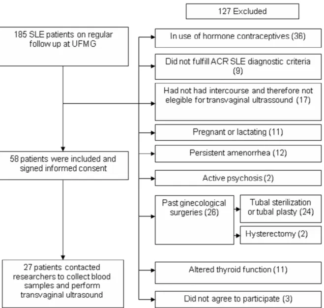
After pre-selecting 185 SLE patients with ages between 18 and 40 years,
127 patients met exclusion criteria, as shown in Figure 1. Fifty-eight patients were
included and agreed to sign the informed consent, and 27 contacted the researchers
to inform the beginning of menstruation. They were submitted to blood sampling and
transvaginal ultrasound at early follicular phase.
The demographic and clinical features of the SLE groups are summarized in
Table 1. The mean age of SLE group was 30.9 years and the mean of disease
duration was 102.7 months. Seventeen (63%) SLE patients had nephritis during the
follow up, and four (14%) also had antiphospholipid syndrome (APS). The median
disease activity index measured by SLEDAI (18) was 2 (range 0-16) and the median
intensity of damage measured by SLICC/ACR –DI (19) was 0 (range 0-8). Treatment
with corticosteroid was necessary in 22 (81.5 %) patients with a mean dose of 5 mg
(range 1.3 – 50). Twenty (74.1%) SLE patients used cyclophosphamide during follow
up, and four (14.8%) were in current use at the time of blood test and ultrasound. The
mean of age when treatment with cyclophosphamide started was 22.6 (range 11-34,
SD±6.6) years. Median cumulated dose of cyclophosphamide was 9.6 (range 1 - 49)
grams.
Table 1: Demographic, clinical and therapeutic characteristics of the 27 SLE patients
included in the study
Characteristic Results
Mean age (yr.) 30.9 (SD±4.8)
Mean age of onset 22.2 (SD±6.1)
Mean Disease duration (mo.) 102.7 (SD±66.7)
Skin color
White
Non white
8 (29.6%)
19 (70.4%)
Education
Elementary
High School
11 (40.7%)
34
College 3 (11.1%)
Marital status Married Other 11 (40.7%) 16 (59.3%) Smoking Past Current Never 1 (3.7%) 1 (3.7%) 25 (92.6%)
Hypothyroidism 1 (3.7%)
Chronic Kidney disease 2 (7.4%)
SLE /ACR Clinical manifestations
Mucocutaneous characteristicsa
Non erosive arthritis
Neuropsychiatric disordersb
Hematologic abnormality
Leukopenia/Lymphopenia
Hemolitic anemia
Trombocytopenia
Serositis (pleurisy or pericarditis)
Nephritisc
Positive ANA
Antibody to Sm
Antibody to DNA
False positive VDRL
Corticosteroid therapy (current) 22 (81.5%)
Mean daily dose of corticosteroid (mg) 5 (1.3 – 50)
Azatioprina therapy (follow-up) 16 (49.2%)
Antimalarial therapy (follow-up) 27 (100%)
Cyclophosphamide therapy (follow-up) 20 (74.1%)
Cyclophosphamide therapy (current) 4 (14.8%)
Mean age of start cyclophosphamide (yr.) 22.6(SD±6.6)
Median cumulate dose of cyclophosphamide (g) 9.6 (1 – 49)
SLEDAI (Median) d 2 (0-14)
SLICC/ACR – DI (Median) e 0 (0-8)
APS 4 (14%)
Menses irregularity
BMI (Mean) f
4 (14%)
23 (SD±3.4)
Values are presented as mean (±SD), n (%), or median (minimum – maximum).
aMucocutaneous characteristics: Malar erythema, Discoid lupus, photosensibility,
mucosal ulcers. bNeuropsychiatric disorders: Psychosis, seizures, cranial
neuropathy and transverse mielitis. cNephritis: Proteinúria > 0,5g/24hs or 3+ rotine
urine test, cylindruria or compatible renal biopsy. dSLEDAI (18): Systemic Lupus
Erythematosus Disease Activity Index. eSLICC (19) (Systemic Lupus International
Collaborating Clinics). fBMI: Body mass index.
Blood sampling for hormone measurements
Blood samples were collected, using sterile equipment, in two vials of serum
gel, each one of 5 mililiters. The material was centrifuged for 5 minutes at 3000
36
by manual pipetting with disposable material. The serum was then stored at - 80
degrees Celsius, until the completion of hormone assays.
Transvaginal ultrasound
Patients and controls underwent transvaginal ultrasound at the same day of
blood collection, during the early follicular phase of menstrual cycle. Ultrasound was
performed by a single examiner (C.P.R.), using a Aloka equipment with a 3 – 7.5
mHz endocavitary transducer. Antral follicle counting included all follicles with a
mean diameter of 2-10 mm calculated from two dimensions. (20)
Hormone assays
AMH was measured using an enzymatically amplified two-site immunoassay
(kit A73819 AMH Gen II ELISA purchased from Beckman Coulter®, Diagnostic
Systems Laboratories, Brea, CA, USA). The results were expressed in nanograms
per milliliter (ng/ml). The lowest amount of AMH in the sample that can be detected
with 95% probality is 0.08 ng/ml, and estimated minimum dose achieved at 20% total
imprecision is 0.16 ng/ml. The coefficient variation of AMH Gen II assay in the
concentration range of our samples was 5.7%. FSH was measured by immunoassay
using kits ECi/ECiQ from Vitros®, and was expressed by internationals microunits
per milliliter (mUI/ml).
Statistical analysis
We calculated that sample size of at least 23 patients was required per group
to achieve a statistical power of 90% with 95% confidence, to detect differences
larger than one standard deviation in serum FSH or AMH, as well as in quantitative
Continuous variables were tested for normal distribution using skewness and
kurtosis parameters. Data with normal distribution were expressed as mean and
standard deviation, while those with non-normal distribution, were presented as
median and percentiles 25 and 75. Between group, differences were tested by
unpaired Student's t test or Mann–Whitney U test, for normal and non-normal data
distribution, respectively. The correlations between ovarian reserve markers and
continuous variables were evaluated by Pearson´s or Spearmann´s coefficient.
Multivariate analysis was performed by logistic regression to test
associations between ovarian reserve markers dichotomized as above versus below
the control group median and demographic, reproductive and clinical characteristics
of patients. We included in the multivariated analysis model all the variables with
p<0.2 in the univariated analysis.
All statistical analyse were performed using the Statistical Package for Social
Sciences (SPSS®, IBM®, USA) for Windows v/17.0, with two-sided p values less
38
Results
The reproductive characteristics of SLE and control groups are shown in
Table 2. Mean age of menarche of SLE patients and control group were 13.15 years
(SD±1.7) and 12.4 years (SD±1.5), respectively. Median cycle length was 28 (28 –
30) days in both groups, and the mean length of menstrual flow was 4.4 (SD±1.1)
days in SLE patients and 4.9 (SD±1.3) days in the control group. There were no
significant differences between groups.
The median FSH concentration was 6.44 (4.19 – 7.69) mUI/ml in SLE
patients and 7.5 (6.03 – 8.09) mUI/ml in the control group, (p=0.135, FIG.2A). AMH
levels were 1.23 (0.24 – 4.63) ng/ml in the SLE group and 1.52 (1.33 – 1.88) ng/ml in
the control group, (p=0.684, FIG.2B). The median AFC by ultrasound was 7 (5 – 13)
follicles in SLE the group and 11 (7 – 12) follicles in control groups, (p=0.076,
FIG.2C).
Table 2. Reprodutive characteristics of controls and SLE patients
Values are presented as mean (±SD) or median (IQR). aUsed T student ,
Mann-Whitney U as appropriated.
SLE (n=27) Controls (n=27) Pa
Age (years) 30.9(SD±4.8) 32.1(SD±3.8) 0.316
Age at menarche (years) 13.2(SD±1.7) 12.4(SD±1.5) 0.076
Menses length (days) 28 (28-30) 28 (28-30) 0.646
Menstrual flow length (days) 4.4(SD±1.1) 4.9(SD±1.3) 0.184
40
Figure 2. Scatters showing comparison of ovarian reserve markers between SLE
patients and controls: AFC (A), FSH (B), and AMH (C). The horizontal bars indicate
the medians.
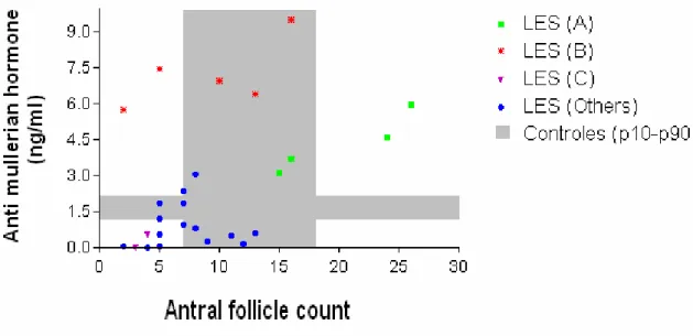
The distribution of SLE patients is illustrated in correlation plots between
ovarian reserve markers in Figures 4A, 4B, and 4C. Different clusters were identified
in SLE patients: cluster A is formed by patients with high AFC and AMH levels,
resembling some characteristics of polycystic ovary syndrome; cluster B has high
AMH levels despite normal AFC; and cluster C includes SLE patients with low
Figure 3. Scatter plots describing correlations between ovarian markers in SLE
patients: AFC vs FSH (A), FSH vs AMH (B), and AMH vs AFC (C). The grey areas
represent the ranges between 10th and 90th percentile in the control group. Different
clusters of SLE patients are represented by different symbols.
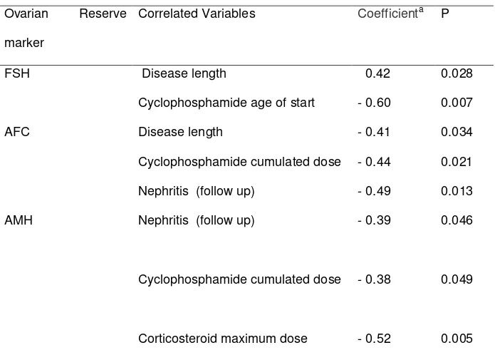
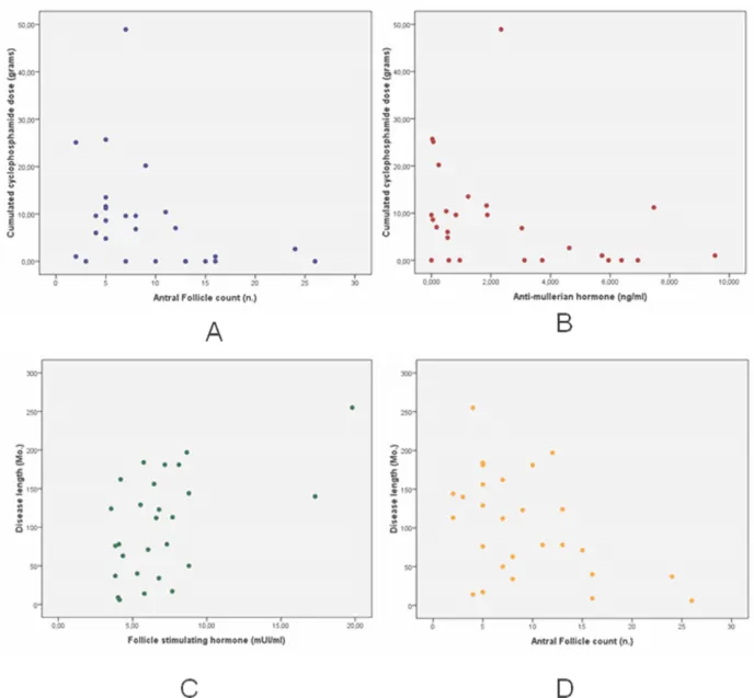
All statistically significant data obtained by the univariate analysis are
shown in table 3. There was significant correlation between cyclophosphamide
cumulate dose and both AMH and AFC (Figure 4A and 4B). Disease length was
correlated with FSH and AFC (Figure 4C and 4D). Nephritis was associated with
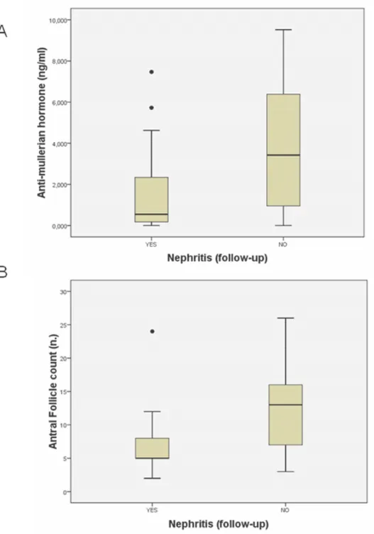
lower AFC (5 vs 13; p=0,013) and lower AMH values (0.54 vs 3.42; p=0,046) as
shown in Figure 5.
In the multivariated logistic regression, AMH independently was associated
with group control (OR 5.1; CI95% 1.286 – 20.405) and higher AFC (OR 1.21; CI95%
42
CI95% 1.055 – 2.109) and endometrial thickness (OR 2.05; CI95% 1.044 – 4.019) as
shown in Table 4. In the SLE group, antral follicle count was independently
associated to low SLICC/ACR-DI (OR 1.613; CI95% 0.025 – 0.841). (Table 5)
Table 3. Statistically significant correlation coefficients by univariate analysis
between ovarian reserve markers and clinical features in 27 SLE cases.
Ovarian Reserve
marker
Correlated Variables Coefficienta P
FSH Disease length 0.42 0.028
Cyclophosphamide age of start - 0.60 0.007
AFC Disease length - 0.41 0.034
Cyclophosphamide cumulated dose - 0.44 0.021
Nephritis (follow up) - 0.49 0.013
AMH Nephritis (follow up) - 0.39 0.046
Cyclophosphamide cumulated dose - 0.38 0.049
Corticosteroid maximum dose - 0.52 0.005
aUsed Spearman rank-order or Mann Whitney as appropriated. AMH - Anti mullerian
Figure 4. Scatter showing correlations between ovarian reserve markers and clinical
features in SLE patients. AFC vs cyclophosphamide cumulated dose (Figure 4A),
AMH vs cyclophosphamide cumulated dose (Figure 4B), FSH vs Disease length
44
Figure 5. Boxplots describing distribution of AMH (A) and AFC (B) in SLE patients
Table 4. Logistic regression analysis to evaluate ovarian reserve and reproductive
characteristics in 54 patients
Dependent variable Covariate OR 95% CI P
AMH > 1.51 ng/ml Control group 5.12 1.286 - 20.405 0.021
FSH 1.00 0.756 – 1.311 NS
AFC 1.21 1.023 - 1.444 0.026
Endometrial
thickness
0.45 0.178 – 1.150 NS
AFC > 9 Control group 3.59 0.915 – 14.101 NS
AMH 1.52 1.055 - 2.109 0.025
FSH 0.91 0.671 – 1.232 NS
Endometrial
thickness
2.05 1.044 - 4.019 0.037
FSH > 6.77 mIU/ml Control group 2.10 0.625 – 7.058 NS
AMH 0.81 0.557 – 1.182 NS
AFC 0.93 0.557 – 1.182 NS
Endometrial
thickness
0.91 0.488 – 1.694 NS
CI - Confidence interval, OR – odds ratio, AMH - Anti mullerian hormone, FSH -
Follicle Stimulating hormone, AFC - Antral follicles count. R square (Nagelkerk):
46
Table. 5 Logistic regression analysis to evaluate ovarian reserve and clinical features
and reproductive characteristics in 27 SLE cases
Dependent variable Covariate OR 95% CI P
AFC > 1.23 ng/ml Major prednisone dose
– follow up
0.95 0.868 – 1.051 NS
Disease length 0.99 0.969 – 1.009 NS
Start age of
cyclophosphamide
1.01 0.863 – 1.193 NS
Cumulated
cyclophosphamide
dose
0.93 0.797 – 1.090 NS
Nephritis 0.92 0.015 – 57.488 NS
SLICC/ACR-DI 0.14 0.025 – 0.841 0.031
AMH > 7 Major prednisone dose
– follow up
0.95 0.894 – 1.000 0.050
Disease length 0.98 0.959 – 1.012 NS
Start age of
cyclophosphamide
1.12 0.896 – 1.408 NS
Cumulated
cyclophosphamide
dose
1.09 0.971 – 1.229 NS
SLICC/ACR-DI 2.00 0.605 – 6.638 NS
FSH > 6.44 mIU/ml Major prednisone dose
– follow up
1.03 0.961 – 1.121 NS
Disease length 1.00 0.991 – 1.025 NS
Start age of
cyclophosphamide
0.86 0.711 – 1.047 NS
Cumulated
cyclophosphamide
dose
1.03 0.916 – 1.151 NS
Nephritis 17.59 0.200 – 1547.158 NS
SLICC/ACR-DI 1.18 0.564 – 2.486 NS
CI - Confidence interval, OR – odds ratio, AMH - Anti mullerian hormone, FSH -
Follicle Stimulating hormone, AFC - Antral follicles count, SLICC – Systemic Lupus
International Collaborating Clinics-American College of Rheumatology/Damage
48
Discussion
In recent years, concurrent with increased survival of SLE patients, also raised
the interest of this population for quality of life. Concerns about the best age for
pregnancy and its viability, as well as doubts about the expected age of menopause
and how to reduce the negative effects of climacteric are part of SLE women’s
thoughts. These questions are justified by some reports in the medical literature of
fewer offspring, and lower average age of menopause in SLE patients compared to
the general population. (7, 21)
In this study we found no difference between the results of three major serum
and ultrasonographic ovarian reserve markers in women with SLE compared with
women without the disease. However, in a broader analysis of the sample, the SLE
patients had about five times more odds to have low AMH values than the control
group. The groups had similar menarche ages and there were no differences
between groups in the length of menstrual cycle or duration of menstrual flow in the
follicular phase. Taken together, these findings suggest that eumenorrheic women
with SLE may already have low AMH levels as an early sign of diminished ovarian
reserve, but only the completion of their follow-up will allow us to verify whether the
women with lower AMH levels are those who eventually will have the earliest
manifestations of ovarian insufficiency.
The two previous studies that specifically evaluated AMH as a marker of
ovarian reserve in patients with SLE found reduced values of this hormone in the
SLE group.(22, 23) In the present study, we noticed a large variation between the
values found in the AMH group of patients with SLE compared the distribution in the
control group. This heterogeneity sample of patients with SLE is more evident in
evaluation of markers of ovarian reserve, such as patients with high levels of AMH
and AFC, resembling women with polycystic ovary syndrome, (24) and women with
clearly reduced ovarian reserve, showing high FSH levels and low values of AMH
and AFC. Five patients had high AMH values associated with the presence of normal
or slightly reduced AFC (“SLE B” group). We were unable to identify any relationship
between this isolated AMH increase and clinical characteristics or disease activity
scores and systemic damage. Thus, it remains unclear why some SLE women have
such high serum AMH concentrations without a correspondent increase in AFC.
One patient with the group "SLE B" profile had existing chronic renal failure in
dialysis, pre-renal transplantation, but without clinical or laboratory evidence of
uremia, low SLEDAI (2 points) and SLICC / ACR-DI (2 points) scores, and had
regular menstrual cycles. A well know complication of chronic renal failure is
hypogonadism, caused by hyperprolactinemia secondary to uremia, which interferes
with the regulation of the hypothalamic-pituitary-gonadal axis, leading to reduced
ovarian function.(25, 26) However, we found no reference in the literature about renal
failure and increased amounts of AMH.
A positive association between AMH and AFC was observed in the group of 54
patients, as well as in the isolated analysis of the SLE group, representing the close
relationship between these markers of ovarian reserve.(27, 28) AFC also showed a
positive association with endometrial thickness, which is surrogate marker of ovarian
estrogen release at the follicular phase, reinforcing the functional significance of the
morphological assessment of growing ovarian follicles.
We found a negative association between AFC and the score for organ
damage (SLICC / ACR-DI) in SLE patients. However, we could not associate the
50
reserve, maybe, showing that the ovarian reserve is more influenced by the sequelae
of chronic inflammation that only by the intensity of the flares. The patients in the
study sample had low levels of SLICC (median = 0) and SLEDAI (median = 2)
characterizing a population with the disease well controlled with low systemic
damage, which may have determinate a limitation to the analysis.
The use of higher doses of corticosteroids during follow up was associated with
lower levels of AMH in SLE patients. This relationship seems to reflect primarily the
inflammatory activity history of the rheumatic disease, rather than a direct effect of
exogenous steroids on the physiology of AMH. We found no differences related to
the current dose of prednisone in the study, nor published reports on the relationship
between AMH and hypercortisolism.
The presence of nephritis and the cumulative dose of cyclophosphamide were
factors individually related to reduced ovarian reserve, by correlations with lower
values of AFC and AMH. Nevertheless, they were not confirmed as independent
determinants of ovarian markers. The SLE patients had a mean age of 31 years,
which is compatible with the group most susceptible to the ovarian effects of
alkylating drugs. However, only four patients (14.8%) were in current treatment with
cyclophosphamide at the time of the study, and the average age of starting
medication was 22 years. Another factor that has not been quantified, but may be
considered was the past use of contraceptives playing the role of a protective factor
for ovarian function. The SLE patients followed at the rheumatology unit are geared
to undertake contraception during the induction treatment of nephritis with
intravenous cyclophosphamide pulses to avoid birth defects secondary to
medication. The anovulation secondary to hormonal contraceptive use is possibly a
We conclude that eumenorrheic patients with SLE often have the same values
of AFC and FSH found in control patients. For better evaluation of AMH in these
patients, we believe it is necessary a serial measurement or even joint evaluation
with other markers like inhibin B, to define early the groups that really have potencial
lost of ovarian function.
The reduction of ovarian reserve in some patients with SLE is closely related to
conditions that demonstrate greater inflammatory intensity of the disease or longer
exposure, such as the presence of nephritis in the course of the disease or use of
higher doses of cyclophosphamide in the treatment, and higher scores of organic
damage.
Acknowledgements
We thank professors Cristina Duarte Lanna and Márcia Mendonça Carneiro
for helpful comments and suggestions. Research supported by CNPq and FAPEMIG,
Brazil.
References
1. Pons-Estel GJ, Alarcon GS, Scofield L, Reinlib L, Cooper GS. Understanding
the epidemiology and progression of systemic lupus erythematosus. Semin
Arthritis Rheum 2010;39:257-68.
2. Medeiros PB, Febronio MV, Bonfa E, Borba EF, Takiuti AD, Silva CA.
Menstrual and hormonal alterations in juvenile systemic lupus erythematosus.
52
3. Pasoto SG, Mendonca BB, Bonfa E. Menstrual disturbances in patients with
systemic lupus erythematosus without alkylating therapy: clinical, hormonal
and therapeutic associations. Lupus 2002;11:175-80.
4. Shabanova SS, Ananieva LP, Alekberova ZS, Guzov, II. Ovarian function and
disease activity in patients with systemic lupus erythematosus. Clin Exp
Rheumatol 2008;26:436-41.
5. Medeiros MM, Silveira VA, Menezes AP, Carvalho RC. Risk factors for ovarian
failure in patients with systemic lupus erythematosus. Braz J Med Biol Res
2001;34:1561-8.
6. Schaller J. Lupus in childhood. Clin Rheum Dis 1982;8:219-28.
7. Ekblom-Kullberg S, Kautiainen H, Alha P, Helve T, Leirisalo-Repo M, Julkunen
H. Reproductive health in women with systemic lupus erythematosus
compared to population controls. Scand J Rheumatol 2009;38:375-80.
8. Gonzalez LA, Pons-Estel GJ, Zhang JS, McGwin G, Jr., Roseman J, Reveille
JD et al. Effect of age, menopause and cyclophosphamide use on damage
accrual in systemic lupus erythematosus patients from LUMINA, a multiethnic
US cohort (LUMINA LXIII). Lupus 2009;18:184-6.
9. Minami Y, Sasaki T, Komatsu S, Nishikori M, Fukao A, Yoshinaga K et al.
Female systemic lupus erythematosus in Miyagi Prefecture, Japan: a
case-control study of dietary and reproductive factors. Tohoku J Exp Med
1993;169:245-52.
10. Silva CA, Hilario MO, Febronio MV, Oliveira SK, Terreri MT, Sacchetti SB et
al. Risk factors for amenorrhea in juvenile systemic lupus erythematosus
11. Moncayo-Naveda H, Moncayo R, Benz R, Wolf A, Lauritzen C. Organ-specific
antibodies against ovary in patients with systemic lupus erythematosus. Am J
Obstet Gynecol 1989;160:1227-9.
12. Pasoto SG, Viana VS, Mendonca BB, Yoshinari NH, Bonfa E. Anti-corpus
luteum antibody: a novel serological marker for ovarian dysfunction in
systemic lupus erythematosus? J Rheumatol 1999;26:1087-93.
13. McDermott EM, Powell RJ. Incidence of ovarian failure in systemic lupus
erythematosus after treatment with pulse cyclophosphamide. Ann Rheum Dis
1996;55:224-9.
14. Park MC, Park YB, Jung SY, Chung IH, Choi KH, Lee SK. Risk of ovarian
failure and pregnancy outcome in patients with lupus nephritis treated with
intravenous cyclophosphamide pulse therapy. Lupus 2004;13:569-74.
15. Broekmans FJ, Kwee J, Hendriks DJ, Mol BW, Lambalk CB. A systematic
review of tests predicting ovarian reserve and IVF outcome. Hum Reprod
Update 2006;12:685-718.
16. Domingues TS, Rocha AM, Serafini PC. Tests for ovarian reserve: reliability
and utility. Curr Opin Obstet Gynecol 2010;22:271-6.
17. Hochberg MC. Updating the American College of Rheumatology revised
criteria for the classification of systemic lupus erythematosus. Arthritis Rheum
1997;40:1725.
18. Bombardier C, Gladman DD, Urowitz MB, Caron D, Chang CH. Derivation of
the SLEDAI. A disease activity index for lupus patients. The Committee on
Prognosis Studies in SLE. Arthritis Rheum 1992;35:630-40.
19. Gladman D, Ginzler E, Goldsmith C, Fortin P, Liang M, Urowitz M et al. The
54
Collaborating Clinics/American College of Rheumatology damage index for
systemic lupus erythematosus. Arthritis Rheum 1996;39:363-9.
20. Rosen MP, Johnstone E, Addauan-Andersen C, Cedars MI. A lower antral
follicle count is associated with infertility. Fertil Steril 2011;95:1950-4, 4 e1.
21. Costenbader KH, Feskanich D, Stampfer MJ, Karlson EW. Reproductive and
menopausal factors and risk of systemic lupus erythematosus in women.
Arthritis Rheum 2007;56:1251-62.
22. Browne H, Armstrong A, Decherney A, Babb R, Illei G, Segars J et al.
Assessment of ovarian function with anti-Mullerian hormone in systemic lupus
erythematosus patients undergoing hematopoietic stem cell transplant. Fertil
Steril 2009;91:1529-32.
23. Lawrenz B, Henes JC, Henes M, Neunhoeffer E, Schmalzing M, Fehm T et al.
Impact of systemic lupus erythematosus on ovarian reserve in premenopausal
women - evaluation by using Anti-Muellerian hormone. Lupus 2011.
24. Lujan ME, Chizen DR, Pierson RA. Diagnostic criteria for polycystic ovary
syndrome: pitfalls and controversies. J Obstet Gynaecol Can 2008;30:671-9.
25. Handelsman DJ, Dong Q. Hypothalamo-pituitary gonadal axis in chronic renal
failure. Endocrinol Metab Clin North Am 1993;22:145-61.
26. Saha MT, Saha HH, Niskanen LK, Salmela KT, Pasternack AI. Time course of
serum prolactin and sex hormones following successful renal transplantation.
Nephron 2002;92:735-7.
27. La Marca A, Broekmans FJ, Volpe A, Fauser BC, Macklon NS. Anti-Mullerian
hormone (AMH): what do we still need to know? Hum Reprod
28. Yang YS, Hur MH, Kim SY, Young K. Correlation between sonographic and
endocrine markers of ovarian aging as predictors for late menopausal
transition. Menopause 2011;18:138-45.
29. Behringer K, Breuer K, Reineke T, May M, Nogova L, Klimm B et al.
Secondary amenorrhea after Hodgkin's lymphoma is influenced by age at
treatment, stage of disease, chemotherapy regimen, and the use of oral
contraceptives during therapy: a report from the German Hodgkin's
5. COMENTÁRIOS E CONCLUSÕES
As medidas dos marcadores de reserva ovariana tradicionais em pacientes
com lúpus eritematoso sistêmico foram semelhantes àquelas encontradas em
pacientes controles. Mais importante, o grupo de pacientes com LES apresenta um
perfil heterogêneo de distribuição dos valores de hormônio anti-mulleriano. A melhor
avaliação destes achados através de dosagens seriadas deste hormônio ou sua
avaliação conjunta com outros marcadores ovarianos, como a inibina B, pode
determinar o papel destes marcadores para melhor seleção precoce de pacientes
com LES sob risco de falência ovariana prematura. Com esta informação, a tomada
de medidas profiláticas se torna mais efetiva e menos onerosa.
A redução da reserva ovariana em algumas pacientes do grupo LES está
intimamente relacionada com condições que demonstram maior intensidade do
processo inflamatório, como a presença de nefrite no decorrer da doença ou uso de
maiores doses de ciclofosfamida no tratamento, além de maiores escores de dano
orgânico.
Concluímos então que:
• Os marcadores de reserva ovariana em pacientes com LES e indivíduos do
grupo controle foram semelhantes.
• Os marcadores de reserva ovariana em pacientes com LES relacionaram-se
com o critério diagnóstico nefrite. A presença deste critério correlacionou-se
com redução da reserva ovariana.
• Os marcadores de reserva ovariana em pacientes com LES
correlacionaram-se com a docorrelacionaram-se acumulada de ciclofosfamida. A recorrelacionaram-serva ovariana mostrou-correlacionaram-se
58
• A reserva ovariana está reduzida em pacientes com LES com maiores
escores de dano orgânico SLICC/ACR-DI. Não encontramos associação
individual ou independente entre valores do SLEDAI e os marcadores de
60
6. REFERÊNCIAS BIBLIOGRÁFICAS
1- Pons-Estel GL, Alarcón GS, Scofield L, et al. Understanding the epidemiology
and progression of the systemic lupus erythematosus. Semin Arthritis Rheum 2010; 39: 257 – 68.
2- Stahl-Hallengren C, Jonsen A, Nived O, Sturfelt G. Incidence studies of systemic lupus erythematosus in Southern Sweden: increasing age, decreasing frequency of renal manifestations and good prognosis. J Rheumatol 2000; 27: 685-91.
3- Pereira Vilar MJ, Sato EI. Estimating the incidence of systemic lupus erythematosus in a tropical region (Natal, Brazil). Lupus 2002;11, 528-32 4- McCarty DJ, Manzi S, Medsger TA Jr, Ramsey-Goldman R, LaPorte RE,
Kwoh CK. Incidence of systemic lupus erythematosus. Race and gender differences. Arthritis Rheum 1995; 38: 1260–70.
5- Hopkinson ND, Doherty M, Powell RJ. The prevalence and incidence of systemic lupus erythematosus in Nottingham, UK, 1989-1990. Br J Rheumatol
1993; 32: 110-15.
6- Nossent JC. Systemic lupus erythematosus on the Caribbean island of Curaçao: an epidemiological investigation. Ann Rheum Dis 1992; 51:1197 –
201.
7- Medeiros PB, Febrônio MV, Bonfá ET. Menstrual and hormonal alterations in juvenile systemic lupus erythematosus. Lupus 2009 Jan; 18(1): 38-43.
8- Pasoto SG, Mendonca BB, Bonfa E. Menstrual disturbances in patients with systemic lupus erythematosus without alkylating therapy: clinical, hormonal and therapeutic associations. Lupus 2002; 11: 175 - 80.
9- Shabanova S, Ananieva LP, Alekberova ZS. Ovarian function and disease activity in patients with systemic lupus erythematosus. Clin Exp Rheumatol.
2008; 26(3):436-41.
10- Medeiros MM, Silveira VA, Menezes AP, Carvalho RC. Risk factors for ovarian failure in patients with systemic lupus erythematosus. Braz J Med Biol Res 2001; 34: 1561 - 68.
12- Elkblom-Kullberg S, Kautiainen H, Alha P. Reproductive health in women with systemic lupus erythematosus compared to population controls. Scand J Rheumatol 2009; 38 (5):375-80.
13- González LA, Pons-Estel GJ, Zhang JS. Effect of age, menopause and cyclophosphamide use on damage accrual in systemic lupus erythematosus patients from LUMINA, a multicentric US cohort (LUMINA LXIII). Lupus 2009; 18(2): 184-6.
14- Lim GS, Petri M & Goldman D (1993). Menstruation and systemic lupus erythematosus: a case-control study. Arthritis and Rheumatism, 36 (Suppl
5): R23
15- Silva CA, Hilário MO, Febrônio MV. Risk factors for amenorrhea in juvenile systemic lupus erythematosus (JSLE): a Brazilian multicentre cohort study.
Lupus 2007; 16(7):531-6.
16- Moncayo-Naveda H, Moncayo R, Benz R, Wolf A & Lauritzen CH. Organspecific antibodies against ovary in patients with systemic lupus erythematosus. American Journal of Obstetrics and Gynecology 1989; 160:
1227 - 29.
17- Pasoto SG, Viana VST, Mendonça BB, Yoshinari NH & Bonfa E. Anti-corpus luteum antibody: a novel serological marker for ovarian dysfunction in systemic lupus erythematosus? Journal of Rheumatology 1999; 26: 1087
- 93.
18- Mc Dermott EM, Powell RJ. Incidence of ovarian failure in systemic lupus erythematosus after treatment with pulse cyclophosphamide. Annals of the Rheumatic Disease 1996; 55: 224 – 29.
19- Park MC, Park YB, Jung SY, Chung IH, Choi KH, Lee SK. Risk of ovarian failure and pregnancy outcome in patients with lupus nephritis treated with intravenous cyclophosphamide pulse therapy. Lupus 2004; 13: 569 - 74. 20- Broekmans FL, Kwee J, Hendricks DJ. A systematic review of tests
predicting ovarian reserve and IVF outcome. Hum Reprod Update. 2006;12
(6):685-718.
21- Domingues TS, Rocha AM, Serafini PC. Tests for ovarian reserve: reliability and utility. Curr Opin Obstet Gynecol 2010; 22(4):271-6.