Risk facto rs fo r o varian failure in
patie nts with syste mic lupus
e rythe m ato sus
Departamento de Clínica Médica, Hospital Universitário Walter Cantídio, Universidade Federal do Ceará, Fortaleza, CE, Brasil
M.M.C. Medeiros, V.A.L. Silveira, A.P.T. Menezes and R.C. Carvalho
Abstract
The aim of the present study was to identify the risk factors for ovarian failure in patients with systemic lupus erythematosus. Seventy-one women aged 17 to 45 years with systemic lupus erythematosus were studied. Patients were interviewed and their medical records re-viewed. Demographic characteristics, clinical and serologic profiles, and menstrual and obstetric histories were recorded. Disease activity was measured by the systemic lupus erythematosus disease activity index. Serum FSH, LH, estradiol, progesterone, TSH, prolactin, and antimicrosomal and antithyroglobulin antibodies were measured. Pa-tients who developed ovarian failure were compared to those who did not. Ovarian failure occurred in 11 patients (15.5%) and nine had premature menopause (11.3%). Cyclophosphamide administration and older patient age were found to be associated with ovarian failure. The cumulative cyclophosphamide dose was significantly higher in patients with ovarian failure than in those without this condition (18.9 vs 9.1 g; P = 0.04). The relative risk for ovarian failure in patients with cumulative cyclophosphamide dose higher than 10 g was 3.2. TSH levels were high in 100% of patients with ovarian failure who had received pulse cyclophosphamide. Ovarian failure, and premature menopause in particular, is common in patients with systemic lupus erythematosus, with the most important risk factors being cyclophos-phamide dose and age. Thyroid problems may be another risk factor for ovarian failure in patients with lupus.
Co rre spo nde nce
M.M.C. Medeiros Rua Paula Ney, 599/302 60140-200 Fortaleza, CE Brasil
Fax: + 55-85-224-7222 E-mail: marmed@ fortalnet.com.br
Received May 23, 2001 Accepted September 26, 2001
Ke y words
·O varian failure ·Systemic lupus erythematosus ·Risk factors ·Cyclophosphamide ·Premature menopause
Intro ductio n
Systemic lupus erythematosus (SLE) is an autoimmune disease which mainly affects women of reproductive age. Menstrual alter-ations ranging from increased cycle flow, generally due to thrombocytopenia, to tem-porary amenorrhea and premature menopause are fairly common in these patients.
The main factors which have been
asso-ciated with the onset of ovarian failure in patients diagnosed with SLE are disease ac-tivity (1), anti-ovarian antibodies (2,3), cyto-toxic agents (4-8) and, more recently, use of thalidomide (9,10). Other potential causes of ovarian insufficiency not specific for lupus are polyglandular insufficiency, viral infec-tions, smoking, oophorectomy, emotional disorders, and idiopathic origin (11).
immunosup-pressive agent of choice for the treatment of various complications of SLE and, there-fore, is the factor most highly associated with ovarian insufficiency. Gonadal toxicity should be of great concern in premenopausal women who take cyclophosphamide. The frequency of ovarian insufficiency in SLE patients treated with this drug ranges from 11 to 59% in different studies and depends on the dose used, the age of the patient and methodological differences (4-8,12-14).
Since estrogens have biological effects which protect the cardiovascular system, as well as the skeletal system from bone loss, estrogen deficiency is linked with a greater risk for cardiovascular disease (15) and os-teoporosis (16), thus contributing to higher morbidity and mortality among patients di-agnosed with lupus. The potential for other risk factors for the reduction of bone mass in this group of patients, such as use of cortico-steroids, lower exposure to sunlight, the con-troversial use of hormone-replacement thera-py (17-19), along with hypoestrogenism sec-ondary to cyclophosphamide-induced ovar-ian failure, renders women diagnosed with SLE far more susceptible to the develop-ment of osteoporosis.
Thus, it is crucial to identify those factors most often associated with ovarian insuffi-ciency in patients with SLE in order that preventive and therapeutic measures may be taken to minimize the risk of these complica-tions.
Mate rial and Me thods
Study population
The study population consisted of female patients diagnosed with SLE, in accordance with the criteria of the American College of Rheumatology (20), attended continuously at the Rheumatology Service of the Univer-sity Hospital of the Federal UniverUniver-sity of Ceará, from January 1999 to March 2000. All patients with a diagnosis of SLE under
treatment at the above-mentioned hospital, aged 16 to 45 years, who agreed to partici-pate were included in the study. Exclusion criteria were patients with known causes of secondary amenorrhea, such as chronic re-nal insufficiency, and reported hysterectomy or oophorectomy.
This is a retrospective cohort study.
Me tho ds
A questionnaire was administered to pa-tients in order to obtain sociodemographic data (age, race, marital status, education, smoking habit), clinical data (duration of disease, clinical manifestations, drugs used to treat lupus), and gynecologic-obstetric his-tory (age at menarche, date of latest men-strual cycle, number of gestations and abor-tions, curettages, gynecological surgeries, and methods of birth control used). At the time of the interview, a disease activity score was determined according to SLE disease activity index (SLEDAI) criteria (21). The following laboratory tests were obtained: blood cell count, hemosedimentation rate, measurement of follicle-stimulating hormone (FSH), luteinizing hormone (LH), estradiol, progesterone, prolactin, thyroid-stimulating hormone (TSH), antinuclear factor, DNA, Sm, antithyroglobulin, and anti-microsomal antibodies, protein count, crea-tinine, and urinalysis. Hormonal determina-tions were performed by radioimmunoas-say.
pulses given every three months for 18-24 months (1 g/m2), with doses adjusted
ac-cording to platelet and leukocyte levels. The protocol was approved by the Hospi-tal Ethics Committee and informed consent was obtained from all patients.
Ovarian insufficiency was considered to have occurred in cases of amenorrhea last-ing longer than 4 months, excludlast-ing preg-nancy. Premature menopause was defined as the presence of amenorrhea for at least 12 months, confirmed by estradiol (0-14 pg/ ml), FSH (42-126 mIU/ml) and LH (11-50 mIU/ml) levels, that had occurred before 40 years of age.
Statistical analysis
The Student t-test for independent
sam-ples, the Fisher exact test and the chi-square and Mann-Whitney tests were applied when appropriate. Risk factors for ovarian insuffi-ciency were studied by multiple logistic re-gression (23).
Re sults
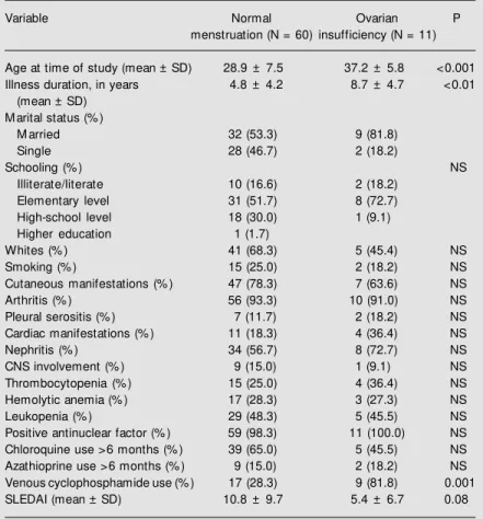
Seventy-one women diagnosed with SLE were studied, with a mean age of 30 years (range: 17-45 years) and approximately 5.4 years of disease. The most common clinical manifestations were articular (93%), cutane-ous (76%), renal (59.1%) and leukopenic (47.9%). Antinuclear factor was positive in 98.5%. Sixty women still had regular men-strual cycles, while 11 (15.5%) had been in amenorrhea for at least 4 months and were thus assigned to the ovarian insufficiency group (Table 1). Eight of the 11 patients with ovarian insufficiency fulfilled criteria for the diagnosis of premature menopause, corre-sponding to a prevalence of 11.3% for pre-mature menopause in the study group. De-mographic and clinical characteristics of the two patient groups are shown in Table 1. The women with ovarian insufficiency presented a higher mean age (37.2 and 28.9 years;
Table 1. Demographic and clinical characteristics of patients w ith systemic lupus erythematosus according to the presence or absence of ovarian insufficiency.
Variable Normal Ovarian P
menstruation (N = 60) insufficiency (N = 11)
Age at time of study (mean ± SD) 28.9 ± 7.5 37.2 ± 5.8 <0.001
Illness duration, in years 4.8 ± 4.2 8.7 ± 4.7 <0.01
(mean ± SD) M arital status (% )
M arried 32 (53.3) 9 (81.8)
Single 28 (46.7) 2 (18.2)
Schooling (% ) NS
Illiterate/literate 10 (16.6) 2 (18.2)
Elementary level 31 (51.7) 8 (72.7)
High-school level 18 (30.0) 1 (9.1)
Higher education 1 (1.7)
Whites (% ) 41 (68.3) 5 (45.4) NS
Smoking (% ) 15 (25.0) 2 (18.2) NS
Cutaneous manifestations (% ) 47 (78.3) 7 (63.6) NS
Arthritis (% ) 56 (93.3) 10 (91.0) NS
Pleural serositis (% ) 7 (11.7) 2 (18.2) NS
Cardiac manifestations (% ) 11 (18.3) 4 (36.4) NS
Nephritis (% ) 34 (56.7) 8 (72.7) NS
CNS involvement (% ) 9 (15.0) 1 (9.1) NS
Thrombocytopenia (% ) 15 (25.0) 4 (36.4) NS
Hemolytic anemia (% ) 17 (28.3) 3 (27.3) NS
Leukopenia (% ) 29 (48.3) 5 (45.5) NS
Positive antinuclear factor (% ) 59 (98.3) 11 (100.0) NS
Chloroquine use >6 months (% ) 39 (65.0) 5 (45.5) NS
Azathioprine use >6 months (% ) 9 (15.0) 2 (18.2) NS
Venous cyclophosphamide use (% ) 17 (28.3) 9 (81.8) 0.001
SLEDAI (mean ± SD) 10.8 ± 9.7 5.4 ± 6.7 0.08
SD: standard deviation; NS: not statistically significant (chi-square test, Fisher exact test, and Student t-test); CNS: central nervous system; SLEDAI: systemic lupus erythematosus disease activity index.
gyneco-logic and obstetric history data are shown in Table 2. There was no significant difference between groups with respect to age at me-narche, fertility, or number of miscarriages. A higher number of women had used oral contraceptives (63.6%) and had had tubal ligation (54.5%) in the group with amenor-rhea.
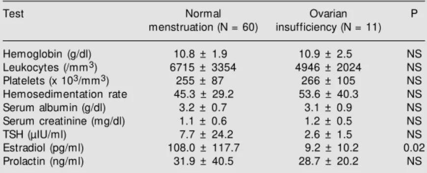
There was no significant difference be-tween groups in terms of laboratory meas-urements of hemoglobin, leukocytes, plate-lets, hemosedimentation rate, serum albu-min, creatinine, TSH or prolactin. In the sample studied, TSH levels were higher than normal (more than 5 µIU) in 11.9% of the women, and approximately 20% were posi-tive for antimicrosomal and antithyroglobu-lin antibodies. Prolactin levels were higher (more than 25 ng/ml) in 21 of the 52 tests performed (40.4%). The only statistically significant difference in laboratory results between groups was with respect to estradiol levels (Table 3).
Of the 26 patients (36.6%) who had used or were still using cyclophosphamide pulse therapy, nine presented ovarian insufficiency at the time of the study, resulting in a preva-lence of 34.6%. Eight patients of this group had performed TSH tests, with elevated lev-els in all of them (100%). Only three of 15 patients (20%) who were menstruating regu-larly presented elevated TSH levels. Some other clinical characteristics of the patients that used cyclophosphamide are shown in Table 4.
The risk of development of ovarian in-sufficiency in women using venous cyclo-phosphamide was 7.8 times higher than that in the group not using the immunosuppres-sive drug (RR = 7.8; 95% CI: 1.8-33.3; P = 0.0007). Patients treated with a cumulative cyclophosphamide dose greater than 10 g were more likely to develop ovarian insuffi-ciency than patients receiving a cumulative dose lower than 10 g (60% vs 18.7%) and had
a 3.2 times higher risk for ovarian insuffi-ciency (RR = 3.2; 95% CI: 1.02-10; P = 0.03).
Table 2. Gynecologic-obstetric data of patients w ith systemic lupus erythematosus according to the presence or absence of ovarian insufficiency.
Variable Normal Ovarian P
menstruation (N = 60) insufficiency (N = 11)
Age at menarche (mean ± SD) 13.0 ± 1.7 12.4 ± 1.7 NS
Number of gestations (mean ± SD) 2 ± 2 3 ± 1.7 NS
Number of miscarriages (mean ± SD) 0.4 ± 0.7 0.2 ± 0.4 NS
Birth control methods used (% )
Oral contraceptive 29 (48.3) 7 (63.6)
Condom 17 (28.3) 0.0
Calendar method 6 (10.0) 0.0
Intrauterine device 1 (1.6) 0.0
Tubal ligation 17 (28.3) 6 (54.5)
None 16 (26.7) 3 (27.3)
SD: standard deviation; NS: not statistically significant (M ann-Whitney test).
Table 3. Laboratory values for patients w ith systemic lupus erythematosus, according to the presence or absence of ovarian insufficiency.
Test Normal Ovarian P
menstruation (N = 60) insufficiency (N = 11)
Hemoglobin (g/dl) 10.8 ± 1.9 10.9 ± 2.5 NS
Leukocytes (/mm3) 6715 ± 3354 4946 ± 2024 NS
Platelets (x 103/mm3) 255 ± 87 266 ± 105 NS
Hemosedimentation rate 45.3 ± 29.2 53.6 ± 40.3 NS
Serum albumin (g/dl) 3.2 ± 0.7 3.1 ± 0.9 NS
Serum creatinine (mg/dl) 1.1 ± 0.6 1.2 ± 0.5 NS
TSH (µIU/ml) 7.7 ± 24.2 2.6 ± 1.5 NS
Estradiol (pg/ml) 108.0 ± 117.7 9.2 ± 10.2 0.02
Prolactin (ng/ml) 31.9 ± 40.5 28.7 ± 20.2 NS
Data are reported as means ± SD. NS: not statistically significant (Student t-test). Reference values: thyroid-stimulating hormone (TSH): 0.3-5.0 µIU/ml; prolactin: up to 25 ng/ml; estradiol (menopause): 0-14 pg/ml.
Table 4. Characteristics of patients w ith systemic lupus erythematosus w ho had used venous cyclophosphamide (N = 26).
Variable Normal Ovarian P
menstruation (N = 17) insufficiency (N = 9)
Age at time of study (mean ± SD) 28.9 ± 6.8 35.8 ± 5.4 0.01
Age at the beginning of CF use 27.0 ± 6.6 31.4 ± 5.7 0.10
(mean ± SD)
Cumulative CF dose in grams 9.1 ± 9.8 18.9 ± 13.1 0.04
(mean ± SD)
SLEDAI (mean ± SD) 18.1 ± 12.0 5.7 ± 7.4 0.01
Disease period in years (mean ± SD) 4.0 ± 3.6 8.9 ± 4.3 0.005
Prolactin (mean ± SD) 49.4 ± 63.8 29.3 ± 21.7 NS
Elevated TSH (% ) 3/15 (20.0)* 8/8 (100.0)* * NS
Smoking (% ) 11.7 11.1 NS
SD: standard deviation; NS: not statistically significant (M ann-Whitney and chi-square tests); CF: cyclophosphamide; SLEDAI: systemic lupus erythematosus disease activity index.
* Three elevated thyroid-stimulating hormone (TSH) readings (>5 µIU/ml) in 15 tests performed in this group.
Logistic regression analysis was em-ployed using complete data for 68 patients in order to study the risk factors for ovarian insufficiency. Ovarian failure was the de-pendent variable, and patient age, duration of disease, age at time of diagnosis, age at onset of menarche, SLEDAI score and cu-mulative dose of cyclophosphamide were found to be the predictive variables. Cyclo-phosphamide dosage alone proved to be an independent risk factor for ovarian insuffi-ciency (OR = 1.31; P = 0.01; 95% CI: 1.06-1.63).
D iscussio n
Ovarian failure, especially premature menopause, should be a constant concern in the management of patiens with SLE. The disease is generally more prevalent among women of reproductive age, and premature interruption of estrogen production may give rise to a higher risk of cardiovascular dis-ease, osteoporosis and infertility, among other estrogen deficiency-related symptoms. Amen-orrhea is the most common menstrual disor-der in SLE and is associated with disease activity, stress and drugs used. Although the toxic effects of cyclophosphamide on ovar-ian function have been observed since the 1960s in patients with rheumatoid arthritis using oral cyclophosphamide (24), many is-sues pertaining to gonadal insufficiency in patients with SLE remain unexplained to this day. There are very few articles in the medical literature that deal with these issues (3-8,12-14,25-27), and the majority focus on the incidence of ovarian failure after cyclo-phosphamide use.
The present study was conducted on a group of 71 women diagnosed with SLE with an ovarian insufficiency rate of 15.5%, with 11.3% of cases fulfilling diagnostic criteria for premature menopause, as con-firmed by hormone level testing. When one considers the fact that in the general popula-tion the mean age of menopause is
approxi-mately 49-51 years (7,28) and that prema-ture menopause is encountered in only 1% (29)of women, it may be concluded that lupus women are at higher risk for premature menopause and its consequences.
The clinical and laboratory characteris-tics of the present patients are in accordance with the medical literature. Ovarian failure was related to the age of the patient at the time of this study, as well as to the use and cumulative dosage of cyclophosphamide. Approximately 80% of the amenorrheic women had used or were using a cyclophos-phamide pulse therapy scheme, and a cumu-lative dose higher than 10 g increased by more than three-fold the risk of development of ovarian failure. The cumulative dose of cyclophosphamide was significantly higher in patients with ovarian failure than in women who were menstruating regularly. The rate of ovarian insufficiency in women treated with cyclophosphamide was approximately 35%. These results agree with those reported in the literature (4-8). Boumpas et al. (4) compared three groups of patients with a diagnosis of SLE: 16 received a monthly cyclophosphamide pulse therapy for a total of seven doses, 23 received 15 doses of cyclophosphamide pulse therapy, and 16 pa-tients were treated with nine monthly doses of methylprednisolone pulse therapy. The rates of permanent amenorrhea within the three groups were 12, 39 and 0%, respec-tively, and the rates of ovarian insufficiency became proportionally higher as the age of the patients increased (£25 years: 12%; 26-30 years: 27%; ³31 years: 62%). In the study performed by Wang et al. (6), 92 patients treated with oral cyclophosphamide pre-sented with a permanent amenorrhea rate of 27%, and patient age at the initiation of treatment and cumulative dose of cyclophos-phamide were associated with ovarian insuf-ficiency. Other studies which employed dos-ages of 1-4 mg kg-1 day-1 of oral
53-71% of the patients (13,30-32). In a more recent retrospective cohort study by Mok et al.(8), of the 70 women treated with cyclo-phosphamide, 18 (26%) developed ovarian insufficiency, with a higher rate in patients who had received the oral immunosuppressor (oral cyclophosphamide: 30%; venous cy-clophosphamide: 13%). Again, patient age at the beginning of the study and cumulative dose of cyclophosphamide were independ-ent risk factors for this complication. In the group with ovarian failure, the cumulative dose of cyclophosphamide was higher than in the group without gonadal insufficiency (28.8 vs 15.4 g). Comparing these results
with those of the present study, it can be observed that the mean dose of 15.4 g in the group without ovarian insufficiency in the Mok et al. (8) study is close to the cumulative dose of 18.9 g observed in the group of women with gonadal failure in the present study. However, it is still impossible to de-termine the contribution of the exact cumu-lative dose considered toxic to the develop-ment of ovarian failure. Only prospective studies can answer this question.
The mechanisms of induction of ovarian toxicity by cyclophosphamide have been well demonstrated through animal studies (33,34). Intraperitoneal injection of 100 mg/kg of cyclophosphamide reduces the ovarian fol-licles of mice by approximately 63%, thereby reducing the estrogen production. Through a feedback mechanism, there is elevation of FSH levels, which accelerates development of new primordial ovarian follicles that are more sensitive to the toxic effects of cyclo-phosphamide. Maintenance of this vicious cycle leads to heightened depletion of the ovarian follicles. Knowledge of these patho-genetic mechanisms have given rise to stud-ies of methods to protect the ovarstud-ies from cyclophosphamide toxicity (26,35).
The reason for an increased risk of ovar-ian failure in women who start cyclophos-phamide therapy at later stages of lupus is that, under normal circumstances, the
num-ber of ovarian follicles decreases markedly with age, until the last decade prior to meno-pause. Use of cyclophosphamide in this phase accelerates depletion of the ovarian follicles. An interesting fact encountered in the present study was the higher clinical activ-ity, measured by SLEDAI, in normally men-struating patients. This result contradicts the traditional idea, stemming from uncontrolled observations, that disease activity would be a cause of amenorrhea in women with SLE (1). It seems that other factors associated with disease activity are the ones that induce ovarian failure. Only prospective studies with larger population samples controlled for vari-ous misleading factors could hope to clarify this issue. A recent study by Mok et al. (27) showed that patients with cyclophosphamide-induced ovarian insufficiency who had been followed for 5 years presented much lower disease reactivity than women with regular menstruation. The authors suggest that ovar-ian insufficiency with hypoestrogenism may be a protective factor against lupus activity. Historically, in pre-corticoid times, it was observed that lupus patients improved upon menopause or when submitted to oophorec-tomy. The pathogenic role of estrogen in SLE has been reinforced by studies based on animal models, using NZB/NZW mice. Es-trogens exhibit various stimulatory effects on the immune system, including increase of prolactin secretion, which has a proinflam-matory role and may play a role in SLE activity (36). In this study, prolactin levels were increased in approximately 40% of patients, and the average values were higher in the group with regular menstruation and, therefore, in the group which presented higher disease activity as measured by SLEDAI.
Twenty to 27% of patients with premature menopause in the general population tested positive for autoantibodies (39,40), the most common types being antithyroglobulin and antimicrosomal agents. Interestingly, when the group of patients who used cyclophos-phamide in the present study was analyzed independently, 100% of the women with ovarian failure were found to present el-evated TSH levels, compared to 20% in the group with normal menstruation. These data
suggest that besides the use of cyclophos-phamide, thyroid disorder may be another risk factor for premature menopause in women diagnosed with SLE and, therefore, thyroid function should be part of the inves-tigation of women with ovarian failure. Al-though published studies have confirmed the existence of subclinical hypothyroidism in SLE (37,38), no study has investigated thyroid diseases as a possible risk factor for ovarian failure in patients with lupus.
Re fe re nce s
1. Lim GS, Petri M & Goldman D (1993). M enstruation and systemic lupus erythe-matosus: a case-control study. Arthritis and Rheumatism, 36 (Suppl 5): R23 (Ab-stract).
2. M oncayo-Naveda H, M oncayo R, Benz R, Wolf A & Lauritzen CH (1989). Organ-specific antibodies against ovary in pa-tients w ith systemic lupus erythemato-sus. American Journal of Obstetrics and Gynecology, 160: 1227-1229.
3. Pasoto SG, Viana VST, M endonça BB, Yoshinari NH & Bonfa E (1999). Anti-cor-pus luteum antibody: a novel serological marker for ovarian dysfunction in systemic lupus erythematosus? Journal of Rheu-matology, 26: 1087-1093.
4. Boumpas DT, Austin HA, Vaughan EM , Yarboro CH, Klippel JH & Balow JE (1993). Risk for sustained amenorrhea in patients w ith systemic lupus erythematosus re-ceiving intermittent pulse cyclophospha-mide therapy. Annals of Internal M edi-cine, 119: 366-369.
5. Gonzalez-Crespo M R, Gomez-Reino JJ, M erino R, Ciruelo E, Gomez-Reino FJ, M oley R, Garcia-Consuegra J, Pinillos V & Rodriguez-Valverde V (1995). M enstrual disorders in girls w ith systemic lupus erythematosus treated w ith cyclophos-phamide. British Journal of Rheumatol-ogy, 34: 737-741.
6. Wang CL, Wang F & Bosco JJ (1995). Ovarian failure in oral cyclophosphamide treatment for systemic lupus erythemato-sus. Lupus, 4: 11-14.
7. M cDermott EM & Pow ell RI (1996). Inci-dence of ovarian failure in systemic lupus erythematosus after treatment w ith pulse cyclophosphamide. Annals of the Rheu-matic Diseases, 55: 224-229.
8. M ok CC, Lau CS & Wong RWS (1998). Risk factors for ovarian failure in patients w ith systemic lupus erythematosus re-ceiving cyclophosphamide therapy. Arthri-tis and RheumaArthri-tism, 41: 831-837. 9. Ordi-Ros J, Cortes F, M artinez N, M auri
M , De Torres I & Vilardell M (1998). Tha-lidomide induces amenorrhea in patients w ith lupus disease. Arthritis and Rheuma-tism, 41: 2273-2275.
10. Ordi-Ros J, Cortes F, Cucurull E, M auri M , Bujan S & Vilardell M (2000). Thalidomide in the treatment of cutaneous lupus re-fractory to conventional therapy. Journal of Rheumatology, 27: 1429-1433. 11. Lieman H & Santoro N (1997). Premature
ovarian failure: a modern approach to di-agnosis and treatment. Endocrinology, 7: 314-321.
12. Warne GL, Fairley KF, Hobbs JB & M artin FIR (1973). Cyclophosphamide induced ovarian failure. New England Journal of M edicine, 289: 1159-1162.
13. Austin HA, Klippel JH, Balow JE, LeRiche NGH, Steinberg AD, Plotz PH & Decker JL (1986). Therapy of lupus nephritis: con-trolled trial of prednisolone and cytotoxic drugs. New England Journal of M edicine, 314: 614-619.
14. Langevitz P, Klein L, Pras M & M any A (1992). The effect of cyclophosphamide pulses on fertility in patients w ith lupus nephritis. American Journal of Reproduc-tive Immunology, 28: 157-158.
15. Van der Schoun YT, van der Graaf Y, Steyyerberg EW, Eijkemans JC & Banga JD (1996). Age at menopause as a risk factor for cardiovascular mortality. Lancet, 347: 714-718.
16. Fogelman I (1996). The effects of oestro-gen deficiency on the skeleton and its
prevention. British Journal of Obstetrics and Gynaecology, 103 (Suppl 14): 5-9. 17. M ok CC, Lau CS, Ho CT, Lee KW, M ok
M Y & Wong RW (1998). Safety of hormo-nal replacement therapy in postmeno-pausal patients w ith systemic lupus ery-t hem aery-t osus. Scandinavian Journal of Rheumatology, 27: 342-346.
18. Lahita RG (1999). The role of sex hor-mones in systemic lupus erythematosus. Current Opinion in Rheumatology, 11: 352-356.
19. Le Thi Huong D, Wechsler B & Piette JC (2000). Effect of pregnancy, menopause and hormone substitution therapy on dis-seminated systemic lupus erythemato-sus. Presse M edicale, 29: 55-60. 20. Tan EM , Cohen AS, Fries JF, M asi AT,
M cShane DJ, Rothfield NF, Schaller JG, Talal N & Winchester RJ (1982). The 1982 revised criteria for the classification of sys-temic lupus erythematosus. Arthritis and Rheumatism, 25: 1271-1277.
21. Gladman DD, Goldsmith CH, Urow itz M B, Bacon P, Bom bardier C, Isenberg D, Kalunian K, Liang M H, M addison P & Nived O (1994). Sensitivity to change of 3 systemic lupus erythematosus disease activity indices: international validation. Journal of Rheumatology, 21: 1468-1471. 22. Fox DA & M cCune WJ (1994). Immuno-suppressive drug therapy of systemic lu-pus erythematosus. Rheumatic Diseases Clinics of North America, 20: 265-299. 23. Kleinbaum DG & Kupper LL (1978).
Ap-plied Regression Analysis and Other M ul-tivariable M ethods. Duxbury Press, Bos-ton, M A, USA.
Rheu-matism, 11: 151-161.
25. Belm ont HM , St orch M , Buyon J & Abramson S (1995). New York University/ Hospital for Joint Diseases experience w ith intravenous cyclophosphamide treat-ment: efficacy in steroid unresponsive lu-pus nephritis. Lupus, 4: 104-108. 26. Blumenfeld Z & Haim N (1997).
Preven-tion of gonadal damage during cytotoxic therapy. Annals of M edicine, 29: 199-206. 27. M ok CC, Wong RWS & Lau CS (1999). Ovarian failure and flares of systemic lu-pus erythematosus. Arthritis and Rheu-matism, 42: 1274-1280.
28. M cKinlay SM , Brambilla DJ & Posner JG (1992). The normal menopause transition. M aturitas, 14: 103-115.
29. Caulam CB (1982). Premature gonadal fail-ure. Fertility and Sterility, 38: 645-655. 30. Steinberg AD & Steinberg SC (1991).
Long-term preservation of renal function in patients w ith lupus nephritis receiving treatment that includes cyclophospha-mide versus those treated w ith pred-nisone only. Arthritis and Rheumatism, 34: 945-950.
31. Balow JE, Aust in HA, Tsokos GC, Antonovych TT, Steinberg AD & Klippel JH (1987). NIH Conference Lupus Nephri-tis. Annals of Internal M edicine, 106: 79-94.
32. Balow JE, Austin III HA, M uenz LR, Joyce KM , Antonovych TT, Klippel JH & Stein-berg AD (1984). Effect of treatment on the evolution of renal abnormalities in lu-pus nephritis. New England Journal of M edicine, 311: 491-495.
33. Ataya KM , Valeriate FA & Ramahi-Ataya A (1981). Effects of cyclophosphamide, aza-thioprine, and 6-mercaptopurine on oo-cyte and follicle number in C57BL/6N m ice. Research Com m unicat ions in Chemical Pathology and Pharmacology, 31: 155-161.
34. Shiromizu K, Thorgeirsson SS & M attison DR (1984). Effect of cyclophosphamide on oocyte and follicle numbers in Sprague-Daw ley rat s, C57BL/6N and DBA/ZN mice. Pediatric Pharmacology, 4: 213-221. 35. M ontz FJ, Wolff AJ & Gambone JCG (1991). Gonadal protection and fecundity rates in cyclophosphamide treated rats.
Cancer Research, 51: 2124-2126. 36. Grossman CJ (1989). Possible underlying
mechanisms of sexual dimorphism in the immune response, fact and hypothesis. Journal of Steroid Biochemistry, 34: 241-251.
37. M agaro M , Zoli A, Altomonte L, M irone L, La Sala L, Barini A & Scuderi F (1992). The association of silent thyroiditis w ith active systemic lupus erythematosus. Clinical and Experimental Rheumatology, 10: 67-70.
38. Chan AT, Al-Saffar Z & Bucknall RC (2001). Thyroid disease in systemic lupus erythe-matosus and rheumatoid arthritis. Rheu-matology, 40: 353-354.
39. Rebar RW & Connolly HV (1990). Clinical features of young w omen w ith hypergo-nadotrophic amenorrhea. Fertility and Ste-rility, 53: 804-810.