Related Cancers: Potential Markers of Progression
Heather Pressler1,4, Tristan M. Sissung2, David Venzon3, Douglas K. Price1, William D. Figg1,2*
1Molecular Pharmacology Section, National Cancer Institute, Bethesda, Maryland, United States of America,2Clinical Pharmacology Program, Medical Oncology Branch, National Cancer Institute, Bethesda, Maryland, United States of America,3Biostatistics and Data Management Section, National Cancer Institute, Bethesda, Maryland, United States of America,4Johns Hopkins University, Baltimore, Maryland, United States of America
Abstract
The organic anion transporting polypeptide (OATP) family of transporters has been implicated in prostate cancer disease progression probably by transporting hormones or drugs. In this study, we aimed to elucidate the expression, frequency, and relevance of OATPs as a biomarker in hormone-dependent cancers. We completed a study examining SLCO1B3, SLCO1B1andSLCO2B1mRNA expression in 381 primary, independent patient samples representing 21 cancers and normal tissues. From a separate cohort, protein expression of OATP1B3 was examined in prostate, colon, and bladder tissue. Based on expression frequency, SLCO2B1 was lower in liver cancer (P = 0.04) which also trended lower with decreasing differentiation (P = 0.004) and lower magnitude in pancreatic cancer (P = 0.05).SLCO2B1also had a higher frequency in thyroid cancer (67%) than normal (0%) and expression increased with stage (P = 0.04).SLCO1B3was expressed in 52% of cancerous prostate samples and increased SLCO1B3 expression trended with higher Gleason score (P = 0.03). SLCO1B3 expression was also higher in testicular cancer (P = 0.02).SLCO1B1expression was lower in liver cancer (P = 0.04) which trended lower with liver cancer grade (P = 0.0004) and higher with colon cancer grade (P = 0.05). Protein expression of OATP1B3 was examined in normal and cancerous prostate, colon, and bladder tissue samples from an independent cohort. The results were similar to the transcription data, but showed distinct localization. OATPs correlate to differentiation in certain hormone-dependent cancers, thus may be useful as biomarkers for assessing clinical treatment and stage of disease.
Citation:Pressler H, Sissung TM, Venzon D, Price DK, Figg WD (2011) Expression of OATP Family Members in Hormone-Related Cancers: Potential Markers of Progression. PLoS ONE 6(5): e20372. doi:10.1371/journal.pone.0020372
Editor:Mikhail V. Blagosklonny, Roswell Park Cancer Institute, United States of America
ReceivedJanuary 20, 2011;AcceptedMay 1, 2011;PublishedMay 19, 2011
This is an open-access article, free of all copyright, and may be freely reproduced, distributed, transmitted, modified, built upon, or otherwise used by anyone for any lawful purpose. The work is made available under the Creative Commons CC0 public domain dedication.
Funding:This study was supported in part by the Intramural Research Program of the National Institutes of Health, National Cancer Institute, Bethesda, MD www.nih.gov. Stipend support was provided for Ms. Pressler by the National Science Foundation www.nsf.gov and Department of Defense Prostate Cancer Research Training Award#PC094258 www.grants.gov. The funders had no role in study design, data collection and analysis, decision to publish, or preparation of the manuscript. No additional external funding received for this study.
Competing Interests:The authors have declared that no competing interests exist.
* E-mail: [email protected]
Introduction
Organic anion-transporting polypeptides (OATPs; encoded by
SLCOgenes) are involved in hepatocyte influx mechanisms and are overexpressed in some cancers [1,2,3]. Known ligands of the OATP family include myriad endogenous substrates such as: steroids (i.e., estrogen sulfate, testosterone, and DHT), bile acids, and peptides [4]. OATP family members also influx a variety of pharmaceuticals, including (but not limited to): antihistamines, blood-glucose lowering drugs, statins, heart medications, and anticancer agents [4]. Therefore, OATPs are emerging as important transporters in the treatment of cancer from the standpoints of drug distribution and disease outcomes, and it is expected that many more endogenous and exogenous substrates will be identified by future studies.
Expression and genetic variation in OATPs have been associated with clinical outcomes in patients with prostate cancer. Both
SLCO1B3 and SLCO2B1have been related to a shorter time to androgen deprivation therapy failure and decreased overall survival in patients with hormone responsive prostate cancer, and castration resistant prostate cancer (CRPC) respectively [1,5,6]. Mechanisti-cally, these associations might be attributed to genetic variation in androgen (or other steroid hormone) influx mechanisms that
increase androgen scavenging during androgen deprivation therapy (ADT) [1]. Although OATP1B3 influx mechanisms are still unknown in colon cancer, there is sufficient evidence suggesting that OATP1B3 is overexpressed in a large proportion of colon tumors, and that this overexpression contributes to cell survival in the presence of oxalaplatin and camptothecin; the latter effect may be related to p53 expression [2].
OATPs have also been shown to contribute to systemic variation in anti-cancer drug treatments. For instance, OATP1B3 was initially identified as a high-affinity hepatocellular transporter of paclitaxel, and is also the most efficient liver uptake transporter of docetaxel [7,8]. OATP1B1 and OATP1B3 also influx SN-38 [4], and this transport may have implications in the treatment of colon cancer [2]. However, the tumor influx mechanisms have yet to be explored for these drugs in relevant diseases, and this effort has been hindered by the lack of studies that have evaluated tumor expression of OATPs.
Materials and Methods
Clinical samples
Samples were obtained from Origene (TissueScan Cancer Survey Panel III, Rockville, MD). Briefly, RNA was isolated from patients of mixed age, clinical diagnosis, and with various tumor stages. cDNA was created, normalized to actin B, checked for contamination and sensitivity (as determined by the manufactur-er). The panel includes 384 samples encompassing 22 cancers and matched normal tissues. The numbers of samples corresponding to different tumor types and the number of samples derived from different tumor grades are reported inTable S1.
Determination of mRNA expression
SLCO1B3 (Hs00251986_m1), SLCO2B1 (Hs00200670_m1), and SLCO1B1 (Hs00272374_m1) expression levels were analyzed using Taqman Gene Expression Assays (Applied Biosystems). Briefly, the primers were mixed with Taqman Master Mix (No Amp Erase) and distilled water then added to the samples. The plates were run in duplicate on Mx3005P QPCR System (Agilent Technologies) for 10 minutes 95uC then cycling for 42 cycles of 30 seconds for 95uC and 1 minute 60uC then FAM (ROX reference) reading after every cycle. Expression of each SLCO gene was normalized to actin by determining the cycle threshold (Ct) for each gene by the following formula: 2‘-(Ct
SLCO-CtActinB)*105(i.e., theDCt). In cases where mRNA samples were below the LLOQ, the samples were noted to haveSLCOmRNA levels at just below LLOQ (i.e. 42.1 cycles) and were normalized to actin as described above. This is considered to be a conservative adjustment as there were a large number of samples with undetectableSLCOmRNA levels that were likely to be significantly lower than the LLOQ.
Determination of protein expression
Tissue section arrays for bladder, colon, and prostate were obtained from Pantomics (Richmond, CA). The prostate tumor tissue array includes benign prostatic hyperplasia and cancerous tissue; the normal and tissue samples were not matched per patient. Bladder and colon cancer arrays were also obtained from the above company and these included uninvolved and cancerous tissue. All tissues were obtained from surgical resection and fixed in 10% neutral buffered formalin for 24 hours. Slides were preparing by baking for 1 hour 60uC before removing the paraffin in xylene, then prepping for staining in 100% ethanol, 95% ethanol, 70% ethanol, and distilled water washes. One slide per set was stained with hematoxylin and eosin. The remaining slides were processed for immunoflourescence by placing in 95uC 10 mM sodium citrate buffer for 5 minutes then allowing to cool for 20 minutes. The slides were blocked for 30 minutes before primary antibody against OATP8 was applied 1:100 (Progen) at 4uC overnight. Slides were washed in Tris-Buffered Saline Tween before applying the secondary FITC goat anti-mouse 1:400 (Abcam) for 2 hours. Slides were washed again in TBST and mounted with VectaShield (Vector Labs) with DAPI. Images were taken on an Olympus BX51 microscope with UPlanFl 40x and 10x lenses. InSight Firewire camera and Spot version 4.5 imaging software were used to capture images. Adobe Photoshop CS3 version 10 was used for after-capture edits where all photos were processed the same.
Statistical considerations
Comparisons between the frequency of SLCO or OATP expression in normal versus tumor tissues was conducted using Fisher’s exact test. Comparisons were made between the magnitude of SLCOmRNA expression versus the corresponding normal tissue using the Wilcoxon rank sum test; these data are
reported as the mean DCT (95% confidence interval; CI) and corresponding fold-change from normal tissue. The Cochran-Armitage trend test was employed to determine if the frequency of
SLCOor OATP expression varied with tumor cell differentiation or stage, while the Jonckheere-Terpstra trend test was used to determine if the magnitude of SLCO mRNA expression varied with tumor cell differentiation or stage. Assessment of protein expression magnitude was not possible, although we did note if tumors expressed detectable levels of OATP1B3 or not. Only two-tailedP-values are reported, and given the exploratory nature of this study,P-values are reported as significant if P,0.05; however, it should be noted that conservative non-parametric and exact tests were performed in order to reduce the potential for false positives.
Results
Expression of SLCO mRNA in normal and tumor tissues
We first hypothesized that SLCO expression would be a biomarker for multiple different hormone-dependent tumor types. To this end,SLCOmRNA expression was determined in samples derived from normal human tissue and corresponding tumor tissue. Expression frequencies were determined for SLCO1B3,
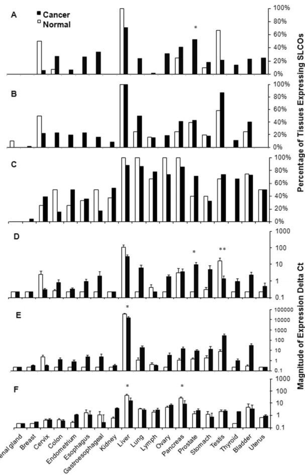
SLCO1B1, and SLCO2B1, and the percent of normal or tumor tissues expressing SLCOs are reported (see Figure 1A–C
respectively). Prostate tumors expressed SLCO1B3 much more frequently than normal prostate tissues (62% vs. 0%,n= 21 and
n= 5 respectively;P= 0.04) and more frequently with increasing Gleason score (P= 0.03). In addition,SLCO1B3expression was less frequent in testicular tumors (21% vs. 67%, n= 19 and n= 6 respectively;P= 0.06). SLCO1B1was frequently expressed signif-icantly higher with decreasing differentiation in colon tumors (P= 0.04). SLCO2B1 was not expressed significantly different in cancer compared to normal tissue. In addition to the above observations, it should be noted that there were several cases where there was no SLCO expression in normal tissues, but expressed in cancerous samples. While not statistically significant due to low power, some tumor tissues haveSLCOexpression and should be evaluated more closely in future studies. These tumors included (forSLCO1B3); colon, endometrium, esophagus, gastro-esophageal, kidney, ovary, and thyroid, (for SLCO1B1); cervix, endometrium, esophagus, gastroesophageal, lung, lymph, ovary, thyroid, bladder, and uterus (for SLCO2B1); colon, bladder (See
Table S1for comprehensive data analysis).
As some tumor tissues had similarSLCOexpression frequencies as normal tissues, we also ascertained if the magnitude ofSLCO
expression was different for SLCO1B1,SLCO1B3, and SLCO2B1
(see Figure 1D–F respectively). Significant differences in expression levels (i.e. mean DCtnormal (95%CI) vs. DCttumor (95%CI)) between normal and tumor tissue were observed for
SLCO1B3in prostate cancer [0.23 (0.23 to 0.23) versus 104 (44 to 164); 45.7-fold increase;P= 0.03] and testicular cancer [174 (0.3 to 350) versus 14.0 (0.3 to 26); 12.8-fold decrease; P= 0.01]. Significant increases were seen for SLCO1B1 in liver cancer [396000 (225000 to 567000) versus 161000 (41000 to 280000); 2.4 fold decrease; P =0.04]. SLCO2B1 expression decreased signifi-cantly in pancreatic cancer [27.5 (9.5 to 45) versus 9.1 (5.5 to 12.8); 3.0 fold decrease;P= 0.05] and liver cancer [47.3 (16.1 to 78) versus 17.0 (4.9 to 29); 2.7 fold decrease;P= 0.04]. A more comprehensive analysis of expression differences is provided in
Table S1.
OATP expression versus tumor differentiation and stage
hypoth-Figure 1. Expression profile of SLCOs in normal and neoplastic tissues.Data are first expressed as a percentage of tissues with mRNA expression of A)SLCO1B3B)SLCO1B1, and C)SLCO2B1, and then as the magnitude of normalized mRNA expression of D)SLCO1B3, E)SLCO1B1, and F)
esized that SLCO expression would be related to increasing tumor differentiation or stage. For prostate tumors, expression of
SLCO1B3increased along with Gleason score up to 67-fold [mean
DCT (95%CI) = 0.23 (0.23 to 0.23), 2.6 (24 to 9.2), 8.5 (20.7 to 17.7), 15.5 (213 to 44), 15.0 (2.4 to 27), for normal tissue, and Gleason = 6, 7, 8, 9 respectively;P= 0.03;Figure 2A].SLCO1B1
expression was related to differentiation in liver cancer (395000 (225000 to 567000), 175000 (4300 to 310000), 47000 (11000 to
83000), and 3500 (N/A) for normal tissue, and well, moderately, and poor differentiated tumor tissue respectively; P= 0.0004,
Figure 2B) up to a274 fold difference.SLCO1B1expression was also related colon cancer (0.23 (0.23 to 0.23), 0.23 (0.23 to 0.23), 1.3 (21.0 to 3.6), and 2.9 (N/A) for normal tissue, and well, moderately, and undifferentiated tumor tissue respectively;
P= 0.05, Figure 2C) up to a 12.5 fold difference. SLCO2B1
expression was also correlated negatively to differentiation in liver
Figure 2. SLCO mRNA expression correlates to differentiation in cancer.SLCO1B3mRNA expression by differentiation in A) prostate P = 0.03.
SLCO1B1expression in B) liver P = 0.0004, C) colon P = 0.05.SLCO1B2expression in D) liver P = 0.005. E)SLCO2B1expression by stage in thyroid P = 0.04. All were found as signifcant correlations by the Jonckheere-Terpstra trend test.
cancer (47 (4.4 to 82), 13 (23.2 to 28), 3.1 (1.4 to 4.7), and 1.7 (N/ A) for normal tissue and well, moderately, and poor differentiated tumor tissue respectively; P= 0.005) up to a 26-fold change (Figure 2D).
The expression and frequency data was also analyzed with respect to stage.SLCO2B1expression increased with stage up to a 29 fold-difference in thyroid cancer (P = 0.04). SeeFigure 2E. For normal and stage I, II, III, IVA the increase was 0.23 (0.23 to 0.23), 1.4 (0.6 to 2.1), 5.6 (20.8 to 12), 6.7 (20.4 to 14), 1.0 (N/A). Due to limited samples numbers for each stage no other relationships were statistically significant. However, other notable trends wereSLCO2B1decreased expression by stage in pancreatic cancer,SLCO1B3increased frequency by stage in prostate cancer,
andSLCO1B1decreased expression by stage in liver cancer. For the full data set seeTable S2.
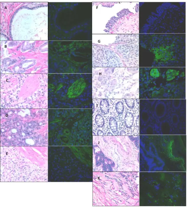
OATP protein expression versus tumor differentiation
To determine if OATP translation is similar to the quantitiative PCR, we conducted immunoflourescence on tissue samples from prostate, colon, and bladder cancer to detect OATP1B3. OATP2B1 was not chosen for protein validation because it is expressed both in normal and cancerous tissues. For prostate tumors, expression of OATP1B3 was primarily observed in prostate tumors and not normal tissues (P =0.001; Figure 3A–E and Table 1) and was highly expressed in the stroma. Moreover, the
frequency of OATP1B3 expression varied significantly with Gleason score (P= 0.001; Table 1). Expression was also more frequent in colon cancer (P =0.06,Figure 3F–HandTable 1) and trended towards an association with a statistically significant higher incidence of expression in colon cancer by Cochran-Armitage test (P =0.02;Table 1). OATP1B3 expression was observed primarily in the vasculature in colon cancer and invasive, cancerous epithelial cells in bladder cancer. Consistent with the mRNA data, OATP1B3 expression was not associated with increasing histological grades of bladder cancer (P= 0.34; see Table 1 and Figure 3I–K), but trended towards an association with bladder cancer grades despite a low number of samples (P= 0.07; seeTable 1).
Discussion
SLCO(and OATP) overexpression has already been shown to be an important factor in colon and prostate cancer (1,2); moreover variation in OATP function [1] appears to be related to clinical outcome of endocrine therapy in prostate cancer [5] as well as overall survival of men CRPC [1]. Previous reports also demonstrated significant decreases in expression of SLCOs in hepatocellular carcinoma; this phenomenon is most likely associated with the reduction of metabolic function due to dedifferentiation in liver tumors derived from patient samples [9]. The present study confirms the above observations and demonstrates for the first time thatSLCOexpression variability is also associated with several other tumor types, including: colon cancer, liver cancer, pancreatic cancer, prostate cancer, testicular cancer, and thyroid cancer. Despite previously published reports,
SLCO1B3, SLCO2B1, andSLCO1B1were not highly expressed in our breast cancer samples [10,11].
We also demonstrated that SLCO1B3 expression levels are significantly related to the Gleason score in prostate cancer.
SLCO1B1expression levels were significantly related to the degree
of differentiation in liver and colon cancers.SLCO2B1expression was also significantly related to liver and thyroid cancer. Immunoflourescent for OATP1B3 showed distinct localization in colon, bladder, and prostate cancers. The vasculature was stained for OATP1B3 only in high-grade colon cancer, cancerous epithelia in bladder cancer, and the stroma in prostate cancer. Thus OATP1B3 could be involved either directly or indirectly supporting carcinogensis depending on the cancer type. Previously mentioned studies suggest that variation in the expression of OATPs is likely involved in underlying disease etiology through
Table 1.OATP Expression in Immunoflourescent Tissue Sections.
Tissue Differentiation Express Total P value
Prostate 0.001
BPH 7(26%) 27 0.001
Gleason 4 4(44%) 9
Gleason 5 4(57%) 7
Gleason 6 5(71%) 7
Gleason 7 4(100%) 4
Gleason 8 16(64%) 25
Gleason 9 9(64%) 14
Colon 0.06
Normal 7(21%) 32 0.02
II–I 3(38%) 8
II 4(50%) 8
III 6(50%) 12
Bladder 0.34
Normal 5(21%) 24 0.07
II–I 1(17%) 6
III–II 3(33%) 9
III 4(50%) 8
P-value = Fischer’s exact orCochran-Armitage trend test. doi:10.1371/journal.pone.0020372.t001
Table 2.Select Substrates of OATPs[3,21].
Substrate
OATP1B1
Anti-Cancer Drugs ACU-154
antamanide Bamet-R2 Bamet-UD2 demethyl phalloidin dihydromicrocystin-LR irinotecan ketoconazole methotrexate paclitaxel PKI-166 SN-38 Hormones estrone-3-sulfate estradiol-17B-glucuronide prostaglandin E2 thyroxine (T4) triiodothyronine (T3) OATP1B3
Anti-Cancer Drugs demethylphalloin
dihydromicrocystin-LR docetaxel imatinib irinotecan methotrexate paclitaxel SN-38 Hormones DHEAS testosterone estrone-3-sulfate estradiol-17B-glucuronide thyroxine (T4) triiodothyronine (T3) OATP2B1
Anti-Cancer Drugs unknown
Hormones DHEAS
estrone-3-sulfate
pregnenolone sulfate
prostaglandin E2
the influx of endocrine factors.
OATPs influx several steroid hormones (i.e. DHEA, testoster-one, DHT, and estrogen-sulfate) that underlie the etiology of several different diseases. For clarity, a table of selected OATP substrates is included (Table 2). Testosterone is a substrate for OATP1B3 and may explain its role in prostate[1], bladder [12,13], and testicular [14] tumor development and progression. OATP1B3 and OATP1B1 also influx estrone-3-sulfate and thyroxine and these hormones play a major role in ovarian [15] and thyroid cancers [16] respectively. The OATP substrates could also inhibit cancer growth, thus be negatively selected against as the cancer progresses. For example, DHEA-S inhibits pancreatic cancer growth [17,18], which might explain the observation that
SLCO2B1expression is reduced in pancreatic cancer. In addition, the present data may also indicate a previously unknown role for the OATP-transported hormones in these cancers. Finally, OATPs transport a wide range of substances, thus there could also be unidentified or uncharacterized substrates that influence disease progression in the above-mentioned cancers.
Although it remains poorly explored, OATP expression may influence treatment success in certain cancer types (Table 2). OATP1B3 influxes docetaxel, paclitaxel, imatinib, irinotecan, SN-38, and methotrexate while OATP1B1 influxes ketoconazole, paclitaxel, and SN-38. OATP2B1 drug substrates remain poorly explored. Interestingly, these drugs are often very effective in treating metastatic diseases that develop as a result of the primary tumors that were evaluated herein. Docetaxel is approved for the treatment of CRPC and non-small cell lung cancer (NSCLC) and OATP1B3 is highly expressed in both prostate and lung tumors suggesting that the effectiveness of docetaxel treatment in these diseases may be, in part, due to sensitivity to docetaxel resulting from the underlying disease etiology. Using prostate tumors as an example, it appears that OATP1B3 is overexpressed during disease development in the primary tumor, facilitates the survival of metastatic prostate lesions during androgen deprivation therapy [1,5,19], and may be involved in the sensitivity of CRPC tumors towards docetaxel due to increased uptake. Moreover,SLCO1B1
overexpression in prostate tumors may also explain the sensitivity of prostate cancer to ketoconazole. Similar relationships may be proposed for use of paclitaxel (in ovarian, lung, and esophageal tumors), imatinib (in gastroesophageal tumors), irinotecan and SN38 (in colon and lung tumors), and methotrexate (in lung tumors). Moreover, previous data have indicated that treatment-and disease-related SLCO1B3 expression in liver result in differences in pharmacokinetic exposure to drugs [20]. We therefore propose the hypothesis that OATPs are involved in multidrug sensitivity through influx mechanisms and drugs
targeting OATP influx may be more effective in certain diseases and treatment contexts based on OATP expression in the tumor and in the liver.
Limitations in this work include low positive tumors for several sample sets, so complex analysis on differentiation and expression could not be adequately assessed. In addition, little to no differentiation data was available for testis, thyroid, lymph, uterus, and gastroesophageal samples. As mentioned above, there are several substrates of OATPs that may explain its role in the identified cancers so further follow up studies should include clinical validation as well as functional studies related to substrate transport. Clinical follow up investigation for individual cancers is clearly warranted and should include examining expression in larger cohorts and confirming the effect of transported substrates on disease progression in thyroid, endometrial, lung, ovary, pancreas, testis, and bladder cancers.
This is the first study indicating thatSLCO1B1,SLCO1B1, and
SLCO2B1is significantly expressed (or expression is reduced) in a variety to tumors, including: colon cancer, liver cancer, pancreatic cancer, prostate cancer, testicular cancer, and thyroid cancer. We also propose that OATP expression may be a biomarker in prostate and colon cancers, and knowledge of tumor expression of OATPs could guide chemotherapy treatment. We conclude OATP expression may have implications on disease etiology and effectiveness of treatment, and should be studied further for its expression in cancer.
Supporting Information
Table S1 Tissues with detectable expression of SLCO1B1, SLCO1B3, and SLCO2B1.
(XLS)
Table S2 Expression of SLCO1B1, SLCO1B3, and SLCO2B1 by cancer stage.
(XLS)
Acknowledgments
We would like to thank Dr. Maria J. Merino for assistance with pathology. We would also like to thank Michael J. Walsh for his help organizing and processing the qPCR data.
Author Contributions
Conceived and designed the experiments: HP TMS DKP WDF. Performed the experiments: HP. Analyzed the data: DV HP. Wrote the paper: HP TMS.
References
1. Hamada A, Sissung T, Price DK, Danesi R, Chau CH, et al. (2008) Effect of SLCO1B3 haplotype on testosterone transport and clinical outcome in caucasian patients with androgen-independent prostatic cancer. Clin Cancer Res 14: 3312–3318.
2. Lee W, Belkhiri A, Lockhart AC, Merchant N, Glaeser H, et al. (2008) Overexpression of OATP1B3 confers apoptotic resistance in colon cancer. Cancer Res 68: 10315–10323.
3. Hagenbuch B GC (2008) Xenobiotic transporters of the human organic anion transporting polypeptides (OATP) family. Xenobiotica 38: 778–801. 4. Sissung TM, Baum CE, Kirkland CT, Gao R, Gardner ER, et al.
Pharmacogenetics of membrane transporters: an update on current approaches. Mol Biotechnol 44: 152–167.
5. Sharifi N, Hamada A, Sissung T, Danesi R, Venzon D, et al. (2008) A polymorphism in a transporter of testosterone is a determinant of androgen independence in prostate cancer. BJU Int 102: 617–621.
6. Wright JL, Kwon EM, Ostrander EA, Montgomery B, Line DW, et al. (2011) Expression of SLCO transport genes in castration resistant prostate cancer and impact of genetic variation in SCLO1B3 and SLCO2B1 on prostate cancer outcomes. Cancer Epidemiol Biomarkers Prev.
7. Baker SD, Verweij J, Cusatis GA, van Schaik RH, Marsh S, et al. (2009) Pharmacogenetic pathway analysis of docetaxel elimination. Clin Pharmacol Ther 85: 155–163.
8. Smith NF, Acharya MR, Desai N, Figg WD, Sparreboom A (2005) Identification of OATP1B3 as a high-affinity hepatocellular transporter of paclitaxel. Cancer Biol Ther 4: 815–818.
9. Vavricka SR, Jung D, Fried M, Grutzner U, Meier PJ, et al. (2004) The human organic anion transporting polypeptide 8 (SLCO1B3) gene is transcriptionally repressed by hepatocyte nuclear factor 3beta in hepatocellular carcinoma. J Hepatol 40: 212–218.
10. Ieiri I, Higuchi S, Sugiyama Y (2009) Genetic polymorphisms of uptake (OATP1B1, 1B3) and efflux (MRP2, BCRP) transporters: implications for inter-individual differences in the pharmacokinetics and pharmacodynamics of statins and other clinically relevant drugs. Expert Opin Drug Metab Toxicol 5: 703–729.
12. Imada S, Akaza H, Ami Y, Koiso K, Ideyama Y, et al. (1997) Promoting effects and mechanisms of action of androgen in bladder carcinogenesis in male rats. Eur Urol 31: 360–364.
13. Miyamoto H, Yang Z, Chen YT, Ishiguro H, Uemura H, et al. (2007) Promotion of bladder cancer development and progression by androgen receptor signals. J Natl Cancer Inst 99: 558–568.
14. Ferlin A, Ganz F, Pengo M, Selice R, Frigo AC, et al. (2010) Association of testicular germ cell tumor with polymorphisms in estrogen receptor and steroid metabolism genes. Endocr Relat Cancer 17: 17–25.
15. Sherman ME, Madigan MP, Lacey JV, Jr., Garcia-Closas M, Potischman N, et al. (2007) Ovarian volumes among women with endometrial carcinoma: associations with risk factors and serum hormones. Gynecol Oncol 107: 431–435.
16. Blechet C, Lecomte P, De Calan L, Beutter P, Guyetant S (2007) Expression of sex steroid hormone receptors in C cell hyperplasia and medullary thyroid carcinoma. Virchows Arch 450: 433–439.
17. Giron RA, Montano LF, Escobar ML, Lopez-Marure R (2009) Dehydroepi-androsterone inhibits the proliferation and induces the death of HPV-positive and HPV-negative cervical cancer cells through an androgen- and estrogen-receptor independent mechanism. FEBS J 276: 5598–5609.
18. Melvin WS, Boros LG, Muscarella P, Brandes JL, Johnson JA, et al. (1997) Dehydroepiandrosterone-sulfate inhibits pancreatic carcinoma cell proliferation in vitro and in vivo. Surgery 121: 392–397.
19. Yang M, Xie W, Mostaghel E, Nakabayashi M, Werner L, et al. (2010) SLCO2B1 and SLCO1B3 may function as pharmacogenomic determinants of resistance to androgen deprivation therapy for prostate cancer. Journal of Clinical Oncology submitted.
20. Franke RM, Carducci MA, Rudek MA, Baker SD, Sparreboom A Castration-dependent pharmacokinetics of docetaxel in patients with prostate cancer. J ClinOncol 28: 4562–4567.

![Table 2. Select Substrates of OATPs[3,21].](https://thumb-eu.123doks.com/thumbv2/123dok_br/16357482.190002/6.918.92.450.687.1070/table-select-substrates-of-oatps.webp)
