Signal-Regulated Kinase Pathway in the
Temporomandibular Joint of Rats
Chuan Ma1,2,3.", Gaoyi Wu1.", Zhaoling Wang1.", Peihuan Wang1
, Longmei Wu4, Guoxiong Zhu1, Huaqiang Zhao2,3*
1Department of Stomatology, Jinan Military General Hospital, Jinan City, Shandong Province, China,2College of Stomatology, Shandong University, Jinan City, Shandong Province, China,3Shandong Provincial Key Laboratory of Oral Biomedicine, Jinan City, Shandong Province, China,4cardiovascular medicine, He Bei medical University, Shijiazhuang City, Hebei Province, China
Abstract
Objectives:To examine the possible involvement and regulatory mechanisms of extracellular signal-regulated kinase (ERK) pathway in the temporomandibular joint (TMJ) of rats subjected to chronic sleep deprivation (CSD).
Methods: Rats were subjected to CSD using the modified multiple platform method (MMPM). The serum levels of corticosterone (CORT) and adrenocorticotropic hormone (ACTH) were tested and histomorphology and ultrastructure of the TMJ were observed. The ERK and phospho-ERK (p-ERK) expression levels were detected by Western blot analysis, and the MMP-1, MMP-3, and MMP-13 expression levels were detected by real-time quantitative polymerase chain reaction (PCR) and Western blotting.
Results:The elevated serum CORT and ACTH levels confirmed that the rats were under CSD stress. Hematoxylin and eosin (HE) staining and scanning electron microscopy (SEM) showed pathological alterations in the TMJ following CSD; furthermore, the p-ERK was activated and the mRNA and protein expression levels of MMP-1, MMP-3, and MMP-13 were upregulated after CSD. In the rats administered with the selective ERK inhibitor U0126, decreased tissue destruction was observed. Phospho-ERK activation was visibly blocked and the MMP-1, MMP-3, and MMP-13 mRNA and protein levels were lower than the corresponding levels in the CSD without U0126 group.
Conclusion:These findings indicate that CSD activates the ERK pathway and upregulates the MMP-1, MMP-3, and MMP-13 mRNA and protein levels in the TMJ of rats. Thus, CSD induces ERK pathway activation and causes pathological alterations in the TMJ. ERK may be associated with TMJ destruction by promoting the expression of MMPs.
Citation:Ma C, Wu G, Wang Z, Wang P, Wu L, et al. (2014) Effects of Chronic Sleep Deprivation on the Extracellular Signal-Regulated Kinase Pathway in the Temporomandibular Joint of Rats. PLoS ONE 9(9): e107544. doi:10.1371/journal.pone.0107544
Editor:Zhuoli Zhang, Northwestern University Feinberg School of Medicine, United States of America
ReceivedMay 11, 2014;AcceptedAugust 11, 2014;PublishedSeptember 16, 2014
Copyright:ß2014 Ma et al. This is an open-access article distributed under the terms of the Creative Commons Attribution License, which permits unrestricted use, distribution, and reproduction in any medium, provided the original author and source are credited.
Data Availability:The authors confirm that all data underlying the findings are fully available without restriction. All relevant data are within the paper.
Funding:The authors have no funding or support to report.
Competing Interests:The authors have declared that no competing interests exist.
* Email: [email protected]
.These authors contributed equally to this work.
"CM, GW and ZW are joint first authors on this work
Introduction
The temporomandibular joint (TMJ) is a specialized synovial joint essential for the function of the mammalian jaw, and it plays an important role in craniofacial growth and function. Tempo-romandibular disorder (TMD) is a functional disorder of the TMJ and has been reported to affect an estimated 9%–15% of the adult population in North America [1]. Although the psychological factoris considered to be an etiology of TMD, only a few studies have focused on whether psychological factors could lead to pathological changes in the TMJ or TMD [2]. Another study has reported that psychosocial factors are important in the etiology and maintenance of TMD [2]. Studies have consistently shown that the majority of patients with TMD report poor sleep quality
and that the subjective ratings of poor sleep are associated with increased severity of clinical pain and psychological distress [4–6]. Many studies of sleep disturbance in TMD are epidemiological reports, clinical case studies, or questionnaire surveys [7,8]. However, few well-controlled experiments have been carried out on sleep disorders in TMD because it is difficult to establish a research model of sleep disturbance on TMD and involves many factors that are complicated to index and quantify.
and osteoarthritis (OA) [12,19]. It is seldom reported that the ERK pathway is activated in the synovial membrane or articular cartilage in TMD patients and in experiments [4].
Therefore, the present study aimed to assess the histomorphol-ogy and ultrastructure of the TMJ and to examine the possible involvement of ERK and its regulatory mechanisms after CSD in a rat sleep deprivation model.
Materials and Methods
Ethics statement
Prior approval from the Animal Care and Use Committee of Jinan Military General Hospital was obtained in accordance with international guidelines for care in animal research. The protocol (Permit Number: IACUC-2013-001) was approved by the Committee on the Ethics of Animal Experiments of Jinan Military General Hospital. All surgery was performed under sodium pentobarbital anesthesia, and all efforts were made to minimize rat suffering.
Experimental design
Two hundred and seventy male 8-week-old Wistar rats (weighing 200–220 g) were purchased from the Laboratory Animal Center of Shandong University (Jinan, China). The
animals were housed in 80 cm645 cm640 cm cages in a
temperature-controlled room at 24uC under a 12-hour light-dark
cycle and given free access to food and water. The animals were acclimated to laboratory conditions for one week, and adapted to the CSD for 30 min per day for five consecutive days before the start of the experiment.
The rats were then randomly divided into three groups (n = 90 per group): the control (CON) group, chronic sleep deprivation (CSD) group, and the chronic sleep deprivation with U0126 injection group (U0126 group). The three groups were equally divided into three subgroups (n = 30 each) according to the observation time points (7, 14, and 21 days). The CSD and U0126 rats were placed on small platforms during the procedure, as described in a subsequent section in this paper. The rats in the
U0126 group were given intra-articular injections of 5mg U0126
(Promega Corporation, USA) dissolved in 50mL of saline into the
TMJs twice a week during the experimental period. The CON rats were placed on a grid under the same conditions.
After 7, 14, and 21 days of sleep deprivation, blood samples were obtained from the cardiac ventricles of CSD and CON group rats between 09:00–12:00 o’clock under anesthesia by intra-peritoneal injections of pentobarbital sodium (50 mg/kg body
weight). The serum was separated by centrifugation (3,0006gfor
10 min at 4uC) and stored immediately at280uC for the hormone
tests described below. All the animals were sacrificed by an overdose of pentobarbital sodium and their bilateral TMJs were removed. The TMJs were dissected, and 10 right joints were randomly selected from each group for hematoxylin and eosin (HE) staining, and 10 left joints were randomly selected for
to induce CSD in this study [5]. As shown in Figure 1, the rats were placed inside a tiled glass water tank containing 15 narrow circular platforms (6.5 cm in diameter) or a grid floor. The grid was made of stainless steel with the rods set 2 cm apart, and it was used to establish an environmental control group for sleep deprivation. The tank is filled with water until approximately 1 cm from the platforms or grid’s surface. The rats that were placed on the grid could lie down without falling into the water, albeit their tails may touch the water. The rats were allowed to move around freely inside the tank by jumping from one platform to another. When they reached the paradoxical phase of sleep, the rats were awakened when their faces would touch the water as a result of the muscle atonia. Thus, sleep deprivation was achieved by depriving the rats of paradoxical sleep. Throughout the study, the temperature of the experimental room was controlled at
2361uC and a 12:12-h light-dark cycle was used (lights on at
07:00 h and off at 19:00 h). Food and water were provided ad libitum by placing chow pellets and water bottles on a grid located at the top of the tank. The water in the tank was changed daily throughout the CSD period.
After the adaptation period, the rats were placed in the MMPM and subjected to sleep deprivation for 18 h (starting at 16:00 o’clock) every day for 21 consecutive days. After each 18-h sleep deprivation period, the animals were allowed to sleep in their individual home cages for 6 h (beginning at 10:00 o’clock).
Serum assay
The serum concentrations of CORT and ACTH were measured by radioimmunoassay using an Access Immunoassay System (Beckman Coulter, USA) according to the manufacturer’s protocols.
Histological staining
The condyle and articular disk were fixed in 10% buffered paraformaldehyde, decalcified with 10% ethylene diamine tetra-acetic acid (EDTA) at 4uC for 4 weeks and embedded in paraffin
wax. The serial 5-mm sections were cut along the sagittal plane and
stained with HE. The central portions of each stained section were examined under a light microscope (DM 2500, Leica, Germany). Image acquisition was performed using the Leica DFC490 system (Leica, Germany).
Preparation of scanning electron microscopic samples
The condyle was fixed with 25 g/L glutaraldehyde for 48 h at
4uC, washed with phosphate buffer (0.1 mol/L), and fixed in
osmic acid (10 mol/L). The samples were dried at the critical point, vacuum sprayed, and observed under a scanning electron microscope (JEM-100SX, JEOL Company, Japan).
Western blotting
homog-enized in a gentle MACSTM Dissociator (Miltenyl Biotec,
Germany). The samples were centrifuged at 14,0006gfor 5 min
to remove debris. The protein concentrations were measured
using a BCA kit (Beyotime, China), and then 50-mg protein
samples were separated by sodium dodecyl sulfate-polyacrylamide gel electrophoresis (SDS-PAGE) and transferred onto a poly-vinylidene fluoride (PVDF) membrane (Millipore, USA). The PVDF sheet was blocked with 5% non-fat dried milk in Tris-buffered saline containing 0.1% Tween-20 at room temperature for 1 h, and incubated with primary rabbit polyclonal antibodies against rat antigens. The following antibodies were used to detect proteins: rabbit anti-MMP-1 polyclonal antibody (1:400 dilution; lot no. bs-4597R, Bioss, China), anti-MMP-3 (1:500 dilution; lot no. BS1238, Bioworld, China), anti-MMP-13 polyclonal antibody (1:500 dilution; lot no. BS-0575R, Bioss), anti-total ERK (1:3,000
dilution; lot no. #1695, Cell Signaling Technology, USA), and
anti-p-ERK (1:1,000 dilution; lot no. #4370, Cell Signaling
Technology). The blots were developed using a horseradish peroxidase-conjugated secondary antibody (Beyotime, China) and enhanced chemiluminescence using an ECL chemiluminescence kit (Beyotime). The blots were exposed to autoradiographic film for 1–2 min for detection.
Reverse transcription and real-time quantitative polymerase chain reaction (RT-qPCR) analysis
Total RNA was extracted using TRIzol reagent (Invitrogen, USA) according to the manufacturer’s instructions. The TMJ tissue was ground into powder in liquid nitrogen using a gentleMACS Dissociator (Miltenyl Biotech, Germany), and reverse transcription and RT-qPCR were carried out using an Ultra SYBR Two Step RT-qPCR Kit (with ROX; CW Biotech, China) according to the manufacturer’s instruction. RT-qPCR was carried out in an Eppendorf Realplex 4 (Eppendorf AG, Germany) with the following settings: 10 min of pre-incubation at 95uC followed by 40 cycles of 20 s at 95uC and 60 s at 55uC. The
25-ml reaction volume contained 26UltraSYBR mixture (with
ROX), forward and reverse primers (10mm), and template cDNA.
Melting curve analysis was carried out using the default program. After each reaction, the cycle threshold (Ct) was recorded when the amplification curve reflected the exponential kinetic
measure-ments. The 22DDCt method was adopted with GAPDH as the
reference gene [6].
The primers for rat MMP-1 (forward: 59
-CTCCCTTGGACT-CACTCATTCTA-39, reverse: 59
-AGAACAT-CACCTCTCCCCTAAAC-39), MMP-3 (forward: 59
-ATGAT-GAACGATGGACAGATGA-39, reverse: 59
-CATTGGCTGAGTGAAAGAGACC-39), MMP-13 (forward:
59-GCGGTTCACTTTGAGGACAC-39, reverse: 59
-TAT-GAGGCGGGGATAGTCTTT-39), and GAPDH (forward: 59
-CAGTGCCAGCCTCGTCTCAT-39, reverse: 59
-AGGGGC-CATCCACAGTCTTC-39) were designed with Primer Premier
Version 5.0 software and their efficiency was confirmed by sequencing their conventional PCR products.
Statistical analysis
All data were expressed as means 6 standard error.
Experi-mental data were analyzed by one-way analysis of variance (ANOVA). Relative indices were analyzed using SPPS version 13.0 software (SPSS, USA). The Student-Newman-Keuls q test was further used to calculate any differences between the groups.
AP-value of less than 0.05 was considered statistically significant.
Results
Increased concentration of serum CORT and ACTH
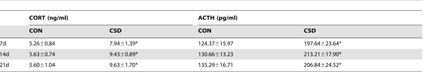
To verify that the experimental rats were under CSD stress, we analyzed the serum CORT and ACTH levels. As shown in Table 1, the CORT concentrations after 7, 14, and 21 days of sleep deprivation were significantly higher in the CSD group than
in the CON group (P,0.05). Similarly, the serum ACTH
concentrations after 7, 14, and 21 days of sleep deprivation were
significantly higher in the CSD group than in the CON group (P,
0.5, indicating that the rats in the CSD group were under sleep deprivation stress.
Histological observations
We selected the intermediate zone of the articular disk and corresponding condylar cartilage for histological observation. As shown in Figure 2, the condyles of the CON group displayed characteristic zonal cellular arrangements with distinct regions in the articular cartilage. No obvious histological changes were found in the condylar cartilages of the CON group. In the CSD group, the fibrous articular surfaces of the condylar cartilages became visibly tougher (white arrow) at 7 days of sleep deprivation (7/10 rats), and a debonding fibrous layer (black arrow) appeared in the majority of samples at 14 (8/10 rats) and 21 days of sleep deprivation (8/10 rats).
Although histopathological changes such as tough fibrous articular surfaces and a fraction of distorted collagen fibers could be observed in the U0126 group, no debonding fibrous layer was observed and fewer TMJ samples showed histological changes (4, 6, and 5 of 10 rats at 7, 14, and 21 days of CSD, respectively) compared with the number of samples in CSD group. Therefore,
Figure 1. The modified multiple platform method.Sleep deprivation was achieved by depriving the rats of paradoxical sleep. (A) Photos of
circular narrow platforms with rats inside. (B) Photos of a grid floor with rats inside. The tank is filled with water up to approximately 1 cm from the surface of the platform or grid.
these findings confirmed that sleep deprivation may cause pathological alterations in rat TMJ, which could be reversed by ERK inhibitor.
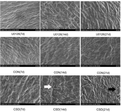
Ultrastructure analysis
To observe the subtle ultrastructural alterations of the TMJ, we used SEM to examine the condyle in the three groups after 7, 14, and 21 days of sleep deprivation. As shown in Figure 3, smooth condylar fibrous articular surfaces and compact bundles of collagen fibers were observed in all the CON subgroups, whereas the CSD rats at 7 days showed more apparent ripples of collagen
fiber bundles. The surfaces of the fibrous chord appeared rough and the uniform distribution of collagen fibers (white arrow) was disturbed in the CSD group at 14 days. In the CSD rats at 21 days, the waves of the collagen fibers became wider and cracks (black arrow) appeared on the surface of the condylar collagen.
Although similar ultrastructural observations were found in the U0126 group, such as twisted bundles of collagen fiber and cracks in a fraction of fibrous surfaces, the width of the collagen fiber waves was lower and the worn strips on the condylar surface showed relatively lower severity than in the CSD group. *P,0.05, significantly different from the control group. Data are represented as the M6SD of n = 10. M, mean; SD, standard deviation; n, sample size. doi:10.1371/journal.pone.0107544.t001
Figure 2. Sagittal section of the rat temporomandibular joint (TMJ) stained with hematoxylin and eosin.The intermediate zone of the
articular disk and corresponding condylar cartilage were selected for histological observation. Upper panel: Central condyles of the U0126 injection group rats at 7, 14, and 21 days of sleep deprivation. Middle panel: Central condyles of the control group rats at 7, 14, and 21 days. Lower panel: Central condyles of the CSD group rats at 7, 14, and 21 days of sleep deprivation (original magnification:6200, scale bar = 50mm).
Figure 3. Ultrastructure of the condyle visualized by scanning electron microscopy (SEM). SEM was used to observe the subtle ultrastructural alterations of the TMJ. Upper panel: Condylar fibrous articular surfaces of the U0126 rats at 7, 14, and 21 days of sleep deprivation. Middle panel: Condylar fibrous articular surfaces of the control group at 7, 14, and 21 days. Lower panel: Condylar fibrous articular surfaces of the CSD group at 7, 14, and 21 days of sleep deprivation (original magnification:62,500).
doi:10.1371/journal.pone.0107544.g003
Figure 4. Expression levels of p-ERK, ERK, MMP-1, MMP-3, and MMP-13 in the condyles.Western blot technique was used to examine the
and mRNA expressions of MMP-1, MMP-3, and MMP-13 were upregulated in the CSD group compared with the control group. As shown in Figure 4, with increasing duration of sleep deprivation, the MMP-1, MMP-3, and MMP-13 protein levels
increased significantly in all the sleep-restricted groups (P,0.05) as
compared with the control group. RT-qPCR revealed the expression of MMP-1, MMP-3, and MMP-13 mRNA (Tables 2), and the expression levels were significantly higher in all the
sleep-restricted groups (P,0.05) as compared with the control group.
These results showed that p-ERK was activated and the MMP expression was upregulated in the condylar cartilage as a result of sleep deprivation.
ERK inhibitor downregulates the expression levels of MMP genes
Next, in order to investigate whether activated p-ERK was involved in the upregulation of MMPs, the articular cavities of one group of rats were injected with the specific ERK inhibitor U0126. The p-ERK, total ERK, MMP-1, MMP-3, and MMP-13 expression levels in the condylar cartilages were then measured. As shown in Figure 4, the p-ERK level in TMJs injected with
U0126 showed an obvious decrease (P,0.01) as compared with
the CSD subgroups that did not receive U0126. The total ERK level, however, showed no obvious changes between the groups that did and did not receive U0126. Both protein (Figure 4) and mRNA (Table 2) expression levels of MMP-3 in all the
U0126-treated groups were significantly downregulated (P,0.05) as
compared with the groups that did not receive U0126. The MMP-1 and MMP-MMP-13 protein (Figure 4) and mRNA (Table 2) expres-sion levels in the U0126 group at 14 and 21 days of sleep
deprivation were downregulated (P,0.05) as compared with the
corresponding levels in the CSD group. As mentioned above, the TMJs that received U0126 injection showed fewer histological alterations in the joint cartilage as compared with those that did not receive U0126.
These findings suggest that the activated ERK pathway was involved in the regulation of the MMP-1, MMP-3, and MMP-13 expression.
Discussion
The present study demonstrated that CSD leads to pathological changes in the TMJ and upregulates the expression and secretion of MMP-1, MMP-3, and MMP-13 by activation of the ERK signaling pathway in TMJ condyles of rats. These findings provide important new evidence indicating that abnormally activated ERK signaling as a result of CSD may contribute to destruction of the TMJ by stimulating the production of MMPs.
MMPM is a well-established method that can effectively cause paradoxical sleep deprivation to rats without resulting in any additional stress such as social isolation or instability [26–28]. The grid on which the rats can lie down and sleep is considered as an adequate environmental control [5]. Many studies have demon-strated that paradoxical sleep deprivation may induce psycholog-ical stress and activate the hypothalamic-pituitary-adrenal (HPA) axis [29,30]. Similar results were found in our study.
Our data showed that CSD could result in pathological and ultrastructural changes in the TMJ of rats. In our present experiment, histopathological changes, such as tough fibrous articular surfaces, fraction of distorted collagen fibers and debonding fibrous layer were observed in the CSD group, which were more serious than U0126 group at each time point. These findings confirmed that sleep deprivation may cause pathological alterations in rat TMJ and partly regulated by ERK pathway. So the ERK inhibitor, U0126, only extenuated the damages, but not reversed, the detailed mechanism would be taken in the further experiments. These findings reflect the correlation between sleep disorders and TMJ dysfunction. The HPA axis was activated and the rats exposed to sleep deprivation experienced a state of stress. Stressed rats have been demonstrated to exhibit obvious gnawing behavior and greater masseter muscle activity [31–33], both of which would exert more and greater jaw motion on the TMJ [34]. The imbalance between biosynthesis and degradation of matrix components may lead to TMJ synovitis and condylar cartilage destruction, both of which are important pathological features of TMD [35]. This is consistent with the findings of our previous study although the experimental rats were exposed to consecutive sleep deprivation [7]. In the previous study, we confirmed that, at the early stage, sleep deprivation could induce increases in the serum level of estradiol and synovitis, and intercellular edema in the synovial membrane of the TMJ [47], which were consistent with our previous findings [8]. These changes corresponded to the
translocation of NF-kB p65 and the mRNA expression level of the
inflammatory factors IL-1b, IL-6, TNF-a, and iNOS in the
synovial membrane after sleep deprivation. At 5th and 7th day after sleep deprivation, the fluorescence signal of p65 in the nuclei of synovial cells significantly increased, which indicated that
NF-kB pathway was activated [47]. Consistently, previous results
showed that the mRNA expression of IL-1b, IL-6 and TNF-a
significantly increased from 3rd day after SD and reached the peak expression at 7th day. Then the acute inflammatory reaction converted to a long-term inflammatory process, which plays a pivotal role in the development of TMD. The cytokine network
plays an important role in TMJ inflammation. TNF-a, IL-1b, and
IL-6 appear to be the major proinflammatory cytokines involved in TMJ pathology [9]. These cytokines can stimulate condylar chondrocytes proliferation and subsequent activation, which were involved in the pathologic process of inflammatory pain, and is associated with persistent inflammation and synovial membrane destruction in osteoarthritis [10].
To explore the molecular mechanism underlying pathological alterations in the TMJ following sleep deprivation, we examined the p-ERK, ERK, and MMP expression levels. In the MAPK pathway, ERK phosphorylation is a sign of ERK activation by
MEK [10,11]. U0126 is a chemically synthesized organic compound that inhibits the kinase activity of MEK [36]. It has been used in both in vivo and in vitro studies of MEK [37,38]. The ERK pathway has been demonstrated to be a key factor in the induction of MMP-1, MMP-3, and MMP-13 in vitro [14,15,39–41]. These MMPs have been found to be upregulated in the synovial membrane and chondrocytes of TMJ diseases [42– 44]. In our study, we found p-ERK activation and upregulation of MMP-1, MMP-3, and MMP-13 in the rats after they experienced sleep deprivation. When injected with U0126, p-ERK activation was blocked and the expression of these MMPs was downregu-lated, accompanied with remission of the pathological destruction of the TMJ. Together, these data indicate that the ERK signaling pathway is activated and the ERK phosphorylation level was significantly elevated following sleep deprivation, and that the increased downstream catabolic proteases and collagenases induced by ERK lead to pathological alterations of the synovial membrane and condylar cartilage in the TMJ. One interesting finding is that although the MMP-1, MMP-3, and MMP-13 expression levels showed obvious decreases after U0126 injection, the MMP-1 and MMP-13 levels in the U0126 group remained higher than those in the control group. A possible explanation for this finding could be that MMP synthesis depends on the simultaneous activation of many of the protein kinase pathways,
including the c-Jun N-terminal kinase (JNK), ERK, p38, and
WNT pathways, as reported previously [10,45,46]. Our previous study also demonstrated changes in the expression levels of c-fos and MAPK kinase 4 (MKK4), which are two key factors that act on the JNK signaling pathway [7]. Therefore, it is understandable that selective inhibition of a single protein kinase pathway induced only partial inhibition of MMP synthesis.
Most TMDs are self-limited with reparative effects and the majority of TMD patients do not exhibit any progressive damages to the TMJ. In our study, we did not observe any reparative effects upon histopathological examination of the rats of the experimental group that were exposed to sleep deprivation for up to 21 days. Therefore, we assume that repair of the TMJ following damage may require more time, and further studies are required to determine whether pathological alterations can be reversed if the experimental sleep deprivation is removed.
In summary, our study demonstrated that the histomorphology of the TMJ was altered by related molecular mechanisms caused when rats were exposed to sleep deprivation. These findings provide evidence for the possible involvement of sleep disturbances in the onset and progression of TMD. Therefore, sleep distur-bances such as poor quality of sleep and sleep loss play important roles in TMD, indicating that such parameters should be taken into consideration in the treatment of TMJ disorders.
Author Contributions
Conceived and designed the experiments: CM GW ZW PW LW HZ. Performed the experiments: CM GW ZW HZ. Analyzed the data: CM PW LW GZ HZ. Contributed reagents/materials/analysis tools: CM GW ZW HZ. Contributed to the writing of the manuscript: CM GW HZ .
References
1. LeResche L, Saunders K, Von Korff MR, Barlow W, Dworkin SF (1997) Use of exogenous hormones and risk of temporomandibular disorder pain. Pain 69: 153–160.
2. Auerbach SM, Laskin DM, Frantsve LM, Orr T (2001) Depression, pain, exposure to stressful life events, and long-term outcomes in temporomandibular disorder patients. J Oral Maxillofac Surg 59: 628–633; discussion 634.
3. Rollman GB, Gillespie JM (2000) The role of psychosocial factors in temporomandibular disorders. Curr Rev Pain 4: 71–81.
10. Firestein GS, Manning AM (1999) Signal transduction and transcription factors in rheumatic disease. Arthritis Rheum 42: 609–621.
11. Dhillon AS, Kolch W (2002) Untying the regulation of the Raf-1 kinase. Arch Biochem Biophys 404: 3–9.
12. Schett G, Tohidast-Akrad M, Smolen JS, Schmid BJ, Steiner CW, et al. (2000) Activation, differential localization, and regulation of the stress-activated protein kinases, extracellular signal-regulated kinase, c-JUN N-terminal kinase, and p38 mitogen-activated protein kinase, in synovial tissue and cells in rheumatoid arthritis. Arthritis Rheum 43: 2501–2512.
13. Reunanen N, Li SP, Ahonen M, Foschi M, Han J, et al. (2002) Activation of p38 alpha MAPK enhances collagenase-1 (matrix metalloproteinase (MMP)-1) and stromelysin-1 (MMP-3) expression by mRNA stabilization. J Biol Chem 277: 32360–32368.
14. Shim YJ, Kang BH, Jeon HS, Park IS, Lee KU, et al. (2011) Clusterin induces matrix metalloproteinase-9 expression via ERK1/2 and PI3K/Akt/NF-kappaB pathways in monocytes/macrophages. J Leukoc Biol 90: 761–769.
15. Lai WC, Zhou M, Shankavaram U, Peng G, Wahl LM (2003) Differential regulation of lipopolysaccharide-induced monocyte matrix metalloproteinase (MMP)-1 and MMP-9 by p38 and extracellular signal-regulated kinase 1/2 mitogen-activated protein kinases. J Immunol 170: 6244–6249.
16. Kubota E, Kubota T, Matsumoto J, Shibata T, Murakami KI (1998) Synovial fluid cytokines and proteinases as markers of temporomandibular joint disease. J Oral Maxillofac Surg 56: 192–198.
17. Srinivas R, Sorsa T, Tjaderhane L, Niemi E, Raustia A, et al. (2001) Matrix metalloproteinases in mild and severe temporomandibular joint internal derangement synovial fluid. Oral Surg Oral Med Oral Pathol Oral Radiol Endod 91: 517–525.
18. Ohno S, Ohno-Nakahara M, Knudson CB, Knudson W (2005) Induction of MMP-3 by hyaluronan oligosaccharides in temporomandibular joint chondro-cytes. J Dent Res 84: 1005–1009.
19. Pelletier JP, Fernandes JC, Brunet J, Moldovan F, Schrier D, et al. (2003) In vivo selective inhibition of mitogen-activated protein kinase kinase 1/2 in rabbit experimental osteoarthritis is associated with a reduction in the development of structural changes. Arthritis Rheum 48: 1582–1593.
20. Landesberg R, Takeuchi E, Puzas JE (1999) Differential activation by cytokines of mitogen-activated protein kinases in bovine temporomandibular-joint disc cells. Archives of Oral Biology 44: 41–48.
21. Suchecki D, Duarte Palma B, Tufik S (2000) Sleep rebound in animals deprived of paradoxical sleep by the modified multiple platform method. Brain Res 875: 14–22.
22. Livak KJ, Schmittgen TD (2001) Analysis of relative gene expression data using real-time quantitative PCR and the 2(-Delta Delta C(T)) Method. Methods 25: 402–408.
23. Slade GD, Diatchenko L, Bhalang K, Sigurdsson A, Fillingim RB, et al. (2007) Influence of psychological factors on risk of temporomandibular disorders. J Dent Res 86: 1120–1125.
24. LeResche L, Mancl LA, Drangsholt MT, Huang G, Von Korff M (2007) Predictors of onset of facial pain and temporomandibular disorders in early adolescence. Pain 129: 269–278.
25. Wu G, Chen L, Zhu G, Su Y, Chen Y, et al. (2011) Psychological stress induces alterations in temporomandibular joint ultrastructure in a rat model of temporomandibular disorder. Oral Surg Oral Med Oral Pathol Oral Radiol Endod 112: e106–112.
31. Ayada K, Watanabe M, Endo Y (2000) Elevation of histidine decarboxylase activity in skeletal muscles and stomach in mice by stress and exercise. Am J Physiol Regul Integr Comp Physiol 279: R2042–2047.
32. Rosales VP, Ikeda K, Hizaki K, Naruo T, Nozoe S, et al. (2002) Emotional stress and brux-like activity of the masseter muscle in rats. Eur J Orthod 24: 107–117. 33. Chen YJ, Huang F, Zhang M, Shang HY (2010) Psychological stress alters ultrastructure and energy metabolism of masticatory muscle in rats. J Biomed Biotechnol 2010: 302693.
34. Chen J, Sorensen KP, Gupta T, Kilts T, Young M, et al. (2009) Altered functional loading causes differential effects in the subchondral bone and condylar cartilage in the temporomandibular joint from young mice. Osteoarthritis Cartilage 17: 354–361.
35. Wattanachai T, Yonemitsu I, Kaneko S, Soma K (2009) Functional lateral shift of the mandible effects on the expression of ECM in rat temporomandibular cartilage. Angle Orthod 79: 652–659.
36. Favata MF, Horiuchi KY, Manos EJ, Daulerio AJ, Stradley DA, et al. (1998) Identification of a novel inhibitor of mitogen-activated protein kinase kinase. J Biol Chem 273: 18623–18632.
37. DeSilva DR, Jones EA, Favata MF, Jaffee BD, Magolda RL, et al. (1998) Inhibition of mitogen-activated protein kinase kinase blocks T cell proliferation but does not induce or prevent anergy. J Immunol 160: 4175–4181. 38. Tolwinski NS, Shapiro PS, Goueli S, Ahn NG (1999) Nuclear localization of
mitogen-activated protein kinase kinase 1 (MKK1) is promoted by serum stimulation and G2-M progression. Requirement for phosphorylation at the activation lip and signaling downstream of MKK. J Biol Chem 274: 6168–6174. 39. Borden P, Solymar D, Sucharczuk A, Lindman B, Cannon P, et al. (1996) Cytokine control of interstitial collagenase and collagenase-3 gene expression in human chondrocytes. J Biol Chem 271: 23577–23581.
40. Mengshol JA, Vincenti MP, Brinckerhoff CE (2001) IL-1 induces collagenase-3 (MMP-13) promoter activity in stably transfected chondrocytic cells: require-ment for Runx-2 and activation by p38 MAPK and JNK pathways. Nucleic Acids Res 29: 4361–4372.
41. Park CH, Lee MJ, Ahn J, Kim S, Kim HH, et al. (2004) Heat shock-induced matrix metalloproteinase (MMP)-1 and MMP-3 are mediated through ERK and JNK activation and via an autocrine interleukin-6 loop. J Invest Dermatol 123: 1012–1019.
42. Wang YL, Li XJ, Qin RF, Lei DL, Liu YP, et al. (2008) Matrix metalloproteinase and its inhibitor in temporomandibular joint osteoarthrosis after indirect trauma in young goats. Br J Oral Maxillofac Surg 46: 192–197. 43. Kanyama M, Kuboki T, Kojima S, Fujisawa T, Hattori T, et al. (2000) Matrix
metalloproteinases and tissue inhibitors of metalloproteinases in synovial fluids of patients with temporomandibular joint osteoarthritis. J Orofac Pain 14: 20–30. 44. Ishimaru JI, Oguma Y, Goss AN (2000) Matrix metalloproteinase and tissue inhibitor of metalloproteinase in serum and lavage synovial fluid of patients with temporomandibular joint disorders. Br J Oral Maxillofac Surg 38: 354–359. 45. Brauchle M, Gluck D, Di Padova F, Han J, Gram H (2000) Independent role of
p38 and ERK1/2 mitogen-activated kinases in the upregulation of matrix metalloproteinase-1. Exp Cell Res 258: 135–144.
46. Ge X, Ma X, Meng J, Zhang C, Ma K, et al. (2009) Role of Wnt-5A in interleukin-1beta-induced matrix metalloproteinase expression in rabbit tempo-romandibular joint condylar chondrocytes. Arthritis Rheum 60: 2714–2722. 47. Gaoyi Wu,Lei Chen, Chuan Ma,Yanliang Wang,Ying Li, Guoxiong Zhu,