Shark (
Callorhinchus milii
)
Giselle Sek Suan Nah1,2, Zhi Wei Lim1, Boon-Hui Tay1, Motomi Osato1,3,4*, Byrappa Venkatesh1,5*
1Institute of Molecular and Cell Biology, Agency for Science, Technology and Research, Singapore, Singapore,2School of Biological Sciences, Nanyang Technological University, Singapore, Singapore,3Cancer Science Institute of Singapore, National University of Singapore, Singapore, Singapore,4Institute of Bioengineering and Nanotechnology, Agency for Science, Technology and Research, Singapore, Singapore,5Department of Pediatrics, Yong Loo Lin School of Medicine, National University of Singapore, Singapore, Singapore
Abstract
TheRunxfamily genes encode transcription factors that play key roles in hematopoiesis, skeletogenesis and neurogenesis and are often implicated in diseases. We describe here the cloning and characterization ofRunx1,Runx2,Runx3andRunxb genes in the elephant shark (Callorhinchus milii), a member of Chondrichthyes, the oldest living group of jawed vertebrates. Through the use of alternative promoters and/or alternative splicing, each of the elephant sharkRunxgenes expresses multiple isoforms similar to their orthologs in human and other bony vertebrates. The expression profiles of elephant shark Runxgenes are similar to those of mammalianRunxgenes. The syntenic blocks of genes at the elephant sharkRunxgene loci are highly conserved in human, but represented by shorter conserved blocks in zebrafish indicating a higher degree of rearrangements in this teleost fish. Analysis of promoter regions revealed conservation of binding sites for transcription factors, including two tandem binding sites for Runx that are totally conserved in the distal promoter regions of elephant shark Runx1-3. Several conserved noncoding elements (CNEs), which are putative cis-regulatory elements, and miRNA binding sites were identified in the elephant shark and humanRunxgene loci. Some of these CNEs and miRNA binding sites are absent in teleost fishes such as zebrafish and fugu. In summary, our analysis reveals that the genomic organization and expression profiles ofRunxgenes were already complex in the common ancestor of jawed vertebrates.
Citation:Nah GSS, Lim ZW, Tay B-H, Osato M, Venkatesh B (2014)RunxFamily Genes in a Cartilaginous Fish, the Elephant Shark (Callorhinchus milii). PLoS ONE 9(4): e93816. doi:10.1371/journal.pone.0093816
Editor:Andre van Wijnen, University of Massachusetts Medical, United States of America ReceivedFebruary 18, 2014;AcceptedMarch 6, 2014;PublishedApril 3, 2014
Copyright:ß2014 Nah et al. This is an open-access article distributed under the terms of the Creative Commons Attribution License, which permits unrestricted use, distribution, and reproduction in any medium, provided the original author and source are credited.
Funding:Funding provided by Biomedical Research Council of A*STAR, Singapore. The funders had no role in study design, data collection and analysis, decision to publish, or preparation of the manuscript.
Competing Interests:The authors have declared that no competing interests exist.
* E-mail: csimo@nus.edu.sg (MO); mcbbv@imcb.a-star.edu.sg (BV)
Introduction
The Runt domain transcription factor, known as the poly-omavirus enhancer-binding protein 2 (PEBP2) or core-binding factor (CBF) is a heterodimer ofaandbsubunits. In humans, the
a-subunit comprises three proteins, RUNX1, RUNX2 and RUNX3 that contain an evolutionarily conserved 128 amino acid Runt domain responsible for DNA binding and heterodimeriza-tion with theb-subunit. Theb-subunit includes a single protein, RUNXb(also known as PEBP2bor CBFb) that does not contain a binding domain but allosterically enhances the DNA-binding activity of the a-subunit and regulates its turnover by protecting it from ubiquitin-proteasome-mediated degradation [1,2].
RUNX1-3 are key transcriptional regulators involved in several major developmental pathways including hematopoiesis, neuro-genesis and skeletoneuro-genesis.RUNX1is among the most frequently mutated genes in human leukemias [3,4,5]. In mice, Runx1 is critical for the generation and maintenance of hematopoietic stem cells (HSC) [6,7]. In addition, Runx1 is involved in the development of skeletal muscle [8], neurons [9] and hair follicles [10]. On the other hand, Runx2 is indispensable for bone development, as evidenced by Runx2-/-mice which lack ossified skeleton and therefore die from respiratory failure shortly after birth [11]. In humans, haploinsufficiency ofRUNX2is associated
with cleidocranial dysplasia (CCD), an autosomal dominant skeletal disorder [12]. Runx3 is expressed in a wide range of tissues and has diverse biological functions. It plays roles in the regulation of epithelial homeostasis in the gastrointestinal tract [13,14], T-cell development during thymopoiesis [15] and differentiation of immune cells including natural killer cells [16], dendritic cells [17] and B cells [18].Runx3is also crucial for the differentiation of proprioceptive dorsal root ganglion (DRG) neurons [19] and chondrocyte maturation during skeletogenesis [20]. In human, RUNX3has been implicated in a multitude of cancers where it can function as a tumor suppressor or oncogene [21,22].
Given the importance of Runx genes in major developmental pathways and human diseases, orthologs of Runx have been characterized in phylogenetically diverse organisms to facilitate better understanding of their origin and functions.Runxgenes have undergone duplications independently in some invertebrate lineages (e.g., fruit fly, mosquito) and in the stem vertebrate lineage followed by acquisition of specialized functions such as hematopoiesis and eye development in Drosophila and bone development in vertebrates [23,24]. Tetrapods contain threeRunx
vertebrateRunxgene that was subsequently lost in tetrapods [25]. In zebrafish, besides Runx1 and Runx3, there are two copies of
Runx2 (Runx2a and Runx2b) which likely arose from the whole-genome duplication event in the teleost fish ancestor [27]. In contrast to multiple copies of a-subunit encoding Runx genes,
Runxb is present as a single copy in all metazoans analysed [24]. Studies into the co-evolution of Runxaand Runxbsubunits have reported similar evolutionary rates of these interacting proteins and evolutionary conservation of the structure of the Runxa -Runxb-DNA complex, suggesting that these proteins have co-evolved to maintain their ability to interact and to coregulate the transcription of target genes [24].
Cartilaginous fishes (Chondrichthyes) are phylogenetically the oldest living group of jawed vertebrates (Gnathostomes) that diverged from bony vertebrates (Osteichthyes) approximately 450 million years (My) ago [28]. By virtue of their phylogenetic position, cartilaginous fishes are a useful reference for the study of the origin and evolution of jawed vertebrate genes and their regulation. Cartilaginous fishes are split into two groups: elasmobranchs comprising sharks, rays and skates; and holoce-phalans represented by chimaeras such as elephant shark (Callorhinchus milii). Coding sequences of Runx genes were previously cloned in an elasmobranch, the dogfish (Scyliorhinus canicula) [29]. Three Runx genes, orthologous to mammalian
Runx1-3 were found to be expressed in the developing cartilage, teeth and placoid scales suggesting that they may be involved in the ancient processes of vertebrate skeletogenesis in this cartilag-inous fish [29]. To improve our understanding of the evolution, function and regulation ofRunxgenes, we have now clonedRunx
genes from the elephant shark and analysed their genomic organization. The elephant shark has the smallest genome among cartilaginous fishes and has been proposed as a model cartilag-inous fish genome [30,31]. Recently the whole genome sequence of the elephant shark was completed [32]. Its comparison with human and other vertebrate genomes indicated that elephant shark is the slowest evolving known vertebrate genome [32]. Furthermore, human and elephant shark were found to share twice as many conserved noncoding elements (CNE), which are putativecis-regulatory elements, as human and teleosts fishes [33]. Thus, elephant shark is a valuable reference genome for understanding the evolution of gene families and cis-regulatory networks in jawed vertebrates.
In this study, we have characterized three members ofRunx
family genes in the elephant shark, as well as the gene encoding the
b-subunit,CmRunxb, by cloning of cDNA and mining the elephant shark genome database. We have determined the tissue-specific mRNA expression of CmRunx and showed that these patterns reflect those in mammalian tissues, which may point to evolutionarily conserved gene functions and developmental pathways. Comparisons of noncoding sequences in the elephant shark and other jawed-vertebrateRunx loci were able to identify CNEs that are conserved over 450 million years of vertebrate evolution and are likely to becis-regulatory elements.
Results and Discussion
Cloning and characterization of elephant sharka-subunit Runx family members
To identifyRunxgenes in the elephant shark, we Blast-searched the elephant shark 1.46coverage sequence assembly [31] using human RUNX protein sequences and identified scaffolds containing fragments of Runx genes. By designing primers complementary to selected exons ofRunx and carrying out RT-PCR and RACE using gill and kidney cDNA as templates, we
were able to obtain full-length coding sequences for three elephant shark Runxa-subunit encoding genes. These sequences were then mapped to the whole-genome assembly of the elephant shark [32], and their precise exon-intron boundaries, transcription start sites (TSS) and UTRs were determined (Fig. 1). To confirm the orthology of the three elephant shark genes, we carried out phylogenetic analysis together with Runx sequences from repre-sentative chordates. The phylogenetic analysis identified the three genes as Runx1, Runx2 and Runx3 (Fig. 2) that are located on scaffolds#152,#106 and#121, respectively.
All three elephant sharkRunxgenes encode a highly conserved Runt domain (Fig. 1). Additionally, like all mammalianRunxgenes characterized to date, each of the elephant shark Runx genes contain two alternative promoters, P1 (distal) and P2 (proximal), that are separated by a characteristically large intron. The exon-intron boundaries of the three elephant shark Runx genes are largely conserved compared to their orthologs in human and other jawed vertebrates. However,CmRunx1 was found to contain an extra exon (exon 4.1) (Fig. 1A) which has not been characterized in any of the known Runx1 genes, except that of the dogfish, S. canicula, suggesting that this exon might be a chondrichthyes-specific feature. In addition, CmRunx2 lacks the equivalents of exons 5.2 and 6.1 found in the mammalian Runx2 gene [34] (Fig. 1B). These exons are also absent in chicken, frog and teleost fishes [26,35], indicating that they were recruited in a mammalian ancestor and likely to perform functions that are specific to mammals. Among the three elephant sharkRunx genes,Runx3is the shortest (CmRunx1, 114 kb;CmRunx2, 139 kb;CmRunx3, 74 kb) similar to its orthologs in mammals (HsRunx1, 261 kb; HsRunx2, 223 kb; HsRunx3, 65 kb; MmRunx1, 225 kb; MmRunx2, 211 kb;
MmRunx3, 57 kb). The short sequence of theRunx3genes is likely due to the loss of an exon equivalent to exon 5.1 inRunx1 and
Runx2genes (Fig. 1).
Mammals express a variety of isoforms for each of the Runx
genes arising from transcription initiation at two alternative promoters (P1 and P2) and alternative splicing of exons. Isoforms generated from the P1 promoter possess unique N-terminal sequences arising from transcription initiation in exon 1 and splicing that bypasses the P2 initiator ATG on exon 2. The use of alternative promoters contributes to the transcriptional and functional complexity of the Runx genes, as evidenced by expression studies and analyses of P1 and P2 promoter knockout mice wherein these transcripts display distinct patterns of expression and exert divergent biological functions during development [36,37]. We could identify 2 to 4 isoforms for each of the elephant shark Runx genes that are homologous to the isoforms in mammals, indicating that the genomic organization and transcriptional profile ofRunxgenes were already complex in the common ancestor of jawed vertebrates.
The availability of the whole-genome sequence of elephant shark enabled us to compare the synteny of genes atRunxloci in elephant shark and other sequenced vertebrate genomes. The synteny of genes at the elephant sharkRunx1, Runx2andRunx3loci is highly conserved in tetrapods (Fig. 3). A comparison of syntenic genes across the threeRunxgene loci indicates that paralogs ofClicand
Rcangenes are present in all threeRunxgene loci. This indicates that three Runx gene loci in the jawed vertebrates are the result of duplication of an ancestral locus that comprisedRunx,ClicandRcan
genes in that order. Of note are the characteristic ‘‘interlocked’’ gene structures ofRunx2and Supt3hin all the jawed vertebrates, except the duplicated locus in zebrafish (Runx2b locus) in which
braFlo1) indicating that their linkage is an ancient feature. It can therefore be inferred that linkage ofSupt3htoRuntgene was retained in the jawed vertebrateRunx2locus whereas the paralogs ofSupt3h
inRunx1andRunx3loci were lost secondarily.
In contrast to the relatively larger blocks of conserved syntenic genes in elephant shark and tetrapod Runx loci, the conserved syntenic blocks in zebrafish Runx loci are shorter (Fig. 3). This
indicates thatRunx loci in zebrafish have experienced a higher degree of rearrangements, and that the arrangement of genes in human Runxloci is more similar to that of elephant shark than zebrafish, underscoring the importance of elephant shark as a useful model reference genome for studying the origin and evolution of human gene loci.
Comparison of elephant shark Runxa-subunit protein sequences
Alignment of elephant shark Runx1, 2 and 3 protein sequences with human RUNX1, 2 and 3 sequences revealed several highly conserved protein domains. Among these is the 128 amino acid Runt domain which is almost totally conserved across elephant shark and human Runx proteins (Fig. 4). Within this domain, amino acid residues for DNA-binding as well as sequence motifs that interact with the b-subunit [38,39] are well conserved. In addition, the nuclear localization signal (NLS), PY and VWRPY motifs, and sites of phosphorylation by Erk2/cdc2 (Fig. 4B) are also highly conserved. The PY and VWRPY motifs mediate transcriptional activity of Runx proteins by recruiting different interaction proteins. The PY motif in the transactivation domain mediates the binding of Runx proteins to WW domain-containing proteins, such as YAP and TAZ [40,41]. Found invariably in all known vertebrate Runx proteins, the C-terminal pentapeptide VWRPY, is responsible for the repressive function of Runx proteins, through the recruitment of transcriptional co-repressors TLE/Groucho [42]. In human RUNX1, the serine residues S249
and S273 are each followed by a proline residue and acts as phosphorylation sites for ERK [43,44]. These phosphorylation sites are also conserved in human RUNX2 (S280 and T305) and elephant shark Runx1 and Runx2. Additionally, the consensus phosphorylation site for CDC2, (S/T)PX(R/K), at which serine residue S451 of human RUNX2 was reported to be phosphor-ylated [45], is remarkably conserved in all human and elephant shark Runx proteins (Fig. 4B).
The N-termini of the P1 and P2 isoforms beginning with MAS(N/D)S and MR(I/V)PV, respectively, are highly conserved across all three Runx proteins between elephant shark and their mammalian orthologs. A distinctive feature present only in CmRunx1 (and its dogfish ortholog) is a short stretch of 18 amino acids downstream of the Runt domain (Fig. 4). This unique sequence is encoded by an extra exon 4.1 that is alternatively spliced in one of the isoforms ofCmRunx1(Fig. 1). Whether these additional amino acids alter the structure and function of the CmRunx1 isoform is unknown, although one might speculate that the presence of this stretch within the nuclear localization signal (Fig. 4B, boxed in green dotted lines) might affect the isoform’s nuclear translocation.
Of the three mammalianRunxgenes, unique toRunx2is the Q/ A stretch, an activation domain located N-terminal to the Runt domain that is composed of successive polyglutamine and polyalanine amino acids. Whilst human RUNX2 contains a stretch of 23 glutamine residues followed by 17 alanine residues, no glutamine repeats but a stretch of 10 alanine residues are found in elephant shark Runx2 (Fig. 4). The QA domain has been proposed to contribute to the osteoblast-specific function of Runx2. This domain controls transactivation function of the Runx2 and also inhibits the heterodimerization of Runx2 with the
b-subunit [46]. In carnivoran mammals, the ratio of the glutamines to alanines (QA ratio) in Runx2 is strongly correlated with facial length [47]. In human, insertion in the alanine tract (23Q/27A) was observed in a CCD patient, suggesting that QA variations of RUNX2 may influence skeletal phenotype [48]. Indeed, glutamine repeat variants in RUNX2 have been recently found to be associated with a lower bone mineral density in the general human population [49]. Therefore, given the significance of the QA domain for skeletal functions of Runx2, it would be of interest to study the physiological relevance of this domain of Runx2 in cartilaginous fishes like the elephant shark which lack ossified endoskeleton.
Altogether, our comparisons indicate that protein domains of elephant shark Runx proteins are well conserved with those of human and demonstrate the highly conserved nature of Runx proteins in all jawed vertebrates.
Expression profile of elephant sharka-subunit Runx genes
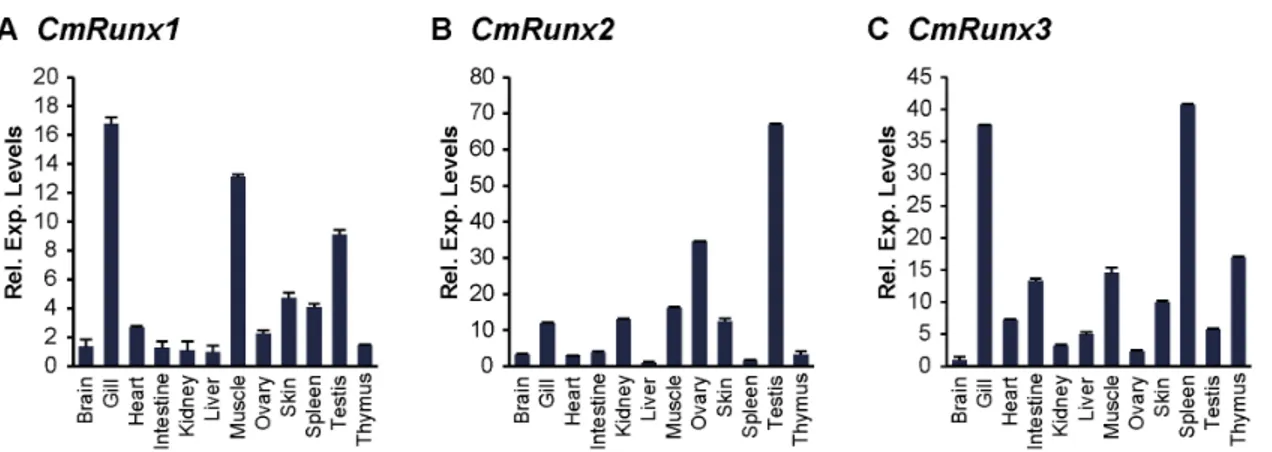
We investigated the expression patterns ofRunxgenes in various tissues of adult elephant shark by quantitative RT-PCR.CmRunx1
is highly expressed in the gills, muscle, testis, skin and spleen (Fig. 5A). HighCmRunx1expression in the gill, a tissue enriched in blood cells, as well as the spleen, a lymphomyeloid tissue of the shark, is concordant with the integral function of Runx1 in hematopoiesis. In addition, high expression of CmRunx1 in the muscle reflects its known role in mammalian skeletal muscle development. AlthoughRunx1is not known to be expressed at high levels in testis of mammals, high expression ofRunx1 has been reported in another cartilaginous fish, the dogfish. Thus, Runx1 might have a function in testis that is specific to cartilaginous fishes. However, this hypothesis remains to be verified. Further-more, a significant level of Runx1 expression in the skin of the
Figure 2. Phylogenetic tree (Bayesian inference) of chordate Runx sequences. Values adjacent to the nodes represent branch support (Bayesian posterior probability). Lancelet (Branchiostoma floridae) Runt (BfRunt) was used as the outgroup. Hs, human; Dr,Danio rerio; Fr,Fugu(Takifugu)rubripes; Cm,Callorhinchus milii; Sc,Scyliorhinus canicula; Ci,Ciona intestinalis.
elephant shark is consistent with previous reports of Runx
expression in placoid scales which are small conical structures in the skin of cartilaginous fishes [29].
Particularly striking is the significant expression ofRunx2in the gonads (ovary and testis) of the elephant shark (Fig. 5B). This expression pattern is reminiscent of that in mice, in which ovary
and testis were reported to be the predominant sites of non-skeletal tissueRunx2expression [50]. In human, recent investigations into the role ofRUNX2in the ovary have highlighted its importance in ovulation and luteinisation [51,52]. These congruent patterns of expression may suggest a role forRunx2 in similar reproductive processes in the elephant shark. Given the indispensable role of
Figure 3. Synteny of genes in theRunxloci of elephant shark and selected bony vertebrates.(A)Runx1locus, (B)Runx2locus and (C)
Runx3locus. Genes are represented by arrows. Genes with conserved synteny are coloured. Clusters of some non-syntenic genes are represented as white boxes and labelled in brackets. The gene order is from Ensembl (www.ensembl.org) and the elephant shark genome assembly (http:// esharkgenome.imcb.a-star.edu.sg/).
Runx2 in osteogenesis, analysis of Runx2 expression in the cartilaginous/skeletal elements of the elephant shark would have been informative, but due to the unavailability of cartilage tissue from elephant shark, we could not verify expression ofRunxgenes in the cartilage. However, in the dogfish, all threeRunxgenes were found highly expressed in the visceral cartilage [29], indicating the possible prominent role ofRunxgenes in the unossified skeleton of cartilaginous fishes.
The immune system of cartilaginous fishes share several features common to other jawed vertebrates. These include tissue sites for immune cell production (thymus and spleen), specialized cell types for innate and adaptive immunity and genes encoding proteins for immune function, such as various immunoglobulin subtypes, TCR, MHC and cytokine-like molecules [53,54]. The elephant shark Runx3 is highly expressed in the gill, spleen and thymus (Fig. 5C), which are all considered to be lymphomyeloid tissues in cartilaginous fishes. Based on the well-established roles ofRunx3in the development and function of diverse immune related cell types in mammals, it appears thatRunx3may have already established roles in the development of the cellular immune system in the common ancestor of jawed vertebrates.
Analysis of elephant sharka-subunit Runx promoter regions
The P1 promoter regions of mammalianRunx1,2and3genes harbour binding sites for transcription factors that are critical for their transcriptional regulation. In particular, P1 promoters of mammalianRunx1, 2and3contain two tandem binding sites for Runx. These sites have been implicated in auto and cross regulation of the three Runx genes [55,56]. To verify if these binding sites are conserved in elephant shark, we compared the P1 promoter regions ofRunx1-3in elephant shark, fugu/zebrafish and tetrapods. Remarkably, the tandem pair of Runx binding sites is totally conserved in P1 promoter regions of elephant sharkRunx1
(Fig. S1A), Runx2 (Fig. 6) and Runx3 (Fig. S1B) loci. The conservation of these binding sites in elephant shark and tetrapods indicate that the auto/cross regulation of the threeRunxgenes is an ancient feature ofRunx genes and was present in the single ancestralRunxlocus that gave rise to the threeRunxloci in jawed vertebrates. Interestingly, in contrast to total conservation of these binding sites in elephant shark, they are less conserved inRunx1
and3loci of fugu and zebrafish (Fig. S1 and 6). It remains to be seen if the divergent binding sites in teleosts are still functional and whetherRunxgenes in teleosts are subject to auto/cross regulation similar to that of elephant shark and tetrapods.
In addition to the two Runx binding sites, the P1 promoter of the mammalian Runx2locus contains well-characterized binding sites for other transcription factors that regulate the expression of
Runx2. These include (i) a vitamin D response element (VDRE) with an overlapping ATF motif, that binds the VDR/RXR heterodimer and mediates the suppression ofRUNX2transcription by the steroid hormone 1,25-(OH)2-vitamin D3 [57]; (ii) a Helix-loop-Helix consensus motif that is critical for the basal transcrip-tion ofRunx2; and (iii) NF1 and AP1 motifs required for osteoblast-specific activity of theRunx2P1 promoter [58] (Fig. 6). All these binding sites, except the VDRE site, are conserved highly in the elephant sharkRunx2gene, but to a lesser extent in the zebrafish and fuguRunx2genes (Fig. 6).
Conserved noncoding elements in human and elephant sharka-subunit Runx loci
Runx genes exhibit highly specific expression in several tissues and cell lineages that is critical for their diverse roles in development and disease. However, the transcriptional regulation of Runx genes has not been well characterized. Comparative genomics is a powerful strategy for identifying evolutionarily conserved cis-regulatory elements. Since functional elements evolve slowly compared to their flanking sequences,cis-regulatory
Figure 4. Runxa-subunit proteins Runx1, Runx2 and Runx3 in elephant shark and human.(A) Schematic representation of Runxa -subunit proteins. The characteristic domains found in all Runx proteins are indicated to show their relative positions along the protein. (B) Alignment of elephant shark and human Runxa-subunit proteins. The first block shows the amino-terminal part of the protein derived from the P1 promoter that differs from that derived from the P2 promoter. The highly conserved Runt domain is boxed with a red line. Within the Runt domain, surfaces involved in DNA contact and interaction with theb-subunit are denoted by black and blue lines, respectively. Cysteine residues involved in the redox regulation of DNA-binding activity are indicated with asterisks. Nuclear localization signal (NLS) is demarcated by a green dashed box. The PY and VWRPY motifs are indicated by orange and blue boxes, respectively. The transactivation domain (TAD) is highlighted in blue and the inhibitory domain (ID) is boxed by purple dotted lines. The nuclear matrix translocalization signal (NMTS) is boxed with a brown line. Minimal consensus sequences for phosphorylation by Erk or cdc2 are boxed by dashed and solid black lines, respectively. The residue targeted for phosophorylation is indicated byG. Cm,Callorhinchus milii; Hs,Homo sapiens.
doi:10.1371/journal.pone.0093816.g004
Figure 5. Expression patterns of elephant sharkRunxgenes.Relative expression levels of (A)CmRunx1, (B)CmRunx2and (C)CmRunx3in various tissues of the elephant shark determined by qRT-PCR.
elements can be identified as conserved noncoding elements (CNEs) in the genomes of related species. Functional assay of CNEs in transgenic systems has indicated that many of them drive reporter gene expression in specific tissues [59]. Indeed, one such CNE predicted in the first intron of Runx1 and conserved in human, mouse, rat, dog, horse and opossum, was shown to drive highly-specific expression to hematopoietic stem cells (HSCs) of mouse [60], indicating that it is an HSC enhancer.
The common ancestor of human and elephant shark diverged about 450 million years ago. Any CNEs conserved in these vertebrates over 900 million years of divergent evolution are likely to be functional elements and must be playing a fundamental role in the regulation of their associated genes. In order to identify such evolutionarily conserved cis-regulatory elements, we aligned the three human Runx loci with their corresponding orthologs from elephant shark and other jawed vertebrates and predicted CNEs that are.65% identical across 50 bp or more. Several CNEs that met these criteria were identified in the threeRunxloci of human and elephant shark (Fig. 7) (Table S2). Notably, two of the CNEs located in the intronic regions of humanRUNX2(Runx2_CNE7 and Runx2_CNE8) overlap two functionally characterized enhancers, denoted as mm657 and mm924 (Fig. 7B), that drive reporter gene expression in the branchial arches and facial mesenchyme respectively of transgenic mouse embryos [61]. The overlap of functional enhancers with the predicted human-elephant shark CNEs provides further support to the notion that CNEs conserved in human and elephant shark are likely to be functional cis-regulatory elements. Thus, they are strong candi-dates for functional assays that can help to identify enhancers of humanRUNXgenes.
Interestingly some of the CNEs conserved in theRunx1-3loci of human and elephant shark are missing in zebrafish and fugu (Fig. 7A and B), which is consistent with previous observations that CNEs in teleosts have been evolving faster than in elephant shark and other vertebrates [62]. This further emphasizes the impor-tance of elephant shark as a model genome of choice for identifying evolutionarily conservedcis-regulatory elements in the human genome.
miRNA binding sites in elephant sharka-subunit Runx genes
Apart from transcriptional control by alternate promoters and
cis-regulatory elements, expression ofRunx1-3is also subjected to post-transcriptional regulation by miRNAs [63,64,65]. Several miRNAs have been shown to regulateRunx1expression levels and consequently its function in hematopoietic differentiation [64]. Among the highly conserved miRNA binding sites in mammalian
Runx139UTR, two clustered sites for miR-27a are remarkably well conserved in the 39UTR of elephant sharkRunx1but not in the teleost fishes, zebrafish and fugu (Fig. 8A). In mammals, miR-27 plays regulatory roles during megakaryocytic and granulocytic differentiation by attenuating Runx1 expression and engaging in feedback loops with Runx1 [64]. Conservation of miR-27 binding sites, coupled with the presence of miR-27 in the elephant shark (GenBank Accession number JX994340) suggests that CmRunx1
may similarly be post-transcriptionally controlled by miR-27 in hematopoietic lineages of the elephant shark.
Eleven Runx2-targeting miRNAs have been reported to control the physiological levels of Runx2 protein during osteogenesis and chondrocyte maturation [65]. Of these, binding sites for miR-23, miR-218 and miR-338 were found conserved in the 39UTR of elephant sharkRunx2, while only that for miR-28 is present in zebrafish and fugu Runx2 (Fig. 8B). Since miR-23 (JX994633), miR-218 (JX994383) and miR-338 (JX994406) are present in the elephant shark, these conserved miRNAs are likely to be involved in the regulation of Runx2 expression in elephant shark, possibly during chondrogenic differentiation through mechanisms similar to that in other jawed vertebrates.
In contrast toRunx1andRunx2, no miRNA binding sites were found to be conserved in the rather short 39UTR of elephant shark
Runx3(data not shown).
Cloning and characterization of elephant sharkb-subunit Runx gene
We cloned the full-length coding sequence of the elephant shark gene encoding theb-subunit of the CBF heterodimeric complex,
Runxb. The exon-intron organization of elephant shark Runxb
(CmRunxb) gene is identical to that of its human ortholog (Fig. 9A) [66]. Furthermore, CmRunxb is transcribed into at least three isoforms that are homologous to the humanRUNXBtype 1, 2 and 3 isoforms. TheCmRunxbtype 1, 2 and 3 isoforms encode proteins of 188, 185 and 156 amino acids, respectively (Fig. 9B).CmRunxb
Type 1 and Type 3 isoforms are largely similar, except for the absence of exon 5 in the Type 3, as a result of exon skipping.
CmRunxbType 1 and Type 2 isoforms differ in their C-terminal ends, resulting from the use of alternative termination codon and splice donor/acceptor sites in exons 5 and 6 (Fig. 9A). At the protein level, CmRunxb and human RUNXb show high conservation of amino acid residues 1-165 (Fig. 9B), of which the N-terminal 135 amino acids required for its heterodimeriza-tion with thea-subunit and DNA binding [67] are almost perfectly conserved. We investigated the expression patterns of Runxb
isoforms in various tissues of adult elephant shark by quantitative RT-PCR. All isoforms display a ubiquitous pattern of expression, with high levels of expression in the gill, heart, ovary and testis of the elephant shark (Fig. 9C). The functional significance of these isoforms that are conserved in human, elephant shark and other jawed vertebrates remains to be investigated. In an attempt to identify conserved cis-regulatory elements for Runxb locus, we searched for CNEs in the human and elephant sharkRunxbloci but found none (data not shown).
Materials and Methods
Ethics statement
Elephant sharks were caught off the coast of Western Port Bay, Victoria (Australia) by licensed commercial fishermen. The samples used in this study were taken from animals that were already dead when the fishermen returned to the fishing jetty. Pieces of tissue were taken from the dead fish, frozen and transported to the laboratory for extraction of DNA and RNA [68,69]. The extraction of DNA and RNA from frozen elephant shark samples was approved by the Institutional Animal Care and Use Committee (IACUC) of the Institute of Molecular and Cell Biology.
Figure 7. Conserved noncoding elements (CNEs) inRunxloci.VISTA plots obtained from the global alignment between the human, mouse, dog, opossum, chicken, lizard, frog, zebrafish, fugu and elephant shark (A)Runx1, (B)Runx2and (C)Runx3loci are shown. Sequence for opossum
Runx2locus is not available. Human sequence is used as the reference sequence. Conserved sequences were predicted at a cut-off of$65% identity across.50 bp windows and are represented by peaks. Blue peaks denote conserved coding exons, pink peaks conserved noncoding regions (CNRs) and cyan peaks untranslated regions (UTRs). Experimentally verified CNEs (mm657 and mm924) in theRunx2locus are indicated with blue arrows below the x-axis and labelled.
doi:10.1371/journal.pone.0093816.g007
Figure 8. miRNA binding sites in the 39UTR ofRunx1andRunx2genes in elephant shark and selected gnathostomes.Schematic diagram of (A)Runx1and (B)Runx239UTRs and miRNA binding sites. The last coding exon is represented by a rectangle and 39UTR by a grey line. Positions of miRNA binding sites are indicated by vertical lines. Binding sites conserved in human and elephant shark are shown in red. Zebrafish and fuguRunx1loci are not shown as they do not contain any conserved miRNA binding sites.
Identification of Runx gene fragments in the elephant shark genome database
The elephant shark 1.46coverage sequence assembly (http:// esharkgenome.imcb.a-star.edu.sg/) was searched with human RUNX1, RUNX2, RUNX3 and RUNXb protein sequences using ‘TBLASTN’ algorithm. The following contigs that contained fragments of various Runx genes were identified in the assembly: Runx1- AAVX01052077, AAVX01261782, AVX01381831 and AAVX01569464; Runx2- AAVX01175228,
AAVX01545426, AAVX01631487, AAVX01048938,
AAVX01307838 and AAVX01048937;Runx3- AAVX01039927, AAVX01288244, AAVX01157201 and AAVX01598430 and
Runxb- AAVX01192354, AAVX01224391 and AAVX01471917. RT-PCR and 59and 39RACE
Primers were designed for representative exons ofRunx genes identified in the elephant shark scaffolds and the full-length coding sequences of the genes were obtained by RT-PCR, 59RACE and/ or 39RACE (primer sequences available upon request). 59and 39
RACE were performed on single-stranded cDNA prepared from total RNA using the SMART RACE cDNA Amplification kit (Clontech, Palo Alto, CA) in a nested PCR. All RT-PCR and RACE products were cloned into the pGEM-T Easy Vector (Promega), and sequenced completely using the BigDye Termi-nator Cycle Sequencing Kit (Applied Biosystems, USA) on an ABI 3730xl capillary sequencer (Applied Biosystems, USA). During the course of this study, the whole-genome sequence of elephant shark became available [32]. The full-length cDNA sequences were mapped to the whole-genome assembly and the sequences of scaffolds that contained the genes (scaffolds#152,#106,#121 and #127) were extracted for further annotation and synteny analysis. Sequences for various isoforms of elephant shark Runx
genes have been deposited in GenBank (Accession numbers KJ150227-KJ150240).
Amino acid alignment and phylogenetic analysis The protein sequences for human and other chordate Runx
genes were retrieved from the National Center for Biotechnology Information (NCBI) database. Multiple sequence alignments were performed by using ClustalW. For phylogenetic analysis, gaps in the alignments were removed using Gblocks Server (ver. 0.91b) with default parameters [70]. The phylogenetic analysis was performed using MrBayes 3.2, employing Dayhoff+G as a substitution model and running 4 chains for 1,000,000 genera-tions. Trees were sampled every 100 generations and according to a saturation curve of likelihood values, the first 2500 trees were discarded as burn-in. TheRuntgene fromBranchiostoma floridaewas used as the outgroup.
Expression profiling of elephant shark Runx genes by qRT-PCR
Total RNA was extracted from various tissues of adult elephant shark using Trizol reagent (Gibco BRL, Grand Island, NY) according to manufacturer’s protocol. Purified total RNA was
reverse-transcribed into cDNA with Superscript II (Invitrogen, Carlsbad, CA). The single strand cDNA was used as a template in qRT-PCR reactions with KAPA SYBR FAST qPCR Kit reagents (KAPA Biosystems, Boston, MA). Sequences of primers used in qRT-PCR are given in Table S1. All primer pairs span at least one intron which helps to distinguish cDNA from genomic DNA products. Expression levels ofRunxgenes were normalized usingb -actingene as the reference.
Prediction of transcription factor binding sites (TFBS) Analysis of TFBSs was carried out using JASPAR (http:// jaspar.binf.ku.dk/cgi-bin/jaspar_db.pl). Only TFBS with predic-tion scores$9 were retained as putative TFBSs.
Prediction of conserved noncoding elements (CNEs) Genomic sequences of the Runx gene loci for the following species were extracted from Ensembl release 73 [71]: human (GRCh37 assembly, February 2009), mouse (GRCm38, Decem-ber 2011), dog (CanFam3.1, SeptemDecem-ber 2011), opossum (Mon-Dom5, October 2006), chicken (GalGal4, November 2011), lizard (AnoCar2.0, May 2010), frog (JGI 4.2, November 2009), zebrafish (Zv9, July 2010) and fugu (FUGU5, October 2011). Repetitive sequences were masked using CENSOR at default settings [72]. Multiple alignments ofRunx gene loci sequences were generated using the global alignment program MLAGAN (http://genome. lbl.gov/vista/index.shtml) with human as the reference sequence. CNEs were predicted using a cutoff of$65% identity across 50-bp windows and visualized using VISTA (http://genome.lbl.gov/ vista/index.shtml).
Supporting Information
Figure S1 P1 promoter region ofRunx1andRunx3of elephant shark and selected bony vertebrates. Multiple sequence alignments of the P1 59UTR regions of (a)Runx1and (b)Runx3are shown. The numbers to the left of the alignment indicate positions relative to the TSS (+1) of elephant shark (C. milii) genes. Corresponding regions from other bony vertebrates were aligned using ClustalW. The tandem Runx binding sites are boxed.
(PDF)
Table S1 Primers used for qRT-PCR. (PDF)
Table S2 CNEs in theRunxloci of human and elephant shark. (PDF)
Acknowledgments
We thank Alison P. Lee and Vydianathan Ravi for critical reading of the manuscript.
Author Contributions
Conceived and designed the experiments: BV MO. Performed the experiments: GSN ZWL BT. Analyzed the data: GSN BV. Wrote the paper: GSN BV.
References
1. Huang G, Shigesada K, Ito K, Wee HJ, Yokomizo T, et al. (2001) Dimerization with PEBP2beta protects RUNX1/AML1 from ubiquitin-proteasome-mediated degradation. The EMBO journal 20: 723–733.
2. Adya N, Castilla LH, Liu PP (2000) Function of CBFbeta/Bro proteins. Semin Cell Dev Biol 11: 361–368.
patterns of elephant sharkRunxbtranscripts. Relative expression levels ofRunxbTypes 1+2 and Type 3 in various tissues of the elephant shark determined by qRT-PCR.
3. Osato M (2004) Point mutations in the RUNX1/AML1 gene: another actor in RUNX leukemia. Oncogene 23: 4284–4296.
4. Osato M, Ito Y (2005) Increased dosage of the RUNX1/AML1 gene: a third mode of RUNX leukemia? Critical reviews in eukaryotic gene expression 15: 217–228.
5. Speck NA, Gilliland DG (2002) Core-binding factors in haematopoiesis and leukaemia. Nature reviews Cancer 2: 502–513.
6. Okuda T, van Deursen J, Hiebert SW, Grosveld G, Downing JR (1996) AML1, the target of multiple chromosomal translocations in human leukemia, is essential for normal fetal liver hematopoiesis. Cell 84: 321–330.
7. Jacob B, Osato M, Yamashita N, Wang CQ, Taniuchi I, et al. (2010) Stem cell exhaustion due to Runx1 deficiency is prevented by Evi5 activation in leukemogenesis. Blood 115: 1610–1620.
8. Wang X, Blagden C, Fan J, Nowak SJ, Taniuchi I, et al. (2005) Runx1 prevents wasting, myofibrillar disorganization, and autophagy of skeletal muscle. Genes Dev 19: 1715–1722.
9. Inoue K, Shiga T, Ito Y (2008) Runx transcription factors in neuronal development. Neural Dev 3: 20.
10. Osorio KM, Lee SE, McDermitt DJ, Waghmare SK, Zhang YV, et al. (2008) Runx1 modulates developmental, but not injury-driven, hair follicle stem cell activation. Development 135: 1059–1068.
11. Komori T, Yagi H, Nomura S, Yamaguchi A, Sasaki K, et al. (1997) Targeted disruption of Cbfa1 results in a complete lack of bone formation owing to maturational arrest of osteoblasts. Cell 89: 755–764.
12. Otto F, Kanegane H, Mundlos S (2002) Mutations in the RUNX2 gene in patients with cleidocranial dysplasia. Human mutation 19: 209–216. 13. Li QL, Ito K, Sakakura C, Fukamachi H, Inoue K, et al. (2002) Causal
relationship between the loss of RUNX3 expression and gastric cancer. Cell 109: 113–124.
14. Brenner O, Levanon D, Negreanu V, Golubkov O, Fainaru O, et al. (2004) Loss of Runx3 function in leukocytes is associated with spontaneously developed colitis and gastric mucosal hyperplasia. Proc Natl Acad Sci U S A 101: 16016– 16021.
15. Taniuchi I, Osato M, Egawa T, Sunshine MJ, Bae SC, et al. (2002) Differential requirements for Runx proteins in CD4 repression and epigenetic silencing during T lymphocyte development. Cell 111: 621–633.
16. Ohno S, Sato T, Kohu K, Takeda K, Okumura K, et al. (2008) Runx proteins are involved in regulation of CD122, Ly49 family and IFN-gamma expression during NK cell differentiation. International immunology 20: 71–79. 17. Fainaru O, Woolf E, Lotem J, Yarmus M, Brenner O, et al. (2004) Runx3
regulates mouse TGF-beta-mediated dendritic cell function and its absence results in airway inflammation. The EMBO journal 23: 969–979.
18. Watanabe K, Sugai M, Nambu Y, Osato M, Hayashi T, et al. (2010) Requirement for Runx proteins in IgA class switching acting downstream of TGF-beta 1 and retinoic acid signaling. Journal of immunology 184: 2785–2792. 19. Inoue K, Ozaki S, Shiga T, Ito K, Masuda T, et al. (2002) Runx3 controls the axonal projection of proprioceptive dorsal root ganglion neurons. Nature neuroscience 5: 946–954.
20. Soung do Y, Dong Y, Wang Y, Zuscik MJ, Schwarz EM, et al. (2007) Runx3/ AML2/Cbfa3 regulates early and late chondrocyte differentiation. Journal of bone and mineral research: the official journal of the American Society for Bone and Mineral Research 22: 1260–1270.
21. Lee CW, Chuang LS, Kimura S, Lai SK, Ong CW, et al. (2011) RUNX3 functions as an oncogene in ovarian cancer. Gynecologic oncology 122: 410– 417.
22. Kudo Y, Tsunematsu T, Takata T (2011) Oncogenic role of RUNX3 in head and neck cancer. Journal of cellular biochemistry 112: 387–393.
23. Braun T, Woollard A (2009) RUNX factors in development: lessons from invertebrate model systems. Blood cells, molecules & diseases 43: 43–48. 24. Sullivan JC, Sher D, Eisenstein M, Shigesada K, Reitzel AM, et al. (2008) The
evolutionary origin of the Runx/CBFbeta transcription factors—studies of the most basal metazoans. BMC evolutionary biology 8: 228.
25. Glusman G, Kaur A, Hood L, Rowen L (2004) An enigmatic fourth runt domain gene in the fugu genome: ancestral gene loss versus accelerated evolution. BMC evolutionary biology 4: 43.
26. Ng CE, Osato M, Tay BH, Venkatesh B, Ito Y (2007) cDNA cloning of Runx family genes from the pufferfish (Fugu rubripes). Gene 399: 162–173. 27. Christoffels A, Koh EG, Chia JM, Brenner S, Aparicio S, et al. (2004) Fugu
genome analysis provides evidence for a whole-genome duplication early during the evolution of ray-finned fishes. Molecular Biology and Evolution 21: 1146– 1151.
28. Benton MJ, Donoghue PC (2007) Paleontological evidence to date the tree of life. Molecular biology and evolution 24: 26–53.
29. Hecht J, Stricker S, Wiecha U, Stiege A, Panopoulou G, et al. (2008) Evolution of a core gene network for skeletogenesis in chordates. PLoS genetics 4: e1000025.
30. Venkatesh B, Tay A, Dandona N, Patil JG, Brenner S (2005) A compact cartilaginous fish model genome. Current biology: CB 15: R82–83. 31. Venkatesh B, Kirkness EF, Loh YH, Halpern AL, Lee AP, et al. (2007) Survey
sequencing and comparative analysis of the elephant shark (Callorhinchus milii) genome. PLoS Biology 5: e101.
32. Venkatesh B, Lee AP, Ravi V, Maurya AK, Lian MM, et al. (2014) Elephant shark genome provides unique insights into gnathostome evolution. Nature 505: 174–179.
33. Venkatesh B, Kirkness EF, Loh YH, Halpern AL, Lee AP, et al. (2006) Ancient noncoding elements conserved in the human genome. Science 314: 1892. 34. Terry A, Kilbey A, Vaillant F, Stewart M, Jenkins A, et al. (2004) Conservation
and expression of an alternative 39exon of Runx2 encoding a novel proline-rich C-terminal domain. Gene 336: 115–125.
35. van der Meulen T, Kranenbarg S, Schipper H, Samallo J, van Leeuwen JL, et al. (2005) Identification and characterisation of two runx2 homologues in zebrafish with different expression patterns. Biochimica et biophysica acta 1729: 105–117.
36. Bee T, Swiers G, Muroi S, Pozner A, Nottingham W, et al. (2010) Nonredundant roles for Runx1 alternative promoters reflect their activity at discrete stages of developmental hematopoiesis. Blood 115: 3042–3050. 37. Liu JC, Lengner CJ, Gaur T, Lou Y, Hussain S, et al. (2011) Runx2 protein
expression utilizes the Runx2 P1 promoter to establish osteoprogenitor cell number for normal bone formation. The Journal of biological chemistry 286: 30057–30070.
38. Akamatsu Y, Ohno T, Hirota K, Kagoshima H, Yodoi J, et al. (1997) Redox regulation of the DNA binding activity in transcription factor PEBP2. The roles of two conserved cysteine residues. The Journal of biological chemistry 272: 14497–14500.
39. Tahirov TH, Inoue-Bungo T, Morii H, Fujikawa A, Sasaki M, et al. (2001) Structural analyses of DNA recognition by the AML1/Runx-1 Runt domain and its allosteric control by CBFbeta. Cell 104: 755–767.
40. Kanai F, Marignani PA, Sarbassova D, Yagi R, Hall RA, et al. (2000) TAZ: a novel transcriptional co-activator regulated by interactions with 14-3-3 and PDZ domain proteins. EMBO J 19: 6778–6791.
41. Yagi R, Chen LF, Shigesada K, Murakami Y, Ito Y (1999) A WW domain-containing yes-associated protein (YAP) is a novel transcriptional co-activator. EMBO J 18: 2551–2562.
42. Javed A, Guo B, Hiebert S, Choi JY, Green J, et al. (2000) Groucho/TLE/R-esp proteins associate with the nuclear matrix and repress RUNX (CBF(alpha)/ AML/PEBP2(alpha)) dependent activation of tissue-specific gene transcription. Journal of cell science 113 (Pt 12): 2221–2231.
43. Tanaka T, Kurokawa M, Ueki K, Tanaka K, Imai Y, et al. (1996) The extracellular signal-regulated kinase pathway phosphorylates AML1, an acute myeloid leukemia gene product, and potentially regulates its transactivation ability. Molecular and cellular biology 16: 3967–3979.
44. Zhang Y, Biggs JR, Kraft AS (2004) Phorbol ester treatment of K562 cells regulates the transcriptional activity of AML1c through phosphorylation. The Journal of biological chemistry 279: 53116–53125.
45. Qiao M, Shapiro P, Fosbrink M, Rus H, Kumar R, et al. (2006) Cell cycle-dependent phosphorylation of the RUNX2 transcription factor by cdc2 regulates endothelial cell proliferation. The Journal of biological chemistry 281: 7118–7128.
46. Thirunavukkarasu K, Mahajan M, McLarren KW, Stifani S, Karsenty G (1998) Two domains unique to osteoblast-specific transcription factor Osf2/Cbfa1 contribute to its transactivation function and its inability to heterodimerize with Cbfbeta. Molecular and cellular biology 18: 4197–4208.
47. Sears KE, Goswami A, Flynn JJ, Niswander LA (2007) The correlated evolution of Runx2 tandem repeats, transcriptional activity, and facial length in carnivora. Evolution & development 9: 555–565.
48. Goodman FR, Mundlos S, Muragaki Y, Donnai D, Giovannucci-Uzielli ML, et al. (1997) Synpolydactyly phenotypes correlate with size of expansions in HOXD13 polyalanine tract. Proceedings of the National Academy of Sciences of the United States of America 94: 7458–7463.
49. Morrison NA, Stephens AA, Osato M, Polly P, Tan TC, et al. (2012) Glutamine repeat variants in human RUNX2 associated with decreased femoral neck BMD, broadband ultrasound attenuation and target gene transactivation. PloS one 7: e42617.
50. Jeong JH, Jin JS, Kim HN, Kang SM, Liu JC, et al. (2008) Expression of Runx2 transcription factor in non-skeletal tissues, sperm and brain. Journal of cellular physiology 217: 511–517.
51. Park ES, Park J, Franceschi RT, Jo M (2012) The role for runt related transcription factor 2 (RUNX2) as a transcriptional repressor in luteinizing granulosa cells. Molecular and cellular endocrinology 362: 165–175. 52. Fan HY, Liu Z, Johnson PF, Richards JS (2011) CCAAT/enhancer-binding
proteins (C/EBP)-alpha and -beta are essential for ovulation, luteinization, and the expression of key target genes. Molecular endocrinology 25: 253–268. 53. Dooley H, Flajnik MF (2006) Antibody repertoire development in cartilaginous
fish. Dev Comp Immunol 30: 43–56.
54. Boehm T, Iwanami N, Hess I (2012) Evolution of the immune system in the lower vertebrates. Annu Rev Genomics Hum Genet 13: 127–149.
55. Drissi H, Luc Q, Shakoori R, Chuva De Sousa Lopes S, Choi JY, et al. (2000) Transcriptional autoregulation of the bone related CBFA1/RUNX2 gene. Journal of cellular physiology 184: 341–350.
56. Spender LC, Whiteman HJ, Karstegl CE, Farrell PJ (2005) Transcriptional cross-regulation of RUNX1 by RUNX3 in human B cells. Oncogene 24: 1873– 1881.
57. Drissi H, Pouliot A, Koolloos C, Stein JL, Lian JB, et al. (2002) 1,25-(OH)2-vitamin D3 suppresses the bone-related Runx2/Cbfa1 gene promoter. Experimental cell research 274: 323–333.
59. Pennacchio LA, Ahituv N, Moses AM, Prabhakar S, Nobrega MA, et al. (2006) In vivo enhancer analysis of human conserved non-coding sequences. Nature 444: 499–502.
60. Ng CE, Yokomizo T, Yamashita N, Cirovic B, Jin H, et al. (2010) A Runx1 intronic enhancer marks hemogenic endothelial cells and hematopoietic stem cells. Stem cells 28: 1869–1881.
61. Attanasio C, Nord AS, Zhu Y, Blow MJ, Li Z, et al. (2013) Fine tuning of craniofacial morphology by distant-acting enhancers. Science 342: 1241006. 62. Lee AP, Kerk SY, Tan YY, Brenner S, Venkatesh B (2011) Ancient vertebrate
conserved noncoding elements have been evolving rapidly in teleost fishes. Molecular biology and evolution 28: 1205–1215.
63. Xu Y, Wang K, Gao W, Zhang C, Huang F, et al. (2013) MicroRNA-106b regulates the tumor suppressor RUNX3 in laryngeal carcinoma cells. FEBS Lett 587: 3166–3174.
64. Rossetti S, Sacchi N (2013) RUNX1: A MicroRNA Hub in Normal and Malignant Hematopoiesis. Int J Mol Sci 14: 1566–1588.
65. Lian JB, Stein GS, van Wijnen AJ, Stein JL, Hassan MQ, et al. (2012) MicroRNA control of bone formation and homeostasis. Nat Rev Endocrinol 8: 212–227.
66. Ogawa E, Inuzuka M, Maruyama M, Satake M, Naito-Fujimoto M, et al. (1993) Molecular cloning and characterization of PEBP2 beta, the heterodimeric
partner of a novel Drosophila runt-related DNA binding protein PEBP2 alpha. Virology 194: 314–331.
67. Kagoshima H, Akamatsu Y, Ito Y, Shigesada K (1996) Functional dissection of the alpha and beta subunits of transcription factor PEBP2 and the redox susceptibility of its DNA binding activity. The Journal of biological chemistry 271: 33074–33082.
68. Davies WI, Tay BH, Zheng L, Danks JA, Brenner S, et al. (2012) Evolution and functional characterisation of melanopsins in a deep-sea chimaera (elephant shark, Callorhinchus milii). PloS one 7: e51276.
69. Tan YY, Kodzius R, Tay BH, Tay A, Brenner S, et al. (2012) Sequencing and analysis of full-length cDNAs, 59-ESTs and 39-ESTs from a cartilaginous fish, the elephant shark (Callorhinchus milii). PloS one 7: e47174.
70. Castresana J (2000) Selection of conserved blocks from multiple alignments for their use in phylogenetic analysis. Molecular Biology and Evolution 17: 540– 552.
71. Flicek P, Ahmed I, Amode MR, Barrell D, Beal K, et al. (2013) Ensembl 2013. Nucleic acids research 41: D48–55.