Nipbl and Mediator Cooperatively Regulate Gene
Expression to Control Limb Development
Akihiko Muto1,2,3, Shingo Ikeda3, Martha E. Lopez-Burks1,2, Yutaka Kikuchi3., Anne L. Calof3,4.*,
Arthur D. Lander1,2.*, Thomas F. Schilling1,2.
1Department of Developmental & Cell Biology, University of California, Irvine, Irvine, California, United States of America,2Center for Complex Biological Systems, University of California, Irvine, Irvine California,3Department of Biological Science, Graduate School of Science, Hiroshima University, Higashi-Hiroshima, Hiroshima, Japan,
4Department of Anatomy & Neurobiology, University of California, Irvine, Irvine, California, United States of America
Abstract
Haploinsufficiency for Nipbl, a cohesin loading protein, causes Cornelia de Lange Syndrome (CdLS), the most common ‘‘cohesinopathy’’. It has been proposed that the effects of Nipbl-haploinsufficiency result from disruption of long-range communication between DNA elements. Here we use zebrafish and mouse models of CdLS to examine how transcriptional changes caused by Nipbl deficiency give rise to limb defects, a common condition in individuals with CdLS. In the zebrafish pectoral fin (forelimb), knockdown of Nipbl expression led to size reductions and patterning defects that were preceded by dysregulated expression of key early limb development genes, includingfgfs,shha,hand2and multiplehoxgenes. In limb buds ofNipbl-haploinsufficient mice, transcriptome analysis revealed many similar gene expression changes, as well as altered expression of additional classes of genes that play roles in limb development. In both species, the pattern of dysregulation ofhox-gene expression depended on genomic location within theHoxclusters. In view of studies suggesting that Nipbl colocalizes with the mediator complex, which facilitates enhancer-promoter communication, we also examined zebrafish deficient for the Med12 Mediator subunit, and found they resembled Nipbl-deficient fish in both morphology and gene expression. Moreover, combined partial reduction of both Nipbl and Med12 had a strongly synergistic effect, consistent with both molecules acting in a common pathway. In addition, three-dimensional fluorescent in situ hybridization revealed that Nipbl and Med12 are required to bring regions containing long-range enhancers into close proximity with the zebrafish hoxda cluster. These data demonstrate a crucial role for Nipbl in limb development, and support the view that its actions on multiple gene pathways result from its influence, together with Mediator, on regulation of long-range chromosomal interactions.
Citation:Muto A, Ikeda S, Lopez-Burks ME, Kikuchi Y, Calof AL, et al. (2014) Nipbl and Mediator Cooperatively Regulate Gene Expression to Control Limb Development. PLoS Genet 10(9): e1004671. doi:10.1371/journal.pgen.1004671
Editor:Gregory S. Barsh, Stanford University School of Medicine, United States of America
ReceivedDecember 13, 2013;AcceptedAugust 14, 2014;PublishedSeptember 25, 2014
Copyright:ß2014 Muto et al. This is an open-access article distributed under the terms of the Creative Commons Attribution License, which permits unrestricted use, distribution, and reproduction in any medium, provided the original author and source are credited.
Funding:This work was supported by the US National Institute of Health (P01-HD052860 and P50-GM076516) and the Cornelia de Lange Syndrome Foundation. This project used the UCI Genomics High-Throughput Facility, which is partially supported by Award Number P30CA062203 from the National Cancer Institute to the Chao Comprehensive Cancer Center. The funders had no role in study design, data collection and analysis, decision to publish, or preparation of the manuscript.
Competing Interests:The authors have declared that no competing interests exist. * Email: [email protected] (ALC); [email protected] (ADL)
.These authors contributed equally to this work.
Introduction
Cohesin, a ring-shaped, DNA-associated protein complex, is best known for its role in tethering sister chromatids together until mitosis [1,2]. However, growing evidence indicates that cohesin, and proteins such as Nipped-B-like (Nipbl) that regulate cohesin loading onto DNA, also play critical roles in gene regulation [3– 13]. In particular, it has been suggested that Nipbl and cohesin mediate interactions between promoters and distant enhancers, a process thought to involve the physical looping out of intervening DNA sequences [14–16]. For example, inDrosophila, Nipped-B (the orthologue of Nipbl) and cohesin regulatecutgene expression by controlling long-range interactions between thecut promoter and a wing-specific remote enhancer [5]. In mice, haploinsuffi-ciency for Nipbl impairs looping that controls the selective expression of beta-globin isoforms by erythroid cells [13].
Recently, it was found that Nipbl co-localizes with the Mediator complex at promoters/enhancers of actively transcribed genes in
mouse embryonic stem cells [17]. Thought to play a pivotal role in transmitting regulatory signals from gene-specific activators/ repressors to RNA polymerase II [18,19], Mediator is a large complex composed of a core that interacts with RNA polymerase II and gene-specific transcriptional regulators, and a Cdk8 submodule (containing Cdk8, CyclinC, Med12 and Med13) and can either negatively [20–22] or positively [23,24] regulate transcription. The reported physical interaction between Mediator and Nipbl at active genes suggests that they function together in promoter-enhancer communication, but exactly how this occurs is unknown.
Much insight into the physiological significance of cohesin’s influence on transcription has come from the study of Cornelia de Lange Syndrome (CdLS) and other ‘‘cohesinopathies’’. CdLS is a congenital syndrome characterized by growth retardation, neuro-logical dysfunction, and structural defects in multiple organs [25– 30], and is caused, in most cases, by haploinsufficiency forNIPBL [31,32]. More recently it has been shown that mutations in
cohesin subunits SMC1A or SMC3 [33,34] or the SMC3 deacetylase, HDAC8 [35], are less common causes of CdLS. Analysis of both patient samples and animal models indicate that Nipbl haploinsufficiency causes small changes (usually less than 1.5-fold) in the expression of many hundreds of genes [3,4,11]. Analysis of both mouse and fish models of Nipbl deficiency suggests that pervasive phenotypic abnormalities result from the collective, and sometimes synergistic, effects of such small changes in gene expression [3,11].
Among the most striking abnormalities in CdLS are limb defects, which range from mild brachydactyly and clinodactyly to severe digit and limb truncations, the latter in about 1/3 of cases [26,28,36]. Limb reduction is one of the few structural defects in CdLS that is not replicated in theNipbl-haploinsufficient mouse model, as these mice exhibit only minor changes in the shape of the olecranon process, and delays in the ossification of limb bones [3]. Hypothesizing that this difference might reflect slight differences in the threshold for triggering such defects in mouse versus man, we decided to look at development of the pectoral fin (the homologue of the mammalian forelimb) in a zebrafish model of Nipbl-deficiency, produced by injection of morpholino oligo-nucleotides (MO) directed against the two zebrafish nipbls[11]. Here we show that Nipbl-deficient fish display a marked reduction in pectoral fin size, which is already apparent early in fin bud development. We demonstrate that Nipbl is required for normal expression of conserved regulators of vertebrate limb growth and patterning, including fgfsin the apical ectodermal ridge (AER), shhin the zone of polarizing activity (ZPA), and severalhoxgenes of the hoxab, hoxcaand hoxdaclusters. We also show that that Nipbl-haploinsufficient mouse limb buds display a pattern of gene expression changes strikingly similar to those observed in Nipbl-deficient pectoral fin buds.
Pectoral fin defects have also been reported inmed12-mutant zebrafish, in which Mediator function is disrupted [37]. Interest-ingly, we find that both the morphological and gene expression changes that occur in Nipbl-deficient fin buds are mimicked when med12is knocked down. In particular, expression of multiplehox
genes in different clusters is affected in a similar position-specific manner in both Nipbl- and Med12-deficient fish embryos, and results of experiments in which we simultaneously knock down both Nipbl and Med12 suggest that they interact genetically. Using 3-dimensional fluorescent in situ hybridization (3D-FISH) in zebrafish fin buds, we further show that Nipbls and Med12 are required for higher-order chromatin organization near thehoxda cluster. Overall, the data point to a shared, conserved role for Nipbl and the Mediator complex in the regulation of long-range enhancer-promoter interactions underlying growth and patterning of the vertebrate limb.
Results
Impaired pectoral fin development in Nipbl-deficient zebrafish
Both nipbl genes in zebrafish, nipbla and nipblb [11], are expressed in developing pectoral fin bud mesenchyme (Fig. S1A, B). To investigate their requirements in forelimb development, we generated ‘‘Nipbl-deficient’’ embryos in which both nipbla andnipblbwere depleted by injecting either of two different sets of antisense morpholinos (MOs) designed against distinct MO target sites, as described previously [11]. Pectoral fins of Nipbl-deficient larvae were 40% shorter in length at 72 hours post fertilization (hpf) than those of control embryos (Figure 1A, B, E). This reduced size was not simply due to developmental delay, since it was much more severe than expected given the delay in development of the eye and lower jaw, which we estimated at approximately 16 hrs (Figure S2, see below). Pectoral fin defects were more severe when MOs to bothnipbl mRNAs were injected, compared with either one alone (Figure S3) [11]; thus, injections of both MOs were used in subsequent experiments. Pectoral fin defects were partially rescued by co-injection of exogenousnipblamRNA, confirming MO specific-ity (Figure 1C–E).
Alcian Blue staining at 5 dpf revealed that pectoral fin cartilages of the endoskeletal discs form in Nipbl-deficient larvae but are smaller (Figure 1F, G). Cell numbers in these discs were reduced by 37% and 33% along anterior-posterior (A-P) and proximo-distal (P-D) axes, respectively (Figure 1H), whereas cell size resembled controls (Figure 1F9, G9) and we found no change in cell death (Figure S4A, B), suggesting impaired growth of cartilage progenitors at earlier stages. In addition, the orderly arrangement and spacing of chondrocytes in the endoskeletal disc cells was noticeably disrupted in Nipbl-deficient fins (Figure 1F9, G9).
In zebrafish embryos, pectoral fin buds first appear at 30 hpf as shallow domes along the A-P axis, and grow and begin to fold posteriorly by 42 hpf. In Nipbl-deficient embryos, pectoral fin buds also initiate at 30 hpf but grow more slowly than controls (Figure 1I–R). TUNEL assays (Figure S4C–E) showed no increase in cell death in Nipbl-deficient fin buds (Figure S4A–E). In contrast, numbers of BrdU+ cells decreased significantly in the mesenchyme of Nipbl-deficient pectoral fin buds (Figure S4F–I). These data suggest that endoskeletal disc size reduction in Nipbl-deficient limb buds reflects cumulative effects of slower rates of cell division.
Since Nipbl is required for embryonic growth in both fish and mice [3,11], we stage-matched embryos using an independent criterion – i.e. the A-P position of the migrating posterior lateral line (pLL) primordium labeled by in situ hybridization (ISH) for fgf10a; Figure S5A, red arrows). In controls, pLL primordia lie just posterior to the pectoral fin buds at 22 hpf, and continue to migrate posteriorly. Based on this staging criterion the develop-mental delay in Nipbl-deficient embryos (summarized in Figure Author Summary
Limb malformations are a striking feature of Cornelia de Lange Syndrome (CdLS), a multi-system birth defects disorder most commonly caused by haploinsufficiency forNIPBL. In addition to its role as a cohesin-loading factor, Nipbl also regulates gene expression, but how partial Nipbl deficiency causes limb defects is unknown. Using zebrafish and mouse models, we show that expression of multiple key regulators of early limb development, includingshha,
hand2 and hox genes, are sensitive to Nipbl deficiency. Furthermore, we find morphological and gene expression abnormalities similar to those of Nipbl-deficient zebrafish in the limb buds of zebrafish deficient for the Med12 subunit of Mediator—a protein complex that mediates physical interactions between enhancers and promoters— and genetic interaction studies support the view that Mediator and Nipbl act together. Strikingly, depletion of either Nipbl or Med12 leads to characteristic changes in
hox gene expression that reflect the locations of genes within their chromosomal clusters, as well as to disruption of large-scale chromosome organization around thehoxda
cluster, consistent with impairment of long-range enhanc-er-promoter interaction. Together, these findings provide insights into both the etiology of limb defects in CdLS, and the mechanisms by which Nipbl and Mediator influence gene expression.
Nipbl and Mediator Regulate Limb Development
S5B) cannot account for the severe limb reductions in Nipbl-deficient larvae (Figure 1S–X).
Nipbls are required forfgfexpression in the AER but not in fin bud mesenchyme
Early limb development is highly conserved from fish to mammals [38–40]. Each fin/limb bud possesses an apical ectodermal ridge (AER) and zone of polarizing activity (ZPA) [40,41], which play important roles in growth and patterning [42– 44]. The AER, a thickened epithelium that rims the distal ends of the buds, is the source of Fgf signals required for P-D limb outgrowth [45–48]. The zebrafish AER expresses 4 fgf genes: fgf4,fgf8a,fgf16andfgf24[49,50]. Of these, expression offgf4, fgf8aandfgf16was dramatically reduced in pectoral fin buds of Nipbl-deficient embryos (Figure 2A–C), which was rescued by over-expression of full-lengthnipblamRNA (Figure S6). This was
not simply due to loss of AER cells sincefgf24expression was not downregulated in the Nipbl-deficient AER (Figure 2D).
Limb bud development also requires expression offgf10aand fgf24 in the mesenchyme [49,51,52]. However, we found no differences in fgf10a expression between wild type and Nipbl-deficient limb buds between 22–48 hpf (Figure S7A), as well as no differences in expression of tbx5a and fgf24, which control the expression offgf10a(Figure S7B–C).
Nipbls are required forshhexpression in the ZPA and its regulation in fin bud mesenchyme
The ZPA acts as an organizing center in the posterior limb/fin bud mesenchyme in part because it produces Shh [38,40,45,50,53–55]. Shh is required for limb A-P polarity, outgrowth and Fgf expression in the AER [55]. Zebrafish Shh (shha) and its receptor (and transcriptional target)ptch2 are first
Figure 1. Nipbl knockdown disrupts pectoral fin development.(A–D) Reduced pectoral fins in live Nipbl-deficient embryos at 76 hpf. Dorsal views, anterior to the left. Uninjected control (A), Nipbl-deficient (nipbla/b-MO) (B), injected with 400 pg ofnipblamRNA alone (C) and co-injected withnipbla/b-MO+nipblamRNA (D). (E) Whisker plots of fin length at 76 hpf; medians: 431.8mm, n = 50 (control), 258.5mm, n = 88 (nipbla/b-MOs),
423.0mm, n = 22 (nipblamRNA alone), and 319.5mm, n = 70 (nipbla/b-MOs+nipblamRNA). p-values are indicated in the graph. (F, F9, G, G9) Alcian blue staining of cartilages in pectoral fins in control (F, F9) and Nipbl-deficient embryos (G, G9) at 120 hpf. F9and G9are higher magnification pictures of boxed areas of endoskeletal discs in F and G, respectively. ac, actinotrichs; cl, cleithrum; ed, endoskeletal disc; sco, scapulocoracoid. (H) Numbers of endoskeletal cells in pectoral fins along proximodistal (PD) and anteroposterior (AP) axes (control; n = 13, Nipbl-deficient embryos; n = 16). PD (Ave6
S.D.): 33.261.5 (control) and 22.464.2 (Nipbl-deficient embryos), p,1028. AP (Ave
6S.D.): 27.961.5 (control) and 17.762.2 (Nipbl-deficient embryos), p,10213
. (I-X) Morphology of developing pectoral fin buds in live embryos. Lateral views, anterior and dorsal to the left and top, respectively. doi:10.1371/journal.pgen.1004671.g001
Nipbl and Mediator Regulate Limb Development
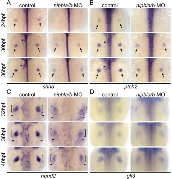
expressed in the ZPA at 24 hpf and expression progressively increases until 36 hpf (Figure 3A–B) [56]. In Nipbl-deficient limb buds, shha and ptch2 expression was reduced at these stages (Figure 3A, B).shhaandptch2expression levels were also reduced in the intestine (where Nipbl is also required for development [11]; Figure 3A, B, asterisks), but unaffected in the notochord and neural tube (Figure 3A, B and unpublished data), suggesting a tissue-specific requirement for Nipbl in the expression of Shh and its receptor.
Hand2 regulatesShhexpression in fin/limb buds [56–58], and we found that hand2 expression was also reduced in Nipbl-deficient fin buds (36 and 40 hpf) compared with stage-matched controls (32 and 36 hpf) (Figure 3C). In mouse limb buds, anterior expression of the transcriptional repressor, Gli3, restricts expres-sion of Hand2 posteriorly [59]. Zebrafish pectoral fin buds also expressgli3[60] but its expression was not affected by reduction of Nipbl (Figure 3D).
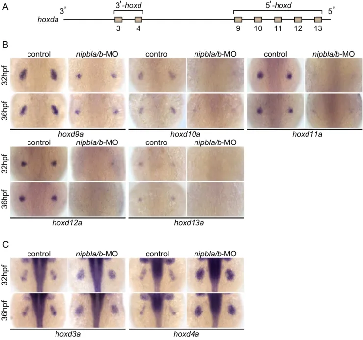
Mammalian Hand2 acts together with the products of 59-Hoxd genes [58] in the regulation ofShhexpression. In pectoral fin buds of Nipbl-deficient embryos, we found that 59-hoxdgenes, including hoxd9a-d13a (Figure 4A), were significantly downregulated (Fig-ure 4B). Importantly, fin bud expression ofhand2,hoxd10a,shha and ptch2 could all be partially rescued by exogenous nipbla mRNA (Figure S8).
Retinoic acid (RA) produced in anterior somites also regulates shha expression in pectoral fin buds (12–22 hpf [42–44,61]), as well as fin bud expression of fgf10a. However, we found no differences in expression of either the RA synthesizing enzyme aldh1a2or the RA degradation enzyme and target gene,cyp26a1, at 13 and 19 hpf in Nipbl-deficient embryos (Figure S9).
Together, these findings indicate that Nipbls regulate the 59 -hoxd/hand2/shha signaling cascade, but do not affect thetbx5a/ fgf24/fgf10a pathway that lies downstream of RA signaling, during vertebrate limb development.
Nipbls regulate expression ofhoxgenes according to their genomic location
Hox genes belong to 13 paralog groups organized in four (mammals) or seven (zebrafish) clusters; theHoxAandDclusters are crucial for limb/fin development [56,62,63]. The most 39 -located genes (39-Hox), such asHoxd1, are expressed earliest in mouse limb buds, whereas expression of 59-located genes (59-Hox, d10-d13) begins later [64,65]. 59-Hoxd gene expression occurs first in proximal limb buds, where it is required forShhexpression in the ZPA to establish A-P patterning [55,66], and is later restricted distally in limb buds, where it is required for proper digit formation [64,65]. Expression ofhoxdgenes in zebrafish fin buds is reminiscent of that in proximal mouse limb buds but appears to lack the second wave of distal expression, consistent with the lack of digits in ray-finned fish [64].
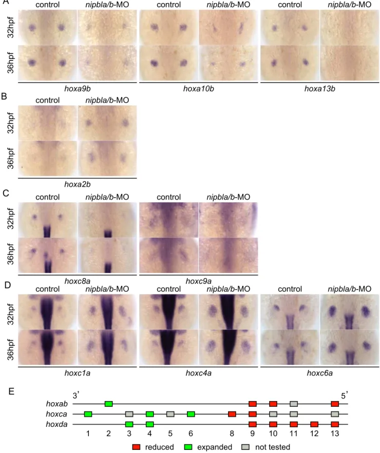
Examination of expression of multiplehoxgenes from theHoxa (hoxab),Hoxc(hoxca),and Hoxd(hoxda) clusters in the fin buds of Nipbl-deficient embryos revealed that changes in expression correlated strongly with positions of genes within clusters (Figures 4–5). Expression of fivehoxdgenes located at the 59ends of the hoxda cluster (hoxd9a-d13a) was severely reduced (Figure 4B), while expression of twohoxdgenes located more 39 in the cluster,hoxd3a and hoxd4a, expanded to encompass the entire bud (Figure 4C). Similarly, expression of 59-genes in the hoxabcluster—such ashoxa9b,a10b, anda13b—was significantly
Figure 2. Reduced expression offgfs in the AER of Nipbl-deficient embryos.Expression offgf4(A),fgf8a(B),fgf16(C) andfgf24(D) in the AER (arrows) at indicated stages in control and Nipbl-deficient embryos examined by ISH. Dorsal views, anterior to the top.
doi:10.1371/journal.pgen.1004671.g002
Nipbl and Mediator Regulate Limb Development
reduced in Nipbl-deficient fin buds, while a 39gene,hoxa2b, was upregulated (Figure 5A, B). Likewise, expression of hoxc8a and hoxc9awas reduced in Nipbl-deficient fin buds while expression of hoxc1a,hoxc4a, andhoxc6aexpanded posteriorly (Figure 5C, D). Thus, in all threehoxclusters expressed in the pectoral fin buds, expression of genes near the 39end of the cluster expands, whereas expression of those closer to the 59 end is reduced (Figure 5E). Interestingly, this position-specific regulation ofhox gene expres-sion is specific to pectoral fin buds, sincehoxexpression patterns in the neural tube were unaffected in Nipbl-deficient embryos (Figure S10).
Shh signaling from the ZPA regulates expression of severalhox genes along the A-P axis of limb buds, and reduced expression of 59-hoxa/hoxd genes as well as posterior expansion of hoxc6a expression, similar to that described above, occurs in Shh-deficient zebrafish [55]. To test if the Shh reductions resulting from Nipbl deficiency might cause the defects in hox gene
expression, we treated wild-type embryos with the Shh signaling inhibitor, cyclopamine (CyA). Although CyA treatment caused some developmental delay, (,4–5 hr, based on the A-P positions
of pLL primordia [compare Fig. S11A with Fig. S5A], and no more than 12 hr based on pectoral fin development), it strongly reduced expression of ptch2 as well as hoxa13b, hoxd10a and hoxd13a, while expression of hoxc4a and hoxc6a expanded posteriorly (compared with stage-matched controls, Figure S11B). These effects of CyA treatment resembled those of Nipbl depletion, but others did not - e.g. hoxd4a expression was severely reduced, andhoxc8aexpression expanded posteriorly in CyA-treated embryos (Figure S11B), in contrast to Nipbl-deficient embryos (Fig. 4C, 5C). Thus, loss of Shh signaling cannot explain all of the changes inhoxgene expression in Nipbl-deficient embryos, suggesting that either Nipbls regulate the expression ofhoxgenes directly, or they do so via regulators other than (or in addition to) Shh.
Figure 3. Reduction of genes involved in theshh-related gene regulatory cassette in developing pectoral fin mesenchyme of Nipbl-deficient embryos.Expression of fin mesenchymal genes at indicated stages in control and Nipbl-deficient embryos examined by ISH. Dorsal views, anterior to the top. (A, B) Expression ofshhaandptch2in pectoral fin buds (arrows) was significantly reduced in Nipbl-deficient embryos, while midline, neural tube expression was unaffected. Expression in endoderm derived-tissues (asterisks) is also reduced. (C) Expression ofhand2was also significantly reduced in stage-matched pectoral fin buds.hand2expression in the fin buds and posterior lateral plate mesoderm is marked by brackets and arrowheads, respectively. (D)gli3expression was not significantly affected in a stage-matched comparison.
doi:10.1371/journal.pgen.1004671.g003
Nipbl and Mediator Regulate Limb Development
Gene expression changes in limb buds ofNipbl
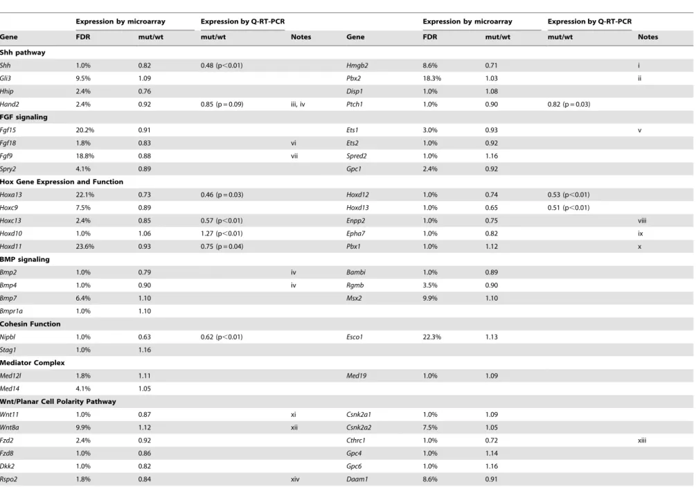
-haploinsufficient mice mirror those in Nipbl-deficient fish Nipbl+/2mutant mice fail to display obvious limb reductions, but do show some limb patterning and bone calcification defects [3]. Given the gene expression changes we found in pectoral fin buds of Nipbl-deficient fish, we decided to investigate if Nipbl-deficient mouse limb buds show some of the same changes. ISH forShhin E10.5 limb buds ofNipbl+/2 mice revealed a marked reduction in Shh expression in the ZPA, similar to Nipbl-deficient fin buds (compare Figure 3A and Figure 6). This was confirmed by both Q-RT-PCR and expression microarray analysis, using RNA extracted from E10.5 limb buds harvested from stage-matched Nipbl+/2 (n = 12) and wildtype (n = 12) littermate embryos (Table 1;
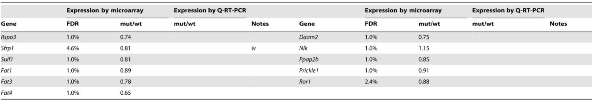
also see Methods). Microarray analysis identified approximate-ly 1000 genes as significantapproximate-ly over- or under-expressed in Nipbl+/2 limb buds (Table 1 and data publically deposited) and, similar to tissues and cells of Nipbl+/2 mice and individuals with CdLS, most gene expression changes were typically less than 1.5-fold [3,4]. Nonetheless, statistically-significant changes in expression (mostly decreases) were observed for multiple genes in the Fgf, Bmp and Shh pathways, as well as numerous genes in the Wnt/planar cell polarity signaling pathway. In addition, multiple genes at the 59and 39 ends of the Protocadherin B cluster were downregulated (not shown), while Stag1 (which encodes a cohesin subunit) was upregulated; both of these changes are hallmarks of Nipbl deficiency in other tissues [3].
Figure 4. Nipbls are required for spatial patterns ofhoxdexpression in pectoral fin buds.(A) Diagram of zebrafishhoxdacluster. (B, C) Expression of 39-hoxdgenes includinghoxd3aandd4a(B) and 59-hoxdgenes includinghoxd9a-d13a(C) was examined by ISH at 32 and 36 hpf to show both time-matched and stage-matched (nipbla/b-MO at 36 hpf and control at 32 hpf) comparisons. Dorsal views, anterior to the top. doi:10.1371/journal.pgen.1004671.g004
Nipbl and Mediator Regulate Limb Development
Figure 5. Nipbls regulatehoxgene expression according to genomic location.(A–D) Expression of genes inhoxab(A,B) andhoxca(C,D) clusters was examined by ISH. (A) 59-hoxa, (B) 39-hoxa, (C) 59-hoxc, and (D) 39-hoxcgenes. Dorsal views, anterior to the top. (E) Diagram summarizing effects of Nipbl reduction onhoxgenes. Genes located closer to 59-ends show reduced expression (red boxes) whereas those closer to 39-ends become expressed across entire fin buds (green boxes).
doi:10.1371/journal.pgen.1004671.g005
Nipbl and Mediator Regulate Limb Development
Similar to Nipbl-deficient fin buds, Nipbl+/2 limb buds displayed reductions in the expression of 59-Hoxgenes (Table 1 and Figure S12). This was particularly obvious for genes at the extreme 59end ofHoxclusters, such asHoxa13,Hoxc13,Hoxd12, andHoxd13, expression of which was reduced between 15% and 35% by microarray, although Q-RT-PCR measurements (Ta-ble 1) and ISH (Figure S12) suggested that the true decrease is probably closer to 50%. Also downregulated were Enpp2 and Epha7, which are known targets of 59-Hox genes (Table 1 and [67,68]).Hand2, which lies upstream of bothShhandHoxgene expression [58,69], was also modestly downregulated (Table 1) similar to Nipbl-deficient fish fin buds (Figure 3C).
Overall, reductions in 59-Hoxgene expression in Nipbl-deficient mouse limb buds were not as large as those observed in Nipbl-deficient zebrafish, most likely reflecting the fact that Nipbl expression is more severely reduced in MO-injected fish embryos than in haploinsufficient mice (which, due to compensatory mechanisms, only show a 37% reduction in Nipbl transcript levels; cf. Table 1). Nonetheless, at least for theHoxDcluster, the downregulation of the most 59- genes inNipbl+/2mouse limbs was accompanied by upregulation of at least some genes lying more 39 in the same cluster (Figure S12A–D).
Med12 and Nipbl regulate spatial expression ofhoxgene expression and act together in pectoral fin development Recent studies indicate that Nipbl and cohesin can co-localize at enhancer and promoter regions with the Mediator complex, suggesting that Nipbl participates with Mediator in linking distant transcriptional regulators to basal transcriptional machinery [17]. Interestingly, in zebrafish, a loss-of-function mutation inmed12, which encodes a subunit of Mediator, disrupts pectoral fin development [37]. We injected embryos with varying amounts of med12-MO ([70]; up to 6 ng/embryo), and observed severe reductions in pectoral fins at 52–120 hpf (Figure 7A–E) that resembled Nipbl-deficient embryos. Moreover, Med12 depletion caused changes in gene expression in pectoral fin buds strikingly similar to those observed in Nipbl-deficient embryos (Figures 7F and S13), particularly changes in expression ofhoxgenes. Notably,
the same 39 genes of thehoxab, hoxca and hoxdaclusters were expanded posteriorly following knockdown of med12, while expression of the same 59genes was reduced (Figure 8A).
The possibility that these similarities reflect a transcriptional relationship between Nipbl and Med12—e.g. Nipbl positively regulates Med12 expression (or vice versa)—was ruled out by direct measurements of transcript levels in the fin buds of MO-injected embryos (Figures 7E and S14). This conclusion also agrees with the mouse microarray data, which show no decrease in expression of any Mediator subunit in Nipbl+/2 limb buds. Indeed, some Mediator genes (Med14, Med19, and Med12l) exhibit modest increases in expression, suggesting, if anything, a negative role for Nipbl in Mediator expression (Table 1).
To test for a genetic interaction between Nipbl and Mediator, nipbla/b-MOs andmed12-MO were co-injected at subthreshold doses, and assayed for changes in pectoral fin development and gene expression. Small amounts ofmed12-MO (0.5 ng/embryo; low-med12-MO) caused only slight reductions in pectoral fin size and 59-hoxa/hoxd gene expression in fin buds (Figure 8B–D). However, when combined with low doses of nipbla/b-MOs (a combination of 0.05 ng/embryo of nipbla-MO and 0.75 ng/ embryo ofnipblb-MO; low-nipbla/b-MO), low-med12-MO caused reductions in 59-hoxgene expression and expansion of 39-hoxgene expression similar to those observed with higher doses of either nipbla/b- ormed12-MOs alone (Figure 8D). These results suggest that Nipbl and Mediator interact functionally to regulate spatial patterning ofhoxgene expression in the developing limb.
Interestingly, depletion of the cohesin subunit Rad21 caused very different defects in pectoral fin development and gene expression than deficiencies for Nipbl or Med12. Rad21 depletion delayed development (by approximately 10 hrs, based on the A-P positions of pLL primordia; Figure S15), consistent with a previous report [71], but when compared with stage-matched controls all fin mesenchymal genes (including 39-hox genes, hoxc6a and hoxd4a) were downregulated (Figure S16). Reductions inhoxgene expression became more severe at later stages, although, interestingly, only in fin buds, and not in the neural tube (Figure S16).
Nipbl and Med12 regulate chromatin conformation around thehoxdacluster
Spatial- and temporal patterns of Hox gene expression are achieved through regulation of chromatin organization around Hoxclusters. In mouse limb buds, for example, remote enhancers located in flanking ‘‘gene deserts’’ found at the telomeric (39) and centromeric (59) sides of the clusters regulate the proximal versus distal expression of59-Hox genes [15,72] ; these enhancers are distinct from cis-regulatory elements within the clusters that regulate co-linear expression along the body axis [73,74]. Although these remote enhancers have been most extensively studied in mammals, some are clearly conserved and functional in teleosts [75–77]. For example, of two distinct regions in the gene desert telomeric to the mouseHoxDcluster recently shown to have proximal limb-specific enhancer activity [72], we located sequenc-es homologous to one, CNS65, about 200 kb telomeric to the hoxdacluster in the zebrafish genome (Figure 9A).
Such results suggest that, in both fish and mice, limb budhox gene expression depends on long-range chromosomal interactions the formation of which may be regulated by Nipbl and Mediator [17]. We tested this hypothesis by looking for changes in chromatin architecture around thehoxdacluster following Nipbl or Mediator depletion, using probes for 3D-FISH with which we can measure physical distances between the hoxda cluster and distant flanking regions on both centromeric and telomeric sides
Figure 6. Reduced ZPA expression of Shh inNipbl+/2 mouse
limb buds.(A–B) Whole mount ISH forShhin the hindlimb buds of E10.5 wild type (A) andNipbl+/2(B) mice. In these dorsal views, anterior
to the top, the left and right ZPA are seen as localized patches of staining on the posteriolateral edge of each bud (arrowheads). The ZPA of the right forelimb bud is also visible in the background (arrows). Scale bar = 0.5 mm. (C) Quantification of ISH patterns from 5 wild type and 5 mutant embryos. Limb bud and ZPA size were estimated from image areas. Hybridization intensity was measured as mean pixel intensity in the ZPA multiplied by ZPA area. Data are normalized to wild type values. * = p,0.05, ** = p,0.01.
doi:10.1371/journal.pgen.1004671.g006
Nipbl and Mediator Regulate Limb Development
Table 1.Gene expression changes inNipbl+/2mouse limb buds.
Expression by microarray Expression by Q-RT-PCR Expression by microarray Expression by Q-RT-PCR
Gene FDR mut/wt mut/wt Notes Gene FDR mut/wt mut/wt Notes
Shh pathway
Shh 1.0% 0.82 0.48 (p,0.01) Hmgb2 8.6% 0.71 i
Gli3 9.5% 1.09 Pbx2 18.3% 1.03 ii
Hhip 2.4% 0.76 Disp1 1.0% 1.08
Hand2 2.4% 0.92 0.85 (p = 0.09) iii, iv Ptch1 1.0% 0.90 0.82 (p = 0.03)
FGF signaling
Fgf15 20.2% 0.91 Ets1 3.0% 0.93 v
Fgf18 1.8% 0.83 vi Ets2 1.0% 0.92
Fgf9 18.8% 0.88 vii Spred2 1.0% 1.16
Spry2 4.1% 0.89 Gpc1 2.4% 0.92
Hox Gene Expression and Function
Hoxa13 22.1% 0.73 0.46 (p = 0.03) Hoxd12 1.0% 0.74 0.53 (p,0.01)
Hoxc9 7.5% 0.89 Hoxd13 1.0% 0.65 0.51 (p,0.01)
Hoxc13 2.4% 0.85 0.57 (p,0.01) Enpp2 1.0% 0.75 viii
Hoxd10 1.0% 1.06 1.27 (p,0.01) Epha7 1.0% 0.82 ix
Hoxd11 23.6% 0.93 0.75 (p = 0.04) Pbx1 1.0% 1.12 x
BMP signaling
Bmp2 1.0% 0.79 iv Bambi 1.0% 0.89
Bmp4 1.0% 0.90 iv Rgmb 3.5% 0.90
Bmp7 6.4% 1.10 Msx2 9.9% 1.10
Bmpr1a 1.0% 1.10
Cohesin Function
Nipbl 1.0% 0.63 0.62 (p,0.01) Esco1 22.3% 1.13
Stag1 1.0% 1.16
Mediator Complex
Med12l 1.8% 1.11 Med19 1.0% 1.09
Med14 4.1% 1.05
Wnt/Planar Cell Polarity Pathway
Wnt11 1.0% 0.87 xi Csnk2a1 1.0% 1.09
Wnt8a 9.9% 1.12 xii Csnk2a2 7.5% 1.05
Fzd2 2.4% 0.92 Cthrc1 1.0% 0.72 xiii
Fzd8 1.0% 0.86 Gpc4 1.0% 1.14
Dkk2 1.0% 0.82 Gpc6 1.0% 1.16
Rspo2 1.8% 0.84 xiv Daam1 8.6% 0.91
Nipbl
and
Mediator
Regulate
Limb
Developm
ent
PLOS
Genetics
|
www.plosgen
etics.org
9
Septemb
er
2014
|
Volume
10
|
Issue
9
|
Table 1.Cont.
Expression by microarray Expression by Q-RT-PCR Expression by microarray Expression by Q-RT-PCR
Gene FDR mut/wt mut/wt Notes Gene FDR mut/wt mut/wt Notes
Rspo3 1.0% 0.74 Daam2 1.0% 0.75
Sfrp1 4.6% 0.81 iv Nlk 1.0% 1.15
Sulf1 1.0% 0.81 Ppap2b 1.0% 0.85
Fat1 1.0% 0.89 Prickle1 1.0% 0.91
Fat3 1.0% 0.78 Ror1 2.4% 0.88
Fat4 1.0% 0.65
Relative gene expression levels in limb buds of stage-matched, E10.5 wildtype andNipbl+/2mice were determined as described (see Materials and Methods), and in certain cases confirmed by Q-RT-PCR. Selected transcripts are
shown.
iInvolved in control of
Shhexpression in limb bud [101].
iiAlso controlsHoxgene expression in the limb bud [102]. iiiAlso controlsHoxgene expression in the limb bud [69]. ivDirect target of Hoxd13 in limb buds [103].
vDirects the position of theShhexpression boundary delineating the experimentally defined ZPA [104]. viMesenchymal, involved in chondrocyte proliferation [105].
vii
AER-Fgf[106].
viiiStrongly activated by HOXA13 [67]. ixHOXA13 target in limb buds [68].
xFunctions as a HOX cofactor during development; complexes with HOXA9; also controlsHoxandShhexpression [102]. xiNon-canonical Wnt [107].
xiiCanonical Wnt [108].
xiiiInteracts with some Wnts and Frizzleds and supports Wnt-Fz-Ror2 complex formation, and at the same reduces Wnt-Fz-LRP complex formation, thus favoring non-canonical Wnt signaling [109]. xivWnt regulator; required for maintenance of AER and Shh signaling [110].
doi:10.1371/journal.pgen.1004671.t001
Nipbl
and
Mediator
Regulate
Limb
Developm
ent
PLOS
Genetics
|
www.plosgen
etics.org
10
Septemb
er
2014
|
Volume
10
|
Issue
9
|
Nipbl and Mediator Regulate Limb Development
(Figure 9). When 3D-FISH was performed on cryosections of 38 hpf pectoral fin buds, using ahoxdprobe and a distant flanking probe (either 39or 59), each nucleus typically contained one or two pairs of closely-spaced fluorescent spots (Figure 9B–D). The separation between spots varied among nuclei (Figure 9E, F), even within a single fin bud, consistent with the dynamic nature of chromatin interactions [78,79]. However, when measured in large numbers ($180 nuclei per condition) we observed a significant increase in inter-probe distances in both Nipbl-deficient and Med12-deficient fin buds, both when centromeric and telomeric flanking probes were used (Figure 9E, F, Table 2). Indeed, a significant percentage of Nipbl- and Med12-deficient nuclei showed inter-probe distances more than double those of controls, and the number of nuclei with probes in close proximity was significantly reduced (Table 2). These effects were not due to changes in nuclear size (Figure 9G). Overall, the results strongly suggest that Nipbl and Mediator regulate expression ofhoxdgenes in developing limbs by modulating the interaction of promoters with remote enhancers.
We also used 3D-FISH to examine chromosome conformation at the hoxda cluster in cells of the hindbrain, where Nipbl deficiency does not alterhoxgene expression (Fig. S10). Similar to pectoral fin buds, we observed close apposition between thehox cluster and its 39- and 59- flanking regions (Figure S17, Table 2), which agrees with data showing that long-rangeHoxDinteractions in the mouse occur in both the limbs and the CNS [15,77,80]. Interestingly, while Nipbl- or Med12-depletion both increased the separation between thehoxcluster and 39flanking sequences in the CNS (similar to the fin buds), they did not alter separation between thehox cluster and 59 flanking sequences in the hindbrain. The potential significance of these results is discussed below.
Discussion
Multiple genes are dysregulated in fin/limb buds of Nipbl-deficient embryos
Limb reductions are among the most striking structural birth defects in CdLS [26,28,36]. Previous studies of both fish and mouse models of Nipbl deficiency, as well as of cell lines derived from human patients with CdLS, strongly suggest that such defects result from the collective and sometimes synergistic effects of numerous small changes in gene expression during development [3,4,11]. Distinct sets of gene expression changes have been found in every tissue studied thus far, providing insights into genetic pathways that underlie defects in different tissues and organs [3,4,11]. Until now, identifying gene expression changes under-lying limb reductions in CdLS has not been possible, since limb reduction is one of the few structural defects in CdLS that is not obviously replicated in the Nipbl-haploinsufficient mouse model [3]. However, by combining studies of zebrafish and mice in the present study, we show that Nipbl levels are critical for limb
development (Figure 1), and that Nipbl regulates expression of specific sets of genes in the embryonic limb, including many key developmental regulators that are conserved between fish and mice. Among theseFgfs,Shh, and 59-Hoxgenes (Figures 2, 3, 5, and Table 1) are of particular note because of the central and conserved roles these genes play in early limb bud growth and patterning.
In the E10.5 mouse embryo, where the larger size of the limb bud (compared with zebrafish) made genome-wide transcriptional profiling feasible, levels of more than 1000 transcripts were significantly altered (Table 1 and data publically deposited). Both the large number of affected genes and the relatively small sizes of the effects were similar to what has been observed in other tissues ofNipbl+/2mice and in cells from individuals with CdLS [3,4]. It may be noteworthy that in the mouse limb a large number of Nipbl-sensitive genes are involved in Wnt/planar cell polarity signaling. Although this finding was not further investigated here, it is possible that disruption of this pathway is related to the disorderly arrangement of endoskeletal cells that we consistently observe in developing, Nipbl-deficient fins (Figure 1F9, G9). It may also be noteworthy that, in Nipbl-deficient mouse limbs, several Mediator subunits are (slightly) upregulated (Table 1). As described below, upregulated Mediator function might potentially provide some compensation for Nipbl deficiency.
Interactions between Nipbl and Mediator in gene regulation
Chromatin binding studies have shown that Nipbl co-localizes with cohesin and the Mediator complex at putative regulatory elements of actively transcribed genes, suggesting that Nipbl and Mediator act together to regulate gene expression [13,17,81]. Here we provide the first in vivo evidence in support of this hypothesis: 1) Med12- and Nipbl-deficient pectoral fin buds display similar size reductions and gene expression changes— particularly within hox gene clusters; 2) subthreshold doses of nipbl- andmed12-MOs synergize to reduce limb size and disrupt gene expression; and 3) bothnipbl- andmed12-MOs cause similar changes in chromatin conformation at thehoxdalocus.
These results support the view that Nipbl and Mediator play roles in the long-range coordination of gene expression. Moreover, the observed differential effects on expression of 39- versus 59-hox genes suggest an important role for Nipbl and mediator in transcriptional coordination at multi-gene loci, a result also supported by position-specific effects seen at the protocadherin beta locus inNipbl-haploinsufficient mice [3], and by studies on the role of Nipbl in long-range control of the beta-globin locus [13].
Interestingly, instead of having position-specific effects, deple-tion of the cohesin subunit Rad21 led to downreguladeple-tion of all 39 -and 59-hox genes that we tested, suggesting that the gene regulatory effects of Nipbl/Mediator are not equivalent to those
Figure 7. Med12 depletion disrupts pectoral fin morphology and gene expression similar to Nipbl depletion.(A, B) Morphology of live embryos at 52 hpf (A, lateral views) and 76 hpf (B, dorsal views). (A) Anterior halves of control andmed12-MO-injected embryos (left column) and higher magnification pictures of their pectoral fin buds (right column). (B) Dorsal views of embryos at 76 hpf. (C) Whisker plots of fin length. Fin lengths (medians) are 430.0mm, n = 18 (control), 275.6mm, n = 20 (med12-MO, 2 ng), and 183.8mm, n = 20 (med12-MO, 4 ng). *: p,1024. (D) Alcian
blue staining of pectoral fin cartilage of control (upper) and Med12-deficient (med12-MO, 4 ng; lower) embryos at 120 hpf. Dorsal view, anterior to the top. Right column, higher magnification pictures of boxed areas of endoskeletal discs. ac, actinotrichs; ed, endoskeletal disc; sco, scapulocoracoid. (E) Controls formed12-MO efficiency. RT-PCR, 36 hpf. Both pairs ofmed12primers (Primer#1 and#2) show that splicing ofmed12mRNA is significantly suppressed bymed12-MO, with a slightly higher efficiency at 6 ng. Primer pair#1 detects both precursor and mature mRNA, whereas primer pair#2 only detects mature mRNA (see Materials and Methods).nipblaandnipblbexpression was not affected by Med12 depletion.ef1awas used as a control. (F) Expression of genes involved in the 59-hox/hand2/shhagene cassette and AERfgfgenes in pectoral fin buds examined by ISH at 36 hpf. Dorsal views, anterior to the top. Similar to Nipbl-deficient embryos,shha,hand2and 59-hoxdgenes in mesenchyme as well asfgf16andfgf8a in the AER are reduced in Med12-deficient embryos (4 ng/embryomed12-MO).
doi:10.1371/journal.pgen.1004671.g007
Nipbl and Mediator Regulate Limb Development
Nipbl and Mediator Regulate Limb Development
of cohesin. Indeed, although cohesin has been implicated in long-range chromatin interactions [82–84], and Rad21 co-localizes at promoters and enhancers with Nipbl and Mediator [17], this co-localization only occurs at a subset of cohesin binding sites. Moreover, recent work suggests that Nipbl, but not cohesin,
co-localizes with certain transcription factors [85]. Such differences may explain the markedly different results that have been observed, in both cell lines and embryos, in the changes in gene expression and chromatin organization that occur in response to depletion of cohesin versus Nipbl [11,85,86].
Figure 9. Nipbls and Med12 play roles in regulation of higher-order chromosome conformation at the Hoxd locus in pectoral fin buds.(A) Diagram of the genomic organization at the zebrafishhoxdalocus. Genes in thehoxdacluster and flanking genes are shown as black boxes. Putative regulatory elements conserved between zebrafish and mouse and probes used for FISH are shown as colored ovals and lines, respectively. (B–D) Typical images of FISH. (B) Low magnification picture of a sagittal section of pectoral fin bud. Scale bar = 10mm. (C,D) Higher
magnification images of nuclei with colocalized (C) and separate signals (D). Hybridized probes are detected as green and red fluorescent dots in DAPI-stained nucleus. Scale bar = 2mm. (E,F) Whisker plots of interprobe distances betweenhoxdand 39probes (E) orhoxdand 59probes (F) at 38 hpf. Medians, numbers of nuclei and embryos, and p-values calculated by the non-parametric Mann-Whitney U-test are shown in Table 2. Dotted lines indicate thresholds for separated (upper) and closed (lower) signals in Table 2. (G) Sizes of nuclei in pectoral fin buds (n = 30 each) were estimated at 38 hpf by measuring major and minor axes. Major axis (Ave6S.D.): 8.5861.63mm (control), 8.2261.76mm (nipbla/b-MOs, p = 0.412),
and 8.1461.43mm (med12-MO, p = 0.280). Minor axis (Ave 6 S.D.): 4.4161.28mm (control), 4.7060.92mm (nipbla/b-MOs, p = 0.314), and
4.5660.73mm (med12-MO, p = 0.577). p-values were calculated by Student’s t-test.
doi:10.1371/journal.pgen.1004671.g009
Figure 8. Functional interactions between Nipbl and Med12 in pectoral fin development.(A)hoxgene expression in pectoral fin buds of Med12-deficient embryos examined by ISH at 36 hpf. Dorsal views with anterior to the top. (B) Lateral views of pectoral fins in living larvae at 76 hpf in controls or injected with 0.5 ngmed12-MO alone (low-med12-MO). (C) Pectoral fin lengths in larvae injected with low-med12-MO alone or combined with low amounts of nipbl-MOs (0.05 ng nipbla-MO+0.75 ng of nipblb-MO; low-nipbla/b-MO). Medians: 410.1mm, n = 16 (control),
382.2mm, n = 24 (low-nipbla/b-MOs), 385.4mm, n = 16 (low-med12-MO alone), and 341.4mm, n = 16 (low-med12-MO+low-nipbla/b-MOs). Asterisks indicate statistical significance (p-values,0.001). (D)hoxexpression in larvae injected with low-med12-MO alone or combined with lownipbla/b-MO. Dorsal views, anterior to the top.
doi:10.1371/journal.pgen.1004671.g008
Nipbl and Mediator Regulate Limb Development
Direct versus indirect effects of Nipbl and Mediator in limb development
Previous studies have proposed that limb development is controlled by a positive feedback loop in which Shh from the ZPA and Fgfs from the AER maintain one another’s expression [38,40,53]. Consistent with this, we found that expression of bothShhandFgfgenes were reduced in Nipbl-deficient limb and fin buds (Figures 2, 3 and Table 1). Asnipblaandnipblbare expressed most highly in fin bud mesenchyme (Figure S1), it is possible that Nipbls regulate the expression of mesenchymal genes such as shha directly, whereas regulation offgfexpression in the AER may be indirect.
On the other hand,hoxgenes could be the major direct targets of Nipbl deficiency, with effects on shha expression being secondary. Both HoxD and Hand2 regulate Shhexpression in early limb/fin buds [57,58,87], and Hox proteins also regulate Hand2expression [88]. InDrosophila, Nipped-B and cohesin bind to genes in thebithorax(Hox) complex (BX-C), specifically in cells that express BX-C genes [81]. More recently, it has been shown that human cohesin binds to theHOXAandHOXBclusters, and disruption of its function reduces expression of multiple HOX genes [83]. Our finding that three distincthoxclusters (A, C, and D) are all affected similarly in Nipbl- and Med12-deficient zebrafish suggests that Nipbl and Mediator play a common role inhoxlocus control. Results of 3D-FISH experiments at thehoxda cluster further suggest that Nipbl/Mediator-dependent regulation of long-range chromatin interactions is an important part of this role, as discussed below.
Regulation of chromatin conformation by Nipbl and Mediator
The position-specific effects of depleting Nipbl or Med12 onhox gene expression in the zebrafish pectoral fin bud—with 59-genes
down-regulated and 39genes up-regulated—suggest a coupling of transcriptional regulation between the two ends of hox clusters. Our 3D-FISH results, which show that Nipbl and Med12 are required in fin buds for long-range interactions on both sides of the hoxdacluster, raise two possibilities for explaining the effects of depleting Nipbl and Med12 onhoxgene transcription (Figure 10). According to one model, disruption of long-range chromosomal interactions leads to a loss of long-range activation at the 59ends and long-range repression at the 39 ends of hox clusters (Figure 10A). Alternatively, disruption of chromosomal conforma-tion may allow the 39remote enhancers to be replaced with other (probably more closely located) regulatory elements, leading to their ectopic activation (Figure 10B). These putative regulatory elements might be fish-specific since, the orthologous 39-Hoxgenes are not upregulated in Nipbl-deficient mouse limb buds.
On the other hand, direct comparisons between mice and fish could be misleading, due to the dynamics ofhoxgene expression. In both tetrapod and zebrafish limb buds,hox gene expression progresses through distinct stages, first being biased toward 39 genes and later toward 59ones [64], as the balance of long-range interactions shifts from telomeric to centromeric [72,77]. If E10.5 mouse hindlimb buds are not at exactly the same stage as the pectoral fin (forelimb) buds examined here, they may not possess the same potential to express 39-Hoxgenes.
A third possibility is that some upregulation ofHoxgenes does take place in theNipbl-deficient mouse limb, similar to fish, but the genes affected are not as close to the 39-end of the cluster. For example, among Hoxd genes, we observed that significant up-regulation ofHoxd10, and possibly alsoHoxd8, accompanies the down-regulationHoxd11,12and13inNipbl+/2limbs.
Interestingly, in the zebrafish hindbrain, the effects of depletion of Nipbl or Med12 on hox gene expression and chromosomal
Table 2.Results from 3D-FISH around the zebrafishhoxdalocus.
median (mm) nuclei (embryos) p* % of nuclei**
closed separated
59-hoxd(centromeric) pectoral fin buds
control 0.278 240 (4) 9.58 5.00
nipbla/b-MO 0.357 240 (4) 4.861027 6.25 15.9
med12-MO 0.362 180 (3) 7.961029 5.56 17.2
hindbrain
control 0.295 165 (4) 8.48 3.64
nipbla/b-MO 0.312 160 (4) 0.092 8.12 8.12
med12-MO 0.306 125 (3) 0.716 9.60 2.40
39-hoxd(telomeric) pectoral fin buds
control 0.220 240 (4) 9.58 1.67
nipbla/b-MO 0.328 420 (7) 5.5610219 4.76 25.0
med12-MO 0.317 180 (3) 2.261029 6.11 28.3
hindbrain
control 0.237 160 (4) 15.0 6.25
nipbla/b-MO 0.349 160 (4) 2.2610211 4.38 26.9
med12-MO 0.337 160 (4) 1.361028 3.13 21.9
* Evaluated by the Mann-Whitney test.
** Proportions of nuclei exhibiting interprobe distances less than half of (closed) and longer than double (separated) the control medians. doi:10.1371/journal.pgen.1004671.t002
Nipbl and Mediator Regulate Limb Development
interactions differ from those observed in fin buds. 3D-FISH in wild-type hindbrain cells reveals chromosomal interactions of hoxdawith both 39- and 59-territories—despite the fact that the hindbrain expresses only 39-hoxgenes. Moreover, the expression of hindbrain hox genes is unaffected in Nipbl- or Med12-deficient embryos, even though long-range interactions on the 39side of the hoxdacluster are markedly diminished. These results suggest: 1) that hindbrainhoxgene expression is not primarily controlled by long-range enhancers (at least not on the 39side), and 2) that long-range interactions ofhoxgenes are not necessarily associated with active transcription (i.e. they may sometimes represent a poised, or latent state). Consistent with the latter idea, in mouse forebrain, whereHoxgenes are not expressed, theHoxdlocus still interacts with many of the same long-range elements as it does in limb bud cells [15]. Similar examples of long-range promoter-enhancer associations that do not necessarily correlate with gene expression have also been described forShhin the mouse limb [14].
Whereas Nipbl and Med12 depletion inhibits both 39- and 59 -chromosomal interactions of thehoxdacluster in the pectoral fin buds, in the hindbrain such depletion fails to affect 59interactions, suggesting a distinct underlying mechanism. In the trunk, the activation ofhoxgene expression is thought to reflect an anterior-to-posterior wave of chromatin decompaction, from 39to 59, such that in anterior structures (such as the hindbrain) 59-hoxgenes and adjacent sequences remain in a highly condensed state [89], associated with high levels of H3K27me3 modification [80]. One possible interpretation of our results is that Nipbl and Med12 play essential roles in long-range interactions, but are not required for the maintenance of the condensed state. The full explanation, however, is likely not this simple, in view of a recent study involving siRNA-treated cell lines and cells from CdLS patients, which shows that chromatin compaction at some loci is highly sensitive to reductions in Nipbl function [86]. Interestingly, the effects on compaction reported in that study were not reproduced
Figure 10. Model ofhoxgene regulation by Nipbls and mediator.Along topological domains, 39- and 59-hoxgenes tend to interact with limb-specific regulatory elements in telomeric and centromeric landscapes, respectively. These interactions are required to establish proper patterns ofhoxgene expression in limb/fin buds and depend on Nipbl/Mediator. The long-range enhancer-promoter interactions are disrupted in the absence of Nipbl and Med12, leading to dysregulation ofhoxgenes. (A) Expanded expression of 39-hoxgenes might be allowed when released from putative remote repressors in Nipbl/Med12-deficient fin buds. (B) Alternatively, disruption of chromosomal conformation may lead to replacement of 39 remote enhancers with (more closely located) putative ectopic enhancers that can activate 39-hoxgenes strongly through long-range interactions. doi:10.1371/journal.pgen.1004671.g010
Nipbl and Mediator Regulate Limb Development
by knockdown of the Smc3 cohesin subunit, underscoring the idea, discussed earlier, that the transcriptional function of Nipbl is distinct from that of cohesin.
Understanding the variability of limb defects caused by Nipbl deficiency
Many of the mesenchymal genes (e.g.shh,hand2, 59-hox) we find downregulated in Nipbl-deficient fin buds are essential for growth and patterning of mouse limbs.Shh2/2 mice, for example, have limb truncations [90,91], andHand2is required forShhexpression in the ZPA [87]. Mice lacking certain genes within theHoxAor HoxDclusters have mild digit defects, while a simultaneous deletion of both HoxA and HoxD clusters causes dramatic forelimb truncations [62,63]. Our finding that expression of Shh and multipleHoxgenes is reduced in the limb buds ofNipbl+/2mutant mice indicates that these genes are common targets of Nipbl in the vertebrate limb, and the dysregulation of their expression is likely to be central to the etiology of limb defects in CdLS.
Nonetheless,Nipbl+/2mice display very mild limb abnormal-ities [3]. One likely explanation for this difference is that haploinsufficiency does not lower Nipbl levels as much as is achieved in MO-injected zebrafish embryos. Indeed, it has been observed that, due to unknown compensatory mechanisms, Nipbl+/2 mice display only a 35–40% reduction in Nipbl transcripts (cf. [3], and Table 1), whereas nipbl MOs can lower nipblaandnipblbtranscript levels to a much larger degree [11].
The idea that the strength of limb phenotypes is related to the degree ofnipbldepletion is further supported by the observation, in zebrafish, that fin reductions are more severe when larger amounts of nipbla-MO are injected, or when both nipbla and nipblb are knocked-down, as opposed to either one alone (Figure S3). In light of this observation, it is noteworthy that only about a third of individuals with CdLS display limb abnormalities at the severe end of the spectrum [26]. A subset of this phenotypic variability likely relates to the strengths of different mutations on Nipbl protein expression (severe forelimb defects tend to correlate with nonsense or frame shift mutations [92,93]). However, it likely also reflects inter-individual variability in the functions of genes that control Nipbl expression or, like components of the Mediator complex, work together with Nipbl in the control of gene expression.
Materials and Methods
Ethics statement
All animals were handled in strict accordance with good animal practice as defined by the relevant national and/or local animal welfare bodies, and all animal work was approved by the University of California, Irvine, Institutional Animal Care and Use Committee.
Fish and mouse maintenance, embryo raising and staging
Zebrafish (AB strain) were maintained and staged as described [94,95]. Embryos were stage-matched based on relative positions of posterior lateral line primordial along the A-P axis, detected by ISH with afgf10aprobe. Pectoral fin buds and the posterior end of the yolk sac extension were used as landmarks (Figure S5). Nipbl+/2 (RRS strain) mice were housed, mated, and staged as described previously [3].
Microinjection of morpholino antisense oligonucleotides (MOs) and mRNA
MOs were designed to block translation (Gene Tools, Inc.), prepared at 20 mg/ml and diluted in 16Danieau buffer [58 mM
NaCl, 0.7 mM KCl, 0.4 mM MgSO4, 0.6 mM Ca (NCO3)2, 5 mM HEPES (pH 7.6)] and stored at220uC. MO sequences are shown elsewhere (allnipbl-MOs andrad21-MO [11], and med12-MO [70]).
Full-length cDNA of nipbla was prepared by fusing partial cDNA fragments amplified by RT-PCR in pCRII-TOPO, fused with SV40 polyA sequence derived from pCS2+ and subcloned into pBS-KS+ for in vitro mRNA synthesis. Full-length capped nipblamRNA was synthesized using mMESSAGE mMACHINE (T3) kit (Ambion) in the presence of rGTP according to the manufacturer’s instructions. Synthesized mRNA was electropho-retically separated and a full-length mRNA was gel-isolated using RECOCHIP (TAKARA). MOs and full-length nipbla mRNA were injected into embryos at the 1–4-cell stage. A combination of nipbla-MO and nipblb-MO were injected to generate Nipbl-deficient embryos, at 0.75 ng/embryo each or otherwise as indicated in figure legends.
Whole mount in situ hybridization (ISH)
Whole mount ISH of zebrafish embryos was performed using digoxigenin (DIG)-labeled antisense RNA probes as previously indicated [11]. Whole mount ISH of E10.5 mouse embryos was performed according to published protocols [96]. The 642 bp mouse Shh probe has been previously described [97]. The Hoxd12andHoxd13probes were a kind gift from Denis Duboule.
Measurement of fin length
Pectoral fin lengths were measured using ImageJ from the proximal base to the distal tip in dorsal views (Whisker plots). The interquartile ranges (IQR) are shown as boxes, with the median as the horizontal lines within the boxes. The upper and lower whiskers are the highest and lowest data points within 1.56the
IQR from the top and bottom of the box, respectively. Individual data including outliers are shown as dots. p-values are calculated by the non-parametric Mann-Whitney U test with the Bonferroni adjustment.
RNA preparation and RT-PCR
Total RNA was extracted from 20 whole zebrafish embryos for each sample, and subjected to cDNA synthesis using ProtoScript M-MuLV First Strand cDNA Synthesis Kit (New England BioLabs). mRNA levels were examined by RT-PCR using ef1a as a control. Primers used in RT-PCR are:
med12-primer #1, sense, 59 -GCTCTGGTCTGGCAC-TACTC-39, antisense, 59-CTGTTGTCTCCTGACACTTG-39; med12-primer#1, sense, 59 -CTAAGCTGCATGCTACAGAG-TAT-39, antisense, 59-CCTTTGCCCG AACCTGTTG-39; nip-bla, sense, 59-GGCTACATGCAGTACAGCCA-39, antisense, 59 -CATCGTACGGGGTTCCACTA-39; nipblb, sense, 59 -CA-GACCCAGAAGGAGAGCT-39, antisense, 59 -CTTGGTC-CGAGTCGTCGTAT-39;ef1a, sense, 59 -TCAGCGCATACAT-CAAGAAGA-39, antisense, 59 -CTGTGCAGACTTTGTGAC-CT-39.
The med12-primer#1 was designed to detect both precursor (including an intron of about 600 bases) and mature mRNA, whereasmed12-primer#2 was designed at junctions of exons to detect only mature mRNA [98].
For Q-RT-PCR of mouse tissue, total RNA was isolated from somite-staged mouse hindlimbs from E10.5 embryos (WTn= 6, mutantn= 7) using the RNeasy minikit (QIAGEN). cDNA was synthesized from RNA using the iScript Reverse Transcription Supermix for RT-qPCR (BioRad). cDNA was PCR amplified using the iQ SYBR green Supermix (BioRad) with a CFX96 Real-Time System (Bio-Rad). Expression changes were normalized to
Nipbl and Mediator Regulate Limb Development
beta-2 microglobulin, and the expression of each gene was calculated using the 22DDCtmethod. A Student’s t test was used for statistical analysis.
Primers:
B2m atgggaagccgaacatactg cagtctcagtgggggtgaat Nipbl agtccatatgccccacagag accggcaacaataggacttg Shh ggaactcacccccaattaca tcatcacagagatggccaag Ptch1 gccacagcccctaacaaaaat acccacaatcaactcctcctg Hand2 ccgacaccaaactctccaag tcttgtcgttgctgctcact Hoxa13 ctggaacggccaaatgtact cctataggagctggcgtctg Hoxc4 ccagcaagcaacccatagtc ctcagagaggcacagcgagt Hoxc6 ccaggaccagaaagccagta ccgagttaggtagcggttga Hoxc13 taccagcactgggctctttc gaatttgctggctgcgtact Hoxd4 ccctgggaaccactgttct ctccctgggctgagactgt Hoxd8 gaggccgagctggtacaata ctagggtttggaagcgactg Hoxd9 gctgaaggaggaggagaagc gcgtctggtatttggtgtagg Hoxd10 ggagcccactaaagtctccc tttccttctcctgcacttcg Hoxd11 aaagagcggcggcacagt aaagaaaaactcgcgttcca Hoxd12 aaggcaccaagtatgactacgc atctgctgctttgtgtagggt Hoxd13 tggaacagccaggtgtactg tggtgtaaggcacccttttc
Cyclopamine (CyA) treatment
CyA was prepared at 10 mM in ethanol and stored at220uC. Zebrafish embryos were incubated in CyA at 50mM in embryo medium starting at 8 hpf in the dark and fixed with 4% paraformaldehyde (PFA) at indicated stages for ISH.
Proliferation and cell death
Cell proliferation was examined by bromodeoxy uridine (BrdU) incorporation assay as previously reported [98]. Incorporated BrdU was detected by staining with a rat monoclonal anti-BrdU antibody (Abcam, 1:100) and an anti-rat Alexa488-conjugated secondary antibody (Invitrogen, 1:200). Nuclei of acid-treated samples were stained with DAPI (0.5mg/mL). Levels of
prolifer-ation were quantified by calculating a ratio of BrdU-positive cells to DAPI-stained total cells. P-values were determined by student t-test
Cell death was examined by the terminal deoxynucleotidyl transferase-mediated dUTP nick end labeling (TUNEL) assay and acridine orange staining. For TUNEL assays, embryos were fixed at indicated stages with 2% PFA for 2 hr at room temperature and then washed in PBS containing 0.1% Triton-X-100. The fixed embryos were dehydrated in a graded series of methanol, permeabilized in cold-acetone for 10 min at 220uC, and then treated with proteinase K (10mg/ml for 10 min at room
temperature). Fragmented genomic DNA in dying cells was detected by using In Situ Cell Death Detection Kit (Roche). Dying cells were also detected by staining whole live embryos with acridine orange (5mg/mL) for 5 min.
Microarray analysis
Total RNA was extracted from hindlimbs (left and right) from each of 12 Nipbl+/+ and 12 Nipbl+/2 mouse embryos (E10.5, somite stages 35–38) [3]. The RNA was further processed by the UCI Genomics High-Throughput Facility for microarray analysis using Affymetrix Mouse Gene 1.0 ST arrays. The 24 probe cell intensity files (.Cel) were pre-processed using the Expression File Creator program of GenePattern (Broad Institute) and statistical analysis was performed using the Comparative Markers Selection module. Raw data will be made freely available to the public through Gene Expression Omnibus (http://www.ncbi.nlm.nih. gov/geo/query/acc.cgi?acc=GSE60932; accession number GSE60932).
Three dimensional-fluorescence in situ hybridization (3D-FISH)
Zebrafish embryos were fixed at indicated stages in 4% PFA and sagittal cryosections cut at a thickness of 20mm. FISH was
performed on sections as described elsewhere [99]. Briefly, sections were permeabilized in 0.5% Triton X-100 in PBS and then genomic DNA was unmasked by 9 cycles of incubation at 90uC using a microwave and cooling for 2 min in 10 mM sodium citrate buffer (pH 6.0). Sections were then permeabilized in acetone for 5 min at220uC and incubated in 50% formamide in 16SSC for at least 4 hours. Pretreated sections were loaded with probe solution prepared in hybridization buffer (50% formamide, 10% dextran sulfate in 16SSC), covered with a cover slip, sealed with rubber cement and prehybridized for at least 2 hr at 37uC. Probes were heat-denatured by incubating the slides at 80uC for 5 min, and hybridized at 37uC for 2–3 days. After washing in 0.16SSC at 60uC, nuclei were stained with DAPI (0.05mg/mL) and slides were mounted for fluorescence microscopy.
Fluorescent probes for FISH were labeled with dUTP conjugated with Alexa 488 or Alexa 568 (Invitrogen) using a Nick Translation Mix (Roche) according to the manufacturer’s instructions and purified as described elsewhere [100]. For labeling, 1mg of BAC DNA purchased from the BACPAC
resource center was used as a template, and 250 ng each of the labeled probes per slide were used for hybridization. Zebrafish BAC clones used for hoxd, 39, and 59 probes are CH73-86I10, CH73-267A7, and CH73-381A1, respectively.
Image analysis
Slides were examined using an Olympus confocal microscope (FV1000) and multiple optical sections along the z-axis were taken in 0.1mm intervals. Captured images were analyzed using ImageJ.
Outlines, areas, and central coordinates along x, y and z axes were measured for each fluorescent signal using the Wand tool in Image J in combination with ROI manager, and spatial distances between two closely located and differently colored signals were calculated. 60 nuclei from pectoral fin buds and 40–45 nuclei from hindbrain were analyzed for each embryo, and 3–7 embryos were used for each condition/probe set tested. Normalized inter-probe distances were plotted in probability histograms showing the mean percentage (6SD) of total nuclei from each sample displaying a given separation between fluorescent dots. Statistical significance was determined by the Mann-Whitney U test.
Supporting Information
Figure S1 Expression of nipbla and nipblb in developing pectoral fin buds. (A) Expression ofnipblaandnipblbin pectoral fin buds examined by ISH. Dorsal views, anterior to the top. (B) Transverse sections of pectoral fin buds at 36 hpf showingnipbla andnipblbexpression in fin mesenchyme (me) rather than apical ectoderm (ec). Expression is also detected in endoderm (en) and neural tube (nt) but not somites (so).
(EPS)
Figure S2 Reduced pectoral fins in Nipbl-deficient embryos. Morphologies of live control (A, B, E, F) and Nipbl-deficient embryos (C, D, G, H) at 60 hpf (A–D) and 76 hpf (E–H). Dorsal (A, C, E, G) and lateral (B, D, F, H) views, anterior to the left. Pectoral fins are reduced in Nipbl-deficient embryos (arrows) and anterior ends of lower jaws are indicated (arrowheads).
(EPS)
Figure S3 Nipbla and Nipblb act cooperatively in pectoral fin development. Morphologies of live control (A), Nipbl-deficient (B) and
Nipbl and Mediator Regulate Limb Development