II Replication and Segregation
Yoshiharu Yamaichi1, Matthew A. Gerding1,2, Brigid M. Davis1, Matthew K. Waldor1,3*
1Channing Laboratory, Brigham and Women’s Hospital, Harvard Medical School, Boston, Massachusetts, United States of America,2Program in Biological and Biomedical Sciences, Graduate School of Arts and Sciences, Harvard Medical School, Boston, Massachusetts, United States of America,3Howard Hughes Medical Institute, Boston, Massachusetts, United States of America
Abstract
There is little knowledge of factors and mechanisms for coordinating bacterial chromosome replication and segregation. Previous studies have revealed that genes (and their products) that surround the origin of replication (oriCII) of Vibrio choleraechromosome II (chrII) are critical for controlling the replication and segregation of this chromosome.rctB, which flanks one side oforiCII, encodes a protein that initiates chrII replication;rctA, which flanks the other side oforiCII, inhibits rctBactivity. The chrIIparAB2operon, which is essential for chrII partitioning, is located immediately downstream ofrctA. Here, we explored howrctAexerts negative control over chrII replication. Our observations suggest that RctB has at least two DNA binding domains—one for binding tooriCIIand initiating replication and the other for binding torctAand thereby inhibiting RctB’s ability to initiate replication. Notably, the inhibitory effect ofrctAcould be alleviated by binding of ParB2 to a centromere-likeparSsite withinrctA. Furthermore, by binding torctA, ParB2 and RctB inversely regulate expression of the parAB2genes. Together, our findings suggest that fluctuations in binding of the partitioning protein ParB2 and the chrII initiator RctB torctAunderlie a regulatory network controlling bothoriCIIfiring and the production of the essential chrII partitioning proteins. Thus, by binding both RctB and ParB2,rctAserves as a nexus for regulatory cross-talk coordinating chrII replication and segregation.
Citation:Yamaichi Y, Gerding MA, Davis BM, Waldor MK (2011) Regulatory Cross-Talk LinksVibrio choleraeChromosome II Replication and Segregation. PLoS Genet 7(7): e1002189. doi:10.1371/journal.pgen.1002189
Editor:William F. Burkholder, Agency for Science, Technology, and Research, Singapore ReceivedJanuary 31, 2011;AcceptedMay 26, 2011;PublishedJuly 21, 2011
Copyright:ß2011 Yamaichi et al. This is an open-access article distributed under the terms of the Creative Commons Attribution License, which permits unrestricted use, distribution, and reproduction in any medium, provided the original author and source are credited.
Funding:This work was supported by NIAID R37 AI-042347 and by HHMI. The funders had no role in study design, data collection and analysis, decision to publish, or preparation of the manuscript.
Competing Interests:The authors have declared that no competing interests exist.
* E-mail: [email protected]
Introduction
Efficient linkage of chromosome replication and chromosome segregation is necessary for all dividing cells. It is particularly important for maintaining balanced genetic content in organisms with more than a single chromosome, which includes a number of bacterial orders (e.g.,Vibrionaceae,Photobacteriaceae[1]). However, there is relatively little knowledge of factors and mechanisms that link replication and segregation of bacterial chromosomes. For the gram-negative enteric pathogenVibrio cholerae, whose genome is comprised of two circular chromosomes [2], distinct mechanisms that control the replication and segregation of each chromosome have been described, but no mechanisms for linking or coordinating these processes have been identified.
The twoV. choleraechromosomes have distinct initiator proteins that are specific for their target chromosomes. The initiator of chromosome I (chrI) replication is DnaA, a conserved AAA+ ATPase protein found in nearly all eubacteria [3–6]. V. cholerae
DnaA binds and melts the origin of replication of chrI (oriCI) but not that oforiCII, the origin of replication of chromosome II (chrII) [7]. It is likely that regulation of DnaA-mediated initiation of V. cholerae chrI parallels DnaA-dependent control of replication initiation inEscherichia coli[6,7].
The initiator of chrII replication is RctB, a protein that is encoded nearoriCIIand conserved among, but restricted to, the
Vibrionaceae/Photobacteriaceae(Figure 1A) [3]. RctB specifically binds
and opensoriCIIDNA in vitro, and its overexpression inV. cholerae
leads to overinitiation of chrII but not chrI [4,7]. RctB can bind and hydrolyze ATP, despite a lack of known ATP binding motifs; however, unlike other ATPase initiator proteins, the ATP-bound form of RctB is inactive fororiCIIreplication [7]. RctB activity is also negatively regulated byrctA, a neighboring gene [8]. Although
rctAis transcribed [3] and was originally annotated as an ORF [2], it does not seem to encode a functional protein; instead at least one role ofrctAappears to be as a DNA site for binding RctB, perhaps thereby titrating the initiator from oriCII [8]. Overall, the regulation of RctB activity and chrII replication initiation, which can be modulated by the factors noted above, by transcription within theoriCII region [8], and by additional proteins such as Dam and SeqA [3,9], is complex and incompletely understood.
Although distinct proteins govern initiation of chrI and chrII replication, the replication of the twoV. choleraechromosomes is thought to be coordinated with the cell cycle, which should facilitate maintenance of genomic balance [10–12]. Genomic integrity is also promoted by chromosome-specific par systems, which have been implicated in the subcellular localization and/or partitioning of the respective oriC regions of each chromosome [13–16]. These systems consist of ParA ATPases, DNA-binding ParB proteins, and cis-acting ParB binding sites,parS([17,18] for review). The two V. cholerae ParB proteins (ParB1 and ParB2, encoded on chrI and chrII, respectively) recognize distinct parS
nucleotide sequence of parS1 is identical to the ‘universal’ parS
sequence originally described inBacillus subtilis[19], the nucleotide sequence ofparS2is restricted to vibrio and photobacteria species [15,20]. All but one of V. cholerae’s 10 consensus parS2 sites lie within chrII, and most of them are located proximal to oriCII. Interestingly, one of theparS2sites, designatedparS2-B, is located withinrctA, suggesting the possibility that this site could provide a basis for coordination of the control of chrII replication and segregation. IndividualparS2 sites are not essential forV. cholerae
viability ([15] and data not shown); however, deletion of the chrII
parAB2locus results in loss of chrII and cell death [16].
Here we explore how RctB interacts with rctA and how rctA
negatively regulates chrII replication. Our observations suggest that RctB has at least two DNA binding domains - one for binding to oriCII and the other for binding to rctA. RctB lacking its C-terminus fails to bindrctAin vitro and its replicative activity is not inhibited byrctAin vivo. Notably, the inhibitory effect ofrctAon RctB could also be alleviated by binding of ParB2 to theparS2site withinrctA. Furthermore, ParB2 and RctB binding torctAinversely alter the expression of the parAB2 genes. Together, our findings suggest that fluctuations in binding of the partitioning protein ParB2 and the chrII initiator RctB torctA underlie a regulatory network controlling bothoriCIIfiring as well the production of the essential partitioning proteins ParA2 and ParB2. Thus, by binding both RctB and ParB2,rctAserves as a nexus for regulatory cross– talk coordinating chrII replication and segregation.
Results
A screen for factors enabling replication of an oriCII-based plasmid containingrctA
Previous studies have established that plasmids harboringoriCII
(defined as the region betweenrctAandrctB([3]; see Figure 1) as the sole origin of replication can replicate inE. colias long as RctB is present, and that such replication can be inhibited by the presence ofrctAeither in cis or in trans [8,21]. We utilized a similar approach to further dissect the molecular basis ofrctA’s inhibition of oriCII-based replication. The replication capacity of plasmids that includedrctB,oriCII, and various additional linked sequences were assessed by their efficiency of transformation into heterolo-gous (E. coli) host strains containing either a control vector or one
that overexpressed RctB (Figure 1B). Using this system, we obtained transformants within 24 hrs of introducing a plasmid that lacked rctA (pYB289), regardless of whether RctB was overex-pressed (Figure 1B[a]). In contrast, no transformants were detectable 24 hrs after introduction of a plasmid that contained
rctA (pYB292) unless the RctB expression construct was also present (Figure 1B[b]), consistent with the suggestion that sequestration of RctB byrctAreduces its replicative activity [8].
Notably, after,48 hrs, rare transformants were obtained with
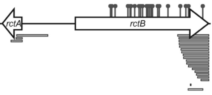
pYB292 even in the absence of RctB overexpression. Most of these colonies could be re-streaked, and plasmid DNA was recovered and sequenced from sixty five transformants. Sixty of these plasmids carried mutations that fell into one of three groups: 1) deletions ofrctA(n = 6), 2) substitutions in therctB sequence that Figure 1. Interactions between RctB, rctA, ParB2 and parS2 control oriCII-based replication. A) Schematic of oriCIIregion of
V. cholerae. DNA fragments used as EMSA probes in Figure 3 are shown by dotted lines and the DNA fragment used in the transcription reporter assay in Figure 7 is shown by the double line. NativeparS2-Band mutated
parS2Xsequences are also shown. Numbers correspond to genomic sequence data (NC_002506). B) Overexpression of RctB and ParB2 enable
oriCII-based replication with origin fragments that includerctAandparS2. Self-ligated DNA fragments containing eitheroriCII-rctB[a],rctA-oriCII-rctB
[b], or rctA(parS2X)-oriCII-rctB [c] were introduced into DH5a cells harboring control vector (pGZ119EH) (open bars), or rctB (pYB285) (closed bars) orparB2(pYB273) (gray bars) expression vectors. Mean and standard deviation of 5 independent experiments are shown. *No transformants obtained after overnight incubation in$3 experiments. doi:10.1371/journal.pgen.1002189.g001
Author Summary
result in amino acid substitutions in RctB (n = 34), and 3) substitutions or deletions inrctB that result in truncations of the carboxyl terminus of RctB (n = 20) (Figure 2 and Table S1). In general, strains carrying these plasmids grew at wild-type rates following the initial 24 hr lag in their detection, suggesting that the mutations within RctB did not impair its replicative capacity. Notably, none of the mutations mapped to theoriCIIsequence per se, an observation that is consistent with the idea that an rctA
transcript or protein does not act in trans on oriCII. In the remaining 5 cases, mutations were likely present in the hostE. coli
chromosome, since the purified oriCII plasmids (which did not harbor mutations) could be re-transformed into the DH5a strain they were isolated from (after plasmid curing) but not into a fresh isolate of DH5a.
The C-terminus of RctB interacts withrctA
The prevalence (20 of 60 clones) of RctB truncations among pYB292 derivatives whose replication did not require RctB overexpression (Figure 2, Table S1) suggested that the C-terminal part of RctB might be required for its interaction with, and inactivation by,rctA. However, the normal replication of plasmids containing such truncations (predicted to remove at least 41, and at most 159, amino acids from the C-terminus of RctB) indicates that truncated RctB retains the capacity to meltoriCIIand initiate chrII replication. Together, these observations raise the possibility that RctB has multiple sites and/or modes for interacting with DNA. To explore this hypothesis, we compared the binding of His-tagged full length RctB and RctB(D500–658), which lacks 159 amino acids of the protein’s C-terminus (hereafter referred to as RctB[DC159]), tooriCIIandrctA, using an electrophoretic mobility shift assay (EMSA). Both proteins readily bound tooriCII, and they appear to have a similar affinity for this sequence, although RctB[DC159] appears to have a greater tendency to form multimeric complexes on the DNA (Figure 3A). In contrast, while wild type RctB bound to the rctA probe, almost no binding of RctB[DC159] was detected (Figure 3A). Together, these observa-tions suggest that RctB has at least two DNA binding domains; one, which bindsoriCII, is contained within RctB[1–499] and can mediateoriCII-based replication, while the other, which bindsrctA, is at least partially contained within, or dependent upon, sequences within RctB[500–658]. We were unable to demonstrate binding of RctB[500–658] torctA, suggesting that additional regions of RctB likely also contribute to rctA binding. Thus, although some sequence similarity has been noted between potential RctB target sites withinoriCIIandrctA[8], our data raises the possibility that RctB actually recognizes two distinct sequences. Additionally, our data provides genetic and biochemical support for the hypothesis
that RctB binding torctAis the basis forrctA’s negative influence on
oriCII-based replication.
ParB2 binding toparS2-B alleviates the negative influence ofrctAon oriCIIreplication
Similar to other chromosomal parS sites, most parS2 sites are located proximal tooriCIIon chrII [15,21]. One of these (designated
parS2-B) is found within the originally annotated rctA sequence (Figure 1A, ref. [15]). AparS2 site is present at a similar position relative tooriCIIin the genomes of multiple other vibrio species [15], despite an overall lack of conservation of the surrounding sequence. We hypothesized that ParB2 binding to thisparS2site might influence binding of RctB torctA, and perhaps thereby regulateoriCII-based replication. This possibility was investigated by measuring the effect of ParB2 on the efficiency with which variousoriCII-related replicons could be transformed intoE. coli. Overexpression of ParB2 had a minimal effect on the transformation of pYB289, consistent with the absence ofrctA/parS2Bwithin this construct (Figure 1B[a]). However, overexpression of ParB2 caused a dramatic increase in the efficiency with which therctA-containing plasmid pYB292 could be introduced into E. coli (Figure 1B[b]). The effect of ParB2 expression was abolished when an alternate plasmid, pYB558, in which the
parS2-Bsite was mutated to parS2X[15], was transformed instead (Figure 1B[c]). Transformants were still obtained with pYB558 when it was introduced into a strain that overexpressed RctB but not when it was introduced into a strain containing an empty vector, suggesting that the mutation inparS2-Bdid not interfere with binding of RctB, and thus that the two proteins do not recognize identical sequences (Figure 1B[b]). Data from the transformation assay was consistent with results from EMSAs, which revealed that ParB2 bound with high affinity to wild typerctAbut notrctAcontainingparS2X, while RctB bound to both probes (Figure 3A). Overall, these data indicate that ParB2 binding toparS2-Bcan mask the negative effect ofrctA
upon replication oforiCII-based replicons.
RctB and ParB2 can simultaneously bindrctA
The simplest explanation for increased transformation efficiency of pYB292 in the presence of overexpressed ParB2 is that binding of ParB2 torctAinterferes with binding of RctB to this site, and thereby makes more RctB available for replication initiation at
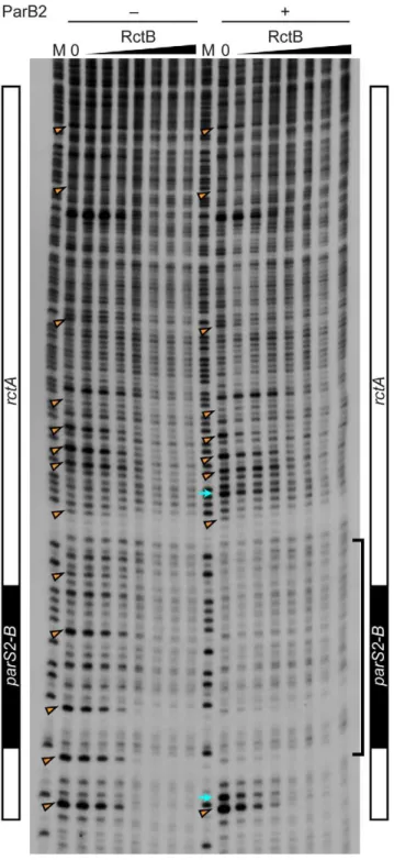
oriCII. However, EMSA analyses did not provide direct support for this hypothesis. Instead, they indicate that RctB and ParB2 can bind simultaneously torctA(Figure 3B and Figure S1). DNase I protection experiments confirmed that RctB can bind torctA, but a specific region of binding was not observed (Figure 4). Instead, when,40–80 ng of RctB were added to the assay, several
non-adjacent nucleotides that were distributed irregularly throughout therctAsequence were protected from DNase I digestion (Figure 4, arrowheads). When higher amounts of RctB (,160–640 ng) were
added, the protection of individual bands became less pronounced and much of the fragment exhibited a degree of protection, including theparS2-Bsite. In contrast, ParB2 protected a,20 bp
continuous stretch of DNA around the parS2-B site (Figure 4). Inclusion of both RctB and ParB2 in the DNAse I protection assays resulted in additive protection, consistent with simultaneous binding of both proteins to rctA. Additionally, DNAse I-hypersensitive sites (Figure 4, arrows) observed in the presence of ParB2 alone became protected upon inclusion of RctB in the reaction, suggesting that RctB can alterrctAstructure even when ParB2 is bound. Collectively the EMSA and footprinting assays show that RctB and ParB2 can simultaneously bind to rctA. However, given the similar patterns of protection of theparS2-B
region by the two proteins, it is difficult to ascertain whether ParB2 interferes with RctB’s binding to this domain withinrctA. Figure 2. Map of locations of mutations that enabled
replication of pYB292.Sites of amino acid substitutions are shown by the pins and deletions are shown as bars. More detailed information is presented in Table S1.
Titration of ParB2 by ectopicparS2sites reduces oriCII-based replication
In order to assess the roles of ParB2,parS2 and rctA at more physiological levels in vivo, we generated an additional construct (pYB404) for the transformation assay that contained all 6 kb of DNA fromparB2throughrctB (Figure 1A), and thereby enabled
expression of ParB2 from its endogenous promoter. In contrast to pYB292, pYB404 replicated inE. colieven without overexpression of RctB or ParB2, despite the presence of rctAin this construct (Figure 5A). Thus, ParB2 produced from its own promoter appears to be sufficient to overcome the negative effect ofrctAonoriCII -mediated replication. However, overexpression of either RctB or Figure 3. Binding of RctB and/or ParB2 to DNA fragments containingrctAandparS2.A) Binding of wild type or mutant RctB or ParB2 proteins to indicated DNA fragments. The amount of protein used in each lane was 0, 0.01, 0.03, 0.1, 0.3, 1, 3, 10, 31, 100, 316, 1000 ng, from left to right. B) Binding of RctB and ParB2 proteins torctA. RctB and ParB2 were premixed and then added to the reaction tube. Amounts of proteins (ng) are indicated and the dilution series from left to right was 0.03, 0.3, 3, 30 and 300 ng. Note that similar banding patterns were observed in experiments where either RctB or ParB2 was added prior to the other protein (see Figure S1).
ParB2 in theE. colistrain did increase the number of transformants obtained, perhaps because limiting amounts of these proteins are present when the plasmid is becoming established (Figure 5A).
We also assessed the influence on transformation efficiency of supplying additional copies of parS2from one of three plasmids with different origins of replication and copy numbers: F (1–2 copies/cell), pSC101 (,5 copies/cell) and p15A (18,22 copies/
cell) [22]. The number of pYB404 transformants obtained was not altered if the ectopic parS2 sequences were in the F-plasmid or even in pSC101 harboring two copies ofparS2separated by 1.4 kb Figure 4. Protection of rctA from DNase I digestion in the
presence of RctB or RctB and ParB2.The DNase I protection assay was performed with 0, 10, 20, 40, 80, 160, 320 and 640 ng RctB bound to a 59-32P-labeled DNA probe containing rctA (including parS2-B, indicated at the side of the gel), in the absence or presence of ParB2 (100 ng). M denotes the G+A chemical sequencing ladder. The bracket indicates the ParB2 footprint. The arrowheads indicate nucleotides protected by RctB in multiple independent experiments. The arrows indicate hypersensitive sites.
doi:10.1371/journal.pgen.1002189.g004
Figure 5. Titration of ParB2 by multicopyparS2preventsoriCII replication.A) Establishment of anoriCIIplasmid containing parAB2-rctA-oriCII-rctB (pYB404) without overexpression of RctB or ParB2. B) pYB404 and pYB289 (oriCII-rctB) were introduced into DH5a cells harboring plasmids containing eitherparS2(closed bar),parS2X(gray bar), control (open bar), or two copies ofparS2 (hatched bar) with various replication origins (indicated on the right, with reported copy numbers shown in parentheses). Means and standard deviations from 3 independent experiments are shown.
(pYB451, yielding ,10 ectopic copies of parS2 (Figure 5B).
However, there was a marked decrease in the efficiency of pYB404 electroporation when the ectopicparS2sites were provided from the moderate-copy-number vector p15A (Figure 5B, p15A ori). In contrast, theparS2Xsequences, which do not bind ParB2, did not alter pYB404 transformation efficiency when supplied from any of the vectors. (Figure 5B, gray bars). Neither wild type nor mutant
parS2 sequences altered the transformation efficiency of a vector (pYB289) that lacksrctA. Thus, the presence of 10parS2sequences (notably, their level within the V. choleraegenome) is compatible with replication of aoriCII-based replicon containingrctA/parS2B. However, an increase to 20 copies (e.g., as should happen followingchrIIreplication) interferes withoriCII-based replication, presumably because ParB2 is titrated away from theparS2-Bsite at the origin and thus can’t counteract rctA-dependent repression. This finding suggests that ParB2 makes an important contribution to controlling replication as well as partitioning ofV. choleraechrII.
ParB2-parS2and RctB-rctAinteractions contribute to oriCIIreplication control inV. cholerae
To further explore the contributions of rctA and parS2-B to regulation of chrII replication, we constructed DrctA V. cholerae
(YBB995) andparS2-B::parS2X V. cholerae(YBB999). These mutant strains did not have detectable growth defects compared to the wild typeV. choleraestrain N16961 (Figure S2), indicating thatrctA
orparS2-Bmediated control oforiCIIis not critical forV. cholerae
viability in rich media. However, quantitative PCR assays measuring the oriCII : oriCIratio revealed that these mutations influence oriCII replication. The DrctA cells exhibited a higher
oriCII : oriCI ratio compared to wild type cells (Table 1), in agreement with a previous report [23]. In contrast, the parS2-B::parS2Xstrain YBB999 had a modest but statistically significant (p = 0.001) reduction in theoriCII:oriCIratio compared to the wild type (Table 1). Both of these results are consistent with our findings using the heterologous host and support the model that ParB2 binding toparS2-Binhibits the negative effect thatrctAexerts on RctB-mediatedoriCIIreplication.
The causes and consequences of chrII overinitiation have not been thoroughly analyzed. However, previous work revealed that extreme overinitiation of oriCII mediated by the RctB mutant RctB[R269S] resulted in a block in V. cholerae cell division, manifest as cell elongation along with a marked decrease in
viability [7]. In contrast, modest overinitiation oforiCIIresulting from overexpression of wild type RctB had virtually no effect on cell viability or morphology (Figure 6A, 6C; see [4,7,23]). Similarly, the modest overinitiation of oriCII caused by the rctA
deletion in YBB995 did not have a detectable affect on cell viability or morphology (Figure 6; [23]). However, deletion ofrctA
sensitizedV. choleraeto the deleterious effects of RctB overexpres-sion. In theDrctA background, RctB overexpression reduced cell viability, particularly in M9 media (Figure 6B), led to an increase in theoriCII: oriCIratio (Table 1), and led to cell filamentation (Figure 6D). In contrast, in theparS2-B::parS2Xbackground, RctB overexpression had little discernible influence on cell division or viability (Figure 6A, 6B), as might be expected given that the basal level of chrII replication initiation in this strain is even lower than in the wild type strain, whose viability was also unimpaired by RctB overexpression. Collectively, these data suggest that V. cholerae can adapt to some variability in RctB levels and availability, and that numerous regulatory processes are geared towards preventing the toxic effects of overinitiating replication of chromosome II.
RctB and ParB2 control transcription ofparAB2
Additional forms of cross-talk between RctB and theparAB2locus are evident from analyses of oriCII-region transcription, which revealed that binding of either ParB2 or RctB torctAalteredparAB2
promoter activity. As has been observed for several additionalparAB
systems [24–26], ParB2 significantly decreased the expression of a
PparAB2-lacZfusion (more than 4-fold; Figure 7). This repression was abolished whenparS2-Bwas mutated toparS2X(Figure 7), strongly suggesting that ParB2 binding toparS2-Bis required for autorepres-sion of the parAB2 locus. In contrast, RctB modestly enhanced expression ofparAB2(Figure 7, p = 0.0003), an effect that does not appear to depend on theparS2-Bsite inrctA. Thus, RctB binding to
rctAmay, despite initially limiting the amount of initiator protein available for replication initiation, ultimately promote replication, as such binding prompts expression of ParB2, which can counter repression of replication. Additionally, these results suggest that cross-talk between pathways controlling replication and partitioning is bidirectional, which is likely to enhance the coordination of these two critical processes.
Discussion
Collectively, our observations suggest that control ofV. cholerae
chrII replication and segregation is linked by a regulatory circuit that involves,6 kb of sequence (and its products) that flankoriCII
and includesparAB2,rctA, andrctB. The primary agent governing replication initiation is RctB; however, initiation can also be influenced by a previously characterized partitioning protein, ParB2, which we now show counteracts rctA’s inhibitory effect upon chrII replication. Analogously, the autoregulatory parAB2
locus is the primary determinant of chrII segregation; however, this process can also be influenced by RctB, which activates
parAB2 expression. It appears likely that the cross-talk between these two systems both prevents extreme fluctuations in protein and chromosome abundance, and enables coordination of chromosome replication and partitioning.
Binding of the chrII replication initiator RctB to the chrII origin and surrounding sequences appears to be more complex than was previously recognized. Our analyses indicate that RctB may in fact have multiple DNA binding modes/domains, which recognize distinct sequences. RctB lacking its C-terminus (as many as 159 amino acids) retained the capacity to bind tooriCIIand to initiate replication at this site. However, both EMSA experiments and Table 1.Ratio oforiCII/oriCIinV. choleraestrains.
Media Strain Genetype oriCII/oriCI
LB* N16961 wild type 1#
YBB995 DrctA 1.5360.06
YBB999 parS2-B::parS2X 0.8560.04
M9{
YBB703 wild type/vector 1#
YBB682 wild type/rctB+
2.4860.24 YBB2003 DrctA/vector 1.3160.06
YBB2004 DrctA/rctB+
3.1460.46
YBB2005 parS2-B::parS2X/vector 0.9460.13 YBB2006 parS2-B::parS2X/rctB+ 1.70
60.17
*Samples were obtained from mid-log phase cultures at an OD600 of,0.7.
{Samples were obtained from cultures grown in M9-Glucose supplemented with 100mM IPTG for 2.5 hrs.
#Defined as 1.
DNase I protection assays (Figure S3) revealed that RctB[DC159] is unable to bind torctA. Residues outside of the C-terminal 159 amino acids are also likely to contribute to rctA binding, although they remain to be identified. The presence of distinct DNA binding domains within the N- and C- terminal parts of RctB introduces the possibility that a single RctB can simultaneously bind torctAand
oriCII. Additional studies are needed to assess whether the binding of RctB to these two sites introduces a bend in the DNA between them. Studies to assess whether the point mutations withinrctBthat enable a bypass ofrctA-mediated replication inhibition do so by altering binding torctAor instead alter other aspects of RctB’s activity or its affinity fororiCIIare also warranted.
To date, precise sequences targeted by RctB have not been identified; it has been speculated that this protein recognizes some short (11 and 12-mer) repeated sequences within the origin and surrounding sequences [3,8]. Our footprinting analyses suggest
that multiple RctB proteins are interacting with the DNA; however, the repeats do not seem to be the principal target of RctB’s C-terminal DNA-binding domain, as many protected sites lie outside of the repeats. The distribution of sites withinrctAthat are protected from DNase I digestion by RctB is unusual, in that RctB appears to interact with multiple non-continuous bases throughout this sequence. One possible explanation for this result is that RctB binding alters the secondary structure ofrctA DNA. Although the DNase I protection assays suggest that multiple RctB proteins interact withrctA, especially at high protein concentra-tions, EMSAs only revealed a single shifted band. The different assay conditions (e.g. the presence of competitor DNA in the EMSAs) may explain this apparent discrepancy. Additional analyses are needed to assess the binding sites for RctB inoriCII.
Our studies confirmed previous reports that rctA inhibits replication oforiCII-based replicons. The inhibitory effect ofrctA Figure 6. Overinitiation oforiCIIcan influence cell viability.A) and B) Plating efficiency of indicated strains on indicated media containing chrolamphenicol. C) and D) Phase-contrast images of YBB2003 (DrctA/vector) (C) and YBB2004 (DrctA/rctB+
) (D) after 4 hr of induction by IPTG. bar = 2mm.
can be overcome by overexpression of RctB (Figure 1B; [8]). Unexpectedly, our work revealed that rctA’s effect can also be mitigated by overexpression of ParB2, which recognizes a parS2
site (parS2-B) within rctA. At least three non-mutually exclusive models can explain how ParB2 abolishes rctA inhibition of replication. One possibility is that ParB2 competes with RctB in binding torctA, resulting in more free RctB that can interact with
oriCII. However, both EMSA and DNase I protection assays demonstrated that ParB2 does not block all RctB binding within
rctAin vitro; at most, only the subset of RctB binding sites inparS2
-Bare blocked by the presence of ParB2. However, these in vitro assays may not fully reflect binding dynamics in vivo in which binding torctAmaybe influenced by the adjacentoriCIIsite and by additional factors such as IHF. An alternative possibility is that RctB binding torctA alters the secondary structure of theoriCII
region in a manner that inhibits replication. ParB2 binding to
parS2-B could counteract RctB-mediated remodeling of oriCII, thereby promoting replication. ParB2 might also alter the extent to whichrctAis transcribed, which has also been shown to influence
rctA’s effectiveness as a replication inhibitor [8]. It is unlikely that the effect of ParB2 upon replication is mediated by a direct interaction between this protein and RctB, as no such interaction was detected using a bacterial two hybrid system (Figure S4). Regardless of the mechanism by which it acts, it is clear that ParB2, previously described as a key agent mediating chrII segregation, also contributes to regulation of chrII replication, thereby enabling linkage of these cellular processes.
We hypothesize that the organization of this regulatory scheme is adapted to accommodate the cell cycle. As ParB2 accumulates, perhaps to amounts that are sufficient to enable chrII segregation, the repressive effects of rctA are relieved, and initiation of chrII replication ensues. Subsequently, ParB2 is re-distributed among the newly synthesized parS2 sites, and its binding to parS2-B is
reduced, enablingrctAto inactivate RctB, and thereby reducing the ability of RctB to initiate replication.
Cross-talk between chrII replication and partitioning is also evident at the level of transcription. The parAB2 locus is autorepressed byparB2, as has been observed for otherparABloci [24–26]; in addition, we demonstrate thatparAB2transcription is activated by RctB. The contrasting effects of these two regulators are likely to rebalance ParAB levels if their abundance becomes aberrantly elevated or reduced.
Undoubtedly, additional factors and mechanisms intersect with these regulatory circuits. For example, previous studies have revealed that RctB can repress its own transcription [27,28] as well as the transcription ofrctA [28]. Transcription of rctA has been reported to inhibit the negative influence of rctA on RctB [8]. Additional regulatory processes also contribute to control of replication initiation. For example, ATP binding inhibits RctB activity by decreasing its ability to bind oriCII [7] and the methylation status oforiCIIalso influences RctB binding tooriCII
[9]. Given the centrality of chromosome replication and segregation to the perpetuation of the species, the existence of multiple and perhaps redundant mechanisms to increase the robustness of the control of these processes is expected. Consistent with this idea, we only observed significant impairment of V. choleraegrowth when RctB was over-expressed in anrctAmutant, a condition that likely allows considerable overinitiation of chrII. Overinitiation also leads to growth impairment and cell filamenta-tion inE. coliandCaulobacter crescentus[29–31].
Although coordinated control of chromosome replication and segregation makes sense to ensure proper chromosome inheritance to daughter cells, little mechanistic information linking these essential processes is available. Recent work by Murray and colleagues revealed that inB. subtilis the ParA ortholog Soj can inhibit or stimulate chromosome replication initiation via interactions with the initiator protein DnaA, while the ParB ortholog Spo0J inhibits initiation of chromosome replication by blocking Soj dimerization [32,33]. A similar regulatory scheme was recently described forV. choleraechrI; Chattoraj and colleagues reported that ParA1 stimulates chrI replication and ParB1 inhibits ParA1 [34]. However, ParA2 appears to govern chrII replication initiation via a distinct mechanism that does not require it to interact with the replication initiator RctB. In contrast to findings for Soj and ParA1, which interact with DnaA, we did not detect interaction between ParA2 and RctB using a bacterial two hybrid system. Our findings, along with previous reports, suggest that further exploration of the roles of Par systems in control of chromosome replication in diverse bacteria is warranted. Since chromosomalpargenes are found in,70% of bacterial genomes
[20], Par proteins andparSsites may commonly exert control over chromosome replication. Finally, it will be interesting to explore whether mechanisms exist to link the replication and/or segregation of the two chromosomes in V. cholerae and other bacteria with multiple chromosomes.
Materials and Methods
Plasmids and strains
Most of the plasmids used in this study are listed in Table 2. The sites and mutations present in the rctA containing oriCII-based plasmids (discussed in Figure 1B[b]) are shown in Table S1. The plasmids used for the bacterial two hybrid analysis are shown in Table S2.
A two-step strategy for construction oforiCII-based plasmids was followed. First, different segments of DNA proximal tooriCIIwere amplified and cloned into pYB199, a derivative plasmid of pKD4 Figure 7. Transcriptional control ofparAB2promoter by RctB
and ParB2. b-galactosidase activities of parAB2 promoter with or without an intactparS2were measured in the presence of a plasmid expressing RctB (pYB284) (black bars) or ParB2 (pSM922) (gray bars), as well as a vector control (pBAD33) (open bars). Averages and standard deviations are shown and * represents significant (p value,0.01) difference between the indicated groups.
[35] which harbors the R6K origin and genes conferring resistance to ampicillin (bla) and kanamycin (aph). Second, the resulting plasmids were digested with XbaI and the fragment containing the oriCIIregion andaphwas gel-purified, self-ligated and then electroporated into E. coli DH5a. Spontaneous suppressor mutants in the rctA containing oriCII-based plasmids (shown in Figure 2 and Table S1) were isolated by electroporating
,100 ng of self-ligated DNA fragments into DH5acells. Colonies
that arose after an,48 hr incubation were re-streaked and then
plasmid DNA was purified and sequenced. In addition, to confirm that mutation in the plasmid enabled establishment, each mutant
plasmid was re-electroporated into DH5a. The mutations in
parS2-B yielding parS2X (Figure 1A) were generated using the QuickChange XL Site Directed Mutagenesis Kit (Stratagene). To construct pYB141, pYB217, pYB447, and pYB448, 15-bp double stranded DNA fragments containing eitherparS2-AorparS2Xwere inserted into the EcoRI site of the vectors (pWKS30 and pXX705). Plasmids pYB193 and pYB216 were constructed in a similar fashion using the NheI and HindIII sites in the pBAD33 vector. To construct pYB451, the second copy of parS2-A was inserted at the AflII site of pYB447, which is,1.4 kb away from
EcoRI site where the first copy ofparS2-A was inserted. Plasmid Table 2.Plasmids used in this study.
Name Description Resistance{
Reference
pBAD33 p15A ori Cm [40]
pCB192-YY pCB192 but no EcoRI site in 39oflacZ Amp [38], This study
pCVD442 vector for allelic exchange Amp [37]
pET-RctB pET28brctB Km [7]
pGZ119EH ColD ori Cm [41]
pSM843 pET28bparB2 Km [15]
pSM922 pBAD33parB2 Cm Sarah McLeod, This study
pWKS30 pSC101 ori Amp [42]
pXX705 F ori Amp [43]
pYB141 pXX705parS2-A Amp This study
pYB190 pCRII Km [16]
pYB193 pBAD33parS2-A Cm This study
pYB199 pKD4 MCS* Amp, Km This study
pYB216 pBAD33parS2X Cm This study
pYB217 pXX705parS2X Amp [15]
pYB264 pYB199rctA oriCII rctB Amp, Km This study
pYB273 pGZ119EHparB2 Cm This study
pYB276 pYB199oriCII rctB Amp, Km This study
pYB285 pGZ119EHrctB Cm [7]
pYB289 oriCII rctB Km [7]
pYB292 rctA oriCII rctB Km This study
pYB294 pBAD33rctB Cm This study
pYB355 pET28brctB[DC159] Km This study
pYB379 pYB199rctA(parS2-B::parS2X)oriCII rctB Amp, Km This study
pYB403 pYB199parB2 parA2 rctA oriCII rctB Amp, Km This study
pYB404 parB2 parA2 rctA oriCII rctB Amp, Km This study
pYB405 pCRIIoriCII Km This study
pYB406 pCRIIrctA Km This study
pYB407 pCRIIrctA(parS2-B::parS2X) Km This study
pYB432 pCVD442-based allelic exchange plasmid to constructDrctA Amp This study pYB445 pCVD442-based allelic exchange plasmid to constructparS2-B::parS2X Amp This study
pYB447 pSC101parS2-A Amp This study
pYB448 pSC101parS2X Amp This study
pYB451 pSC101parS2-A parS2-A Amp This study
pYB452 pCB192-YYPparAB2-rctA lacZ Amp This study
pYB453 pCB192-YYPparAB2-rctA(parS2-B::parS2X)lacZ Amp This study
pYB558 rctA(parS2-B::parS2X)oriCII rctB Km This study
*MCS; multi cloning site.
pCB192-YY, a derivative of the transcriptional fusion vector pCB192 [36] in which the EcoRI site in the 39 end oflacZwas removed by introduction of a silent mutation (GAATTC to GAATTT), was used to create transcriptional fusions to the
parAB2promoter. TheparAB2promoter region was amplified and cloned into the HindIII-EcoRI site of pCB192-YY. All the relevant DNA sequences of all plasmids used in this study were determined. The sequences of the oligonucleotides used in this study are listed in Dataset S1.
Mutations were introduced on to theV. cholerae chromosome (DrctA, andparS2-B::parS2X) via allele exchange using pCVD442-based plasmids as described [37].V. cholerae strains used in this study are listed in Table 3.
Transformation efficiency experiments
DH5acells harboring either pGZ119 (vector),or Isopropyl-b -D-1-thiogalactopyranoside (IPTG) inducible rctB orparB2, pYB285 or pYB273 respectively, were grown in LB broth containing 100mM IPTG till mid-log phase to prepare electrocompetent cells. Similarly, DH5a cells harboring plasmid-borne parS2
sequences or control plasmids were grown until mid-log phase to prepare chemical competent cells. 40 ng of self-ligated DNA or 10 ng of pYB404 DNA were introduced into the competent cells. As a control, 10 ng of plasmid pYB190 was also introduced into competent cells in each experiment. The number of colonies obtained in the pYB190 transformations were used to normalize transformation efficiencies. Means and standard deviations were derived from 3–5 independent experiments for all plasmids tested.
Beta-galactosidase assays
Assays were performed in triplicate with log phase cultures as described previously [38]. Two tailed, two-sample equal t-tests were used to compare the results from 3 independent experiments (total 9 samples each) for the statistical analysis.
Electrophoretic mobility shift assay (EMSA)
Wild type RctB-His6, RctB[DC159]-His6, and ParB2-His6 proteins were purified as previously described [15]. Sequences used for oriCII, rctA, and rctA (parS2-B::parS2X) (see Figure 1A) EMSA probes were initially cloned into the pCR-Blunt II-TOPO vector (Invitrogen), yielding pYB405, pYB406, and pYB407, respectively. pYB190, a pCR-Blunt II TOPO derivative contain-ing an irrelevant 10 bp, was used to construct the negative control probe. To prepare radio-labeled probes, appropriate DNA regions were amplified from the plasmids with universal M13 forward and
reverse primers [22], end labeled with [c-32P] ATP with polynucleotide kinase (New England Biolabs), purified from 6% DNA retardation gels (Invitrogen), and ethanol precipitated. In the binding reactions, 5,000 cpm of probe DNA containing different concentrations of RctB and/or ParB2 in a reaction buffer of 20 mM Tris-Cl (pH 7.5), 1 mM EDTA, 150 mM NaCl, 12.5mg/ mL poly (dI-dC), and 0.1 mg/mL BSA were incubated for 10 min at room temperature. The reactions were then electrophoresed in a 6% DNA retardation gel in 0.56 TAE and visualized by autoradiography.
DNase I foot print assay
DNase I footprint assays were performed as previously described with minor modifications [39]. The rctA probe was made by PCR using 59-32P-radiolabeled rctA 59-FP
(CGTTTAAATAACCCACATATTCTTCGATAAGG) and
rctA 39-FP (ATACCTATTCGCTGGAGGAAAGATAGG)
primers on a plasmid encoding parAB2-rctA-oriCII-rctB(pYB403). The probe was purified from 6% DNA retardation gels, eluted, and ethanol precipitated. 1,200,000 cpm of probe was incubated with different amounts of RctB without and with 100 ng of ParB2 in 20mL of 20 mM Tris-Cl pH 8.0, 125 mM NaCl, 1 mM DTT for 10 min at room temperature. 0.1 U of DNase I (Applied Biosystems) was added to each reaction and incubated at room temperature for 30 sec. The digestions were quenched by the addition of 6mL of 660 mM Tris-Cl pH 9.5, 66 mM EDTA, 3.3% SDS and placed on ice. Samples were ethanol precipitated, resuspended in recrystalized formamide, and 20,000 cpm of each was run on an 8% polyacrylamide gel with 8 M urea (National Diagnostics) in 16TBE. The gels were then dried and visualized by autoradiography.
Quantitative PCR assay
Genomic DNA was prepared from each strain by phenol-chloroform extraction followed by ethanol precipitation. The genomic DNA was then digested with PstI and 10 pg was used for each quantitative PCR (qPCR) reaction. Genomic DNA from an N16961 stationary culture was used to generate the standard curve. qPCR was performed with the StepOnePlus Real-Time PCR system (Applied Biosciences) using SYBR Green Master mix (Applied Biosciences) according to the manufacturer’s protocol. The primer pairs used for oriCI and oriCII were described previously [4]. Each qPCR run was done in triplicate and the ratio was calculated from three independent experiments.
Growth curves and microscopy
V. choleraecells harboring a plasmid borne copy of an IPTG-induciblerctBor a control vector were grown in either LB or M9-Glucose media containing 100mM IPTG at starting OD600 of 0.003. Subsequently, OD600 and CFU were monitored hourly. Growth curves shown in Figure S2 are representative of at least three independent experiments. A small aliquot of cells was removed at 4 hr, fixed with 3% paraformaldehyde, and then examined with 1006 alpha-plan lens on a Zeiss Axioplan 2 microscope.
Supporting Information
Dataset S1 List of oligonucleotides used in this study. (XLS)
Figure S1 Binding of RctB and ParB2 proteins torctA. A) and C), RctB was added to the reaction tube 8 min prior to addition of ParB2. B) and D), ParB2 was added to the reaction tube 8 min Table 3.V. choleraestrains used in this study.
Name Description Source/Reference
N16961 [2]
YBB682 N16961/pYB285 [7]
YBB703 N16961/pGZ119EH [7]
YBB995 N16961DrctA This study YBB999 N16961parS2-B::parS2X This study
YBB2003 YBB995/pGZ119EH This study
YBB2004 YBB995/pYB285 This study YBB2005 YBB999/pGZ119EH This study
YBB2006 YBB999/pYB285 This study
prior to addition of RctB. Amount of proteins in titration was 0.03, 0.3, 3, 30 and 300 ng, from left to right.
(TIF)
Figure S2 Growth curves ofV. cholerae strains. OD600 nm (A) and Colony forming units (CFU) (B) ofV. cholerae N16961 (gray circles), YBB995 (DrctA; open triangles) and YBB999 ( parS2-B::parS2X; closed squares) cells grown in LB media at indicated time points are shown.
(TIF)
Figure S3 Protection of rctA from DNase I digestion by RctB[DC159]. The DNase I protection assay was performed with 0, 10, 20, 40, 80, 160, 320 or 640 ng RctB[DC159] bound to a 59-32P-labeled DNA containingrctA (includingparS2-B, indicated at the side of thegel). M denotes the G+A chemical sequencing ladder.
(TIF)
Figure S4 Interactions between RctB and ParB2. A pair of plasmids that express RctB or ParB2 fused to the T18 and T25 subunits of adenylate cyclase was simultaneously introduced into
E. coliBTH101. After transformation, 2mL of cells were spotted
onto LB plates containing ampicillin (100mg/mL), kanamycin (50mg/mL), IPTG (100mM), and bromo-chloro-indolyl-galacto-pyranoside (X-gal, 60mg/mL) and incubated overnight at 30uC. (TIF)
Table S1 List of mutantoriCIIplasmids. (DOC)
Table S2 List of plasmids used for bacterial two hybrid assay. (DOC)
Acknowledgments
We are grateful to Sarah M. McLeod and Monica P. Hui for plasmids and purified ParB2 protein. We also thank Waldor lab members for helpful discussions and technical advice.
Author Contributions
Conceived and designed the experiments: YY MKW. Performed the experiments: YY MAG. Analyzed the data: YY MAG BMD MKW. Contributed reagents/materials/analysis tools: YY MAG. Wrote the paper: YY BMD MKW.
References
1. Okada K, Iida T, Kita-Tsukamoto K, Honda T (2005)Vibrioscommonly possess two chromosomes. J Bacteriol 187: 752–757.
2. Heidelberg JF, Eisen JA, Nelson WC, Clayton RA, Gwinn ML, et al. (2000) DNA sequence of both chromosomes of the cholera pathogenVibrio cholerae. Nature 406: 477–483.
3. Egan ES, Waldor MK (2003) Distinct replication requirements for the twoVibrio choleraechromosomes. Cell 114: 521–530.
4. Duigou S, Knudsen KG, Skovgaard O, Egan ES, Løbner-Olesen A, et al. (2006) Independent control of replication initiation of the two Vibrio cholerae
chromosomes by DnaA and RctB. J Bacteriol 188: 6419–6424.
5. Mott ML, Berger JM (2007) DNA replication initiation: mechanisms and regulation in bacteria. Nat Rev Microbiol 5: 343–354.
6. Zakrzewska-Czerwin´ska J, Jakimowicz D, Zawilak-Pawlik A, Messer W (2007) Regulation of the initiation of chromosomal replication in bacteria. FEMS Microbiol Rev 31: 378–387.
7. Duigou S, Yamaichi Y, Waldor MK (2008) ATP negatively regulates the initiator protein ofVibrio choleraechromosome II replication. Proc Natl Acad Sci U S A 105: 10577–10582.
8. Venkova-Canova T, Srivastava P, Chattoraj DK (2006) Transcriptional inactivation of a regulatory site for replication ofVibrio choleraechromosome II. Proc Natl Acad Sci U S A 103: 12051–12056.
9. Demarre G, Chattoraj DK (2010) DNA adenine methylation is required to replicate bothVibrio choleraechromosomes once per cell cycle. PLoS Genet 6: e1000939. doi:10.1371/journal.pgen.1000939.
10. Egan ES, Løbner-Olesen A, Waldor MK (2004) Synchronous replication initiation of the twoVibrio choleraechromosomes. Curr Biol 14: R501–R502. 11. Rasmussen T, Jensen RB, Skovgaard O (2007) The two chromosomes ofVibrio
choleraeare initiated at different time points in the cell cycle. EMBO J 26: 3124–3131.
12. Stokke C, Waldminghaus T, Skarstad K (2011) Replication patterns and organization of replication forks inVibrio cholerae. Microbiology 157: 695–708. 13. Fogel MA, Waldor MK (2006) A dynamic, mitotic-like mechanism for bacterial
chromosome segregation. Genes Dev 20: 3269–3282.
14. Saint-Dic D, Frushour BP, Kehrl JH, Kahng LS (2006) A parA homolog selectively influences positioning of the large chromosome origin inVibrio cholerae. J Bacteriol 188: 5626–5631.
15. Yamaichi Y, Fogel MA, McLeod SM, Hui MP, Waldor MK (2007) Distinct centromere-likeparSsites on the two chromosomes ofVibriospp. J Bacteriol 189: 5314–5324.
16. Yamaichi Y, Fogel MA, Waldor MK (2007)pargenes and the pathology of chromosome loss inVibrio cholerae. Proc Natl Acad Sci U S A 104: 630–635. 17. Gerdes K, Howard M, Szardenings F (2010) Pushing and pulling in prokaryotic
DNA segregation. Cell 141: 927–942.
18. Salje J (2010) Plasmid segregation: how to survive as an extra piece of DNA. Crit Rev Biochem Mol Biol 45: 296–317.
19. Lin DC, Grossman AD (1998) Identification and characterization of a bacterial chromosome partitioning site. Cell 92: 675–685.
20. Livny J, Yamaichi Y, Waldor MK (2007) Distribution of centromere-likeparS
sites in bacteria: insights from comparative genomics. J Bacteriol 189: 8693–8703.
21. Yamaichi Y, Duigou S, Shakhnovich EA, Waldor MK (2009) Targeting the Replication Initiator of the SecondVibrioChromosome: Towards Generation of
Vibrionaceae-Specific Antimicrobial Agents. PLoS Pathog 5: e1000663. doi:10.1371/journal.ppat.1000663.
22. Sambrook J, Russell D (2001) Molecular Cloning. Cold Spring Habor Laboratory Press.
23. Srivastava P, Chattoraj DK (2007) Selective chromosome amplification inVibrio cholerae. Mol Microbiol 66: 1016–1028.
24. de la Hoz AB, Ayora S, Sitkiewicz I, Ferna´ndez S, Pankiewicz R, et al. (2000) Plasmid copy-number control and better-than-random segregation genes of pSM19035 share a common regulator. Proc Natl Acad Sci U S A 97: 728–733. 25. Carmelo E, Barilla` D, Golovanov AP, Lian LY, Derome A, et al. (2005) The unstructured N-terminal tail of ParG modulates assembly of a quaternary nucleoprotein complex in transcription repression. J Biol Chem 280: 28683–28691.
26. Ringgaard S, Ebersbach G, Borch J, Gerdes K (2007) Regulatory cross-talk in the doubleparlocus of plasmid pB171. J Biol Chem 282: 3134–3145. 27. Pal D, Venkova-Canova T, Srivastava P, Chattoraj DK (2005) Multipartite
regulation ofrctB, the replication initiator gene ofVibrio choleraechromosome II. J Bacteriol 187: 7167–7175.
28. Egan ES, Duigou S, Waldor MK (2006) Autorepression of RctB, an initiator of
Vibrio choleraechromosome II replication. J Bacteriol 188: 789–793.
29. Katayama T, Takata M, Sekimizu K (1997) CedA is a novelEscherichia coli
protein that activates the cell division inhibited by chromosomal DNA over-replication. Mol Microbiol 26: 687–697.
30. Fujimitsu K, Su’etsugu M, Yamaguchi Y, Mazda K, Fu N, et al. (2008) Modes of overinitiation,dnaAgene expression, and inhibition of cell division in a novel cold-sensitive hda mutant ofEscherichia coli. J Bacteriol 190: 5368–5381. 31. Collier J, Shapiro L (2009) Feedback control of DnaA-mediated replication
initiation by replisome-associated HdaA protein inCaulobacter. J Bacteriol 191: 5706–5716.
32. Murray H, Errington J (2008) Dynamic control of the DNA replication initiation protein DnaA by Soj/ParA. Cell 135: 74–84.
33. Scholefield G, Whiting R, Errington J, Murray H (2011) Spo0J regulates the oligomeric state of Soj to trigger its switch from an activator to an inhibitor of DNA replication initiation. Mol Microbiol 79: 1089–1100.
34. Kadoya R, Baek JH, Sarker A, Chattoraj DK (2011) Participation of a Chromosome Segregation Protein ParAI of Vibrio cholerae in Chromosome Replication. J Bacteriol 193: 1504–1514.
35. Datsenko KA, Wanner BL (2000) One-step inactivation of chromosomal genes inEscherichia coliK-12 using PCR products. Proc Natl Acad Sci U S A 97: 6640–6645.
36. Schneider K, Beck CF (1986) Promoter-probe vectors for the analysis of divergently arranged promoters. Gene 42: 37–48.
37. Donnenberg MS, Kaper JB (1991) Construction of aneaedeletion mutant of enteropathogenic Escherichia coli by using a positive-selection suicide vector. Infect Immun 59: 4310–4317.
38. Miller JH (1992) A short course in bacterial genetics Cold Spring Habor Laboratory Press.
39. Bruist MF, Glasgow AC, Johnson RC, Simon MI (1987) Fis binding to the recombinational enhancer of the Hin DNA inversion system. Genes Dev 1: 762–772.
41. Lessl M, Balzer D, Lurz R, Waters VL, Guiney DG, et al. (1992) Dissection of IncP conjugative plasmid transfer: definition of the transfer region Tra2 by mobilization of the Tra1 region in trans. J Bacteriol 174: 2493–2500. 42. Wang RF, Kushner SR (1991) Construction of versatile low-copy-number
vectors for cloning, sequencing and gene expression inEscherichia coli. Gene 100: 195–199.