Endoprosthesis Implantation at the Entry Pathway of the Right
Atrium Monitored by Intracardiac Ultrasound
Rogério Tadeu Tumelero, Norberto Toazza Duda, Alexandre Pereira Tognon, Saulo Martins Coccio Filho
Interventional Cardiology of the Hospital São Vicente de Paulo, Faculdade de Medicina da Universidade de Passo Fundo – Passo Fundo, RS - Brazil
Mailing address: Rogério Tadeu Tumelero •
Rua José Bonifácio, 230/302 – 99070-070 – Passo Fundo, RS - Brazil E-mail: [email protected]
Manuscript received March 22, 2006; revised manuscript received June 2, 2006; accepted June 22, 2006.
Introduction
Obstructions of the upper venous system have a varied physiopathogenesis, including the occurrence of spontaneous thrombosis (1 to 4% of all venous thromboses, the presence of hemodialysis or chemotherapy catheters, and even extrinsic compression caused by intrathoracic tumors1-3). The use
of endoprosthesis for the treatment of obstructions in the venous system has become common practice, and it has been indicated for situations of risk of pulmonary embolism, superior vena cava syndrome, and loss of vascular access4.
Within this context, the use of intravascular/intracardiac ultrasound may be useful in evaluating the extension and severity of the obstructive lesion, besides providing important information about the mural and intraluminal morphology of the venous system. The authors report the case of a patient with obstruction of the superior vena cava secondary to thrombosis of totally implantable catheter, which was impairing the right atrium. The patient had a polytetrafluoroethylene (PTFE)-covered endoprosthesis implanted in the superior vena cava/right atrium system, which was monitored by intravascular/intracardiac ultrasound.
Case Report
The case of a 61-year-old female patient with previous radical mastectomy due to infiltrating ductal carcinoma and a totally implantable catheter implanted for chemotherapy is described. Eleven months after catheter implantation, the patient began to experience edema of the face and upper limbs, as well as cyanosis of the face. Computed tomography of the chest showed the presence of thrombosis in the superior vena cava and upper portion of the right atrium, confirming the diagnosis of superior vena cava syndrome (SVCS). No extrinsic compression was observed.
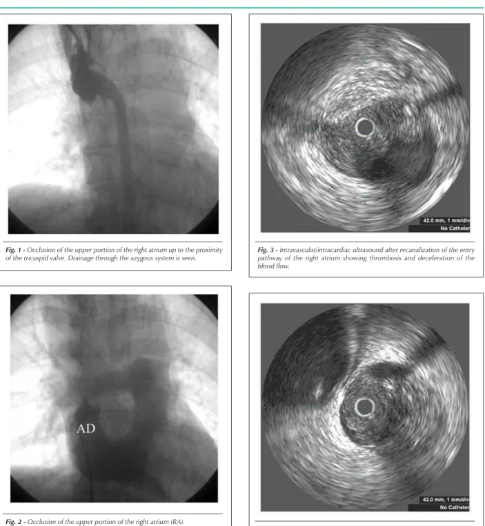
Twenty days after the onset of symptoms, the implantable catheter was removed, and no remission of the symptoms was observed. Patient was referred to venography and right cardiac catheterization. An injection into the right subclavian vein and right femoral vein allowed visualization of the complete occlusion in the upper portion of the right atrium, immediately after the origin of the azygous system up to the tricuspid valve region. The upper venous system drained into the azygous system. Pressures in the right ventricle and pulmonary artery were within normal ranges (mean pulmonary arterial pressure = 20 mmHg) (Figure 1).
Due to the severe clinical impairment, surgical risk and guarded prognosis, surgery was contraindicated. The percutaneous procedure was indicated for implantation of the PTFE-covered endoprosthesis through the femoral artery, without predilation in order to prevent thrombi displacement to the pulmonary system. The right femoral and right subclavian veins were punctured, and the right atrium was injected with contrast material, confirming the obstruction of the entry pathway. Non-fractioned heparin (10,000 UI) was administered endovenously. A 0.014” Shinobi wire (Cordis, FL, USA) was used to move past the occlusion; recanalization was performed by dilation with a 3mm x 20mm balloon. A 0.035”, 300cm DAIG guidewire (St. Jude Medical, MN, USA) was positioned through the femoral vein. A 9F sheath was introduced over the guidewire to the right internal jugular vein, and a 9Mhz intravascular/intracardiac ultrasound catheter (Boston Scientifics, MN, USA) was positioned.
Using the Galaxy system (Boston Scientifics, MN, USA), ultrasound images were obtained which, combined with the angiographic images, enabled assessment of the presence and distribution of the thrombus, its relationship with the azygous system and with the tricuspid valve (figures 2 to 5), as well as the dimensions of the endoprosthesis to be used. Balloon predilation remained a non-viable option due do the ultrasonographic findings showing a large thrombus. A 17 x 27 mm (16F) Apolo endoprosthesis (Nano Endovascular, SC,
Key words
Superior vena cava syndrome; endoprotesis; ultrasonography, interventional.
BR) was implanted through the femoral puncture access, followed by balloon postdilation (a Multitrack 18x50mm balloon, Numed, NY, USA). At the end of the procedure, new angiographic images (figure 6) and ultrasonographic images (figure 7) were obtained. Despite the reestablishment of an adequate blood flow to the right atrium entry pathway, as well as flow maintenance in the azygous system, a thrombus was detected at the proximal rim of the endoprosthesis.
Follow-up pulmonary arteriography did not show any thrombi images, and the pulmonary artery pressures remained within normal ranges. While still in the hemodynamics
room, the patient experienced a considerable improvement of cyanosis and noticeable reduction of facial edema. She was discharged two days after the procedure, and was on acetylsalicylic acid (200mg) and anticoagulation with oral warfarin 5mg for 6 months, with a target INR (International Normalized Ratio) between 2.0 and 3.0.
At the 4th and 10th months of follow-up, the patient
underwent a hemodynamic study with catheterization of the right chambers plus angiography associated with intravascular/ Fig. 1 -Occlusion of the upper portion of the right atrium up to the proximity
of the tricuspid valve. Drainage through the azygous system is seen.
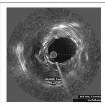
Fig. 3 -Intravascular/intracardiac ultrasound after recanalization of the entry pathway of the right atrium showing thrombosis and deceleration of the blood flow.
Fig. 2 -Occlusion of the upper portion of the right atrium (RA).
intracardiac ultrasound, which showed that the initial results had been maintained and that the thrombus at the proximal rim of the endoprosthesis had disappeared (figures 8 and 9). After two years, the patient was on 200mg of ASA, asymptomatic and performed routine daily activities. Chest X-ray does not show displacement of the endoprosthesis.
Discussion
Thrombosis in the upper venous system is a rare occurrence, Fig. 6 - Venous angiography showing recanalization of the superior vena cava/right atrium and the image of a thrombus at the upper portion of the endoprosthesis.
Fig. 8 - Follow-up venous angiography made at 10 months showing maintenance of recanalization of the superior vena cava/right atrium and no image of a thrombus in this portion.
Fig. 5 -Intravascular/intracardiac ultrasound after recanalization of the entry pathway of the right atrium (RA) showing the tricuspid valve (TV) free of the thrombus.
Fig. 7 -Intravascular/intracardiac ultrasound showing adequate apposition of the endoprosthesis.
and is usually associated with the presence of catheters implanted in the venous system either for chemotherapy or hemodialysis; thrombi may also occur in certain situations after radiotherapy1. There are also reports of superior vena
cava syndrome in patients with pacemakers2,3. The diagnosis
of this syndrome may be difficult when the symptoms are mild and associated with minor obstructions or caused by extensive collateral circulation. The presence of edema helps to make most diagnoses.
Treatment in these cases includes thrombolysis, balloon
TV
RA
Fig. 9 - Intravascular/intracardiac ultrasound showing maintenance of adequate apposition of the endoprosthesis and no image of a thrombus on its proximal edge.
dilation, stenting , surgery, or a combination of these procedures. However, the best strategy has not yet been established. Chemical thrombolysis performed with thrombolytic agents (rTPa, streptokinase) does not provide assurance as to recanalization, has poor success rates, and may pose a risk of bleeding. Surgery in these cases is considered a major procedure which demands extracorporeal circulation, thus raising the morbidity-mortality risk in a group of patients with non-established diagnoses and high comorbidities, mainly those with neoplasms or chronic renal insufficiency that are undergoing hemodialysis. Current trends consist of employing the percutaneous technique, whose superiority has been described in literature, both for recanalization of occlusions and superior vena cava stenosis without involvement of the right atrium and/or tricuspid valve4. Balloon angioplasty for the treatment
of superior vena cava obstructions provides immediate adequate results5. On the long run, however, its efficacy is
compromised by elastic retraction. On the other hand, the use of self-expandable stents or balloon-expandable stents significantly reduces the occurrence of elastic retraction, thus eliminating one of the factors responsible for restenosis. Self-expandable stents, in particular, adapt better to the anatomy of overlying structures by determining a type of negative elastic retraction, as they continue to expand over time6 besides adapting to sites subjected to an ample range
of movements, such as joints or angulations. The use of PTFE-covered endoprosthesis for the treatment of superior vena cava syndrome with the involvement of the right atrium has not yet been described in literature, and in the case reported, the initial success was maintained throughout the two-year follow-up. The considerable clinical experience with aortic endoprostheses without oral anticoagulation leads us to the discussion of high and low pressure territories, and the need for anticoagulation. Presumably,
until reendothelialization is achieved in the venous system, oral anticoagulation is essential mainly to avoid pulmonary embolism. It is not known if the endoprosthesis coating poses any risk for embolic events. Therefore, prolonged oral anticoagulation therapy was administered for six months. Oral anticoagulation was interrupted after this period, and the reduced risk of thrombosis was attributed to the endothelialization of the endoprosthesis. Besides the images obtained by angiography and intravascular ultrasound, the maintenance of normal pressure levels within the pulmonary artery indicated absence of significant embolism episodes. One should consider, however, the possibility of microembolism in the pulmonary territory, which is known to have intense fibrinolytic action.
Another application of percutaneous intervention in the venous system includes the palliative treatment of the superior vena cava syndrome of neoplastic origin. Several case reports are described in literature and, as such, the technique has been shown to be safe, easily-performed, with high rates of success, and providing rapid relief of symptoms when compared to conventional treatment, chemotherapy, or radiotherapy alone, and it may become the procedure of choice for these situations7-10. The use of endovascular biopsy
could be associated with stenting to allow a definite diagnosis to guide anti-tumoral therapy11.
The use of intravascular/intracardiac ultrasound for the percutaneous treatment of venous system obstructions has been studied, providing details not visible on venography. With this technique, the formation of collaterals proximal to a venous obstruction may be differentiated from intraluminal trabeculations12. The extrinsic compression and resulting
deformity of the venous lumen, on the other hand, may be directly visualized. Ultrasound seems to be better than venography for assessing the grade of the obstruction, as venography would underestimate the grade of the lesion, particularly in cases of extensive occlusions12. Moreover,
ultrasound provides data on the morphology of the luminal wall, presence of thrombi or debris and, in case of stenosis secondary to multiple punctures, the fibrous component may also be visualized12-13. It also enables determination of
the extension and grade of the obstructive lesion, mainly to define the extension and the diameter of the balloons and/or stents to be employed, besides providing data on the proximal and distal edges of the stent and its adequate apposition 12-13. The use of intravascular/intracardiac ultrasound helps to
establish the adequate positioning of the prosthesis without compromising the valve.
Conclusion
References
1. Bornak A, Wicky S, Ris HB, Probst H, Milesi I, Corpataux JM. Endovascular treatment of stenoses in the superior vena cava syndrome caused by non-tumoral lesions. Eur Radiol. 2003;13 (5): 950-6.
2. Chan AW, Bhatt DL, Wilkoff BL, Roffi M, Mukherjee D, Gray BH, et al. Percutaneous treatment for pacemaker-associated superior vena cava syndrome. Pacing Clin Electrophysiol. 2002; 25 (11): 1628-33.
3. Teo N, Sabharwal T, Rowland E, Curry P, Adam A. Treatment of superior vena cava obstruction secondary to pacemaker wires with balloon venoplasty and insertion of metallic stents. Eur Heart J. 2002; 23 (18): 1456-70.
4. Schindler N, Vogelzang RL. Superior vena cava syndrome: experience with endovascular stents and surgical therapy. Surg Clin North Am. 1999; 79: 683-94.
5. Lock JE, Bass JL, Castaneda-Zuniga W, Fuhrman BP, Rashkind WJ, Lucas RV. Dilation angioplasty of congenital or operative narrowing of venous channels. Circulation. 1984; 70: 457-64.
6. Kanzaki M, Sakuraba M, Kuwata H, Ikeda T, Oyama K, Mae M, et al. Stenting in obstruction of superior vena cava: clinical experience with the self-expanding endovascular prosthesis. Kyobu Geka. 2004; 57: 347-50.
7. Bierdrager E, Lampmann LE, Lohle PN, Schoemaker CM, Schijen JH, Palmen FM, et al. Endovascular stenting in neoplastic superior vena cava syndrome
prior to chemotherapy or radiotherapy. Neth J Med. 2005; 63 (1): 20-3.
8. Kim YI, Kim KS, Ko YC, Park CM, Lim SC, Kim YC, et al. Endovascular stenting as a first choice for the palliation of superior vena cava syndrome. J Korean Med Sci. 2004; 19 (4): 519-22.
9. Greillier L, Barlesi F, Doddoli C, Durieux O, Torre JP, Gimenez C, et al. Vascular stenting for palliation of superior vena cava obstruction in non-small-cell lung cancer patients: a future “standard” procedure? Respiration. 2004; 71 (2): 178-83.
10. Urruticoechea A, Mesia R, Dominguez J, Falo C, Escalante E, Montes A, et al. Treatment of malignant superior vena cava syndrome by endovascular stent insertion. Experience on 52 patients with lung cancer. Lung Cancer. 2004; 43 (2): 209-14.
11. Lee-Elliott CE, Abubacker MZ, Lopez AJ. Fast-track management of malignant superior vena cava syndrome. Cardiovasc Intervent Radiol. 2004; 27 (5): 470-3.
12. Neglén P, Raju S. Intravascular ultrasound scan evaluation of the obstructed vein. J Vasc Surg. 2002; 35: 694-700.