v. 50 no. 2 - abr./jun. 2013 Arq Gastroenterol 157
ARQGA/1660
GASTROENTEROLOGIA EXPERIMENT
AL / EXPERIMENT
AL GASTROENTEROLOGY
INTRODUCTION
Ethanol ingestion causes acute gastric mucosal lesions in humans(5). Ethanol-induced gastric damage is characterized by epithelial cellular loss, mucosal edema and subepithelial hemorrhage(6, 9). The molec-ular mechanism of ethanol-induced gastric injury has not yet been completely elucidated.
Hydrogen sulide (H2S) is a gaseous transmitter involved in a multitude of physiologic functions, which includes immune and inlammatory processes(2), perception, and pain mediation, as well as control of gastric mucosal integrity, vascular tone, and the control of gastrointestinal motility(4). H
2S is synthe-sized endogenously from L-cysteine, primarily via two enzymes: cystathionine-γ-lyase (CSE) and cys-tathionine-β-synthetase (CBS)(9). CBS and CSE are expressed in a different extent in neurons in the brain and in the enteric nervous system(9). Recent studies suggest that H2S may similarly contribute to mucosal defense(3, 10). The gastric mucosa expresses both CSE
IMMUNOHISTOCHEMICAL APPROACH REVEALS
LOCALIZATION OF CYSTATHIONINE-
γ
-LYASE
AND CYSTATHIONINE-ß-SYNTHETASE IN
ETHANOL-INDUCED GASTRIC MUCOSA
DAMAGE IN MICE
Jand-Venes Rolim
MEDEIROS
1, Pedro Marcos Gomes
SOARES
2,
Gerly Anne de Castro
BRITO
2and Marcellus Henrique Loiola Ponte de
SOUZA
2ABSTRACT – Context - Hydrogen sulphide (H2S) has been proved to be a neuromodulator and contributes to the maintenance of gastric mucosal integrity in damage caused by anti-inlammatory nonsteroidal drugs. Previously, we demonstrated that H2S synthesis is essential to gastric protection against ethanol. Objective - To better understanding the role of H2S and the detailed localization of its production in both normal and injured stomach due to ethanol injection, we studied the expression of cystathionine-γ-lyase (CSE) and cystathionine-β-synthetase (CBS) isoforms in gastric mucosa of mice treated with saline or 50% ethanol. Methods - Mice were treated by gavage with saline or 50% ethanol (0.5 mL/25 g). After 1 hour, mice were sacriiced, and gastric tissue was evaluated by histological and immunohistochemical analysis speciic for CSE and CBS. Results - We have demonstrated a non-speciic expression of CBS in the normal gastric mucosa and expression of CSE occurring mainly in the parietal cells of the animals treated with ethanol. Conclusion - Thus, we demonstrated that the expression of CBS appears to be constitutive and diffuse across the gastric epithelium, while the expression of CSE appears to be induced in parietal cells by damage agents such as ethanol.
HEADINGS – Gastric mucosa. Cystathionine-β-Synthase. Cystathionine-γ-Lyase. Ethanol. Immunohistochemistry. Mice.
Declared conflict of interest of all authors: none
1 Biotechnology and Biodiversity Center Research (BIOTEC), Federal University of Piauí, Campus de Parnaíba, Parnaíba, PI, Brazil; 2 Brazilian Semi-Arid Institute of
Biomedicine (INCT-IBISAB), Departament of Physiology and Pharmacology, Federal University of Ceará, Fortaleza, CE, Brazil.
Correspondence: Prof. Jand-Venes Rolim Medeiros - Biotechnology and Biodiversity Center Research (BIOTEC), Universidade Federal do Piauí, Campus de Parnaíba - Av. São Sebastião, 2819 - Reis Veloso - 64202-020 – Parnaíba, PI, Brazil. E-mail: [email protected]
and CBS and has the ability to generate H2S, but CSE seems to be the main enzyme involved in the H2S generation by the gastric mucosa(3, 4).
The mechanism through which H2S protects the stomach from damage due to ethanol has not been completely elucidated. Therefore, the aim of this study was to determine the effect of ethanol on the immuno-expression of CSE/CBS in the gastric mucosa of mice.
METHODS
Animals procedures
Medeiros JVR, Soares PMG, Brito GAC, Souza MHLP.
Immunohistochemical approach reveals localization of cystathionine-γ-lyase and cystathionine-β-synthetase in ethanol-induced gastric mucosa damage in mice
158 Arq Gastroenterol v. 50 no. 2 - abr./jun. 2013
Tissue collection and immunohistochemical assay The present study used 10 mice and was divided into two groups: normal group (treated only saline) or 50% ethanol (0.5 mL/25 g). One hour later, the animals were sacriiced after being weighed and anesthetized with 40 mg/kg of intraperito-neal thiopental (Abbott Laboratories, Chicago, IL, USA). The stomachs were rapidly removed and a sample of the corpus region was ixed in 10% neutral buffered formalin solution, sectioned and embedded in parafin to histological analysis.
Speciic immunohistochemistry for cystathionine γ-lyase (CSE) and cystathionine ß-synthase (CBS) was performed according to Costa et al.(1). The gastric mucosal cells were identiied by their speciic morphology, localization and staining behavior and their total number was counted at ×250 magniication. Each microscopic slide contained three portions of the 5 cm gastric mucosa segment. A pathol-ogist blinded to the experimental conditions used a light microscope to count all the immunopositive cells. Three full countings were performed, and the last counting results were used in the analysis. The gastric chief cell, parietal cells and mucosal cells were counted.
Statistical analysis
All values are expressed as means ± SEM. ANOVA and student test t were used to determine statistical signiicance
of differences between groups. Differences were considered to be signiicant when P<0.05.
RESULTS
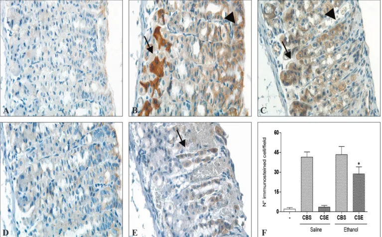
Figure 1 show that ethanol administration increased the CSE immunoexpression exclusively in the parietal cell and all gastric mucosal cells express CBS. In control group (saline alone) the immunostaining to CBS was especially seen in chief cells, although it was also observed some im-munostaining in the mucosal cells (Figure 1 – panel B and Table 1). However, the animal treatment with ethanol 50% was accompanied by a wider parietal cells staining (Figure 1 – panel C and Table 1). Unlike CBS, there was no signii-cant immunostaining of CSE in the control group (Figure 1 – panel D and Table 1). However, in animals treated with ethanol 50%, CSE was notably expressed in parietal cells (Figure 1 – panel E).
A
B
C
FIGURE 1. Immunohistochemical features of cystathionine γ-lyase (CBS) and cystathionine β–synthetase (CSE) in the gastric mucosa of normal mice and ethanol-induced gastric damage. (A) Negative control (immune-negative cells); (B-C) Immunostaining for CBS in the stomach of an animal treated only with saline (panel B) or treated with ethanol 50% (panel C) with arrows marking indicating the chief cells and arrowheads indicate labeling in mucosal cells (panel B) or parietal cells (panel C). (D-E) There was no signiicant immunostaining of CSE in the control group (panel D). However, in animals treated with ethanol 50%, was observed to mark CSE in parietal cells (panel E) and some mucosal cells. (F) Counting of CBS and CSE positive immunostained cells (ive animals from each group). The results are presented as means ± S.E.M. * < 0.05 vs saline + CSE group
Medeiros JVR, Soares PMG, Brito GAC, Souza MHLP. Immunohistochemical approach reveals localization of cystathionine-γ-lyase and cystathionine-β-synthetase in ethanol-induced gastric mucosa damage in mice
v. 50 no. 2 - abr./jun. 2013 Arq Gastroenterol 159
DISCUSSION
It was demonstrated that H2S contributes to the main-tenance of gastric mucosal integrity in damage caused by nonsteroidal anti-inlammatory drugs(10) and ethanol(3). In the present study, we evaluated the ethanol effect on the immunohistochemical expression of CBS and CSE in gastric mucosa of mice. Cystathionine-γ-lyase (CSE) and cystathi-onine-β-synthase (CBS) can use L-cysteine as an alternative substrate to form hydrogen sulide (H2S). The gastric mucosa expresses both CSE and CBS and has the ability to generate H2S(10), and CSE seems to be the main enzyme involved in the H2S generation by the gastric mucosa(9, 10).
Detailed localization of CSE and CBS in stomach has not yet been evaluated and general agressors has ability to modulate CSE expression, but they do not affect CBS expression. Our results clearly showed that administration of ethanol resulted in an increased immunostaining of CSE but not of CBS in the gastric wall. We results demonstrated that the control group (saline) showed only immunostailing of CBS, that was expression mainly in chief and mucosal cells. However, after ethanol administration, there was also expression in parietal cells. Likewise, the ethanol promotes expressions of CSE in parietal cells. Our indings show that ethanol promotes an increase in coexpression of CSE espe-cially in the parietal cells, suggesting that these cells may be mainly responsible for the synthesis of H2S involved stomach gastroprotection. In fact, we previously demonstrated that H2S donor and H2S precursor (L-cysteine) decreased etha-nol-induced gastropathy(10). Thus, this increase expression of CSE may be a mechanism of defense against the ethanol-in-duced gastric damage.
Little is known about the cellular consequences of an elevated CSE expression or about the associated increase in endogenously produced H2S. CSE-derived H2S inhibits cell proliferation(8, 14) and induces cell death predominantly by an apoptotic mechanism in polymorphonuclear cells(11, 13). Deiciency of CSE activity in humans is presumed to cause cystathioninemia (cystathioninuria), an autosomal recessive inborn error probably with no consistent clinical conse-quence(12, 14).
The CSE transcript, protein and enzymatic activity were detected in liver, kidney, and, at much lower levels, in small intestine and stomach of both rats and mice. Immunohis-tochemical analyses revealed predominant CSE expression in hepatocytes and kidney cortical tubuli(7). These results suggest important physiological roles for CSE in mice. We demonstrated that, after ethanol administration, almost all gastric mucosal cells express CBS while the expression of CSE occurs almost exclusively in the parietal cell. These results showed that the gastric epithelium therefore could be one source of H2S in the gastric wall. Furthermore, the coexpression of CSE and CBS in gastric mucosal might be an indication that they are equally involved in the production of H2S(4, 7).
Our data supports that CSE expression increased during the gastric damage induced by ethanol and it suggests the involvement of H2S during this event. Because the CSE/H2S pathway is also involved in regulating gastrophaty induced by ethanol these observations raise the possibility of H2 S-mod-ifying agents being used to improve resistance to gastric mucosa injury. Overall, our incomplete understanding of the role and mechanisms of action of H2S in many aspects of physiology and pathophysiology of the gastrintestinal tract provides a ield for extensive research in the future.
CONCLUSIONS
We conclude that the expression of CBS appears to be constitutive and diffuse across the gastric epithelium, while the expression of CSE appears to be induced in parietal cells by damage agents such as ethanol. Thus, future studies need to address the relevance of CBS and CSE produced H2S for the signaling and gastroprotect effects.
ACKNOWLEDGMENTS
We appreciate the excellent technical support of Maria Silvandira. The authors gratefully acknowledge the inancial support from Research Foundation for the State of Piauí (FAPEPI) and National Council of Technological and Sci-entiic Development (CNPq) (Brazil).
TABLE 1. Ethanol effect on the immunohistochemical expression of CBS and CSE in gastric mucosa cells of mice
Experimental group Enzyme Number of chief
cells
Number of parietal cells
Number of mucosal cells
Saline CBS 19.3 ± 2.1 8.9 ± 1.7 24.5 ± 4.3
Saline CSE 2.2 ± 1.0 3.1 ± 1.2 3.2 ± 2.3
Ethanol CBS 23.8 ± 5.2 19.2 ± 4.2a 9.6 ± 2.3a
Ethanol CSE 8.8 ± 3.3 21.9 ± 2.2b 12.5 ± 1.0b
Medeiros JVR, Soares PMG, Brito GAC, Souza MHLP.
Immunohistochemical approach reveals localization of cystathionine-γ-lyase and cystathionine-β-synthetase in ethanol-induced gastric mucosa damage in mice
160 Arq Gastroenterol v. 50 no. 2 - abr./jun. 2013
Medeiros JVR, Soares PMG, Brito GAC, Souza MHLP. Abordagem imunoistoquímica revela a localização de cistationina-γ-liase e cistationina-β -sin-tetase na lesão da mucosa gástrica induzida por etanol em camundongos. Arq Gastroenterol. 2013,50(2):157-60.
RESUMO – Contexto – O sulfeto de hidrogênio (H2S) tem sido mostrado como um neuromodulador e contribuidor para a manutenção da integridade da mucosa gástrica na lesão causada por drogas antiinlamatórias não esteroidais.Previamente, demonstramos que a síntese de H2S é essencial para a proteção da mucosa gástrica contra a administração de etanol. Objetivo – Para compreender o papel do H2S e a localização detalhada de sua produção no estômago normal e após lesão induzida pela administração de etanol, estudou-se a expressão das isoformas cistationina-γ-liase (CSE) e cistationina-β-sintetase (CBS) na mucosa gástrica de camundongos tratados com salina ou etanol 50%. Métodos – Os camundongos foram tratados por gavagem com salina ou etanol 50% (0,5 mL/25 g). Após 1 hora, os camundongos foram sacriicados e os tecidos gástricos foram avaliados por análise histológica e imunoistoquímica especíica para CBS e CSE. Resultados – Foi demonstrado expressão não especíica de CBS na mucosa gástrica normal e expressão de CSE ocorrendo principalmente nas células parietais dos animais tratados com etanol. Conclusão – Assim, demonstramos que a expressão de CBS parece ser constitutiva e difusa através do epitélio gástrico, enquanto a expressão de CSE parece ser induzida nas células parietais por agentes lesivos como o etanol.
DESCRITORES– Mucosa gástrica. Cistationina-β-Sintase. Cistationina-γ-Liase. Etanol. Imunoistoquímica. Camundongos.
REFERENCES
1. Costa NR, Silva RO, Nicolau LA, Lucetti LT, Santana AP, Aragão KS, Soares PM, Ribeiro RA, Souza MH, Barbosa AL, Medeiros JV. Role of soluble guanylate cyclase activation in the gastroprotective effect of the HO-1/CO pathway against alendronate-induced gastric damage in rats. Eur J Pharmacol. 2013;700:51-9. 2. Dal-Secco D, Cunha TM, Freitas A, Alves-Filho JC, Souto FO, Fukada SY,
Grespan R, Alencar NM, Neto AF, Rossi MA, Ferreira SH, Hothersall JS, Cunha FQ. Hydrogen sulide augments neutrophil migration through enhancement of adhesion molecule expression and prevention of CXCR2 internalization: role of ATP-sensitive potassium channels. J Immunol. 2008;181:4287-98.
3. Fiorucci S, Antonelli E, Distrutti E, Rizzo G, Mencarelli A, Orlandi S, Zanardo R, Renga B, Di Sante M, Morelli A, Cirino G, Wallace JL. Inhibition of hydro-gen sulide hydro-generation contributes to gastric injury caused by anti-inlammatory nonsteroidal drugs. Gastroenterology. 2005;129:1210-24.
4. Fiorucci S, Distrutti E, Cirino G, Wallace JL. The emerging roles of hydrogen sulide in the gastrointestinal tract and liver. Gastroenterology. 2006;131:259-71. 5. Gottfried EB, Korsten MA, Lieber CS. Alcohol-induced gastric and duodenal
lesions in man. Am J Gastroenterol. 1978;70:587-92.
6. Guslandi M. Effect of ethanol on the gastric mucosa. Dig Dis. 1987;5:21-32. 7. Ishii I, Akahoshi N, Yu XN, Kobayashi Y, Namekata K, Komaki G, Kimura H.
Murine cystathionine gamma-lyase: complete cDNA and genomic sequences, promoter activity, tissue distribution and developmental expression. Biochem J. 2004;381:113-23.
8. Mariggiò MA, Minunno V, Riccardi S, Santacroce R, De Rinaldis P, Fumarulo R. Sulide enhancement of PMN apoptosis. Immunopharmacol Immunotoxicol. 1998;20:399-408.
9. Medeiros JVR, Gadelha GG, Lima SJ, Garcia JA, Soares PMG, Santos AA, Brito GAC, Ribeiro RA, Souza MHLP. Role of the NO/cGMP/KATP pathway in the protective effects of sildenail against ethanol-induced gastric damage in rats. Br J Pharmacol. 2008;153:721-7.
10. Medeiros JV, Bezerra VH, Gomes AS, Barbosa AL, Lima Jr. RC, Soares PM, Brito GA, Ribeiro RA, Cunha FQ, Souza MH. Hydrogen sulfide prevents ethanol-induced gastric damage in mice: role of ATP-sensitive potassium chan-nels and capsaicin-sensitive primary afferent neurons. J Pharmacol Exp Ther. 2009;330:764-70.
11. Valitutti S, Castellino F, Musiani P. Effect of sulfurous (thermal) water on T-lym-phocyte proliferative response. Ann Allergy. 1990;65:463-8.
12. Vargas JE, Mudd SH, Waisbren SE, Levy HL. Maternal γ-cystathionase deiciency: absence of both teratogenic effects and pregnancy complications. Am J Obstet Gynecol. 1999;181:753-5.
13. Wang R. Two’s company, three’s a crowd: can H2S be the third endogenous gaseous transmitter? Faseb J. 2002;16:1792-8.
14. Yang G, Cao K, Wu L, Wang, R. Cystathionine gamma-lyase overexpression inhibits cell proliferation via a H2S-dependent modulation of ERK1/2 phosphor-ylation and p21Cip/WAK-1. J Biol Chem 2004;279:49199-205.