Hyperglycemia Impairs Neutrophil-Mediated
Bacterial Clearance in Mice Infected with the
Lyme Disease Pathogen
Ashkan Javid1, Nataliya Zlotnikov1, Helena Pětrošová1, Tian Tian Tang1, Yang Zhang1,
Anil K. Bansal1, Rhodaba Ebady1, Maitry Parikh1, Mijhgan Ahmed1, Chunxiang Sun1, Susan Newbigging2, Yae Ram Kim1, Marianna Santana Sosa1, Michael Glogauer1, Tara J. Moriarty1
*
1Matrix Dynamics Group, Faculty of Dentistry, University of Toronto, Fitzgerald Building, Room 241, 150
College Street, Toronto, Ontario, M5S 3E2, Canada,2Mount Sinai Hospital/Research Institute, The Toronto
Centre for Phenogenomics, 25 Orde Street, Toronto, Ontario, M5T 3H7, Canada
*tara.moriarty@utoronto.ca
Abstract
Insulin-insufficient type 1 diabetes is associated with attenuated bactericidal function of neutrophils, which are key mediators of innate immune responses to microbes as well as pathological inflammatory processes. Neutrophils are central to immune responses to the Lyme pathogenBorrelia burgdorferi. The effect of hyperglycemia on host susceptibility to
and outcomes ofB.burgdorferiinfection has not been examined. The present study
investi-gated the impact of sustained obesity-independent hyperglycemia in mice on bacterial clearance, inflammatory pathology and neutrophil responses toB.burgdorferi.
Hyperglyce-mia was associated with reduced arthritis incidence but more widespread tissue coloniza-tion and reduced clearance of bacterial DNA in multiple tissues including brain, heart, liver, lung and knee joint.B.burgdorferiuptake and killing were impaired in neutrophils isolated
from hyperglycemic mice. Thus, attenuated neutrophil function in insulin-insufficient hyper-glycemia was associated with reducedB.burgdorfericlearance in target organs. These
data suggest that investigating the effects of comorbid conditions such as diabetes on out-comes ofB.burgdorferiinfections in humans may be warranted.
Introduction
Lyme disease, also known as Lyme borreliosis, is the most common vector-borne disease in temperate climates, with an estimated annual incidence of ~300,000 cases in the United States [1]. Lyme disease is caused by members of theBorrelia burgdorferisensu lato species complex, which are transmitted to humans and other vertebrate hosts during the blood meal of infected ticks. Following tick transmission,B.burgdorferidisseminates widely through the host and col-onizes multiple organs and tissues [2–4]. Clinical manifestations of Lyme disease include ery-thema migrans, arthritis, carditis, and neuroborreliosis; the majority of these manifestations are thought to result from pathological host immune responses to infection, rather than dam-age caused byB.burgdorferi[5,6].B.burgdorferican persist for extended periods in the host, a11111
OPEN ACCESS
Citation:Javid A, Zlotnikov N, Pětrošová H, Tang TT, Zhang Y, Bansal AK, et al. (2016) Hyperglycemia Impairs Neutrophil-Mediated Bacterial Clearance in Mice Infected with the Lyme Disease Pathogen. PLoS ONE 11(6): e0158019. doi:10.1371/journal. pone.0158019
Editor:Brian Stevenson, University of Kentucky College of Medicine, UNITED STATES
Received:March 22, 2016
Accepted:June 8, 2016
Published:June 24, 2016
Copyright:© 2016 Javid et al. This is an open access article distributed under the terms of the
Creative Commons Attribution License, which permits unrestricted use, distribution, and reproduction in any medium, provided the original author and source are credited.
Data Availability Statement:All relevant data are within the paper. The same data in spreadsheet format may be obtained upon contacting the corresponding author attara.moriarty@utoronto.ca.
likely as a result of reduction in bacterial burden to levels that do not elicit host-damaging immune responses, changes in the nature of immune responses to bacteria, and/or gradual res-olution of inflammatory pathology [7]. Thus, factors that alter immune responses toB. burg-dorferiinfection and bacterial burden in tissues can affect disease progression and outcomes. Lyme disease incidence has been increasing in North American and European countries, where the prevalence of diabetes is also growing [1,8]. Both Type I (insulin-insufficient) and Type II (obesity-associated insulin-resistant) diabetes increase susceptibility to infection with many pathogens and worsens infection outcomes in animal models and humans, most notably forStaphylococcus aureus,Burkholderia pseudomalleiandMycobacterium tuberculosis infec-tions [9–13]. An important feature of aberrant immune responses in obesity-independent hyperglycemia is neutrophil dysfunction. Hyperglycemia dysregulates neutrophil activation and impairs the ability of neutrophils to phagocytose and killS.aureus,B.pseudomallei, and non-pathogenicEscherichia coli in vitro, and to controlS.aureusinfectionin vivo[13–18]. Neutrophils are important contributors to both innate host defense againstB.burgdorferiand inflammatory pathology in Lyme disease. They are recruited to the heart and joints early in infection and are a major constituent of inflammatory infiltrates in Lyme arthritis, where they contribute to immunopathology and control bacterial burden [19–24].
Collectively, these observations prompted us to examine whether obesity-independent hyperglycemia caused by insufficient insulin levels (Type 1 diabetes) alters the outcomes ofB.
burgdorferiinfection in mouse models of Lyme disease. To determine if hyperglycemia influ-encesB.burgdorferiinfection and its disease outcomes, we investigated the effects of obesity-independent hyperglycemia onB.burgdorfericlearance and dissemination, disease severity, and bacterial uptake and killing by neutrophils in mouse models of Type 1 diabetes and Lyme disease.
Material and Methods
Ethics Statements
This study was carried out in accordance with the principles outlined in the most recent poli-cies andGuide to the Care and Use of ExperimentalAnimals by The Canadian Council on Animal Care. All animal work was approved by the University of Toronto Animal Care Com-mittee in accordance with institutional guidelines (Protocol 010430). Work withB.burgdorferi
was carried out in accordance with University of Toronto, Public Health Agency of Canada, and Canadian Food Inspection Agency guidelines (University of Toronto biosafety permit 12a-M30-2). The authors declare that there are no conflicts of interest.
Animals
Male C57BL/6NCrl and C3H/HeNCrl mice purchased from (Charles River, Montréal, QC) and male heterozygote C57BL6J-Ins2Akita/Jmice (Jackson Labs, Sacramento, CA) were housed in groups of 3 or 4 per cage under pathogen-free conditions with environmental enrichment, and fedad libitumthroughout experiments with standard rodent chow (Tekland 2018 Rodent Chow, Harlan Laboratories, Mississauga, ON). Equal numbers of mice were randomly assigned to experimental and control groups upon arrival. Mice were monitored daily throughout experiments for lethargy, weight loss, failure to groom, lameness and dehydration.
Breeding, genotyping and phenotyping of Akita mice
C57BL6J-Ins2Akita/Jmale mice were crossed with C57BL/6J female mice (Jackson Labs) bred in-house to generate heterozygous Ins2Akitamice. DNA for genotyping was extracted from tail
(TJM); University of Toronto Faculty of Dentistry Bertha Rosenstadt Endowment Fund (TJM) and Enrichment Endowment Fund (TJM); and Canada Foundation for Innovation/Ontario Research Fund: 27881 (TJM). Salaries: Postdoctoral fellowships: Heart & Stroke/Richard Lewar Centre of Excellence (HP); graduate scholarships: University of Toronto (UofT) (AJ, NZ, TTT, RE), UofT Faculty of Dentistry Harron (RE), Queen Elizabeth II (TTT);
undergraduate scholarships: CIHR Institute of Musculoskeletal Health and Arthritis (MA, MP, YZ), NSERC (TTT), UofT Faculty of Dentistry (MA), UofT Work Study (YRK, TTT, MA), and Ontario Summer Experience Program (YRK).
Competing Interests:The authors have declared
snips using the Qiagen DNeasy tissue extraction kit, following manufacturer’s instructions (Qiagen, Toronto, ON). PCR genotyping was performed by amplification using primers oIMR1093 (5’-TGCTGATGCCCTGGCCTGCT-3’) and oIMR1094 (5’-TGGTCCCACATAT GCACATG-3’), followed by overnight digestion of PCR products with Fnu4HI. Digestion products were resolved on 3% agarose gels to visualize DNA bands corresponding to wild-type (140 bp) and mutant (280 bp) gene sequences. Hyperglycemia phenotyping was performed by measuring non-fasting blood glucose in blood obtained from the saphenous vein using a cali-brated Aviva Nano glucometer and glucose strips (Accu-Check/Roche, Laval, QC). Due to vari-able time to onset of the hyperglycemia phenotype in Akita mice, infections were performed in age-matched animals ranging from 4 to 8 weeks of age. Control mice for Akita experiments were normoglycemic homozygous wild-type littermates. Characteristics of all mouse strains used in this study are summarized inTable 1.
STZ induction of hyperglycemia
Five-week-old male mice (all experiments except for arthritis studies) or 3-week-old male C3H/HeN mice (arthritis studies) were rendered diabetic by a multiple low-dose streptozotocin (STZ) treatment as described previously [26]. Briefly, mice fedad libitumwere injected intra-peritoneally with either 40μg STZ (Cedarlane Laboratories, Burlington, ON) per gram body
weight in 0.1 M sodium citrate or equal volume of buffer without STZ (vehicle) once daily for 5 (C57BL/6) or 7 (C3H/HeN) consecutive days. Mice were considered hyperglycemic when non-fasting blood glucose levels reached 15 mmol/L. Non-non-fasting blood glucose was>25 mmol/L in all STZ-treated mice at time of infection and sacrifice. Non-fasting blood glucose in blood obtained from the saphenous vein was measured using a calibrated Aviva Nano glucometer and glucose strips (Accu-Check/Roche). STZ-treated animals were fed semi-liquefied mash of standard diet to prevent dehydration as previously described [27]. Blood glucose and body weight were measured in all animals before experimental treatment, every 2–3 days during STZ treatment, on the day of infection withB.burgdorferiand at the time of sacrifice.
Borrelia burgdorferi strains
, cultivation and mouse infections
Infections were performed with freshly inoculated cultures of log phase GCB726, a B31 5A4 NP1-derived infectious strain ofB.burgdorferitransformed with GFP-expressing plasmid pTM61 [28]. Cultures were grown in Barbour-Stoenner-Kelly-II (BSK-II) medium prepared as previously described [29] supplemented with 6% heat-inactivated rabbit serum (Cedarlane Laboratories) and 100μg/mL gentamycin (Bioshop Canada Inc, Burlington, ON) at 36°C and 1.5% CO2.
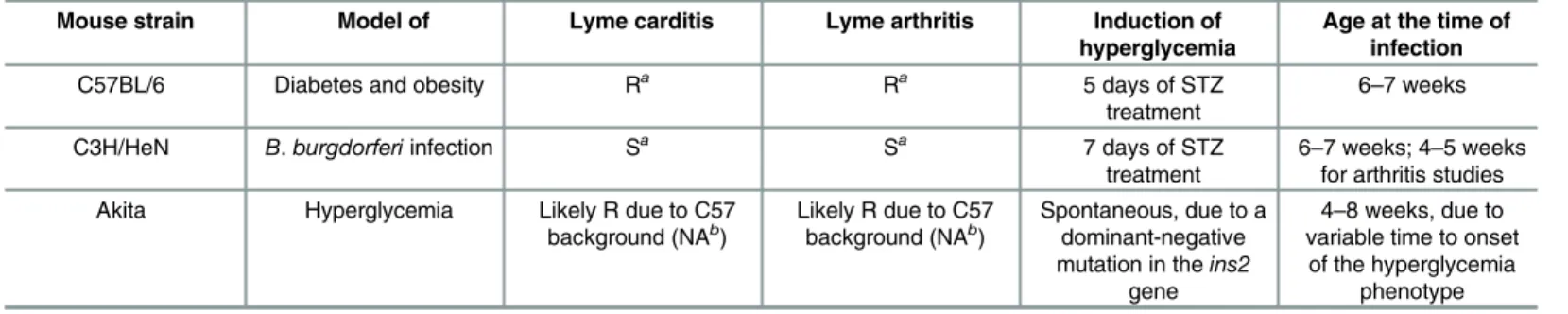
Table 1. Mouse strains used in this study.
Mouse strain Model of Lyme carditis Lyme arthritis Induction of
hyperglycemia
Age at the time of infection
C57BL/6 Diabetes and obesity Ra Ra 5 days of STZ
treatment 6–7 weeks
C3H/HeN B.burgdorferiinfection Sa Sa 7 days of STZ
treatment 6–7 weeks; 4–5 weeksfor arthritis studies Akita Hyperglycemia Likely R due to C57
background (NAb) Likely R due to C57background (NAb) Spontaneous, due to adominant-negative
mutation in theins2
gene
4–8 weeks, due to variable time to onset of the hyperglycemia
phenotype
aR and S stand for resistant and sensitive, respectively [25]. bNA–information not available.
Five days after the last STZ treatment (“washout period”), mice from vehicle- and STZ-treated groups received a subcutaneous injection at the dorsal lumbar midline with either 1x10⁴B.burgdorferisuspended in BSK-II medium or with BSK-II medium alone (mock-infected control). For all experiments except for arthritis studies, C57BL/6 and C3H/HeN mice were 6–7 weeks of age at time of infection. We used 6–7 week-old animals in most experiments because STZ treatment disrupts weight gain during maturation to adulthood and can be lethal in young mice [30] at the age typically used forB.burgdorferiinfectivity studies (3–4 weeks). For arthritis studies, mice were less than 5 weeks of age at time of infection. Due to variable time to onset of the hyperglycemia phenotype in Akita mice [31], infections in heterozygotes and homozygous wild-type normoglycemic controls were performed in age-matched animals ranging from 4–8 weeks of age.
At 4 weeks post-infection, non-fasting peripheral blood glucose and body weight were mea-sured, animals were then anesthetized with 2% isoflurane, and blood was drawn by cardiac puncture for complete blood count (CBC) analysis. Animals were then euthanized by cervical dislocation and tissues and neutrophils were harvested for histology, quantitative real-time PCR (qPCR), andin vitroneutrophil function assays.
DNA extraction and qPCR for measurement of bacterial DNA copy
number
Total DNA concentration and qPCR determination of bacterial burden were measured in blood, brain, bladder, ear, heart, liver, lung, knee joint (patella), and ventral thoracic skin harvested from animals. Total DNA was extracted using the Qiagen DNeasy tissue extraction kit, following man-ufacturer’s instructions. Concentration and purity of extracted DNA were measured using a NanoDrop spectrophotometer (Thermo Fisher Scientific, Toronto, ON). qPCR measurement of
flaBDNA copy number was performed as described previously [32,33] using a CFX96 real-time PCR machine (Bio-Rad Laboratories, Mississauga, ON). Briefly, each qPCR assay was performed with duplicate standards containing 101–106copies of plasmid pTM222 encoding theflaB seg-ment for qPCR amplification on the same plate as sextuplicate reactions containing DNA extracted from each tissue sample. Reactions were performed in 1X iQ SsoFast EvaGreen Super-mix (Bio-Rad) prepared according to manufacturer’s instructions and contained 400 nM of each offlaBprimers T1 (5'-GCAGCTAATGTTGCAAATCTTTTC-3') and T2 (5'- GCAGGTGCTG GCTGTTGA-3'). qPCR was performed using 2μl of extracted DNA, in a total reaction volume
of 20μl. PCR conditions: Step 1: 98°C 2 min; Step 2: 40 cycles of 98°C 5 s, 59.2°C 5 s; Step 3: 65°C
5 s; Step 4: melting curve analysis over melting range 65°C to 95°C. Standards were used to calcu-late the exact number of copies offlaBsequence in samples. Each plate included negative control wells (DNA extraction elution buffer). The R2value of the standard curve obtained on every run was examined to ensure run quality and pipetting accuracy; runs with R2values below 0.85 were repeated. The average copy number obtained from all qPCR repeats for each sample was used in subsequent graphing and statistical analysis. Copy numbers from samples with aberrant melting or amplification curves compared to replicate measurements for the same sample were not included in calculations of average copy number.flaBDNA copy numbers for samples were nor-malized to total DNA concentration (copy number perμg of DNA) to control for differences in
DNA extraction efficiency among samples. Tissues were considered qPCR-positive if at least one
flaBDNA copy was detected perμg of total DNA isolated from tissues.
Histology, arthritis and carditis scoring
both above and below the joint) and placed immediately in 1.5 ml 10% neutral-buffered forma-lin (Sigma Chemicals, St Louis, MO), which was changed after 24 hours of fixation [34]. Sam-ples were embedded in paraffin, sectioned, and stained with hematoxylin and eosin (H&E) by the histology services of the University of Toronto Faculty of Dentistry, Hospital for Sick Chil-dren and Toronto Centre for Phenogenomics. Tibiotarsal joints were decalcified for 96 hours in TBD-2 Decalcifier solution (Thermo-Fisher Scientific), prior to embedding, sectioning and H&E staining. The stained tissue sections included the tibiotarsal joint and were sectioned lon-gitudinally with view of all long bones and joints. A veterinary pathologist analyzed tissue sec-tions in a blinded fashion under light microscopy for any abnormal lesions. A score of 0, 1 or 2 was assigned to each section based on severity of inflammation in the joints, and corresponded to: no significant pathological findings (0), mild pathology (a few neutrophils within the joint space and large basophilic‘smudge’-like particles) (1), and severe pathology (large numbers of neutrophils within the joint space, large basophilic‘smudge’-like particles and extension of inflammation into the subcutaneous tissue around the joint) (2).
Scoring of inflammation in hearts was performed by modification of a previously described protocol for quantifying multifocal cardiac inflammation [35]. Briefly, the number of nuclei in five 100 mm2regions of interest in 2–3 matched H&E-stained sagittal sections per heart were enumerated using a counting grid and the average number of nuclei per region of interest in each section was calculated. Nuclei were counted in each atrium and ventricle and the heart apex. The majority of tissue included in each region of interest was derived from the myocardium.
Complete Blood Count (CBC)
Twentyμl of uncoagulated whole blood were drawn by cardiac puncture in anesthetized mice
using needles and syringes coated with 4% sodium citrate (Sigma). CBC analysis was per-formed by a Hemavet 950 (Drew Scientific, Dallas, TX) or by an IDEXX veterinary reference lab (Markham, ON), using a Sysmex Hematology Analyzer Model XT2000V. We indepen-dently verified both methods and similar results were obtained. MULTI-TROL calibration con-trols (Drew Scientific) were run before each series of Hemavet measurements.
Bone marrow neutrophil isolation
Bone marrow neutrophils from femurs and tibias were harvested and cleaned as described pre-viously [36]. Briefly, bone marrow was flushed with 10 ml ice-cold minimum essential medium eagle alpha modification (α-MEM) and samples were homogenized gently using a 20-gauge needle and spun down for 10 minutes at 700 xg at 4°C. The pellets were resuspended in 1 ml 1X Dulbecco’s phosphate buffered saline without calcium chloride and magnesium chloride (dPBS-/-) (Sigma), neutrophils were isolated on a discontinuous 82%/65%/55% Percoll gradi-ent (Sigma) prepared in dPBS-/- and cgradi-entrifuged for 30 min at 1,015xg at 4°C. The neutrophil layer (between 65% and 82%) was washed and counted using a Z1 Coulter Particle Counter (Beckman Coulter, Fullerton, CA). As described previously [37], purity was determined by Romanowsky Diff-Quik staining of methanol-treated air-dried smears according to manufac-turer’s instructions (Sigma), and was>90%.
Isolation of peritoneally-recruited neutrophils
into the abdominal cavity without perforating organs, massaged for 5–10 minutes, and fluid was withdrawn. Samples were centrifuged at 700 xg 4°C for 10 min in a fixed angle rotor, washed twice with 10 ml ice-cold 1X dPBS-/-, resuspended in 1 ml dPBS-/- and counted using a Z1 Coulter Parti-cle Counter, as described above. Sample purity measured by Diff-Quik staining was>90%. Mice which were used for isolation of peritoneally-recruited neutrophils were not used for any other experiment (e.g. measurement of bacterial burden, blood glucose, histology or isolation of bone marrow neutrophils) to prevent artefacts associated with acute inflammation.
Ex vivo
neutrophil bacterial uptake and killing assays
At the end of the experimental or control treatments, animals were sacrificed and neutrophils were harvested from bone marrow and peritoneum and co-incubatedin vitrowith opsonized
E.coliorB.burgdorferi. Opsonization was performed with pre-immune serum from congenic normoglycemic mice. After co-incubation, numbers of intact extracellular bacteria (B. burgdor-feri) or colony-forming units (CFUs:E.coli) relative to input bacterial numbers were measured to assess neutrophil uptake and killing of bacteria. Opsonized bacteria incubated under the same conditions in the absence of neutrophils were used as a control.
E.coliDH5αwas cultured in LB Lennox-broth overnight at 37°C, OD600was measured using
an Ultraspec 3000 (Biochrom Ltd., Cambridge, UK), and 3x10⁶bacteria were opsonized for 30 minutes at 37°C with 5μl serum from normoglycemic uninfected mice. Opsonized bacteria were
incubated with 1x10⁶neutrophils at a multiplicity of infection (MOI) of 3:1 for 1 hour at 37°C.
After incubation, bacteria-neutrophil mixtures were diluted 1:104and 1:105in dPBS-/- and 100μl of dilutions were plated in triplicate on LB Lennox-agar plates (Bioshop) and incubated
overnight at 37°C. CFUs were enumerated for all replicates and dilutions, and normalized to CFUs for similar dilutions of complement-opsonized input bacteria stored on ice until plating.
B.burgdorferi(1x10⁷) cultivated to log phase (<7x10⁷/ml) were opsonized at 36°C and 1.5% CO2for 30 minutes in 5μl blood serum from normoglycemic uninfected C3H/HeN mice.
Neu-trophils (1x10⁶) were incubated with opsonized bacteria at a MOI of 10:1 and incubated 16 h in Roswell Park Memorial Institute (RPMI) media (Sigma) containing 5% heat-inactivated fetal bovine serum at 36°C and 1.5% CO2. Numbers of intact bacteria remaining after overnight
incubation were counted with a Petroff-Hausser counting chamber (Hausser Scientific, Hor-sham, PA) and normalized to counts for complement-opsonized input bacteria stored at 4°C until uptake counting was performed.B.burgdorferiviability was determined by LIVE-DEAD staining (ThermoFisher Scientific), performed according to manufacturers’instructions.
Statistical analyses
Statistical analyses of all measured parameters in experimental groups were performed using GraphPad Prism v6.0 graphing and statistical analysis software (GraphPad Software, La Jolla, CA). Normally distributed data were analyzed using two-way ANOVA with Holm-Sidak post-tests or paired t-post-tests. Non-normally distributed data were log-transformed and analyzed by one-way Kruskal-Wallis ANOVA with Dunn’s postests, or by two-tailed Mann-Whitney t-tests. Normality testing was performed using the D'Agostino & Pearson omnibus normality test.Pvalues of<0.05 were considered significant.
Results
Hyperglycemic mouse models of
B
.
burgdorferi
infection
low dose streptozotocin (STZ)-treated C57BL/6 mice [39]. STZ is an antibiotic which induces irreversible insulin insufficiency by killing pancreaticβ-cells [26]. C57BL/6 mice are readily infected byB.burgdorferibut are resistant to infection-induced inflammatory pathologies in joints (arthritis) and heart (carditis) [2]. In subsequent experiments, hyperglycemia was induced by STZ treatment in the arthritis- and carditis-susceptible C3H/HeN mouse model of Lyme disease, to measure effects of hyperglycemia on Lyme disease pathology. Induction of hyperglycemia in C3H/HeN mice requires more extended treatment with STZ [26], and the effects of hyperglycemia on immune responses to bacterial infection in this background have not been characterized. To exclude potential hyperglycemia-independent effects of STZ treat-ment, infections were also conducted in C57BL/6-derived insulin-insufficient heterozygote Akita mice, which carry a dominant-negative mutation in the insulin 2 gene resulting in spon-taneous emergence of type 1 diabetes in young adult animals [31,40]. Infections were also con-ducted in age-matched littermates which did not carry the Akita mutation. Due to variable time to onset of hyperglycemia in Akita animals, mice used for Akita experiments were from a broader age range than those used in STZ experiments. Mouse strains used in this study and their characteristics are summarized inTable 1.
To induce hyperglycemia, we treated 5-week old mice with 40μg of STZ for 5 (C57BL/6) or
7 (C3H/HeN) consecutive days, followed by a 5 day“washout”period to ensure that STZ was no longer present in tissues at the time of infection [26]. Age- and strain-matched control mice were treated with vehicle only (buffer without STZ). Mice were infected with a B31
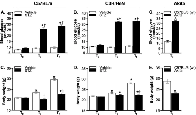
5A4-derivedB.burgdorferistrain [33] or mock-infected with cultivation medium alone, and sacrificed four weeks after infection. STZ treatment increased non-fasting blood glucose levels to>25 mmol/L in both C57BL/6 and C3H/HeN mice, similarly to previously reported values [41]. Thus, STZ-treated mice were hyperglycemic. Blood glucose levels were comparable at time of infection (Ti) and sacrifice (Tf) (Fig 1A and 1B), indicating that a stable state of
hyper-glycemia was maintained over the course of infection and that regeneration ofβ-cells likely did not occur [42]. Blood glucose levels in all normoglycemic infected mice also remained stable throughout the experiment, indicating thatB.burgdorferiinfection did not affect blood glucose (Fig 1A and 1B). Heterozygous Akita mice were significantly more hyperglycemic than STZ-treated C57BL/6 counterparts, but blood glucose levels in normoglycemic control animals in STZ and Akita experiments also differed significantly (p<0.05, compare Ti values inFig 1A
and 1C). This was possibly because the age ranges of mice used in these experiments differed, or because the C57BL/6 strains used in STZ and Akita experiments originated with different suppliers (Table 1: Charles River BL/6 NCrl for STZ experiments, BL/6J Jackson Labs for Akita experiments). Body weight of STZ-treated and Akita heterozygotes, but not vehicle-treated mice or age-matched wild-type Akita littermates, was significantly reduced at the time of sacri-fice compared to baseline (Fig 1D–1F), consistent with previous reports of reduced weight gain during maturation to adulthood in hyperglycemic mice [30]. As for blood glucose, body weights differed significantly in hyperglycemic Akita and STZ-treated C57 mice and in normo-glycemic controls (p<0.05, compare Ti values inFig 1D and 1F). Together, these data indicated that STZ treatment and Akita heterozygosity induced hyperglycemia.
More widespread
B
.
burgdorferi
colonization and reduced bacterial
clearance in tissues of hyperglycemic mice
reported thatB.burgdorferican be cultivated and detected by quantitative PCR (qPCR) in all of these tissues [2–4,43–46]. Heart, skin, joint, bladder and ear are among the most common targets examined in murine pathogenesis studies, because these tissues often display higherB.
burgdorferiburden and are the sites of disease pathology in mice (heart, joint). Tissues such as brain and liver are much less commonly positive for these bacteria, but can still be infrequently colonized. We collected samples from tissues typically targeted byB.burgdorferi(bladder, blood, ear, heart, knee joint and ventral thoracic skin), tissues that exhibit physiological dys-function in hyperglycemia (liver, brain) [47,48], and tissues where hyperglycemia is associated with impaired immune responses to bacterial infection (lung) [49].B.burgdorferiDNA copy number was quantified by measuring absolute copy number of bacterialflaBDNA sequence per microgram of extracted DNA by qPCR, as previously described [32,33] (Fig 2). We did not determine viability ofB.burgdorferiisolated from individual organs.
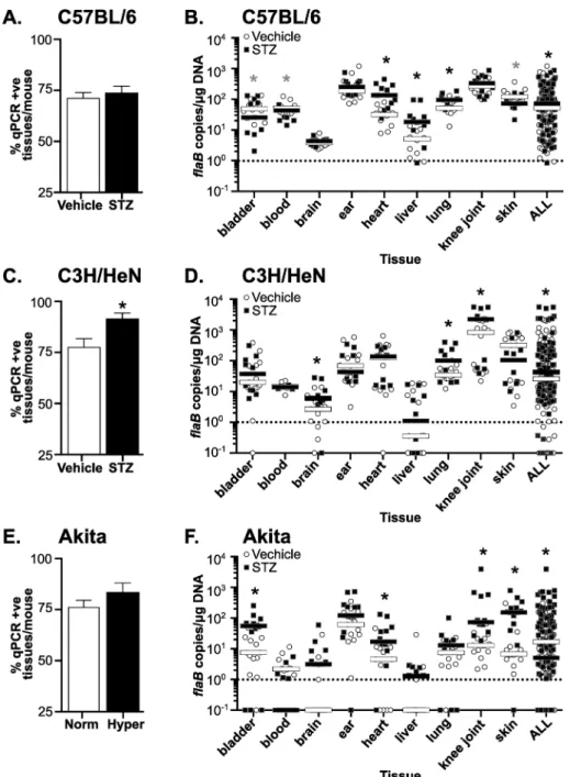
We first assessed whether hyperglycemia affected the extent ofB.burgdorferidissemination to and colonization of tissues by calculating the average percentage of tissues per mouse in which at least oneflaBcopy/μg total extracted DNA was detected (Fig 2A, 2C and 2E). The
per-centage of tissues/mouse which were positive forflaBDNA was calculated by assigning a score of 1 to each tissue with at least 1flaBcopy/μg DNA and a score of 0 to tissues where<1flaB copy was measured. The sum of these scores for all tissues in each mouse (bladder, blood, brain, ear, heart, liver, lung, knee joint and skin) was divided by the total number of tissues tested in each mouse to determine the percentage of tissues/mouse which wereflaB-positive. The mean percentage offlaB-positive tissues per mouse was somewhat elevated in STZ-treated C57BL/6 mice (Fig 2A) and hyperglycemic Akita heterozygotes (Fig 2C), but not significantly. Fig 1. Hyperglycemic mouse models ofB.burgdorferiinfection.(A-C) Non-fasting blood glucose levels in STZ- and
vehicle-treated C57BL/6 (A) and C3H/HeN (B) mice, and in Akita mice and age-matched wild type C57BL/6 mice (C). (D-F) Body weight of STZ- and vehicle-treated C57BL/6 (D) and C3H/HeN (E) mice, and of Akita mice and age-matched wild type C57BL/6 mice (F). T0stands for baseline values (before STZ treatment), Ticorresponds to time of infection, and Tfstands for
time of sacrifice. N = 17–21 mice per group. Statistical analysis: (A-B, D-E) Two-way ANOVA with Holm-Sidak post-tests. (C, F) Two-tailed t-test.*indicates p<0.05 vs. T0within group;†indicates p<0.05 vs. Vehicle within time point. ^ indicates
p<0.05 vs wild-type.
However, significantly more tissues per mouse wereflaB-positive in STZ-treated C3H/HeN mice (Fig 2B), suggesting a more widespread colonization in this mouse strain in the context of Fig 2.B.burgdorferitissue colonization and bacterial DNA copy number in hyperglycemic and normoglycemic mice.(A, C and E) Percentage of tissues/mouse positive forB.burgdorferi flaBDNA in infected normoglycemic and hyperglycemic mice at 4 weeks post-infection. Percentage of qPCR-positive tissues/mouse in C57BL/6 (A), C3H/HeN (C), and Akita (E) mice are shown. (B, D and F) MedianB. burgdorferi flaBcopy number/μg total DNA in indicated tissues and in all tissues combined (ALL) in infected
normoglycemic and hyperglycemic mice at 4 weeks post-infection. Shown are individual values and medians (bars) in tissues of C57BL/6 (B), C3H/HeN (D) and Akita (F) mice. Values are plotted on a log scale to facilitate visualization of a large range of values. Dotted lines in each graph indicate the cutoff point (1flaB
copy/μg DNA) below which tissues were considered negative. Statistical analysis: Kruskal-Wallis ANOVA with Dunn’s post-test. For all panels, N = 10–13 mice per experimental group and strain. Fold differences in medians are summarized inTable 2.*indicates p<0.05 vs. normoglycemic controls.
hyperglycemia. Overall, hyperglycemia was associated with a significant 11% average increase in the number of qPCR-positive tissues per mouse across all mouse strains (p<0.05).
We next compared medianB.burgdorferi flaBDNA copy numbers in tissues of hyperglyce-mic and normoglycehyperglyce-mic hyperglyce-mice (Fig 2B, 2D and 2F). Median values were compared because copy number values in most tissues and experimental groups were not normally distributed. Values shown inFig 2B, 2D and 2Fwere plotted on log scales to facilitate graphing of a wide range of copy number values on the same plots. As expected,flaBcopy number in liver and brain were lower than in other tissues for normoglycemic mice (Fig 2B, 2D and 2F). Unexpect-edly,flaBcopy numbers in skin and knee joints, and in all tissues combined (ALL) differed sig-nificantly in vehicle-treated C57BL/6 mice compared to wild-type Akita C57BL/6 littermates (p<0.05, compareFig 2B and 2F). This indicated that in addition to strain/supplier-origin-spe-cific differences in blood glucose levels and body weight in these animals, bacterial burden was also affected.
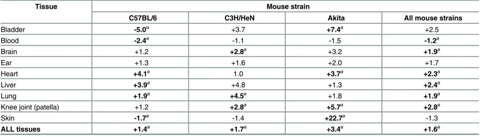
In hyperglycemic STZ-treated C57BL/6 mice, median copy number for all tissues combined was significantly increased by 1.4-fold compared to normoglycemic controls (Fig 2B,Table 2: ALL). As observed in hyperglycemic C57BL/6 mice, overall tissue copy number was signifi-cantly increased by 1.7-fold in hyperglycemic STZ-treated C3H/HeN mice compared to nor-moglycemic controls (Fig 2D,Table 2: ALL). Copy number across all tissues was also
significantly elevated by 3.4-fold in hyperglycemic Akita mice (Fig 2F,Table 2: ALL). Across all mouse strains combined, copy number for all tissues was 1.6-fold greater in hyperglycemic than normoglycemic mice. Tissues where copy number was consistently and significantly ele-vated in hyperglycemia were brain, heart, liver, lung and knee joint, where median copy num-bers were 1.9 to 2.8-fold greater across all mouse strains (Table 2). Together, these results indicated that hyperglycemia was associated with more widespreadB.burgdorfericolonization and reduced clearance of bacterial DNA debris.
Reduced incidence of arthritis but not carditis in
B
.
burgdorferi
-infected
hyperglycemic mice
Lyme carditis is an inflammatory condition, which is characterized in susceptible mouse strains (C3H, but not C57BL/6 mice) by increased leukocyte infiltration and fibroblast proliferation [25,50]. Prolonged hyperglycemia can cause reduced cellular density in the heart resulting from accumulation of excess extracellular matrix [51]. To distinguish between possible effects
Table 2. Median fold-differences inB.burgdorferiDNA copy number in hyperglycemic vs normoglycemic mice.
Tissue Mouse strain
C57BL/6 C3H/HeN Akita All mouse strains
Bladder -5.0a +3.7 +7.4a +2.5
Blood -2.4a -1.1 -1.5 -1.2a
Brain +1.2 +2.8a +3.2 +1.9a
Ear +1.3 +1.6 +2.0 +1.7
Heart +4.1a 1.0 +3.7a +2.3a
Liver +3.9a +4.8 +1.3 +2.4a
Lung +1.9a +4.5a +1.8 +1.9a
Knee joint (patella) +1.2 +2.8a +5.7a +2.8a
Skin -1.7a -1.4 +22.7a -1.3
ALL tissues +1.4a +1.7a +3.4a +1.6a
aindicates p<0.05 hyperglycemic vs normoglycemic, Kruskal-Wallis ANOVA with Dunn’s post-test
of hyperglycemia (hypocellularity) andB.burgdorferi-induced carditis (hypercellularity), we adapted a nuclei-counting method for measuring infiltration of inflammatory cells in multifo-cal cardiac inflammation that enabled us to quantitatively distinguish these conditions [35]. Numbers of nuclei in five 100 mm2regions of interest in two-three matched hematoxylin- and eosin-stained sagittal sections per heart were enumerated using a counting grid. Nuclei were counted in each atrium and ventricle and the heart apex, and the majority of tissue included in each region of interest was derived from the myocardium.
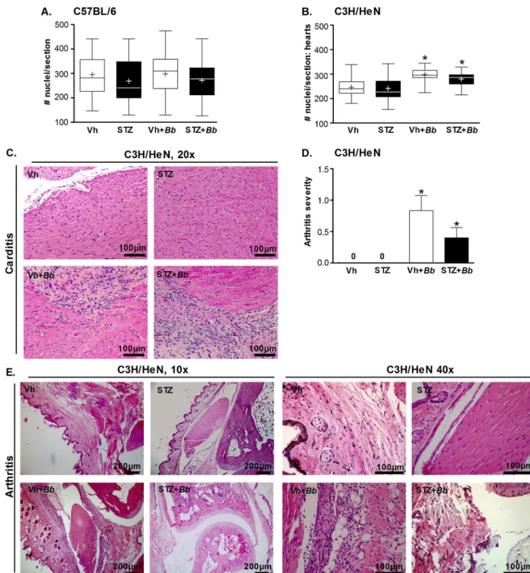
No significant changes in cellularity measured by this method were detected in hyperglyce-mic and normoglycehyperglyce-mic carditis-resistant C57BL/6 (Fig 3A). In carditis-sensitive C3H/HeN mice, hypercellularity indicative of carditis was significantly elevated in both hyperglycemic and normoglycemic infected animals. However, STZ treatment, which did not alter bacterial DNA copy number in the hearts of C3H/HeN mice, had no additional effect on carditis sever-ity (Fig 3B). Representative histology sections of C3H/HeN mice are shown inFig 3C. Since Akita mice are C57BL/6-derived, and therefore likely resistant to both carditis and arthritis, we did not perform histological analysis on samples from these mice. Collectively, these results indicated thatB.burgdorferiinfection resulted in inflammation in the heart of C3H/HeN, but not C57BL/6 mice, and that hyperglycemia did not alter the levels of cardiac inflammation in either mouse strain. Since bacterial DNA burden was not elevated in hearts of hyperglycemic C3H/HeN mice, we concluded that hyperglycemia did not affect carditis severity in a fashion that was independent of bacterial clearance.
In contrast to heart, bacterial copy number was significantly greater in knee joints of hyper-glycemic C3H/HeN animals (Fig 2D). Arthritis is a prominent pathology observed followingB.
burgdorferiinfection in juvenile (3 week-old) C3H mice, and is secondary to infiltration of neu-trophils and other leukocytes into joints [2]. To investigate the effect of hyperglycemia on arthritis, we induced hyperglycemia by STZ treatment in 3-week old C3H/HeN mice, followed byB.burgdorferiinfection at just under 5 weeks of age. As for the older mice used in all other experiments in our study, STZ treatment induced significant hyperglycemia in 3-week old mice (>30 mmol/L; p<0.05 compared to vehicle-treated controls; data not shown). Four weeks after infection (5–6 weeks of sustained hyperglycemia), arthritis in tibiotarsal joints was scored by a murine veterinary pathologist blinded to the identity of experimental samples (Fig 3D).B.burgdorferiinfection caused arthritis in both normoglycemic and hyperglycemic mice, and arthritis was absent in mock-infected controls (Fig 3D). Arthritis incidence was signifi-cantly decreased in hyperglycemic infected animals compared to normoglycemic counterparts (40.0 vs 58.3% incidence; p<0.05), but reduction in arthritis severity in hyperglycemic mice was not significant (Fig 3D). Representative joint histology images are displayed inFig 3E. Thus, although bacterial burden was significantly elevated in the joints of hyperglycemic C3H/ HeN mice, inflammatory pathology was less frequently observed.
Impaired
B
.
burgdorferi
killing by activated neutrophils isolated from
hyperglycemic mice
Neutrophils contribute to control ofB.burgdorferiburden and inflammatory pathology in joints [23], and their function is often impaired in the context of hyperglycemia [13–18]. This prompted us to determine if the ability of neutrophils from hyperglycemic mice to control bac-terial survivalex vivowas altered. To determine whether the bactericidal function of neutro-phils towardB.burgdorferiwas disrupted in hyperglycemia, we measured uptake and killing of
Fig 3.B.burgdorferi-induced carditis and arthritis in hyperglycemic and normoglycemic mice.(A-B) Cardiac cellularity in C57BL/6 (A) and
C3H/HeN (B) mice. Experimental groups: normoglycemic mock-infected (Vh), hyperglycemic mock-infected (STZ), normoglycemic infected (Vh +Bb), hyperglycemic infected (STZ+Bb). The numbers of nuclei in five 100 mm2regions of interest in 2–3 matched H&E-stained sagittal sections
per heart were enumerated using a counting grid. Nuclei were counted in each atrium and ventricle and the heart apex, and the majority of tissue included in each region of interest was derived from the myocardium. Summary values are shown for the average numbers of nuclei/section/ mouse. Tukey box plots represent the 25–75% range, line and plus symbols (+) correspond to medians and means, respectively, and error bars span minimum to maximum values. N = 11–15 mice per group. Statistics: Two-way ANOVA with Holm-Sidak post-test.*indicates p<0.05 vs
C57BL/6-derived Akita heterozygotes made age-matching challenging, we performed these experiments only in STZ-treated C57BL/6 and C3H/HeN mice, where age of animals was iden-tical and inter-subject variation was minimized.
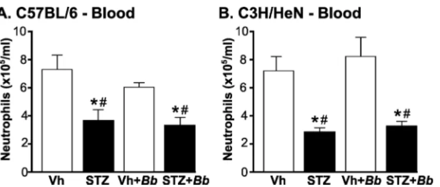
We first determined numbers of peripheral neutrophils in blood of C3H/HeN and C57BL/6 mice at 4 weeks post-infection after 5–6 weeks of sustained hyperglycemia (Fig 4). Neutrophil counts in mice were significantly reduced in both hyperglycemic C57BL/6 and C3H/HeN mice independent of infection status (Fig 4A and 4B). Importantly, significant neutropenia was pres-ent in hyperglycemic mice infected withB.burgdorferi(Fig 4A and 4B), indicating thatB. burg-dorferiinfection was not sufficient to overcome deficits in neutrophil production and/or mobilization in bone marrow of diabetic animals.
We next examined the ability of neutrophils harvested from C57BL/6 (Fig 5A, 5C, 5E and 5G) and C3H/HeN (Fig 5B, 5D, 5F and 5H–5J) mice after 4 weeks ofB.burgdorferiinfection (5–6 weeks of sustained hyperglycemia) to control uptake and survival ofB.burgdorferi opso-nized with pre-immune serum (Fig 5E–5J). Pre-immune serum was used to investigate the effects of hyperglycemia on neutrophil function independent of its effects on adaptive immune responses, and to maintain consistency with the methods of a previous study measuring effects of hyperglycemia on neutrophil killing ofE.coli[18]. Opsonization in this context therefore refers to exposure of bacteria to serum complement proteins [52].
Uptake was measured by determining the percentage ofB.burgdorferiwhich remained fol-lowing co-incubation with neutrophils, compared to mock-treated bacteria incubated without neutrophils.B.burgdorferisurvival was determined by LIVE-DEAD staining, which identifies cells with permeable membranes. Killing of pre-immune serum-opsonizedE.coli, which is impaired in neutrophils from hyperglycemic mice [18] was also measured (Fig 5A–5D), both as a control, and to determine if ongoingB.burgdorferiinfection altered the ability of neutro-phils to kill other bacteriaex vivo. Hyperglycemia primarily affects the bactericidal function of activated neutrophils isolated from the circulation and tissues, possibly because hyperglycemia dysregulates neutrophil transition to the fully activated state required for bactericidal activities
scoring system: 0: no pathology, 1: mild pathology, 2: severe pathology. N = 10–11 mice/group. Shown are mean±SEM severity scores.
Statistics: Kruskal-Wallis ANOVA with Dunn’s post-test.*indicates p<0.05 vs. mock-infected within vehicle- or STZ-treated groups. (E)
Representative H&E-stained tibiotarsal joint sections of C3H/HeN mice. Scale bars: 100–200μm.
doi:10.1371/journal.pone.0158019.g003
Fig 4. Neutropenia inB.burgdorferi-infected and mock-infected hyperglycemic mice.(A, B) Mean
±SEM numbers of circulating neutrophils in blood of C57BL/6 (A) and C3H/HeN mice (B). Neutrophils were
enumerated by particle counting with a Coulter counter in blood drawn by cardiac puncture fromB.
burgdorferi-infected and mock-infected mice at 4 weeks post-infection (5–6 weeks of sustained hyperglycemia). N = 6–9 mice/group. Experimental groups: Normoglycemic mock-infected (Vh), hyperglycemic mock-infected (STZ), normoglycemicB.burgdorferi-infected (Vh+Bb), hyperglycemicB. burgdorferi-infected (STZ+Bb). Statistical analysis: Two-way ANOVA with Holm-Sidak post-tests.*indicates p<0.05 vs. Vehicle; # indicates p<0.05 vs. normoglycemicB.burgdorferi-infected (Vh+Bb) mice.
[7–10]. To determine whether control of bacterial viabilityex vivodepended on tissue-specific activation factors encountered during recruitment (peritoneal exudate) or systemic factors pro-moting neutrophil activation in bone marrow, we performed assays with neutrophils harvested from bone marrow (Fig 5A, 5B, 5E and 5F) as well as neutrophils obtained from peritoneal exudates following acute sodium periodate treatment (Fig 5C and 5D and 5G–5J).
Neither hyperglycemia norB.burgdorferiinfection affectedB.burgdorferiuptake (Fig 5E and 5F) orE.colisurvival (Fig 5A and 5B) following co-incubation with bone marrow neutro-phils. By contrast, significantly moreE.colisurvived following incubation with peritoneal neutro-phils from hyperglycemic C57BL/6 and C3H/HeN mice than normoglycemic controls (Fig 5C and 5D). Interestingly,E.colisurvival following incubation with peritoneal neutrophils isolated from both normoglycemic and hyperglycemic infected C57BL/6 mice was similar to mock-infected normoglycemic animals, but was significantly lower than in mock-mock-infected STZ-treated animals (Fig 5C).B.burgdorferiuptake was also impaired in hyperglycemic animals (Fig 5G and 5H). Approximately 2-fold moreB.burgdorferisurvived following co-incubation with peritoneal neutrophils from hyperglycemic C3H mice, as determined by LIVE-DEAD staining (Fig 5I and 5J). These data confirmed previous reports that hyperglycemia impairs the ability of activated neutrophils to killE.coli[18], and indicated that hyperglycemia also inhibits the ability of acti-vated neutrophils to phagocytose and killB.burgdorferi.
Discussion
These studies demonstrated that hyperglycemia induces neutropenia and inhibits neutrophilB.
burgdorferikilling in mice, which are associated with impaired bacterial clearance in multiple tissues and protection against Lyme arthritis. Type I and Type II diabetes affect susceptibility to and outcomes of infection with diverse pathogens in both humans and animal models [9,11–18,53–55]. Type I and II diabetes have different effects on immune function, due to the presence of comorbid obesity and insulin resistance in Type II diabetes [56]. However, one of the most prominent immune deficiencies arising from hyperglycemia itself is impaired activa-tion of neutrophils, which is associated with reduced phagocytosis, bacterial killing and bacte-rial clearance from tissues [13,15–18,57–61]. Our data indicate thatB.burgdorferiis another member of the growing list of bacteria for which hyperglycemia-dependent neutrophil dys-function has been associated with reduced bacterial killing and clearance in infection.
We did not investigate numbers of cultivatableB.burgdorferiin tissues, and therefore do not know if clearance inhibition in mice reflected deficits in killing of bacteria or in removal of their debris. Furthermore, we estimated neutrophilB.burgdorferikillingex vivousing LIVE--DEAD staining, which identifies bacteria with membranes that are permeable to propidium iodide, and not viability per se. Nevertheless, the hyperglycemia-associated ~2-fold reduction Fig 5. Bacterial uptake and survival following co-incubation with neutrophils isolated from hyperglycemic and normoglycemic mice.Bone marrow (A-B and E-F) or peritoneally-recruited (C-D and G-J) neutrophils were
isolated fromB.burgdorferi-infected and mock-infected C57BL/6 (A, C, E, G) and C3H/HeN (B, D, F, H-J) mice at 4 weeks post-infection (5–6 weeks of sustained hyperglycemia), and coincubated with complement-opsonizedE.coli
(A-D) orB.burgdorferi(E-J).E.colisurvival (A-D) andB.burgdorferiuptake by neutrophils (E-H) were measured by comparing numbers ofE.coliCFUs and intactB.burgdorferifollowing neutrophil co-incubation with values for
complement-opsonized input bacteria. (I-J)B.burgdorferikilling was measured by LIVE-DEAD staining. Panel I shows sample images for bacteria incubated in the absence (no PMN) and presence (+PMN) of neutrophils. Panel J shows quantification ofB.burgdorferikilling. Summary values: mean±SEM. N = 4–6 mice/group. Experimental
groups: No neutrophil (PMN) control: opsonized bacteria incubated in absence of neutrophils, normoglycemic mock-infected (Vh), hyperglycemic mock-mock-infected (STZ), normoglycemicB.burgdorferi-infected (Vh+Bb), hyperglycemic
B.burgdorferi-infected (STZ+Bb). Statistical analysis: Two-way ANOVA with Holm-Sidak post-tests.*indicates p<0.05 vs. Vehicle; # indicates p<0.05 vs. normoglycemicB.burgdorferi-infected (Vh+Bb) mice;†indicates p<0.05
vs. hyperglycemic mock-infected mice (STZ); & indicates p<0.05 vs no PMN control (panel J only).
in neutrophilB.burgdorferikilling measured by LIVE-DEAD staining inex vivoexperiments was very similar to the ~1.7-fold reduction in clearance ofB.burgdorferiDNA from tissues of hyperglycemic mice. Together, these observations suggest that hyperglycemia-dependent defi-cits in control of bacterial burdenin vivowere at least partly due to inhibition of neutrophilB.
burgdorferikilling.
Our finding that hyperglycemia affected the bacterial uptake function of neutrophils recruited to tissues but not of bone marrow neutrophils also agrees with previous reports that hyperglycemia primarily affects the function of circulation and tissue neutrophils, likely due to dysregulation of neutrophil transition to the fully activated state required for bactericidal activ-ities [15–18,57]. In addition, across all mouse strains and conditions used in our studies, hyper-glycemia resulted in more widespreadB.burgdorfericolonization and reduced clearance ofB.
burgdorferidebris both in typical targets of this bacterium (heart and joint), but also in organs frequently affected by hyperglycemia (brain, lung and liver) [3,47,49]. Finally, we found that chemically-induced hyperglycemia in two different mouse strains and genetically-induced hyperglycemia were all associated with reducedB.burgdorfericlearance. Collectively, these data suggest that impairedB.burgdorfericlearance is associated with hyperglycemia itself, and not with hyperglycemia-independent effects of streptozotocin, the Akita mutation, or mouse strain-specific differences in immune function.
Impaired bacterial phagocytosis and killing in hyperglycemia have been associated with reduced production of lysosomal enzymes, bactericidal/permeability-increasing protein (BPI), and reactive oxygen species, reduced production and activation of elastase, reduced neutrophil extracellular trap (NET) formation and degranulation, as well as glycation-dependent hin-drance of C3 binding to bacteria during opsonophagocytosis [18,62–65]. Our studies did not investigate the specificB.burgdorferiphagocytic and killing activities which were disrupted in neutrophils from hyperglycemic mice. SinceB.burgdorferiis protected from the cytotoxic effects of the neutrophil respiratory burstin vivoandin vitro[66–71], it is most likely that inhi-bition of other neutrophil activities is responsible for reducedB.burgdorferikilling in hypergly-cemia, such as reductions in NET formation, C3-dependent phagocytosis and production and activation of BPI and elastase [66,72–76].
Despite reduced clearance ofB.burgdorferifrom joints, the incidence of the most prominent neutrophil-based response to infection with this pathogen, Lyme arthritis, was unexpectedly reduced. Neutrophils play an important role in the immunopathology of Lyme arthritis and contribute to, but are not essential for, control of bacterial burden in joints [19,20,22–24]. Dif-ferences in Type I interferon (IFN)-dependent neutrophil recruitment to joints are associated with differences in arthritis severity in C3H and C57 mouse strains, which respectively develop severe and mild arthritis in response toB.burgdorferiinfection [2,23,77]. Therefore, it is possi-ble that in our studies neutropenia and impaired neutrophil activation in hyperglycemia pro-tected joints from neutrophil-dependent tissue damage in response toB.burgdorferi.
The studies reported here demonstrated that hyperglycemia impairs neutrophil responses toB.burgdorferiand clearance of these bacteria from multiple tissues, and affects the patholog-ical outcomes ofB.burgdorferiinfection. While much work remains to determine the mecha-nisms by which hyperglycemia disrupts immune responses toB.burgdorferi, these findings suggest that investigating the potential effects of comorbid diabetes on susceptibility to and outcomes ofB.burgdorferiinfection in humans may be warranted.
Acknowledgments
histology services of Faculty of Dentistry and CMHD Pathology Core Lab/Centre for Phenoge-nomics), training (C. McCulloch, I. Talior-Volodarsky, F. Lakschevitz), and invaluable help with manuscript preparation and critical review (F. Thong, Moriarty lab members).
Author Contributions
Conceived and designed the experiments: AJ HP SN MG TJM. Performed the experiments: AJ NZ HP TTT YZ AKB RE MP MA CS SN YRK MSS TJM. Analyzed the data: AJ NZ HP TTT YZ AKB RE MP MA CS SN YRK MSS TJM. Contributed reagents/materials/analysis tools: MG TJM. Wrote the paper: AJ NZ HP TTT RE MG TJM.
References
1. Mead PS. Epidemiology of Lyme disease. Infect Dis Clin North Am. 2015; 29: 187–210. doi:10.1016/j. idc.2015.02.010PMID:25999219
2. Barthold SW, Beck DS, Hansen GM, Terwilliger GA, Moody KD. Lyme borreliosis in selected strains and ages of laboratory mice. J Infect Dis. 1990; 162: 133–138. doi:10.1093/infdis/162.1.133PMID: 2141344
3. Barthold S, Persing D, Armstrong A, Peeples R. Kinetics ofBorrelia burgdorferidissemination and evo-lution of disease after intradermal inoculation of mice. Am J Pathol. 1991; 139: 263–73. PMID:1867318
4. Yang L, Weis JH, Eichwald E, Kolbert CP, Persing DH, Weis JJ. Heritable susceptibility to severe Bor-relia burgdorferi-induced arthritis is dominant and is associated with persistence of large numbers of spirochetes in tissues. Infect Immun. 1994; 62: 492–500. PMID:8300208
5. Stanek G, Wormser GP, Gray J, Strle F. Lyme borreliosis. Lancet. 2012; 379: 461–473. doi:10.1016/ S0140-6736(11)60103-7PMID:21903253
6. Norris SJ, Coburn J, Leong JM, Hu LT, Hook M. Pathobiology of Lyme diseaseBorrelia. In: Samuels DS, Radolf JD, editors. Borrelia: Molecular Biology, Host Interaction and Pathogenesis. Norwich, UK: Horizon Scientific Press; 2010. pp. 293–325.
7. Weis J, Bockenstedt L. Host response. In: Radolf JD, Samuels DS, editors.Borrelia: Molecular biology, host interaction, and pathogenesis. Norfolk, UK: Caister Academic Press; 2010. pp. 413–442.
8. Finucane MM, Stevens GA, Cowan MJ, Danaei G, Lin JK, Paciorek CJ, et al. National, regional, and
global trends in body-mass index since 1980: systematic analysis of health examination surveys and epidemiological studies with 960 country-years and 91 million participants. Lancet. 2011; 377: 557– 567. doi:10.1016/S0140-6736(10)62037-5PMID:21295846
9. Joshi N, Caputo GM, Weitekamp MR, Karchmer AW. Infections in patients with diabetes mellitus. N Engl J Med. 1999; 341: 1906–1912. doi:10.1056/NEJM199912163412507PMID:10601511
10. Rich J, Lee JC. The pathogenesis ofStaphylococcus aureusinfection in the diabetic NOD mouse.
Dia-betes. 2005; 54: 2904–2910. doi:10.2337/diabetes.54.10.2904PMID:16186391
11. Leegaard A, Riis A, Kornum JB, Prahl JB, Thomsen VØ, Sørensen HT, et al. Diabetes, glycemic
con-trol, and risk of tuberculosis: a population-based case-control study. Diabetes Care. 2011; 34: 2530– 2535. doi:10.2337/dc11-0902PMID:21972407
12. Casqueiro J, Casqueiro J, Alves C. Infections in patients with diabetes mellitus: A review of pathogene-sis. Indian J Endocrinol Metab. 2012; 16: S27–S36. doi:10.4103/2230-8210.94253PMID:22701840
13. Yano H, Kinoshita M, Fujino K, Nakashima M, Yamamoto Y, Miyazaki H, et al. Insulin treatment directly restores neutrophil phagocytosis and bactericidal activity in diabetic mice and thereby improves surgi-cal siteStaphylococcus aureusinfection. Infect Immun. 2012; 80: 4409–4416. doi:
10.1128/IAI.00787-12PMID:23027538
14. Fadini GP, Menegazzo L, Rigato M, Scattolini V, Poncina N, Bruttocao A, et al. NETosis delays diabetic wound healing in mice and humans. Diabetes. 2016; doi:10.2337/db15-0863
15. Riyapa D, Buddhisa S, Korbsrisate S, Cuccui J, Wren BW, Stevens MP, et al. Neutrophil extracellular traps exhibit antibacterial activity againstBurkholderia pseudomalleiand are influenced by bacterial
and host factors. Infect Immun. 2012; 80: 3921–3929. doi:10.1128/IAI.00806-12PMID:22927051
16. Hanses F, Park S, Rich J, Lee JC. Reduced neutrophil apoptosis in diabetic mice during staphylococcal infection leads to prolongedTNF-αproduction and reduced neutrophil clearance. Fowler VG, editor.
PLoS ONE. 2011; 6: e23633. doi:10.1371/journal.pone.0023633PMID:21912601
18. Joshi MB, Lad A, Bharath Prasad AS, Balakrishnan A, Ramachandra L, Satyamoorthy K. High glucose modulates IL-6 mediated immune homeostasis through impeding neutrophil extracellular trap forma-tion. FEBS Lett. 2013; 587: 2241–2246. doi:10.1016/j.febslet.2013.05.053PMID:23735697
19. Brown CR, Blaho VA, Loiacono CM. Susceptibility to experimental Lyme arthritis correlates with KC and monocyte chemoattractant protein-1 production in joints and requires neutrophil recruitment via CXCR2. J Immunol. 2003; 171: 893–901. doi:10.4049/jimmunol.171.2.893PMID:12847259
20. Brown CR, Blaho VA, Loiacono CM. Treatment of mice with the neutrophil-depleting antibody RB6-8C5 results in early development of experimental Lyme arthritis via the recruitment of Gr-1- polymorphonu-clear leukocyte-like cells. Infect Immun. 2004; 72: 4956–4965. doi:10.1128/IAI.72.9.4956–4965.2004 PMID:15321987
21. Ritzman AM, Hughes-Hanks JM, Blaho VA, Wax LE, Mitchell WJ, Brown CR. The chemokine receptor CXCR2 ligand KC (CXCL1) mediates neutrophil recruitment and is critical for development of experi-mental Lyme arthritis and carditis. Infect Immun. 2010; 78: 4593–4600. doi:10.1128/IAI.00798-10 PMID:20823213
22. Sahay B, Singh A, Gnanamani A, Patsey RL, Blalock JE, Sellati TJ. CD14 signaling reciprocally con-trols collagen deposition and turnover to regulate the development of Lyme arthritis. Am J Pathol. 2011; 178: 724–734. doi:10.1016/j.ajpath.2010.10.025PMID:21281805
23. Lochhead RB, Sonderegger FL, Ma Y, Brewster JE, Cornwall D, Maylor-Hagen H, et al. Endothelial cells and fibroblasts amplify the arthritogenic type I IFN response in murine Lyme disease and are major sources of chemokines inBorrelia burgdorferi-infected joint tissue. J Immunol. 2012; 189: 2488– 2501. doi:10.4049/jimmunol.1201095PMID:22851707
24. Duray PH, Steere AC. Clinical pathologic correlations of Lyme disease by stage. Ann N Y Acad Sci.
1988; 539: 65–79. doi:10.1111/j.1749-6632.1988.tb31839.xPMID:2847622
25. Barthold SW, Cadavid D, Philipp MT. Animal models of borreliosis. In: Radolf JD, Samuels DS, editors.
Borrelia: Molecular biology, host interaction, and pathogenesis. Norfolk, UK: Caister Academic Press; 2010. pp. 359–411.
26. Wu KK, Huan Y. Streptozotocin-induced diabetic models in mice and rats. In: Enna SJ, Williams M, Bar-ret JF, Ferkany JW, Kenakin T, Porsolt RD, editors. Current Protocols in Pharmacology. Hoboken, NJ, USA: John Wiley & Sons, Inc.; 2008.
27. Thomas J, Garg ML, Smith DW. Dietary resveratrol supplementation normalizes gene expression in the hippocampus of streptozotocin-induced diabetic C57BL/6 mice. J Nutr Biochem. 2014; 25: 313– 318. doi:10.1016/j.jnutbio.2013.11.005PMID:24456733
28. Moriarty TJ, Norman MU, Colarusso P, Bankhead T, Kubes P, Chaconas G. Real-time high resolution 3D imaging of the Lyme disease spirochete adhering to and escaping from the vasculature of a living host. PLoS Pathog. 2008; 4: e1000090. doi:10.1371/journal.ppat.1000090PMID:18566656
29. Barbour AG. Isolation and cultivation of Lyme disease spirochetes. Yale J Biol Med. 1984; 57: 521–5. PMID:6393604
30. Ventura-Sobrevilla J, Boone-Villa VD, Aguilar CN, Román-Ramos R, Vega-Avila E, Campos-Sepúl-veda E, et al. Effect of varying dose and administration of streptozotocin on blood sugar in male CD1 mice. Proc West Pharmacol Soc. 2011; 54: 5–9. PMID:22423571
31. Yoshioka M, Kayo T, Ikeda T, Koizumi A. A novel locus,Mody4, distal to D7Mit189 on chromosome 7 determines early-onset NIDDM in nonobese C57BL/6 (Akita) mutant mice. Diabetes. 1997; 46: 887– 94. doi:10.2337/diab.46.5.887PMID:9133560
32. Lee W-Y, Moriarty TJ, Wong CHY, Zhou H, Strieter RM, van Rooijen N, et al. An intravascular immune response toBorrelia burgdorferiinvolves Kupffer cells andiNKT cells. Nat Immunol. 2010; 11: 295–
302. doi:10.1038/ni.1855PMID:20228796
33. Moriarty TJ, Shi M, Lin Y-P, Ebady R, Zhou H, Odisho T, et al. Vascular binding of a pathogen under shear force through mechanistically distinct sequential interactions with host macromolecules. Mol Microbiol. 2012; 86: 1116–1131. doi:10.1111/mmi.12045PMID:23095033
34. Bleau C, Karelis AD, St-Pierre DH, Lamontagne L. Crosstalk between intestinal microbiota, adipose tis-sue and skeletal muscle as an early event in systemic low grade inflammation and the development of obesity and diabetes. Diabetes Metab Res Rev. 2014; doi:10.1002/dmrr.2617
35. Diebold RJ, Eis MJ, Yin M, Ormsby I, Boivin GP, Darrow BJ, et al. Early-onset multifocal inflammation in the transforming growth factorβ1-null mouse is lymphocyte mediated. Proc Natl Acad Sci USA. 1995; 92: 12215–12219. PMID:8618872
37. Itou T, Collins LV, Thorén FB, Dahlgren C, Karlsson A. Changes in activation states of murine polymor-phonuclear leukocytes (PMN) during inflammation: a comparison of bone marrow and peritoneal exu-date PMN. Clin Vaccine Immunol. 2006; 13: 575–583. doi:10.1128/CVI.13.5.575–583.2006PMID: 16682479
38. Glogauer M, Marchal CC, Zhu F, Worku A, Clausen BE, Foerster I, et al.Rac1deletion in mouse neu-trophils has selective effects on neutrophil functions. J Immunol. 2003; 170: 5652–5657. doi:10.4049/ jimmunol.170.11.5652PMID:12759446
39. Van Belle TL, Taylor P, von Herrath MG. Mouse models for type 1 diabetes. Drug Discov Today Dis Models. 2009; 6: 41–45. doi:10.1016/j.ddmod.2009.03.008PMID:20407588
40. Wang J, Takeuchi T, Tanaka S, Kubo SK, Kayo T, Lu D, et al. A mutation in the insulin 2 gene induces diabetes with severe pancreaticβ-cell dysfunction in the Mody mouse. J Clin Invest. 1999; 103: 27–37. doi:10.1172/JCI4431PMID:9884331
41. Toye AA, Lippiat JD, Proks P, Shimomura K, Bentley L, Hugill A, et al. A genetic and physiological study of impaired glucose homeostasis control in C57BL/6J mice. Diabetologia. 2005; 48: 675–686. doi:10.1007/s00125-005-1680-zPMID:15729571
42. King AJF. The use of animal models in diabetes research. Br J Pharmacol. 2012; 166: 877–894. doi: 10.1111/j.1476-5381.2012.01911.xPMID:22352879
43. Barthold SW, de Souza MS, Janotka JL, Smith AL, Persing DH. Chronic Lyme borreliosis in the labora-tory mouse. Am J Pathol. 1993; 143: 959–971. PMID:8362988
44. Schaible UE, Gay S, Museteanu C, Kramer MD, Zimmer G, Eichmann K, et al. Lyme borreliosis in the severe combined immunodeficiency (scid) mouse manifests predominantly in the joints, heart, and liver. Am J Pathol. 1990; 137: 811–820. PMID:2221014
45. Pachner AR, Itano A.Borrelia burgdorferiinfection of the brain: characterization of the organism and
response to antibiotics and immune sera in the mouse model. Neurology. 1990; 40: 1535–1540. PMID: 2215944
46. Cerar T, Korva M, Avšič-Županc T, Ružić-SabljićE. Detection, identification and genotyping ofBorrellia
spp. in rodents in Slovenia by PCR and culture. BMC Vet Res. 2015; 11: 188. doi: 10.1186/s12917-015-0501-yPMID:26253121
47. Perry RJ, Samuel VT, Petersen KF, Shulman GI. The role of hepatic lipids in hepatic insulin resistance and type 2 diabetes. Nature. 2014; 510: 84–91. doi:10.1038/nature13478PMID:24899308
48. Grayson BE, Seeley RJ, Sandoval DA. Wired on sugar: the role of the CNS in the regulation of glucose homeostasis. Nat Rev Neurosci. 2013; 14: 24–37. doi:10.1038/nrn3409PMID:23232606
49. Pezzulo AA, Gutiérrez J, Duschner KS, McConnell KS, Taft PJ, Ernst SE, et al. Glucose depletion in the airway surface liquid is essential for sterility of the airways. Rojas M, editor. PLoS ONE. 2011; 6: e16166. doi:10.1371/journal.pone.0016166PMID:21311590
50. Armstrong AL, Barthold SW, Persing DH, Beck DS. Carditis in Lyme disease susceptible and resistant strains of laboratory mice infected withBorrelia burgdorferi. Am J Trop Med Hyg. 1992; 47: 249–258. PMID:1503192
51. Bugger H, Abel ED. Rodent models of diabetic cardiomyopathy. Dis Model Mech. 2009; 2: 454–466. doi:10.1242/dmm.001941PMID:19726805
52. Hawley KL, Olson CM, Carreras-González A, Navasa N, Anguita J. Serum C3 Enhances Complement Receptor 3-Mediated Phagocytosis ofBorrelia burgdorferi. Int J Biol Sci. 2015; 11: 1269–1271. doi:10. 7150/ijbs.13395PMID:26435692
53. Farnsworth CW, Shehatou CT, Maynard R, Nishitani K, Kates SL, Zuscik MJ, et al. A humoral immune defect distinguishes the response toStaphylococcus aureusinfections in mice with obesity and type 2 diabetes from that in mice with type 1 diabetes. Infect Immun. 2015; 83: 2264–2274. doi:10.1128/IAI. 03074-14PMID:25802056
54. Jackson LA, Hilsdon R, Farley MM, Harrison LH, Reingold AL, Plikaytis BD, et al. Risk factors for group B streptococcal disease in adults. Ann Intern Med. 1995; 123: 415–420. doi: 10.7326/0003-4819-123-6-199509150-00003PMID:7639440
55. Lin Y-T, Wang F-D, Wu P-F, Fung C-P.Klebsiella pneumoniaeliver abscess in diabetic patients: asso-ciation of glycemic control with the clinical characteristics. BMC Infect Dis. 2013; 13: 56. doi:10.1186/ 1471-2334-13-56PMID:23363608
56. Odegaard JI, Chawla A. Connecting type 1 and type 2 diabetes through innate immunity. Cold Spring Harbor Perspectives in Medicine. 2012; 2: a007724–a007724. doi:10.1101/cshperspect.a007724 PMID:22393536
58. Perner A, Nielsen SE, Rask-Madsen J. High glucose impairs superoxide production from isolated blood neutrophils. Intensive Care Med. 2003; 29: 642–645. doi:10.1007/s00134-002-1628-4PMID:12552364
59. Kannan Y, Tokunaga M, Moriyama M, Kinoshita H, Nakamura Y. Beneficial effects of troglitazone on neutrophil dysfunction in multiple low-dose streptozotocin-induced diabetic mice. Clin Exp Immunol. 2004; 137: 263–271. doi:10.1111/j.1365-2249.2004.02532.xPMID:15270842
60. Bian Z, Guo Y, Ha B, Zen K, Liu Y. Regulation of the inflammatory response: enhancing neutrophil
infil-tration under chronic inflammatory conditions. J Immunol. 2012; 188: 844–853. doi:10.4049/jimmunol. 1101736PMID:22156344
61. Tagzirt M, Corseaux D, Pasquesoone L, Mouquet F, Roma-Lavisse C, Ung A, et al. Alterations in neu-trophil production and function at an early stage in the high-fructose rat model of metabolic syndrome. Am J Hypertens. 2014; 27: 1096–1104. doi:10.1093/ajh/hpu021PMID:25103937
62. McManus LM, Bloodworth RC, Prihoda TJ, Blodgett JL, Pinckard RN. Agonist-dependent failure of neu-trophil function in diabetes correlates with extent of hyperglycemia. J Leukoc Biol. 2001; 70: 395–404. PMID:11527989
63. Gubern C, López-Bermejo A, Biarnés J, Vendrell J, Ricart W, Fernández-Real JM. Natural antibiotics and insulin sensitivity: the role of bactericidal/permeability-increasing protein. Diabetes. 2006; 55: 216– 224. doi:10.2337/diabetes.55.01.06.db05-1108PMID:16380496
64. Stegenga ME, van der Crabben SN, Blümer RME, Levi M, Meijers JCM, Serlie MJ, et al. Hyperglycemia enhances coagulation and reduces neutrophil degranulation, whereas hyperinsulinemia inhibits fibrino-lysis during human endotoxemia. Blood. 2008; 112: 82–89. doi:10.1182/blood-2007-11-121723PMID: 18316629
65. Hair PS, Echague CG, Rohn RD, Krishna NK, Nyalwidhe JO, Cunnion KM. Hyperglycemic conditions inhibit C3-mediated immunologic control ofStaphylococcus aureus. J Transl Med. 2012; 10: 35. doi: 10.1186/1479-5876-10-35PMID:22390383
66. Peterson PK, Clawson CC, Lee DA, Garlich DJ, Quie PG, Johnson RC. Human phagocyte interactions with the Lyme disease spirochete. Infect Immun. 1984; 46: 608–611. PMID:6500703
67. Garcia R, Gusmani L, Murgia R, Guarnaccia C, Cinco M, Rottini G. Elastase is the only human
neutro-phil granule protein that alone is responsible forin vitrokilling ofBorrelia burgdorferi. Infect Immun. 1998; 66: 1408–1412. PMID:9529060
68. Brown CR, Reiner SL. Development of Lyme arthritis in mice deficient in inducible nitric oxide synthase. J Infect Dis. 1999; 179: 1573–1576. doi:10.1086/314774PMID:10228086
69. Crandall H, Ma Y, Dunn DM, Sundsbak RS, Zachary JF, Olofsson P, et al.Bb2Bb3regulation of murine
Lyme arthritis is distinct fromNcf1and independent of the phagocyte nicotinamide adenine
dinucleo-tide phosphate oxidase. Am J Pathol. 2005; 167: 775–785. doi:10.1016/S0002-9440(10)62050-0 PMID:16127156
70. Esteve-Gassent MD, Elliott NL, Seshu J.sodAis essential for virulence ofBorrelia burgdorferiin the murine model of Lyme disease. Mol Microbiol. 2009; 71: 594–612. doi:10.1111/j.1365-2958.2008. 06549.xPMID:19040638
71. Troxell B, Zhang J-J, Bourret TJ, Zeng MY, Blum J, Gherardini F, et al. Pyruvate protects pathogenic spirochetes from H2O2killing. PLoS ONE. 2014; 9: e84625. doi:10.1371/journal.pone.0084625PMID:
24392147
72. Benach JL, Habicht GS, Gocinski BL, Coleman JL. Phagocytic cell responses toin vivoandin vitro
exposure to the Lyme disease spirochete. Yale J Biol Med. 1984; 57: 599–605. PMID:6393611
73. Benach JL, Fleit HB, Habicht GS, Coleman JL, Bosler EM, Lane BP. Interactions of phagocytes with the Lyme disease spirochete: role of the Fcreceptor. J Infect Dis. 1984; 150: 497–507. doi:10.1093/
infdis/150.4.497PMID:6386995
74. Montgomery RR, Lusitani D, de Boisfleury Chevance A, Malawista SE. Human phagocytic cells in the early innate immune response toBorrelia burgdorferi. J Infect Dis. 2002; 185: 1773–1779. doi:10. 1086/340826PMID:12085324
75. Lusitani D, Malawista SE, Montgomery RR. Calprotectin, an abundant cytosolic protein from human polymorphonuclear leukocytes, inhibits the growth ofBorrelia burgdorferi. Infect Immun. 2003; 71: 4711–4716. doi:10.1128/IAI.71.8.4711–4716.2003PMID:12874352
76. Menten-Dedoyart C, Faccinetto C, Golovchenko M, Dupiereux I, Van Lerberghe P-B, Dubois S, et al. Neutrophil extracellular traps entrap and killBorrelia burgdorferisensu stricto spirochetes and are not
affected byIxodes ricinustick saliva. J Immunol. 2012; 189: 5393–5401. doi:10.4049/jimmunol. 1103771PMID:23109724