Mycobacterium tuberculosis
-Induced
Polarization of Human Macrophage
Orchestrates the Formation and
Development of Tuberculous Granulomas
In
Vitro
Zikun Huang1☯, Qing Luo1☯, Yang Guo1, Jie Chen1, Guoliang Xiong2, Yiping Peng3,
Jianqing Ye1, Junming Li1*
1Department of Clinical Laboratory, the First Affiliated Hospital of Nanchang University, Nanchang, Jiangxi, China,2Department of Clinical Laboratory, Jiangxi Chest Hospital, Nanchang, Jiangxi, China,
3Department of Tuberculosis, Jiangxi Chest Hospital, Nanchang, Jiangxi, China
☯These authors contributed equally to this work.
Abstract
The tuberculous granuloma is an elaborately organized structure and one of the main histo-logical hallmarks of tuberculosis. Macrophages, which are important immunologic effector and antigen-presenting cells, are the main cell type found in the tuberculous granuloma and have high plasticity. Macrophage polarization during bacterial infection has been elucidated in numerous recent studies; however, macrophage polarization during tuberculous granulo-ma forgranulo-mation and development has rarely been reported. It regranulo-mains to be clarified whether differences in the activation status of macrophages affect granuloma formation. In this study, the variation in macrophage polarization during the formation and development of tu-berculous granulomas was investigated in both sections of lung tissues from tuberculosis patients and anin vitrotuberculous granuloma model. The roles of macrophage polarization in this process were also investigated.Mycobacterium tuberculosis(M.tuberculosis) infec-tion was found to induce monocyte-derived macrophage polarizainfec-tion. In thein vitro tubercu-lous granuloma model, macrophage transformation from M1 to M2 was observed over time followingM.tuberculosisinfection. M2 macrophages were found to predominate in both ne-crotic and non-nene-crotic granulomas from tuberculosis patients, while both M1 and M2 polar-ized macrophages were found in the non-granulomatous lung tissues. Furthermore, it was found that M1 macrophages promote granuloma formation and macrophage bactericidal activityin vitro, while M2 macrophages inhibit these effects. The findings of this study pro-vide insights into the mechanism by whichM.tuberculosiscircumvents the host immune system as well as a theoretical foundation for the development of novel tuberculosis thera-pies based on reprogramming macrophage polarization.
OPEN ACCESS
Citation:Huang Z, Luo Q, Guo Y, Chen J, Xiong G, Peng Y, et al. (2015)Mycobacterium tuberculosis-Induced Polarization of Human Macrophage Orchestrates the Formation and Development of Tuberculous GranulomasIn Vitro. PLoS ONE 10(6): e0129744. doi:10.1371/journal.pone.0129744
Academic Editor:Giovanni Delogu, The Catholic University of the Sacred Heart, ITALY
Received:January 3, 2015
Accepted:May 12, 2015
Published:June 19, 2015
Copyright:© 2015 Huang et al. This is an open access article distributed under the terms of the
Creative Commons Attribution License, which permits unrestricted use, distribution, and reproduction in any medium, provided the original author and source are credited.
Data Availability Statement:All relevant data are within the paper.
Introduction
Tuberculosis, caused byMycobacterium tuberculosis, remains one of the major causes of mor-tality worldwide and is responsible for 1–2 million deaths each year [1,2]. In most cases,M. tu-berculosisestablishes latent infection in humans. Only 5%–10% of infected individuals develop clinical symptoms, indicating that the host immune system constrainsM.tuberculosis replica-tion in all other infected individuals. On the other hand,M.tuberculosisproduces neither tox-ins nor evasive enzymes; thus, the pathological lesion of tuberculosis also results from the host’s immune response. Therefore, the immune response of the host critically influences the progression ofM.tuberculosisinfection.
The most direct and focused site of interplay between the host immune system andM. tu-berculosisis the tuberculous granuloma, which is one of the main histological hallmarks of tu-berculosis. The tuberculous granuloma is an organized structure consisting mainly of
macrophages, epithelioid cells, multinucleated giant cells and T lymphocytes [3]. It is generally considered that the main function of the tuberculous granuloma is to localize the infected bac-teria and to prevent the spread of infection. It has been reported that the development and prognosis of tuberculous granulomas determine the outcome inM.tuberculosisinfections to some extent [4–6], although the details of this function remain unclear. In most cases, macro-phages cannot completely eradicateM.tuberculosis, but endocytose the bacilli to restrain their replication. More importantly, following the phagocytosis ofM.tuberculosis, macrophages se-crete a variety of chemoattractant cytokines, which help recruit uninfected macrophages, monocytes and lymphocytes from the circulation to the site of infection, initiating the forma-tion of tuberculous granulomas [7]. Therefore, deciphering the functions of the various types of macrophages in tuberculous granulomas is important to elucidate the mechanism of tuber-culous granuloma formation and development, and to improve our understanding of the gen-eral host immune response againstM.tuberculosis.
Macrophages are a complex cell population with a high level of plasticity [8]. Once activat-ed, macrophages can present a myriad of different, sometimes contradictory, functions such as promotion of inflammatory responses and angiogenesis, tumor growth inhibition, tissue re-modeling and cell destruction [9–11]. Previous studies have demonstrated that the function of macrophages correlates with their microenvironment, especially the types and concentrations of cytokines. Following the Th1/Th2 classification system, the terms M1 and M2 were intro-duced into macrophage nomenclature to define the two main polarized activation states of macrophages.
Generally, M1 polarization of macrophages is induced by LPS or IFN-γ(also known as
clas-sical activation), leading to the proinflammatory response and increased microbicidal and tumoricidal capacity. In microenvironments dominated by IL-4 or IL-13, macrophages under-go M2 polarization (also known as alternative activation), which mediates tissue repair and im-mune escape of pathogens and tumors, leading to persistent infection and tumor development [9,12]. In addition to their different functionalities, M1 and M2 macrophages display different phenotypes and cytokines secretion profiles. In humans, M1 macrophages are characterized by high levels of CXCL10, CXCL11, CCR7, TNF-α, IL-6 and IL-12, while M2 macrophages exhibit
elevated levels of CCL17, CCL18 and IL-10 [10].
Recent studies have shown that the activation status or polarization of macrophages is criti-cal for the development of host immune responses against invading pathogens [13]. However, some pathogens, such as hepatitis B virus [14], facilitate their immune escape and pathogenesis by modulating macrophage polarization. Recently, it was reported thatM.tuberculosisalso has the potential to modulate macrophage polarization. In macaque and human tuberculous granulomas, Mattila et al. [15] found substantial coexpression of proinflammatory and
anti-inflammatory macrophage markers in granulomatous macrophages. This is the first re-port focusing on the polarization of granulomatous macrophages in aM.tuberculosisinfection model to demonstrate that macrophage activation in tuberculous granuloma is not binary, but occurs along a spectrum. However, due to the restriction in macaque and human specimens, Mattila et al. focused mainly on the observation of phenotypic markers of macrophages in tu-berculous granulomas at a fixed time-point. The variation in the functional markers and polar-ization status of granulomatous macrophages at different stages of infection, and the impact on the formation and development of tuberculous granulomas are still unclear. Most recently, Ma-rino et al. [16] predicted that macrophage polarization drives granuloma outcome duringM.
tuberculosisinfection using a computational biology approach, although this hypothesis requires confirmation.
In this study, the variation in macrophage polarization status during the formation and de-velopment of tuberculous granulomas was investigated in both histological analyses of sections of lung tissues from tuberculosis patients and thein vitrotuberculous granuloma model. A pre-liminary investigation of the role of macrophage polarization in the formation and develop-ment of tuberculous granulomas was also conducted. To our knowledge, this is the first report describing the dynamic changes in macrophage polarization status within tuberculous granulo-ma. Additionally, our study revealed that the polarization of macrophages orchestrates the for-mation and development of the tuberculous granuloma, which might improve our
understanding of the role of macrophages role in immune responses against tuberculosis.
Materials and Methods
Human samples and cell culture
Eighteen lung tissue resection samples were collected from active tuberculosis patients. Each tissue sample was divided into three parts; one part was paraffin-embedded and ultrathin sec-tions were prepared for histopathological and acid-fast staining examination; another part was frozen and sliced with a cryostat for immunohistochemical analysis; the last part was divided into the granulomatous tissue and the surrounding non-granulomatous tissue under a dissect-ing microscope. Total RNA was extracted from these two divided tissues usdissect-ing TRIzol reagent (Invitrogen, USA) for quantitative PCR analysis.
Peripheral blood samples were collected from healthy PPD-negative donors with no previ-ous contact with tuberculosis patients. Peripheral blood mononuclear cells (PBMCs) were iso-lated from these blood samples according to previously reported procedures [17,18]. Briefly, 15 ml whole blood was added to 15 ml of Hank’s balanced salt solution (HBSS; Invitrogen, USA), and then gently layered onto 15 ml Ficoll-Hypaque solution (Sigma, USA) in a 50-ml polypropylene tube. This was then centrifuged at 2,000 rpm for 20 min at 20°C. The cells at the interface were carefully transferred with a Pasteur pipette to a fresh 50-ml tube and HBSS was added. After further centrifugation at 1,500 rpm for 10 min at 20°C, the supernatant was dis-carded and cells were resuspended in HBSS. After centrifugation the cells were then suspended in RPMI 1640 medium (Gibco, USA) at 5×10⁶/ml and cultured at 37°C in a humidified incuba-tor under 5% CO2.
To develop monocyte-derived macrophages (MDM), monocytes were purified using anti-CD14 magnetic beads (Miltenyi Biotec, USA) and then cultured in RPMI 1640 medium con-taining 10% human serum and 0.05% glutamine. MDMs were identified on the basis of mor-phology and flow cytometric (FCM) evaluation of anti-CD68 staining. CD68-positive cells with typical macrophage morphology were used as MDM in subsequent experiments.
To induce MDM polarization, CD68-positive cells were treated with 10 ng/ml IFN-γ(R&D
10 ng/ml IL-4 (PeproTech, USA) to achieve M2 macrophage. The non-polarized MDM were cultured without treatment (M0 phenotype). After 48 h, the supernatant was collected and ad-herent cells were harvested for further analysis.
Bacterial strains and culture conditions
M.tuberculosis-GFP was constructed by transforming plasmid pUV15 (a kind gift from Profes-sor Michael Niederweis, Department of Microbiology, University of Alabama, Birmingham, AL, USA) intoM.tuberculosisH37Rv (ATCC 27294) and screening on Middlebrook 7H10 agar (BD company, USA) containing 50μg/ml hygromycin B.
M.tuberculosisH37Rv (ATCC 27294) andM.tuberculosis-GFP were grown to early mid-log phase in Middlebrook 7H9 broth (BD, USA) with or without hygromycin B, supplemented with 10% albumin-dextrose-catalase at 37°C. The bacterial aggregates were disrupted by gentle agitation with 3-mm-diameter glass beads (Merck) and diluted in phosphate-buffered saline (PBS) and allowed to stand for 15 min, before the supernatant were collected and adjusted to an OD600of 0.5 (approximately 107individualized bacteria/ml). Bacteria were plated for
deter-mination of viable counts on Middlebrook 7H10 agar with or without hygromycin B.
Generation of the
in vitro
model of tuberculous granuloma
Thein vitromodel of tuberculous granuloma was generated as previously reported [17–20]. Briefly, PBMCs isolated according to the method described previously, were placed in a 24-well plate (106cells/well) (Corning Life Sciences, USA), and cultured in RPMI 1640 medi-um at 37°C under 5% CO2. The cells were then infected withM.tuberculosisat a multiplicity
of infection (MOI) of 1:100 (bacteria/cells). The morphology and size of cellular aggregates were observed and recorded during granuloma formation. To measure the distribution of
M.tuberculosisin thisin vitromodel of tuberculous granuloma, PBMC were also infected with
M.tuberculosis-GFP, stained with rhodamine-phalloidin and analyzed by laser scanning confocal microscopy.
For histological assays, the tuberculous granulomas were harvested following reported pro-cedures [18]. Briefly, after the medium was removed from theM.tuberculosis-infected cells prepared as described previously, 1 ml 10% formalin fixative was added to each well. After 2 h, cell aggregates were then stained with 100μl hematoxylin for 2 min. The stain was then
re-moved and 0.5 ml 2% agarose was added into each well. Plates were cooled at 4°C for 10–15 min to allow the agarose to solidify before agarose plugs containing the cellular aggregates were care-fully removed from the plates, embedded in paraplast paraffin and sectioned (5μm thickness)
using a microtome
For FCM or RT-PCR analyses, cells were digested with trypsin, collected by centrifugation, and digested with 0.5 mg/ml collagenase (Sigma, USA) and 30μg/ml DNase I (Sigma). The
in-dividualized MDM cells were then purified using the adherence method.
Histological processing
For hematoxylin and eosin (H&E) staining, the tissue sections were stained with hematoxylin for 10 min. After gentle rinsing in running water for 1 min, the tissue sections were differentiat-ed in 1% hydrochloric acid alcohol solution (1–3 s) followed by staining with eosin (5–10 s). The sections were dehydrated and then soaked in xylene twice before being analyzed under a microscope.
For immunohistochemical analysis, the tissue sections were immersed in 3% H2O2and then
rinsed with PBS to deactivate the endogenous peroxidase. Non-specific staining was blocked by incubation with 5% bovine serum albumin (Invitrogen, USA) for 30 min. The sections were then incubated with mouse anti-human CD68 monoclonal antibody (ZSGB-Bio, Beijing, China), rabbit anti-human iNOS polyclonal antibody (Abcam, UK) or mouse anti-human CD206 monoclonal antibody (BioLegend, USA) at 4°C overnight. After extensive washing in PBS, the tissue sections were incubated with goat anti-rabbit or goat anti-mouse secondary an-tibodies (IgG/HRP) at 37°C in darkness for 15 min. After further extensive washing in PBS, the sections were covered with 200μl DAB (ZSGB-Bio) and incubated for 3–5 min. The sections
were then gently rinsed in running water to remove excess DAB, and were stained with hema-toxylin for 3 min, before they were differentiated in the 1% hydrochloric acid alcohol solution for 3–5 s and incubated in saturated lithium carbonate solution for 3–5 s. Subsequently, the sections were dehydrated in a gradient alcohol series and soaked in the xylene twice before they were analyzed under a microscope.
RNA isolation and real-time RT-PCR
Total RNA was extracted using TRIzol reagent (Invitrogen, USA). The cDNA was synthesized with PrimeScript reverse transcription reagents (TaKaRa, Japan). The reverse transcription was carried out at 37°C for 30 min before the reaction was terminated at 85°C for 5 s.
Real-time PCR was carried out in the StepOnePlus thermal cycler (Applied Biosystems) using the SYBR Premix Ex Taq II reagents (TaKaRa) under the following conditions: denatur-ing for 30 s at 95°C followed by 40 cycles of 15 s at 95°C and 30 s at 60°C and dissociation for 15 s at 95°C followed by 1 min at 60°C and 15 s at 95°C. The primers for targeted genes are listed inTable 1. GAPDH was set as the internal control. Each experiment was carried out in triplicate, and the results from each trial were normalized before the analysis of the changes of relative gene expression level was performed.
Flow cytometry analysis and ELISA
Cells were stained for cell surface markers by incubation for 30 min at room temperature with an-tibodies against CD86 (PE-CD86, clone IT2.2), HLA-DR (FITC-HLA-DR, clone L243), CD206 (PE-CD206, clone 19.2) and CD163 (FITC-CD163, clone GHI/61). Each antibody was titered to achieve optimal cell staining. Appropriate isotype control antibodies (PE-IgG2b and FITC-IgG2b) were used at the same concentration. All antibodies were purchased from eBioscience Company (USA). After staining, cells were washed twice with PBS and resuspended in 0.5 ml PBS before the FCM analysis (Beckman Coulter, USA). Each experiment was repeated at least three times, and all data were analyzed with FlowJo Software (Tree Star). Cells were gated on the macrophage popula-tion based on size (forward-angle light scatter) and density (side-angle light scatter).
Table 1. Primers for target genes.
Genes Forward (50
–30) Reverse (50–30)
GAPDH GCACCGTCAAGGCTGAGAAC TGGTGAAGACGCCAGTGGA
CXCL10 GGCCATCAAGAATTTACTGAAAGCA TCTGTGTGGTCCATCCTTGGAA
CXCL11 CCTTGGCTGTGATATTGTGTGCTA CCTATGCAAAGACAGCGTCCTC
CCL17 TGAGGACTGCTCCAGGGATG AACGGTGGAGGTCCCAGGTA
CCL18 AAGAGCTCTGCTGCCTCGTCTA CCCTCAGGCATTCAGCTTCA
CCR7 GGTGGTGGCTCTCCTTGTCATT GCTTTAAAGTTCCGCACGTCCTT
Cytokine assays
The supernatant from cell culture plates was collected at several time-points. The collected su-pernatants were filtered with 0.22μm low protein-binding filter and then stored at -80°C.
Cyto-kines IL-6, IL-10, IL-12 and TNF-αin the supernatants were measured using ELISA kits (R&D
Systems) according to the manufacturer’s instructions.
Examination of bactericidal activity
Cells were infected withM.tuberculosis(MOI = 5) and cultured at 37°C under 5% CO2for 4 h.
After centrifugation, the supernatant was collected and the infected cells were washed three times with pre-warmed HBSS to remove the non-phagocytized bacteria. The cells were lysed with 0.05% SDS in PBS at several time-points. Bacteria in the supernatants and cell lysates were collected by centrifugation. The collected bacteria were mixed and used to inoculate Middleb-rook 7H10 solid medium containing 10% OADC (BD). The inoculated bacteria were cultured at 37°C for 3 weeks before colony counting.
Ethics statement
Written informed consent was obtained from all participants before their enrollment into the study. The experiments conducted in this study were approved by the Ethics Committee of the First Affiliated Hospital of Nanchang University (China). The Institutional Ethics Committee specifically approved this study (Approval number: 2011–003). All investigation protocols were carried out according to the principles of the Declaration of Helsinki.
Statistical analysis
All data were analyzed with SPSS17.0 software, and results were recorded as mean ± standard deviation (SD). Comparisons between two groups were performed by two-tailed unpaired Stu-dent’st-test and all graphs were generated using Graph Prism 5.0 software (San Diego, CA, USA).P<0.05 was considered to indicate statistical significance.
Results
M
.
tuberculosis
induces polarization of human monocyte-derived
macrophages
in vitro
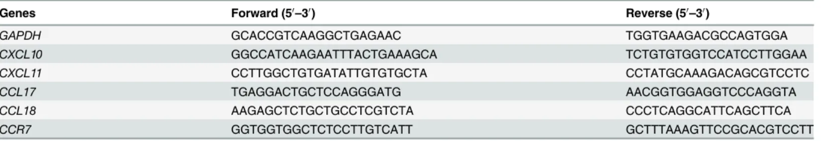
Before the exploration of macrophage polarization during the course ofM.tuberculosis infec-tion, we first determined the phenotypic and functional markers associated with human M1 and M2 macrophages, including CD86, CD163, CD206, HLA-DR, CXCL10, CXCL11, CCR7, CCL17, and CCL18, as well as the expression of related cytokines in the culture supernatant, including IL-6, IL-10, IL-12, and TNF-α. The results showed that following IFN-γ/LPS
induc-tion, the expression of HLA-DR, CD86, CXCL10, CXCL11, and CCR7 significantly increased, and the levels of IL-6, IL-12, and TNF-αin the culture supernatant were also significantly
en-hanced. On the other hand, IL-4 induction of MDM upregulated the expression levels of mo-lecular markers such as HLA-DR, CD206, CCR7, CCL17, and CCL18. Expression of IL-10 in the culture supernatants was also significantly increased (Fig 1). Based on these findings, CD86, CXCL11, and TNF-αwere selected as M1 molecular markers in subsequent studies, and
CD206, CCL17, and IL-10 were used as molecular markers for M2.
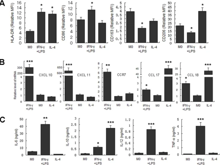
uninfected M0 cells. Expression of other markers, including CXCL11, CCL17, TNF-αand
IL-10, were significantly upregulated, indicating a mixed activation. With increased time of infec-tion, the expression of M1 macrophage-related markers, including CD86, CXCL11, and
TNF-αdecreased, while the expression of M2 macrophage-related markers including CD206,
CCL17 and IL-10 gradually increased (Fig 2). These results suggested thatin vitroinfection of human MDMs withM.tuberculosisinduces mixed M1/M2 activation, with a gradual transfor-mation to M2 with increased time of infection.
Macrophages undergo M1 to M2 transformation in the
in vitro
model of
tuberculous granuloma
The activation status of macrophages during the formation and development of granulomas was investigated using thein vitromodel of tuberculous granuloma established byM.
Fig 1. Determination of polarization markers of differently activated MDM.MDM were activated by IFN-γ/LPS, IL-4 or left untreated for 2 days. Markers
reported for M1 and M2 macrophages were determined. A. Phenotypic markers were measured by FCM. Mean fluorescent intensity (MFI) was measured and depicted as relative expression as compared to isotype control. Represented is the mean of three donors±SD. Significance was determined as compared to untreated group.*P<0.05,**P<0.01,***P<0.001. B. Effector molecules of differently activated macrophages were determined by real-time RT-PCR. Represented is mean±SD of relevant level of mRNA in activated MDM compared to M0 macrophage. C. Cytokines in the supernatant were measured by ELISA. Represented is the mean of three donors±SD. Significance was determined as compared to untreated group.*P<0.05,**P<0.01,
***P<0.001.
tuberculosisH37Rv infection of human PBMC according to methods described previously [17–
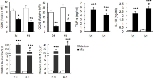
20]. The formation and development of granulomas was monitored by light microscopy to day 9 post-infection. Visible cell aggregation occurred on day 3 post-infection withM.tuberculosis
(Fig 3A,b). With prolonged infection, the volume of cell aggregates gradually increased, and granuloma-like structures with multilayer cell aggregations (Fig 3A,c-e) were observed on day 6. To better observe the distribution ofM.tuberculosisin thisin vitromodel, PBMCs infected withM.tuberculosis-GFP and stained with rhodamine-phalloidin were observed using laser scanning confocal microscopy. GFP-labeledM.tuberculosiswas found to be distributed be-tween, and possibly within, the aggregated cells (Fig 3A,f). No cellular aggregation was ob-served in the control groups at this stage. To analyze the cellular components of thisin vitro
granuloma, the cell aggregates were harvested, sectioned and stained with H&E and May-Grünwald-Giemsa (MGG) staining. Macrophages and lymphocytes were the main cellular components of these structures(Fig 3A,gandh). The histological characteristics and bacterial distribution of these aggregates of cells were consistent with previous reports ofin vitro tuber-culous granulomas and mimic the“true granuloma”[17–20].
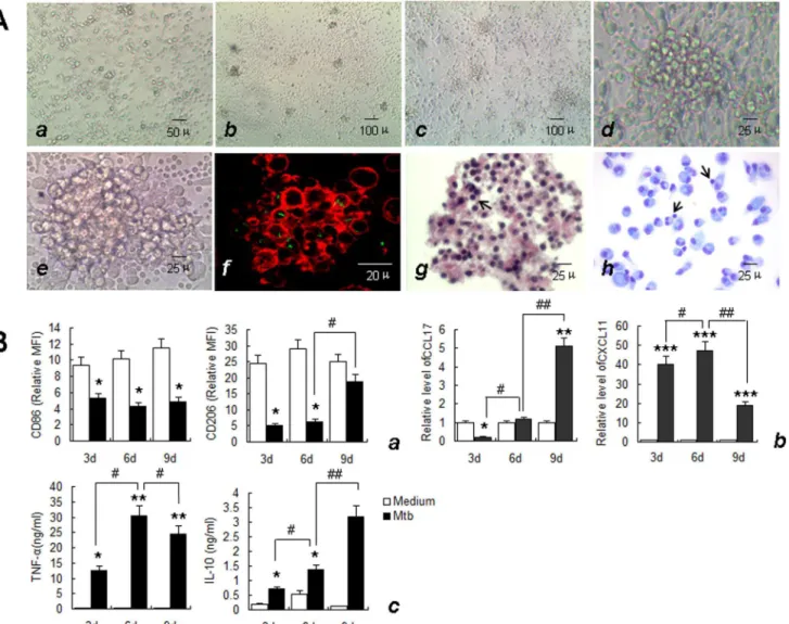
The activation status of macrophages in this model was then evaluated. In the early stages of granuloma-like structure formation (on day 3), the expression of the M1 macrophage-associat-ed molecular markers, CXCL11 and TNF-αwere significantly upregulated, with the exception
of CD86, which was expressed at lower levels, while the expression levels of the M2-related mo-lecular markers CD206 and CCL17 were significantly decreased. IL-10 expression significantly increased compared with that of M0 cells, but was similar to the level in cells following IFN-γ/
LPS induction (Fig 3B). Thus, M1 macrophages were predominant in the early stages of granu-loma-like structure formation. During the typical granugranu-loma-like structure formation stage (6d), the expression of both M1 and M2-related markers, including CXCL11, CCL17, TNF-α
and IL-10 increased significantly compared with that observed in the early (3d) stages, with the
Fig 2. Determination of polarization markers ofM.tuberculosisinfected MDM.MDM were infected withM.tuberculosisH37Rv at MOI of 5 and polarization markers were determined at indicated times. A. Phenotypic markers were measured by FCM. MFI was measured and depicted as relative expression as compared to isotype control. Represented is the mean of three donors±SD. Significance was determined as compared to uninfected group.
*P<0.05,**P<0.01,***P<0.001. B. Effector molecules of macrophages were determined by real-time RT-PCR. Represented is mean±SD of fold change of mRNA level in infected macrophages to uninfected MDM. C. Cytokines in the supernatant were measured by ELISA. Represented is the mean of three donors±SD. Significance was determined as compared to uninfected group.*P<0.05,**P<0.01,***P<0.001.Significance in corresponding marker expression between 6d and 3d was also calculated and represented by the following symbols:#P<0.05.
exception of CD86 and CD206, which showed no significant changes. With increased time of infection, the expression of M1-type molecular markers such as CXCL11 and TNF-α
signifi-cantly decreased, with the exception of CD86, which showed no significant change. In contrast, expression levels of the M2-type molecular markers CD206, CCL17 and IL-10 continued to in-crease (Fig 3B), suggesting that M2 macrophages predominate during the late stages of granu-loma-like structure formation.
Fig 3. Characteristics ofin vitrotuberculous granuloma model and the expression of granulomatous macrophage markers.A. Uninfected PBMCs at day 6 (a, ×200) and the morphology and structure of in vitro tuberculous granuloma model at day 3 (b, ×100), day 6 (c, ×100; d, ×400) and day 9 (e, ×400) in light microscopy. The actin filament in cells was labeled with rhodamine-phalloidin and the bacteria between and possibly within the cells was shown in green (f), (×1000). There were mainly macrophages and some lymphocytes (arrows) in granuloma observed by HE (g), (×400), and May-Grünwald-Giemsa staining (h), (×400). Size scale is indicated. B. Supernatant was collected and Macrophages were isolated from in vitro tuberculous granuloma as described in Materials and Methods at indicated times. The phenotypic markers on macrophages were determined by FCM. MFI was measured and depicted as relative expression as compared to isotype control. Represented is the mean of three donors±SD. (a). Effector molecules were determined by real-time RT-PCR. Represented is mean±SD of relative level of mRNA in activated MDM compared to M0 macrophage (b). Cytokines in the supernatant were measured by ELISA. Represented is the mean of three donors±SD (c). Significance was determined as compared to untreated group.*P<0.05,**P<0.01,
***P<0.001. Significance in corresponding marker expression between 9d, 6d and 3d was also calculated and represented by the following symbols: #
P<0.05,# #P<0.01.
Macrophages in granulomatous and non-granulomatous human tissues
exhibit differences in polarization status
Our results indicated that the activation state of macrophages underwent M1-to-M2 transition during the formation and development ofin vitrotuberculous granulomas. Compared with the
in vitromodel,in vivotuberculous granulomas are formed in a much more complex environ-ment. To further investigate the activation status of macrophages in the tuberculous granulo-ma, lung tissues from 18 pulmonary tuberculosis patients who received pneumonectomy were collected, sectioned and analyzed by H&E staining, acid-fast staining and immunohistochemis-try. Typical tuberculous granulomas were observed in most biopsy tissues as shown by H&E staining (Fig 4A,a). Acid-fast staining showed numerous acid-fastM.tuberculosis(stained red) distributed between, and possibly within, the aggregated cells (Fig 4A,b). Immunohisto-chemical staining for CD68 showed a large number of CD68-positive macrophages in the non-necrotic granulomas and the surrounding regions of caseous necrosis (Fig 4B).
To determine the polarization status of macrophages in the granulomatous lung tissues, as defined by the expression level of inducible nitric oxide (iNOS) and CD206, three serial sec-tions of the same tissue zone were stained with antibodies against CD68, iNOS and CD206. Re-sults showed that macrophages in both necrotic and non-necrotic granulomas were iNOSmed CD206high, indicating M2 dominant activation. However, macrophages in non-granulomatous tissues showed a different polarization status, characterized by a iNOShignCD206high pheno-type (Fig 4C). The expression levels of CXCL10, CXCL11, CCR7, CCL17 and CCL18 in the tu-berculous granuloma were measured by quantitative real-time RT-PCR and were compared with the levels detected in lung tissues without granulomas. The expression levels of M2-related markers (CCL17 and CCL18) significantly increased in the granulomatous lung tissues, where-as the expression of M1-related markers wwhere-as not consistent between the two groups (Fig 4D). These results demonstrated the coexistence of M1 and M2 macrophages in infected lung tis-sues, whereas M2 macrophages predominated in granulomatous tissues from patients with active tuberculosis.
Macrophage polarization modulates granuloma formation and the
bactericidal activity of macrophages
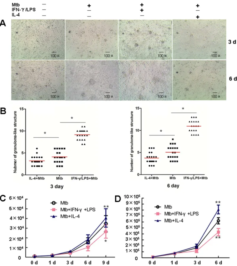
To further explore whether the polarization status of macrophage affects the progression of granulomas, IFN-γ+LPS and IL-4 were used to induce M1 and M2 macrophage polarization,
respectively, prior toM.tuberculosisinfection. Aggregates of cells were observed by light mi-croscopy at the indicated time-points. Enumeration of the granuloma-like structures in 20 ran-domly selected low-power fields (LPF, ×100) showed that visible cell aggregation in both groups had at day 3 post-infection. Compared with those in the untreated control group, the quantity of granuloma-like structures in the IFN-γ+LPS-treated group was significantly greater
(9.15 per LPF vs. 4.1 per LPF at day 3; 10.95 per LPS vs. 5.05 per LPF at day 6). In contrast, cell aggregation was significantly suppressed in the IL-4-treated group, and significantly fewer granuloma-like structures were observed compared with the untreated control group (3.20 per LPF vs. 4.1 per LPF at day 3; 3.75 per LPS vs. 5.05 per LPF at day 6). With the extension of the infection time, the surface area of the granuloma-like structures in both groups increased. Compared with the non-pretreated infection group, the surface area of the granuloma-like structures was relative larger and the structure tighter in the IFN-γ+LPS-treated group, while
Next, we examined the bactericidal effect of granulomatous cell populations onM. tubercu-losis. IFN-γ+LPS-pretreatment effectively improved the bactericidal function of granulomatous
cells onM.tuberculosis, while IL-4 pretreatment significantly inhibited this bactericidal effect (Fig 5B). To further confirm that the difference in bactericidal activity is related to the polariza-tion state of the macrophages, we used IFN-γ+LPS and IL-4 to induce polarization of human
MDM to the M1 and M2 phenotypes, respectively, prior toM.tuberculosisinfection. Measure-ment of the bacterial load at different time-points post-infection showed significantly lower colony-forming units (CFU) in the M1 (IFN-γ+LPS) group, while that in the M2 (IL-4) group
was notably higher (Fig 5C) compared with that in the M0 (un-induced) group. These results were consistent with those obtained using thein vitrogranuloma model, indicating that M1 macrophages possess higher bactericidal activity, compared with that of M2 macrophages, which is significantly attenuated.
Discussion
Increasing evidence has shown that macrophages play a dual role inM.tuberculosisinfection. On one hand, macrophages are the main immune effector cells and antigen-presenting cells re-sponsible for anti-tuberculosis immunity; on the other hand, macrophages are a target ofM.
tuberculosisand represent a“safe harbor”for the bacteria in infected humans [21,22]. Recent studies suggest that macrophages have great plasticity, and can be polarized differently under different conditions, thereby exhibiting different functionalities [23,24]. The tuberculous gran-uloma is the main pathological feature of tuberculosis and the most direct site of interaction of
M.tuberculosiswith immune cells. Macrophages are the major cell population in the tubercu-lous granuloma; therefore, a better understanding of macrophage polarization duringM. tuber-culosisinfection provide insights into the mechanism by whichM.tuberculosiscircumvents the host immune system.
Recently, Mattila et al. [15] found that substantial proinflammatory and anti-inflammatory macrophages coexist in the granulomatous lesions in the lung ofM.tuberculosis-infected ma-caques and tuberculosis patients, suggesting thatM.tuberculosishas the potential to modulate the macrophage polarization. However, this study mainly focused on the observation of pheno-typic markers of the macrophages in tuberculous granulomas at a fixed time-point. The varia-tion in funcvaria-tional markers and polarizavaria-tion status of granulomatous macrophages at different stages of infection, and the impact on the formation and development of tuberculous granulo-mas are still unclear.
In this study, anin vitrotuberculous granuloma model was generated based on the reported protocol [17–19], and the phenotypic and functional markers associated with macrophage po-larization were determined at different stages of granuloma formation. The results showed that, in the early stages of tuberculous granuloma formation, the polarization status of macro-phages was mainly M1 type. The expression levels of M1-associated markers gradually de-creased with time, while the M2-associated markers gradually inde-creased. During the late stage of tuberculous granuloma formation in thisin vitromodel, the polarization status of
Fig 4. Activation status of macrophages in human tuberculous granuloma. A.HE and acid-fast staining of tuberculous granuloma from the human lung tissue of tuberculosis (TB) patients. The structure of tuberculous granuloma with caseous necrosis was obviously observed by HE staining (a), (×200). Macrophages with irregular shapes were frequently observed (b), (×1000). Bacteria (stained in red, arrows) between and possibly within the granuloma cells were shown by the acid-fast staining (c), (×1000).B.The immunostaining of aggregate of CD68-positive macrophages in granuloma with caseous necrosis (a) or without caseous necrosis (b), (×100). CD206-positive multinucleated giant cells were frequently observed (c), (×400).C.The immunostaining of serial sections of the same tissue zone showed the expression levels of CD68, CD206 and iNOS in granulomas (×100) and nongranulomatous tissue (×400). D. The levels of CXCL10, CXCL11, CCR7, CCL17 and CCL18 mRNA in granulomatous lung tissues of tuberculosis patients were determined by real-time RT-PCR, in comparison with lung tissues containing no granuloma. The bars represent means±SD of fold changes of mRNA expression levels in
granulomatous tissues (G, showed as black boxes) compared to tissues containing no granuloma (NG, showed as blank boxes). (**P<0.01,***P<0.001).
Fig 5. Macrophage polarization modulates granuloma formation and the bactericidal activity of macrophages. A.PBMC were left untreated, or infected withM.tuberculosis, or pretreated with IFN-γ+LPS following infection withM.tuberculosis, or pretreated with IL-4 following infection withM.
macrophage was mainly M2 type. Thus, this study, for the first time, delineates the sequence of macrophage polarization during tuberculous granuloma formation. Given that modulation of macrophage polarization is one of the main mechanisms by whichM.tuberculosiscircumvents the host immune response, leading to immunopathology [25], this study may provide a theo-retical foundation for the development of novel therapies of tuberculosis based on reprogram-ming macrophage polarization.
Recent studies have demonstrated that the molecular markers associated with macrophage polarization are affected by both activators and genetic background [26]. Therefore, before evaluation of macrophage polarization during the course ofM.tuberculosisinfection, the clas-sical induction method was applied to determine the phenotypic and functional markers of M1 and M2 macrophages in humans. In brief, IFN-γ+LPS and IL-4 were used to induce
transfor-mation of MDM to M1 and M2 types, respectively, and a series of molecular markers were measured. CD86, CXCL11, and TNF-αwere selected as M1 molecular markers, while CD206,
CCL17, and IL-10 were used as M2 markers in the subsequent studies. However, CD86 expres-sion was persistently lower in the infected macrophages, which was inconsistent with the polar-ization states reflected by other molecular markers. CD86 is a positive costimulatory molecule and plays an important role in the activation of adaptive immune responses. Thus, it can be speculated that low CD86 expression byM.tuberculosis-infected cells may be one of the mech-anisms by whichM.tuberculosiscircumvents the adaptive immune responses of the host. Therefore, we believe that CD86 is not a suitable marker of polarization during the course of
M.tuberculosisinfection. In the subsequent human tissue studies, we used iNOS, which is re-ported to be a M1 phenotypic marker in a several studies [27,28] to ascribe the M1 phenotype.
Our findings in the lung tissues obtained from pulmonary tuberculosis patients showed that iNOS was expressed at low levels, while CD206 was expressed at high levels by macrophages distributed in both the necrotic and non-necrotic granulomas. These findings are in accor-dance with those reported by Mattila et al. [15], demonstrating M2 dominant activation of macrophages in granulomatous tissues from patients with active tuberculosis. Furthermore, we found that both iNOS and CD206 were expressed at high levels in non-granulomatous lung tis-sues from pulmonary tuberculosis patients, demonstrating the coexistence of M1 and M2 mac-rophages in the lung tissues of tuberculosis patients.
Next, we investigated the impact of macrophage polarization on tuberculous granuloma for-mation as well as the bactericidal effect of macrophages at different polarization states in thein vitrogranuloma model. The results showed that velocity of granuloma structure formation ve-locity was similar for macrophages in different polarization states. However, M1 macrophages significantly promoted the formation of tuberculous granuloma, demonstrated by the increase in the number of granuloma-like structures and the increase in macrophage volume. In con-trast, IL-4 pretreatment significantly inhibited the formation of tuberculous granulomas. Our study also demonstrated that compared with M0 macrophages, M1 macrophages significantly lowered theM.tuberculosisload, while M2 macrophages had the opposite effect.
structures in each group was counted in 20 randomly selected low-power field (LPF, ×100).*P<0.05.C. Thein vitromodel of tuberculous granuloma was generated as described in Materials and Methods. At 48h beforeM.tuberculosisinfection, PBMC were pretreated with IFN-γ+LPS, or IL-4, or left untreated.
Supernatant were collected and cells were lysed at indicated time points. Bacteria in supernatant and cell lysate were collected, mixed and inoculated onto the Middlebrook 7H10 solid medium containing 10% OADC. The bacterial load was determined by CFU counting. Represented is the mean of at least three tests±SD. Significance was determined as compared to untreated group.*P<0.05,**P<0.01.D. MDM, pretreated with IFN-γ+LPS, or IL-4, or left untreated
for 2 day, were infected withM.tband cultured as described in Materials and Methods. Supernatant were collected and cells were lysed at indicated time points. Bacteria in supernatant and cell lysate were collected, mixed and inoculated onto the Middlebrook 7H10 solid medium containing 10% OADC. The bacterial load was determined by CFU counting. Represented is the mean of at least three tests±SD. Significance was determined as compared to untreated group.*P<0.05,**P<0.01.
To our knowledge, no previous studies have been reported to investigate whether polarized macrophages influence the formation and development of tuberculous granulomas. Our study, for the first time, reveals that M1 macrophages induced by IFN-γ+LPS promote granuloma
formation, while M2 macrophages induced by IL-4 have an inhibitory effect. Our findings fur-ther demonstrate that macrophage polarization status plays a critical role in regulating the for-mation and development of tuberculous granulomas, although the mechanism of this
regulation requires further investigation. A recent study by Cyktor et al. revealed that IL-10 in-hibited the formation of granulomas induced byM.tuberculosisin a mouse model [29]. Based on our observation that IL-10 was greatly elevated in the M2 macrophage model induced by IL-4, it can be speculated that the inhibitory role of M2 macrophages in preventing the forma-tion and development of granulomas may be partially mediated by IL-10.
In summary, our findings demonstrate the coexistence of M1 and M2 polarized macro-phages in the tuberculous granuloma and indicate that M1 macromacro-phages promote tuberculous granuloma formation, while M2 macrophages inhibit this process.
Author Contributions
Conceived and designed the experiments: JL. Performed the experiments: ZH QL YG JC GX YP JY. Analyzed the data: JL ZH QL. Contributed reagents/materials/analysis tools: GX YP JY. Wrote the paper: JL ZH. Obtained signed informed consents from participants: JL YP QL.
References
1. Eurosurveillance editorial team (2013) WHO publishes Global tuberculosis report 2013. Euro Surveill 18: pii: 20615.
2. World Health Organization (2014) Global Tuberculosis Report 2014. World Health Organization, Ge-neva, Switzerland. Available:http://www.who.int/tb/publications/global_report/en/.
3. Egen JG, Rothfuchs AG, Feng CG, Winter N, Sher A, Germain RN (2008) Macrophage and T cell dy-namics during the development and disintegration of mycobacterial granulomas. Immunity 28: 271–
284. doi:10.1016/j.immuni.2007.12.010PMID:18261937
4. Lugo-Villarino GD, Benard HA, Neyrolles O (2012) Emerging trends in the formation and function of tu-berculosis granulomas. Frontiers in immunolog. 3: 405. doi:10.3389/fimmu.2012.00405PMID:
23308074
5. Russell DG (2007) Who puts the tubercle in tuberculosis? Nature reviews 5: 39–47. PMID:17160001 6. Saunders BM, Britton WJ (2007) Life and death in the granuloma: immunopathology of tuberculosis.
Immunology and cell biology 85: 103–111. PMID:17213830
7. Miranda MS, Breiman A, Allain S, Deknuydt F, Altare F (2012) The tuberculous granuloma: an unsuc-cessful host defence mechanism providing a safety shelter for the bacteria? Clinical & developmental immunology 2012: 139127.
8. Biswas SK, Chittezhath M, Shalova IN, Lim JY (2012) Macrophage polarization and plasticity in health and disease. Immunologic research 53: 11–24. doi:10.1007/s12026-012-8291-9PMID:22418728 9. Gordon S, Martinez FO (2010) Alternative activation of macrophages: mechanism and functions.
Im-munity 32: 593–604. doi:10.1016/j.immuni.2010.05.007PMID:20510870
10. Mantovani A, Sica A, Sozzani S, Allavena P, Vecchi A, Locati M (2004) The chemokine system in di-verse forms of macrophage activation and polarization. Trends in immunology 25: 677–686. PMID: 15530839
11. Martinez FO, Sica A, Mantovani A, Locati M (2008) Macrophage activation and polarization. Front Biosci 13: 453–461. PMID:17981560
12. Li W, Katz BP, Spinola SM (2012) Haemophilus ducreyi-induced interleukin-10 promotes a mixed M1 and M2 activation program in human macrophages. Infection and immunity 80: 4426–4434. doi:10. 1128/IAI.00912-12PMID:23027536
14. Bility MT, Cheng L, Zhang Z, Luan Y, Li F, Chi L, et al. (2014) Hepatitis B virus infection and immuno-pathogenesis in a humanized mouse model: induction of human-specific liver fibrosis and M2-like mac-rophages. PLoS Pathogens 10: e1004032. doi:10.1371/journal.ppat.1004032PMID:24651854 15. Mattila JT, Ojo OO, Kepka-Lenhart D, Marino S, Kim JH, Eum SY, et al. (2013) Microenvironments in
tu-berculous granulomas are delineated by distinct populations of macrophage subsets and expression of nitric oxide synthase and arginase isoforms. J Immunol 191: 773–784. doi:10.4049/jimmunol.1300113
PMID:23749634
16. Marino S, Cilfone NA, Mattila JT, Linderman JJ, Flynn JL, Kirschner DE (2014) Macrophage polariza-tion drives granuloma outcome during Mycobacterium tuberculosis infecpolariza-tion. Infect Immun doi:10. 1128/IAI.02494-14
17. Puissegur MP, Botanch C, Duteyrat JL, Delsol G, Caratero C, Altare F (2004) An in vitro dual model of mycobacterial granulomas to investigate the molecular interactions between mycobacteria and human host cells. Cell Microbiol 6: 423–433. PMID:15056213
18. Birkness KA, Guarner J, Sable SB, Tripp RA, Kellar KL, Bartlett J, et al. (2007) An in vitro model of the leukocyte interactions associated with granuloma formation in Mycobacterium tuberculosis infection. Immunology and cell biology 85: 160–168. PMID:17199112
19. Wang H, Maeda Y, Fukutomi Y, Makino M (2013) An in vitro model of Mycobacterium leprae induced granuloma formation. BMC infectious diseases 13: 279. doi:10.1186/1471-2334-13-279PMID:
23782413
20. Kapoor N, Pawar S, Sirakova TD, Deb C, Warren WL, Kolattukudy PE (2013) Human granuloma in vitro model, for TB dormancy and resuscitation. PLoS One 8: e53657. doi:10.1371/journal.pone. 0053657PMID:23308269
21. Liu PT, Modlin RL (2008) Human macrophage host defense against Mycobacterium tuberculosis. Curr Opin Immunol 20: 371–376. doi:10.1016/j.coi.2008.05.014PMID:18602003
22. Leemans JC, Thepen T, Weijer S, Florquin S, van Rooijen N, van de Winkel JG, et al. (2005) Macro-phages play a dual role during pulmonary tuberculosis in mice. J Infect Dis 191: 65–74. PMID: 15593005
23. Geissmann F, Manz MG, Jung S, Sieweke MH, Merad M (2010) Development of monocytes, macro-phages, and dendritic cells. Science 327: 656–661. doi:10.1126/science.1178331PMID:20133564 24. Mosser DM, Edwards JP (2008) Exploring the full spectrum of macrophage activation. Nat Rev
Immu-nol 8: 958–969. doi:10.1038/nri2448PMID:19029990
25. Lugo-Villarino G, Vérollet C, Maridonneau-Parini I, Neyrolles O (2011) Macrophage polarization: con-vergence point targeted by mycobacterium tuberculosis and HIV. Front Immunol 15: 43.
26. Sans-Fons MG, Yeramian A, Pereira-Lopes S, Santamaría-Babi LF, Modolell M, Lloberas J, et al. (2013) Arginine transport is impaired in C57Bl/6 mouse macrophages as a result of a deletion in the promoter of Slc7a2 (CAT2), and susceptibility to Leishmania infection is reduced. J Infect Dis 207: 1684–1693. doi:10.1093/infdis/jit084PMID:23460752
27. Redente EF, Higgins DM, Dwyer-Nield LD, Orme IM, Gonzalez-Juarrero M, Malkinson AM (2010) Dif-ferential polarization of alveolar macrophages and bone marrow-derived monocytes following chemi-cally and pathogen-induced chronic lung inflammation. J Leukoc Biol 88: 159–168. doi:10.1189/jlb. 0609378PMID:20360403
28. Davis MJ, Tsang TM, Qiu Y, Dayrit JK, Freij JB, Huffnagle GB, et al. (2013) Macrophage M1/M2 polari-zation dynamically adapts to changes in cytokine microenvironments in Cryptococcus neoformans in-fection. MBio 4: e00264–13. doi:10.1128/mBio.00264-13PMID:23781069
29. Cyktor JC, Carruthers B, Kominsky RA, Beamer GL, Stromberg P, Turner J (2013) IL-10 inhibits mature fibrotic granuloma formation during Mycobacterium tuberculosis infection. J Immunol 190: 2778–2790.