Dietary Regulation of the Gut Microbiota
Engineered by a Minimal Defined Bacterial
Consortium
Ting-Chin David Shen1, Christel Chehoud2, Josephine Ni1, Evelyn Hsu3, Ying-Yu Chen1, Aubrey Bailey2, Alice Laughlin2, Kyle Bittinger2, Frederic D. Bushman2, Gary D. Wu1*
1Division of Gastroenterology, Perelman School of Medicine, University of Pennsylvania, Philadelphia, Pennsylvania, United States of America,2Department of Microbiology, Perelman School of Medicine, University of Pennsylvania, Philadelphia, Pennsylvania, United States of America,3Division of
Gastroenterology and Hepatology, Department of Pediatrics, University of Washington School of Medicine, Seattle, Washington, United States of America
Abstract
We have recently reported that Altered Schaedler Flora (ASF) can be used to durably engi-neer the gut microbiota to reduce ammonia production as an effective modality to reduce morbidity and mortality in the setting of liver injury. Here we investigated the effects of a low protein diet on ASF colonization and its ability to engineer the microbiota. Initially, ASF inoc-ulation was similar between mice fed a normal protein diet or low protein diet, but the out-growth of gut microbiota differed over the ensuing month. Notable was the inability of the dominantParabacteroidesASF taxon to exclude other taxa belonging to the Bacteroidetes phylum in the setting of a low protein diet. Instead, a poorly classified yet highly represented Bacteroidetes family, S24-7, returned within 4 weeks of inoculation in mice fed a low protein diet, demonstrating a reduction in ASF resilience in response to dietary stress. Neverthe-less, fecal ammonia levels remained significantly lower than those observed in mice on the same low protein diet that received a transplant of normal feces. No deleterious effects were observed in host physiology due to ASF inoculation into mice on a low protein diet. In total, these results demonstrate that low protein diet can have a pronounced effect on engi-neering the gut microbiota but modulation of ammonia is preserved.
Introduction
The gut microbiota responds to multiple environmental stressors such as diet [1–4], antibiotic use [5], inflammation of the intestinal tract [6], and infection of the host with enteric pathogens [7]. By studying the gut microbiota in pediatric patients with Crohn’s disease, we have recently shown that the effects of these factors may be independent even if present simultaneously [8]. Amongst these, the impact of diet has received considerable attention as a potential modifiable factor that shapes the composition and/or function of the gut microbiota to prevent and/or treat disease [9]. The high-level efficacy of fecal microbiota transplantation (FMT) in the
a11111
OPEN ACCESS
Citation:Shen T-CD, Chehoud C, Ni J, Hsu E, Chen Y-Y, Bailey A, et al. (2016) Dietary Regulation of the Gut Microbiota Engineered by a Minimal Defined Bacterial Consortium. PLoS ONE 11(5): e0155620. doi:10.1371/journal.pone.0155620
Editor:John F. Rawls, University of North Carolina at Chapel Hill, UNITED STATES
Received:January 24, 2016
Accepted:May 2, 2016
Published:May 13, 2016
Copyright:© 2016 Shen et al. This is an open access article distributed under the terms of the Creative Commons Attribution License, which permits unrestricted use, distribution, and reproduction in any medium, provided the original author and source are credited.
Data Availability Statement:Sequence data sets are available from NCBI’s Sequence Read Archive (SRA) database (accession numbers SRP058968 and SRP068327). Additional data are deposited under the following DOI in the Dryad repository: doi:10.5061/dryad.n2s40.
treatment ofClostridium difficileinfections (CDI) is proof of concept that inoculating a host with a consortium of microbes has a meaningful effect on the composition of the gut micro-biota [10]. The use of feces could be considered an untargeted approach with potential risks [11], but growing evidence suggests that the use of defined microbial consortia could be devel-oped to treat disease [12,13]. We have recently shown that the gut microbiota can be durably reconfigured to reduce fecal urease activity and ammonia production through oral inoculation of Altered Schaedler Flora (ASF), a defined microbial consortium that contains minimal urease gene content [13]. ASF comprises eight murine gut commensal bacterial strains (S1 Table) assembled in the 1970s and standardized by the National Cancer Institute in 1978 [14–16]. It is now commonly used to create gnotobiotic mice and/or to enhance the health of immunode-ficient mouse strains.
Examples of co-metabolism between the gut microbiota and its mammalian host requiring host-derived substances include bile acids, mucous, and urea. The latter is particularly impor-tant for nitrogen flux between the host and the gut microbiota [17,18]. As the primary source of nitrogen, dietary protein is essential to the synthesis of nucleic acids, amino acids, and other nitrogenous compounds. The catabolism of dietary protein by the host leads to hepatic forma-tion of urea, a nitrogenous waste product that is excreted through the urine or delivered into the colon, where hydrolysis by bacterial urease results in the production of carbon dioxide and ammonia. Ammonia is a shared substrate for the synthesis of proteins, amino acids, and other small molecules by both the host and its microbiota. Although generally thought to be nutri-tionally beneficial to the host by enhancing nitrogen recycling, the production of ammonia by the gut microbiota can have deleterious effects in the setting of altered hepatic function, result-ing in the development of neurotoxicity [19–21]. Under such conditions, a low protein diet (LPD) can be used to reduce systemic ammonia levels [22,23].
By inoculating mice with ASF after the endogenous microbiota has been reduced through the use of antibiotics and polyethylene glycol (PEG), the composition of the gut microbiota can be durably modified in composition as well as function. Functionally, there was a long-lasting reduction in fecal ammonia that was effective in reducing morbidity and mortality in the thioa-cetamide model of liver injury [13]. Since (1) the absorption of fecal ammonia produced by the gut microbiota may be an important source of nitrogen for the host especially in the setting of dietary protein restriction [17], and (2) low protein diets are used clinically in patients with hyperammonemic inborn errors of metabolism [24], there are a number of questions about the impact of diet on the engineering of the gut microbiota to reduce ammonia production. What is the effect of a LPD on the ability of a defined bacterial consortium to colonize in the gut? Does a LPD have an effect on the composition of the engineered microbiota? Will the ammonia reduction by microbiota engineering be sustained and exhibit lower levels than those achiev-able by a LPD alone? And lastly, will a significant reduction in gut microbiota ammonia pro-duction be deleterious to the host on a LPD?
Here, we address these questions by inoculating mice on a LPD with either feces from con-ventionally-reared mice (Normal Feces, or NF) or with ASF, monitoring the resultant composi-tion of the gut microbiota over time by 16S tagged sequencing, assessing funccomposi-tionality by quantifying fecal ammonia levels, and investigating the impact on the host by metabolic profil-ing. Although a LPD has no effect on the ability of ASF to colonize the gut of the host upon inoculation, the resultant engineered state of the microbiota is altered primarily due to the re-emergence of S24-7, a specific bacterial taxonomic family within the Bacteroidetes phylum. Despite this alteration, fecal ammonia levels remain diminished and without consequence to the metabolic physiology of the host on a LPD.
Results
LPD impacts host physiology and nitrogen metabolism but modestly
alters the composition of the gut microbiota
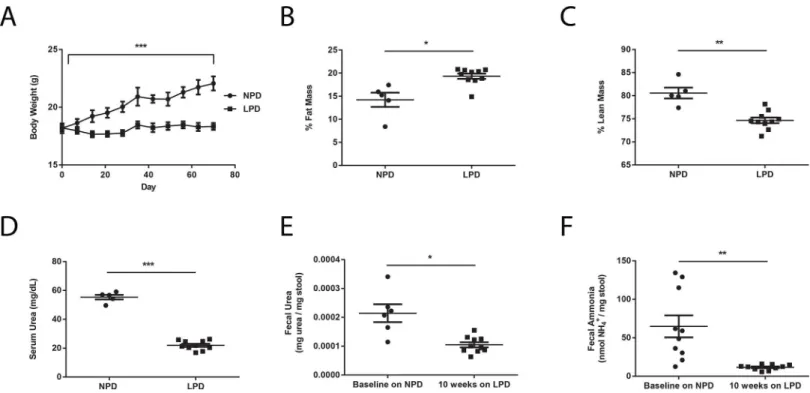
We first set out to investigate the effects of a LPD on both the murine host and the gut micro-biota. Fifteen adult female C57BL6J mice were placed on an open source irradiated purified rodent diet containing normal amount of dietary protein at 21% by kilocalories (AIN-76A), henceforth referred to as normal protein diet (NPD,Table 1) for one week upon arrival into the University of Pennsylvania SPF vivarium. Subsequently, ten of the 15 mice were switched to an irradiated low protein diet (LPD,Table 1) formulated using AIN-76A as the base that contains 3% protein by kilocalories. The LPD was made isocaloric by proportionally increasing carbohydrate content while keeping fat content unchanged. The remaining five mice continued to be fed the NPD. We monitored physiological changes in these mice using body weight and food intake measurements as well as body composition determination via nuclear magnetic resonance (NMR) imaging. We found that compared to NPD-fed mice, LPD-fed mice exhib-ited poor weight gain despite equivalent caloric consumption (Fig 1A). LPD-fed mice also demonstrated increased fat mass and decreased lean mass (Fig 1B and 1C). Corresponding to a reduction in serum urea concentration compared to NPD-fed mice (Fig 1D), LPD-fed mice exhibited significant reductions in fecal urea and fecal ammonia levels after ten weeks on the LPD (Fig 1E and 1F). These results are consistent with the fundamental role that dietary pro-tein plays in host nitrogen balance. Reduction in dietary propro-tein may have an effect on the gut microbiota by reducing the delivery of urea to the colonic environment leading to the reduction in fecal ammonia levels.
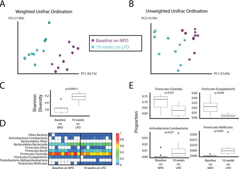
16S tagged sequencing (1045 to 5305 reads per sample, median = 3448 reads) revealed mod-est yet distinct differences in the composition of the fecal microbiota in mice after placement on the LPD. These difference can be visualized in principal coordinates analysis of weighted (Fig 2A) and unweighted (Fig 2B) UniFrac distance. The LPD led to a significant increase in the diversity of the gut microbial community as assessed by the Shannon diversity index (Fig
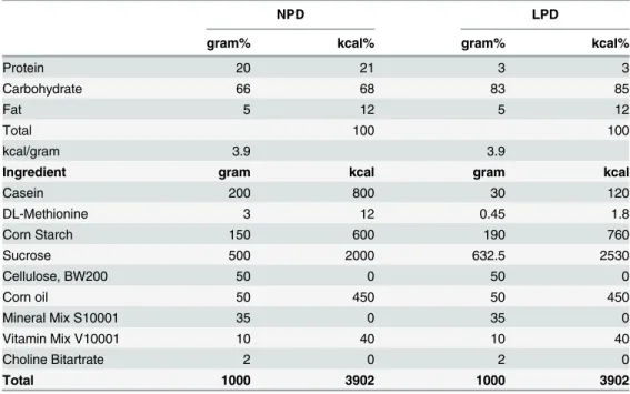
Table 1. Components of normal protein diet (NPD) and low protein diet (LPD).
NPD LPD
gram% kcal% gram% kcal%
Protein 20 21 3 3
Carbohydrate 66 68 83 85
Fat 5 12 5 12
Total 100 100
kcal/gram 3.9 3.9
Ingredient gram kcal gram kcal
Casein 200 800 30 120
DL-Methionine 3 12 0.45 1.8
Corn Starch 150 600 190 760
Sucrose 500 2000 632.5 2530
Cellulose, BW200 50 0 50 0
Corn oil 50 450 50 450
Mineral Mix S10001 35 0 35 0
Vitamin Mix V10001 10 40 10 40
Choline Bitartrate 2 0 2 0
Total 1000 3902 1000 3902
2CandS1 Fig). The LPD also led to significant increases in the relative abundance of Molli-cutes and Coriobacteria and a decrease in the FirmiMolli-cutes classes Erysipelotrichi and Clostridia (Fig 2D and 2E).
LPD has no effect on the initial colonization of ASF into the host
microbiota
We have previously shown that there is a reduction in gut bacterial biomass upon treatment of mice with oral antibiotics (vancomycin and neomycin) and PEG, thus permitting the coloniza-tion of ASF upon inoculacoloniza-tion by oral gavage [13]. However, the effect of a LPD on the coloniza-tion of ASF into the gut of a prepared host remains unknown. After preparacoloniza-tion with
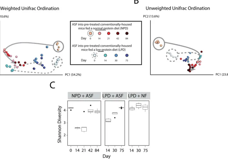
antibiotics and PEG, we orally inoculated five of the LPD-fed mice with ASF (herein referred to as“ASF-transplanted”). As a control group, we transplanted the other five LPD-fed mice using feces from conventionally-reared donor mice (herein referred to“NF-transplanted,”for Normal Feces). Using 16S rRNA tagged sequencing, we tracked taxonomic alterations in the gut microbiota over time. We found that NF-transplanted mice exhibited minimal change in the composition of their gut microbiota (S2 Fig). However, the gut microbiota of ASF-trans-planted mice underwent a shift in composition in a similar fashion to that previously observed in NPD-fed mice transplanted with ASF [13], as shown inFig 3A and 3B.In particular, the shifts along PC1 in both cohorts of mice represent changes due to the initial ASF inoculum (compare days 0 to 14 in both groups), whereas differences between the two cohorts of mice along PC2 may represent the effect of diet. These findings suggest that a LPD does not affect the initial colonization of ASF into the host microbiota.
Fig 1. Changes in murine physiology and nitrogen metabolism on a LPD.Differences in (A) body weight (n = 5 in NPD group, n = 10 in LPD group), (B) % fat mass, (C) % lean mass, and (D) serum urea concentrations between NPD-fed and LPD-fed mice. (E) Fecal urea and (F) fecal ammonia levels at baseline on the NPD and after placement on the LPD. Values represent mean±SEM. Statistical significance in body weight determined by two-way ANOVA
with repeated measures; statistical significance in other parameters determined by paired and unpaired two-tailed Student’s t test.*p<0.05,**p<0.01, ***p<0.001.
Diet affects the resilience of the gut microbiota engineered by inoculation
with ASF
By tracking the composition of mice inoculated with ASF, we determined the effect of a LPD on the ability of ASF to engineer a different microbiota composition. In the setting of a NPD, we previously observed that ASF transplantation led to the development of a new steady state community after one month composed of both ASF and the return of selected taxa of the Fir-micutes phylum, but no non-ASF Bacteroidetes [13]. We proposed thatParabacteroides
ASF519, the dominant taxon in ASF in feces, may have prevented the return of other Bacteroi-detes taxa by competitive niche exclusion. Tracking compositional changes in the gut micro-biota over time, we found that in the setting of the LPD, the gut micromicro-biota engineered by ASF transplantation developed into an alternative rich community with diversity similar to that on
Fig 2. Effect of a LPD on the composition of the gut microbiota.Principal coordinates analysis (PCoA) ordination of mice before and after placement on the LPD for 10 weeks. Changes in community membership were analyzed using (A) weighted and (B) unweighted Unifrac. (C) The interquartile range of Shannon diversity values is shown for mice on the NPD who were later put on the LPD (Wilcoxon rank sum test p-value = 0.0001299). (D) Heatmap showing the relative abundance of bacterial lineages over time in mice who were on the NPD at baseline and then after ten weeks on the LPD. Rows indicate bacterial lineages annotated at the class taxonomic level on the left. The color key on the right of the figure indicates relative abundance. Columns summarize the sequencing results from individual fecal specimens. Each column represents a different mouse. The columns are grouped by diet. (E) Bacterial lineages that change on the LPD. Four bacterial classes significantly differed between the NPD and LPD (FDR-corrected Wilcoxon test p-value<0.05). Relative
abundance of each class in both diet groups is shown. Box and whiskers show the interquartile range; black circles mark the outlier samples.
the NPD (Fig 3C). However, ASF519 did not suppress the return of other Bacteroidetes. Instead, Bacteroidetes S24-7, a poorly classified yet common bacterial taxon in the commensal murine gut microbiota [25,26], returned after ASF transplantation and reached an equilibrium state with ASF519 (Fig 4andS3 Fig). We plotted the progression of the transplanted ASF community over time. We found that ASF reached a new steady state in the setting of the LPD at around 4 weeks after transplantation, similar to what we previously observed in the setting of the NPD [13] (Fig 3A and 3B). However, this steady state more closely resembled the endogenous microbiota, likely as a result of the return of S24-7 on the LPD (best observed in
Fig 3Aalong PC1–compare the solid to dotted grey line). Overall, these findings suggest that ability of ASF lineages to compete is reduced in the presence of a LPD.
The ASF-engineered gut microbiota lowers fecal ammonia more
effectively than LPD alone
We have previously shown that ASF transplantation durably reduces fecal ammonia by decreasing fecal urease activity [13]. Since a LPD itself mainly reduces fecal ammonia by
Fig 3. Effect of a LPD on the initial colonization of ASF and subsequent resilience over time.Principal coordinates analysis (PCoA) ordination of mice after transplantation with ASF. Changes in community membership were analyzed using (A) weighted and (B) unweighted Unifrac. Dietary groups are color coded as indicated with the shades of colors indicating progression in time. Day 0 samples have gray circles around them (solid for NPD, dashed for LPD). The arrows were added to help visualize the progression of time after ASF transplantation. (C) The interquartile range of Shannon diversity values are shown for mice on the NPD and LPD inoculated with either ASF or Normal Feces (NF). Black circles mark the outlier samples.
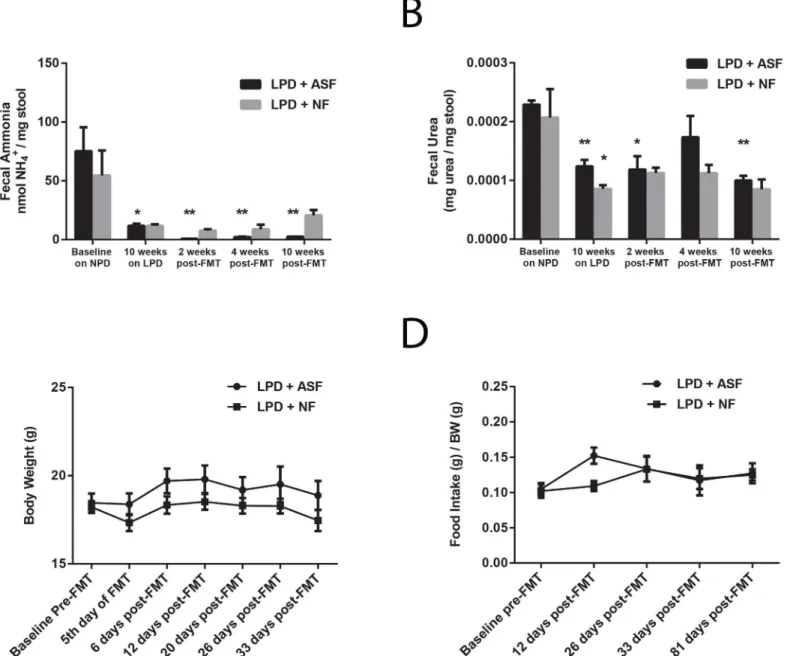
decreasing the delivery of urea to the colon (Fig 1E and 1F), we sought to determine whether the ASF-engineered microbiota would be able to reduce fecal ammonia levels below those achieved by a LPD alone. We measured fecal urea and fecal ammonia in mice at baseline on the NPD, after ten weeks on the LPD, and compared ASF and NF transplantation on the LPD. As shown inFig 5A,after the initial reduction in fecal ammonia levels induced by the LPD, ASF transplantation reduced fecal ammonia further than did NF transplantation. The ability of the ASF-engineered microbiota to lower fecal ammonia levels below those achieved by the LPD alone is likely due to the reduction in fecal urease activity since there was no difference in fecal urea levels after NF and ASF transplantation (Fig 5B). These results indicate that the functionality of the ASF-engineered gut microbiota is not significantly altered in the setting of a LPD despite alterations in its composition.
The low fecal urease and fecal ammonia-producing microbiota
engineered by ASF inoculation does not exacerbate host metabolic
dysfunction induced by LPD
Urea is a nitrogenous waste product, but it is thought to contribute to host nutrition via urea nitrogen recycling by intestinal bacterial urease in both ruminants and non-ruminants, leading to microbial and/or host synthesis of peptides, amino acids, and other small molecules [17]. We asked whether this role of urea may become important for host physiology in the setting of a LPD, where systemic nitrogen is reduced. After transplanting the above cohorts of mice with ASF or NF, we continued to monitor their body weight, food intake, and survival. Remarkably,
Fig 4. S24-7 returns after ASF transplantation into mice on a LPD but not on a NPD.(A) Relative abundance of bacterial taxa are shown. Each column represents a single sample of a pre-treated, ASF-transplanted mice on the LPD (LPD + ASF). Progression of inoculation is shown across multiple days post inoculation with ASF. Relative abundance of (B) Parabacteroides (including ASF 519) and (C) S24-7. Box and whiskers show the interquartile range; black circles mark the outlier samples.
despite the absence of colonic urea nitrogen recycling, ASF-transplanted mice did not differ significantly from NF-transplanted mice (Fig 5C and 5DandS4 Fig). Thus, in the setting of a LPD, ASF transplantation does not lead to significant detrimental changes to host physiology and metabolism.
Discussion
The success of FMT in the treatment of recurrentClostridium difficileinfections provides proof of concept that the gut microbiota can be a target for the treatment of disease in humans.
Fig 5. ASF transplantation alters colonic urea nitrogen recycling without significantly affecting host physiology.(A) ASF transplantation reduces fecal ammonia below the level achieved by the LPD alone (n = 4–5 per group,*p<0.05 compared to baseline,**p<0.01 compared to 10 weeks on the LPD).
(B) No difference in fecal urea level between ASF- and NF-transplanted mice (n = 2–5 per group,*p<0.05 compared to baseline,**p<0.01 compared to
baseline). No difference in (C) body weight or (D) food intake between ASF- and NF-transplanted mice (n = 5 per group). Values represent mean±SEM. Significance determined by two-tailed Student’s t-test.
The use of fecal transfer will likely be replaced by the use of defined microbial consortia with specific biological properties. As proof of concept, we have shown in murine models that a defined consortium of eight bacteria, known as Altered Schaedler Flora (ASF), can be used to engineer the gut microbiota with altered functionality, namely a reduction in fecal urease activ-ity and ammonia production [13]. Critical to the success of this strategy is the substantial reduction in the biomass of the baseline microbiota to provide a niche into which the bacterial inoculum can colonize.
An important consideration for engineering the gut microbiota is resilience to environmen-tal stress. An optimally engineered microbiota would be a rich community that stays intact in the presence of environmental stress. Diet is an important environmental stressor on the gut microbiota that should be considered when engineering gut microbial communities. As one example, the low ammonia-producing microbiota engineered by ASF, which has functional durability for several months in mice fed an irradiated diet, shows reduced resilience when the mice are fed a non-irradiated diet [13]. We chose to study the impact of dietary protein on the resilience of the ASF-engineered microbiota for several reasons: 1) Dietary protein has been shown to influence the composition of the gut microbiota in gnotobiotic mice [27]; 2) Protein consumption regulates the production of hepatic urea that may affect colonic urea delivery to the gut microbiota [17,28]; 3) Protein-restricted diets are an important therapeutic modality for patients with hyperammonemic inborn errors of metabolism [23,29].
Unlike the modulation of fat and fiber in mice, which have been shown to have a strong effect on the composition of the murine gut microbiota [1,27], we show that severe restriction of dietary protein had a modest effect. Within the Firmicutes phylum the Clostridia and Erysi-pelotrichi classes decreased significantly on a LPD, consistent with the preference of taxa within Firmicutes, particularly Clostridia species, to metabolize amino acids and peptides [30,
31]. Alternatively, since we show that a LPD reduces serum urea concentrations with reduced delivery to the colon resulting in lower fecal ammonia levels, an alteration in nitrogen flux via ammonia into the gut microbiota [32,33] may also have an effect on the composition of the bacterial microbiota.
Since we balanced protein with carbohydrate in the composition of the purified rodent diets, it is difficult to ascertain if the differences in the composition of the gut microbiota are due primarily to alterations in protein or in carbohydrate. We found that two bacterial phyla present at low abundances increased significantly on a LPD. Specifically, the classes Mollicutes (Tenericutes phylum) and Coriobacteria (Actinobacteria phylum) increased on a LPD. Previ-ous work has shown that Mollicutes proliferated on a typical Western diet characterized by high-fat/high-sugar content, likely because of their ability to import and process simple sugars [34]. Thus, an increase in the abundance of Mollicutes that we observed on a LPD could be due to the increase in carbohydrate content rather than the reduction in protein content. Another study also showed that gut colonization by Actinobacteria and Tenericutes was strongly corre-lated with decreased hepatic levels of glycogen and glucose [35], further suggesting the inter-play between the host and these two phyla may be closely related to carbohydrate metabolism.
demonstrated in theBacteroidesgenus where successful competition for carbohydrate sub-strates plays an important role [36]. By contrast, on a LPD, the resultant engineered microbiota appears to be more similar to baseline primarily due to the reemergence of a single bacterial taxon belonging to the Bacteroidetes family, S24-7.
S24-7 has been previously recognized as a dominant taxonomic group in the murine micro-biota. It was first characterized by Salzmanet al., who referred to the taxon as“mouse intestinal bacteria”[25]. The S24-7 taxon is phylogenetically distinct from other named genera in the order Bacteroidales. The taxon has been reported as altered in several recent mouse studies: it was increased in proportion following partial hepatectomy [37], associated with co-infection byHymenolepisspp. [38], and decreased in proportion following antibiotic treatment for par-enteral nutrition-associated liver injury [39]. However, to our knowledge, no study has previ-ously characterized competition between S24-7 and other Bacteroidetes species in mice. The S24-7 taxon is typically encountered at very low (<1%) abundance in fecal samples from
human populations. However, one study of a previously uncontacted Amerindian population reported the taxon to be enriched in isolated Yanomami Amerindians relative to Guahibo Amerindian, Malawian, and U.S. subjects [40]. The average abundance of the taxon in Yano-mami Amerindians was reported to be nearly 5% of total bacteria, suggesting a potential role for S24-7 in the human gut.
Upon reduction of bacterial biomass through a combination of antibiotics and PEG, S24-7 is no longer detectable and shows no return over time after mice have been inoculated with ASF. Since bothParacteroides(ASF519) and S24-7 are closely related within the Bacteroidetes phylum, we speculate that S24-7 may be co-excluded from the luminal gut environment by ASF519 through competitive niche exclusion, a mechanism that has been hypothesized as the basis for the inversely-related proportions of Bacteroides and Prevotella in the human gut microbiota [2,41], a predominant feature of“enterotypes”[42]. From a mechanistic stand-point, the basis of competitive niche exclusion may involve the competition of metabolic sub-strates as has been demonstrated for Bacteroides species in a reductionist model system [36]. Since S24-7 reappears and co-exists at approximately equal levels with ASF519 in LPD-fed mice, the alteration of substrate availability via diet may have altered the luminal environment of the gut that reduces the need for competition between these two taxa. For example, a LPD may have altered the balance of nitrogen flux into the gut microbiota via the uptake of ammo-nia. Indeed, despite the return of S24-7 and the similarities between the composition of the gut microbiota of a conventionally-housed mouse and the ASF-engineered community established in LPD-fed mice, fecal ammonia levels remained much lower in ASF-transplanted mice than those transplanted with normal feces. This suggests that S24-7 may be urease negative. Further elucidation of such mechanism(s) will require genomic characterization of S24-7 along with an evaluation of its biological properties.
The quantification of fecal ammonia was used to determine the impact of microbiota com-position on the function of the community. Despite the modest alterations in the gut micro-biota induced by the consumption of a LPD, there was a significant reduction in fecal ammonia levels reflecting the reduced abundance of urea substrate available for hydrolysis by the gut microbiota. This observation emphasizes the notion that diet may have an indirect impact on the gut microbiota by alteration of the host similar to the outgrowth of a pathobiont due to the enhanced production of sulfated bile acids in mice fed milk fat [43]. Importantly, engineering of the gut microbiota using ASF led to a reduction in fecal ammonia levels signifi-cantly greater than that observed on a LPD.
intake [17,44]. This might be a significant limitation of a strategy focused on reducing gut microbiota ammonia production for the treatment of hyperammonemia and hepatic encepha-lopathy [13]. Although LPD-fed mice did not exhibit growth, as would be expected, ASF trans-plantation with subsequent robust reduction of fecal ammonia levels did not lead to any effects on food intake, growth, or mortality relative to LPD-fed mice transplanted with normal feces who had much higher levels of fecal ammonia. Since patients with hyperammonemic inborn errors of metabolism are placed on a LPD to prevent metabolic crises, our observations provide preliminary evidence that the engineering of gut microbiota to reduce fecal ammonia produc-tion may be well tolerated in this patient populaproduc-tion. However, addiproduc-tional safety studies are needed.
In summary, we show that diet has a significant effect on the ability of a defined microbial consortium to engineer the composition of the gut microbiota. Specifically, LPD alters the co-exclusion of two dominant taxa within the Bacteroidetes phylum. Given the alterations in the syntropic host-microbiota interactions in nitrogen flux that occur in the levels of urea delivery from the host to the gut microbiota, the reduced production of ammonia via bacterial urease, and the uptake of ammonia by both the host and the gut microbiota, a LPD may be a particu-larly important environmental stressor that will impact upon the composition of an engineered microbiota. Nevertheless, the functionality of the engineered gut microbiota, as quantified by a reduction in fecal ammonia levels, remained intact. Together with the absence of detrimental effects on host physiology in the setting of a LPD, the reduction in fecal ammonia levels via engineering of the gut microbiota may be an effective therapeutic strategy for patients with hyperammonemic inborn errors of metabolism.
Materials and Methods
Animals
C57B6J female mice 8 to 12 weeks old (The Jackson Laboratory) were used in this study. Fecal pellets collected from five ASF-colonized CB17 SCID mice (Taconic) served as the source of the ASF inoculum whereas five conventionally-colonized C57B6J mice (The Jackson Labora-tory) served as the source of the normal feces (NF) inoculum used in the FMT procedures as previously described [13]. Fecal homogenates were prepared by diluting 0.1 g feces 10-fold in PBS. Mice were prepared for FMT by oral delivery of antibiotics in drinking water (1.125 g aspartame, 0.15 g vancomycin, and 0.3 g neomycin in 300 mL sterile water) for 72 hours. Dur-ing the final 12 hours, the water supply was exchanged with a 10% PEG solution (Merck), and the mice were fasted. Mice were then inoculated daily with fecal homogenates by oral gavage for 7 days. All mice were housed five per cage in a conventional specific-pathogen free (SPF) facility (and transferred from one conventional facility to another conventional facility within the University of Pennsylvania 10 weeks after the start of experiment for NMR imaging) and fed irradiated AIN-76A (Research Diets D10001, 21% protein by kilocalories–NPD, see
exhibited symptoms indicative of severe illness or moribundity requiring medical treatment or euthanasia. All animal studies were performed with the approval of the Institutional Animal Care and Use Committee of the University of Pennsylvania (Protocol Number: 803408).
16S V1-V2 Sequencing
DNA was isolated from stool as previously described [2,45]. 100 ng of DNA was amplified with barcoded primers annealing to the V1-V2 region of the 16S rRNA gene (forward primer, 50-AGAGTTTGATCCTGGCTCAG-30; reverse primer, 50-CTGCTGCCTYCCGTA-30; [46,47] using AccuPrime Taq DNA Polymerase System with Buffer 2 (Life Technologies). PCR reac-tions were performed on a thermocycler using the following condireac-tions: initiation at 95°C for 5 min followed by 20 cycles of 95°C × 30 s, 56°C × 30 s, and 72°C × 1 min 30 s, then a final exten-sion step at 72°C for 8 min. The amplicons from each DNA sample, which was amplified in quadruplicate, were pooled and purified with Agencourt AMPure XP beads (Beckman Coulter) following the manufacturer’s instructions. Purified DNA samples were then sequenced using the 454/Roche GS FLX Titanium chemistry (454 Life Sciences).
16S rRNA Gene Sequence Analysis
16S rRNA gene sequence data was processed with QIIME v 1.8.0 [48] using default parameters. Sequences were clustered into operational taxonomic units (OTUs) at 97% similarity and then assigned Greengenes taxonomy [49] using the uclust consensus taxonomy classifier. Sequences were aligned using PyNAST [50] and a phylogenetic tree was constructed using FastTree [51]. Weighted and unweighted UniFrac [52] distances were calculated for each pair of samples for assessment of community similarity and generation of principal coordinate analysis (PCoA) plots. Statistical analyses for bacterial abundance difference was performed using non-paramet-ric Wilcoxon test, and p-values were corrected for multiple comparisons using the Benjamini and Hochberg procedure.
Measurement of fecal ammonia
Fecal ammonia concentrations were determined using an Ammonia Assay Kit (ab83360, Abcam, Cambridge, MA). Fecal pellets were suspended in the assay buffer provided at a con-centration of 1 mg/10 uL, homogenized, and centrifuged at 13,000 x g for 10 minutes at room temperature to remove insoluble material. Ammonia concentration was then determined according to the kit protocol.
Measurement of serum and fecal urea
Urea concentrations were determined using the QuantiChrom™Urea Assay Kit (DIUR-500, Bioassay Systems, Hayward, CA). Serum samples were assayed directly. Fecal pellets were sus-pended in ddH2O at a concentration of 1 mg/10uL, homogenized, and centrifuged at 2,500 x g
for 10 minutes at room temperature to remove insoluble material. Urea concentration was then determined according to the kit protocol.
Supporting Information
S1 Fig. Diversity in each mouse after a LPD.Shannon diversity is shown for all ten mice
while on the NPD (blue) and after ten weeks on the LPD (salmon). (EPS)
S2 Fig. Principal coordinates analysis ordination of mice on the LPD after transplantation
(B) unweighted Unifrac. (EPS)
S3 Fig. Relative abundance of bacterial taxa after ASF transplantation.Each bar represents
a single sample. Samples represent pre-treated ASF-inoculated mice on the NPD (NPD + ASF). Progression is shown across multiple days post-inoculation with ASF.
(EPS)
S4 Fig. Murine mortality on a LPD.Kaplan Meier curve showing no significant difference in
survival between ASF- and NF-transplanted mice on the LPD (n = 5 per group at start of experiment).
(TIF)
S1 Table. ASF consortium membership.ASF consortium number designation is indicated in
the left column, and bacterial taxonomy at the genus or species level is indicated in the right column.
(DOCX)
Acknowledgments
Metabolic phenotyping was performed by Mouse Phenotyping, Physiology and Metabolism Core at the University of Pennsylvania.
Author Contributions
Conceived and designed the experiments: TDS EH GDW. Performed the experiments: TDS JN EH YYC AL. Analyzed the data: TDS CC JN AB KB. Contributed reagents/materials/analysis tools: FDB GDW. Wrote the paper: TDS CC KB FDB GDW.
References
1. Hildebrandt MA, Hoffmann C, Sherrill-Mix SA, Keilbaugh SA, Hamady M, Chen YY, et al. High-fat diet determines the composition of the murine gut microbiome independently of obesity. Gastroenterology. 2009; 137(5):1716–24 e1-2. Epub 2009/08/27. S0016-5085(09)01457-7 [pii] doi:10.1053/j.gastro. 2009.08.042PMID:19706296; PubMed Central PMCID: PMC2770164.
2. Wu GD, Chen J, Hoffmann C, Bittinger K, Chen YY, Keilbaugh SA, et al. Linking long-term dietary pat-terns with gut microbial enterotypes. Science. 2011; 334(6052):105–8. Epub 2011/09/03. sci-ence.1208344 [pii] doi:10.1126/science.1208344PMID:21885731.
3. David L, Maurice C, Carmody R, Gootenberg D, Button J, Wolfe B, et al. Diet rapidly and reproducibly alters the human gut microbiome. Nature. 2014; 505(7484):559–63. doi:10.1038/nature12820PMID: 24336217
4. Carmody RN, Gerber GK, Luevano JM Jr., Gatti DM, Somes L, Svenson KL, et al. Diet dominates host genotype in shaping the murine gut microbiota. Cell Host Microbe. 2015; 17(1):72–84. Epub 2014/12/ 24. doi:10.1016/j.chom.2014.11.010PMID:25532804; PubMed Central PMCID: PMC4297240.
5. Dethlefsen L, Huse S, Sogin ML, Relman DA. The pervasive effects of an antibiotic on the human gut microbiota, as revealed by deep 16S rRNA sequencing. PLoS biology. 2008; 6(11):e280. doi:10.1371/ journal.pbio.0060280PMID:19018661; PubMed Central PMCID: PMC2586385.
6. Frank DN, St Amand AL, Feldman RA, Boedeker EC, Harpaz N, Pace NR. Molecular-phylogenetic characterization of microbial community imbalances in human inflammatory bowel diseases. Proc Natl Acad Sci U S A. 2007; 104(34):13780–5. Epub 2007/08/19. 0706625104 [pii] doi:10.1073/pnas. 0706625104PMID:17699621; PubMed Central PMCID: PMC1959459.
7. Yurist-Doutsch S, Arrieta MC, Vogt SL, Finlay BB. Gastrointestinal microbiota-mediated control of enteric pathogens. Annu Rev Genet. 2014; 48:361–82. doi:10.1146/annurev-genet-120213-092421 PMID:25251855.
9. Albenberg LG, Wu GD. Diet and the intestinal microbiome: associations, functions, and implications for health and disease. Gastroenterology. 2014; 146(6):1564–72. doi:10.1053/j.gastro.2014.01.058 PMID:24503132.
10. van Nood E, Vrieze A, Nieuwdorp M, Fuentes S, Zoetendal EG, de Vos WM, et al. Duodenal infusion of donor feces for recurrent Clostridium difficile. N Engl J Med. 2013; 368(5):407–15. Epub 2013/01/18. doi:10.1056/NEJMoa1205037PMID:23323867.
11. Hecht GA, Blaser MJ, Gordon J, Kaplan LM, Knight R, Laine L, et al. What Is the Value of a Food and Drug Administration Investigational New Drug for Fecal Microbiota Transplantation in Clostridium diffi-cile Infection? Clinical gastroenterology and hepatology: the official clinical practice journal of the Amer-ican Gastroenterological Association. 2013. Epub 2013/10/24. S1542-3565(13)01641-8 [pii] doi:10. 1016/j.cgh.2013.10.009PMID:24148361.
12. Lawley TD, Clare S, Walker AW, Stares MD, Connor TR, Raisen C, et al. Targeted restoration of the intestinal microbiota with a simple, defined bacteriotherapy resolves relapsing Clostridium difficile dis-ease in mice. PLoS Pathog. 2012; 8(10):e1002995. Epub 2012/11/08. doi:10.1371/journal.ppat. 1002995PPATHOGENS-D-12-01455 [pii]. PMID:23133377; PubMed Central PMCID: PMC3486913.
13. Shen TD, Albenberg L, Bittinger K, Chehoud C, Chen YY, Judge CA, et al. Engineering the gut micro-biota to treat hyperammonemia. J Clin Invest. 2015; 125(7):2841–50. doi:10.1172/JCI79214PMID: 26098218.
14. Dewhirst FE, Chien CC, Paster BJ, Ericson RL, Orcutt RP, Schauer DB, et al. Phylogeny of the defined murine microbiota: altered Schaedler flora. Appl Environ Microbiol. 1999; 65(8):3287–92. Epub 1999/ 07/31. PMID:10427008; PubMed Central PMCID: PMC91493.
15. Wannemuehler MJ, Overstreet AM, Ward DV, Phillips GJ. Draft genome sequences of the altered schaedler flora, a defined bacterial community from gnotobiotic mice. Genome announcements. 2014; 2(2). doi:10.1128/genomeA.00287-14PMID:24723722; PubMed Central PMCID: PMC3983311.
16. Wymore Brand M, Wannemuehler MJ, Phillips GJ, Proctor A, Overstreet AM, Jergens AE, et al. The Altered Schaedler Flora: Continued Applications of a Defined Murine Microbial Community. ILAR jour-nal / Natiojour-nal Research Council, Institute of Laboratory Animal Resources. 2015; 56(2):169–78. Epub 2015/09/02. doi:10.1093/ilar/ilv012PMID:26323627; PubMed Central PMCID: PMC4554250.
17. Stewart G, Smith C. Urea nitrogen salvage mechanisms and their relevance to ruminants, non-rumi-nants and man. Nutr Res Rev 2005; 18(1):49–62. doi:10.1079/NRR200498PMID:19079894
18. Fuller MF RP. Nitrogen cycling in the gut. Annu Rev Nutr 1998; 18:385–411. PMID:9706230
19. Vilstrup H, Amodio P, Bajaj J, Cordoba J, Ferenci P, Mullen K, et al. Hepatic Encephalopathy in Chronic Liver Disease: 2014 Practice Guideline by the American Association for the Study of Liver Diseases and the European Association for the Study of the Liver. Hepatology. 2014; 60(2):715–35. doi:10. 1002/hep.27210PMID:25042402
20. Riordan SM, Williams R. Treatment of hepatic encephalopathy. N Engl J Med. 1997; 337(7):473–9. Epub 1997/08/14. doi:10.1056/NEJM199708143370707PMID:9250851.
21. Saudubray JM, Nassogne MC, de Lonlay P, Touati G. Clinical approach to inherited metabolic disor-ders in neonates: an overview. Semin Neonatol. 2002; 7(1):3–15. Epub 2002/06/19. doi:10.1053/siny. 2001.0083S1084275601900831 [pii]. PMID:12069534.
22. Nguyen DL MT. Protein restriction in hepatic encephalopathy is appropriate for selected patients: a point of view. Hepatol Int 2014; 8(2):447–51. doi:10.1007/s12072-013-9497-1PMID:25525477
23. Singh R. Nutritional management of patients with urea cycle disorders. J Inherit Metab Dis. 2007; 30 (6):880–7. PMID:18034368
24. Brusilow S, Maestri N. Urea cycle disorders: diagnosis, pathophysiology, and therapy. Adv Pediatr. 1996; 43:127–70. PMID:8794176
25. Salzman NH, de Jong H, Paterson Y, Harmsen HJ, Welling GW, Bos NA. Analysis of 16S libraries of mouse gastrointestinal microflora reveals a large new group of mouse intestinal bacteria. Microbiology. 2002; 148(Pt 11):3651–60. Epub 2002/11/13. doi:10.1099/00221287-148-11-3651PMID:12427955.
26. Serino M, Luche E, Gres S, Baylac A, Berge M, Cenac C, et al. Metabolic adaptation to a high-fat diet is associated with a change in the gut microbiota. Gut. 2012; 61(4):543–53. Epub 2011/11/24. doi:10. 1136/gutjnl-2011-301012PMID:22110050; PubMed Central PMCID: PMC3292714.
27. Faith JJ, McNulty NP, Rey FE, Gordon JI. Predicting a human gut microbiota's response to diet in gno-tobiotic mice. Science. 2011; 333(6038):101–4. Epub 2011/05/21. science.1206025 [pii] doi:10.1126/ science.1206025PMID:21596954; PubMed Central PMCID: PMC3303606.
29. Adam S, Almeida MF, Assoun M, Baruteau J, Bernabei SM, Bigot S, et al. Dietary management of urea cycle disorders: European practice. Mol Genet Metab. 2013; 110(4):439–45. doi:10.1016/j.ymgme. 2013.09.003PMID:24113687
30. Neis E, Dejong C, Rensen S. The role of microbial amino acid metabolism in host metabolism. Nutri-ents. 2015; 7(4):2930–46. doi:10.3390/nu7042930PMID:25894657
31. Barker H. Amino acid degradation by anaerobic bacteria. Annu Rev Biochem. 1981;(50: ):23–40.
32. Metges C, El-Khoury A, Henneman L, Petzke K, Grant I, Bedri S, et al. Availability of intestinal microbial lysine for whole body lysine homeostasis in human subjects. Am J Physiol 1999; 277(4):E597–607. PMID:10516118
33. Metges C, Petzke K, El-Khoury A, Henneman L, Grant I, Bedri S, et al. Incorporation of urea and ammo-nia nitrogen into ileal and fecal microbial proteins and plasma free amino acids in normal men and ileos-tomates. Am J Clin Nutr 1999; 70(6):1046–58. PMID:10584050
34. Turnbaugh P, Bäckhed F, Fulton L, Gordon J. Diet-induced obesity is linked to marked but reversible alterations in the mouse distal gut microbiome. Cell Host Microbe 2008; 3(4):213–23. doi:10.1016/j. chom.2008.02.015PMID:18407065
35. Claus S, Ellero S, Berger B, Krause L, Bruttin A, Molina J, et al. Colonization-induced host-gut microbial metabolic interaction. MBio. 2011; 2(2):e00271–10. doi:10.1128/mBio.00271-10PMID:21363910
36. Lee SM, Donaldson GP, Mikulski Z, Boyajian S, Ley K, Mazmanian SK. Bacterial colonization factors control specificity and stability of the gut microbiota. Nature. 2013; 501(7467):426–9. Epub 2013/08/21. nature12447 [pii] doi:10.1038/nature12447PMID:23955152.
37. Liu HX, Rocha CS, Dandekar S, Wan YY. Functional analysis of the relationship between intestinal microbiota and the expression of hepatic genes and pathways during the course of liver regeneration. Journal of hepatology. 2015. Epub 2015/10/11. doi:10.1016/j.jhep.2015.09.022PMID:26453969.
38. Kreisinger J, Bastien G, Hauffe HC, Marchesi J, Perkins SE. Interactions between multiple helminths and the gut microbiota in wild rodents. Philosophical transactions of the Royal Society of London Series B, Biological sciences. 2015; 370(1675). Epub 2015/07/08. doi:10.1098/rstb.2014.0295PMID: 26150661; PubMed Central PMCID: PMC4528493.
39. Harris JK, El Kasmi KC, Anderson AL, Devereaux MW, Fillon SA, Robertson CE, et al. Specific micro-biome changes in a mouse model of parenteral nutrition associated liver injury and intestinal inflamma-tion. PLoS One. 2014; 9(10):e110396. Epub 2014/10/21. doi:10.1371/journal.pone.0110396PMID: 25329595; PubMed Central PMCID: PMC4203793.
40. Clemente JC, Pehrsson EC, Blaser MJ, Sandhu K, Gao Z, Wang B, et al. The microbiome of uncon-tacted Amerindians. Science advances. 2015; 1(3). Epub 2015/08/01. doi:10.1126/sciadv.1500183 PMID:26229982; PubMed Central PMCID: PMC4517851.
41. Faust K, Sathirapongsasuti JF, Izard J, Segata N, Gevers D, Raes J, et al. Microbial co-occurrence relationships in the human microbiome. PLoS Comput Biol. 2012; 8(7):e1002606. Epub 2012/07/19. doi:10.1371/journal.pcbi.1002606PCOMPBIOL-D-12-00158 [pii]. PMID:22807668; PubMed Central PMCID: PMC3395616.
42. Arumugam M, Raes J, Pelletier E, Le Paslier D, Yamada T, Mende DR, et al. Enterotypes of the human gut microbiome. Nature. 2011; 473(7346):174–80. Epub 2011/04/22. nature09944 [pii] doi:10.1038/ nature09944PMID:21508958.
43. Devkota S, Wang Y, Musch MW, Leone V, Fehlner-Peach H, Nadimpalli A, et al. Dietary-fat-induced taurocholic acid promotes pathobiont expansion and colitis in Il10-/- mice. Nature. 2012; 487 (7405):104–8. Epub 2012/06/23. nature11225 [pii] doi:10.1038/nature11225PMID:22722865; PubMed Central PMCID: PMC3393783.
44. Picou D, Phillips M. Urea metabolism in malnourished and recovered children receiving a high or low protein diet. Am J Clin Nutr. 1972; 25:1261–6. PMID:5086048
45. Wu GD, Lewis JD, Hoffmann C, Chen YY, Knight R, Bittinger K, et al. Sampling and pyrosequencing methods for characterizing bacterial communities in the human gut using 16S sequence tags. BMC Microbiol. 2010; 10:206. Epub 2010/08/03. 1471-2180-10-206 [pii] doi:10.1186/1471-2180-10-206 PMID:20673359; PubMed Central PMCID: PMC2921404.
46. Hoffmann C, Minkah N, Leipzig J, Wang G, Arens MQ, Tebas P, et al. DNA bar coding and pyrosequen-cing to identify rare HIV drug resistance mutations. Nucleic acids research. 2007; 35(13):e91. doi:10. 1093/nar/gkm435PMID:17576693; PubMed Central PMCID: PMCPMC1934997.
47. Hamady M, Walker JJ, Harris JK, Gold NJ, Knight R. Error-correcting barcoded primers for pyrosequen-cing hundreds of samples in multiplex. Nat Methods. 2008; 5(3):235–7. doi:10.1038/nmeth.1184 PMID:18264105; PubMed Central PMCID: PMCPMC3439997.
04/13. nmeth.f.303 [pii] doi:10.1038/nmeth.f.303PMID:20383131; PubMed Central PMCID: PMC3156573.
49. McDonald D PM, Goodrich J, Nawrocki EP, DeSantis TZ, Probst A, Andersen GL, Knight R, Hugen-holtz P. An improved Greengenes taxonomy with explicit ranks for ecological and evolutionary analy-ses of bacteria and archaea. ISME J 2012; 6(3):610–8. doi:10.1038/ismej.2011.139PMID:22134646
50. Caporaso JG BK, Bushman FD, DeSantis TZ, Andersen GL, Knight R. PyNAST: a flexible tool for align-ing sequences to a template alignment. Bioinformatics. 2010; 26(2):266–7. doi:10.1093/bioinformatics/ btp636PMID:19914921
51. Price MN, Dehal PS, Arkin AP. FastTree 2—approximately maximum-likelihood trees for large align-ments. PLoS One. 2010; 5(3):e9490. Epub 2010/03/13. doi:10.1371/journal.pone.0009490PMID: 20224823; PubMed Central PMCID: PMC2835736.