Targets Using the Analog-Sensitive Kinase Method in
Zebrafish
Elena Cibria´n Uhalte1.¤a
, Marieluise Kirchner1., Nicole Hellwig1¤b
, Jasmina J. Allen2,3¤c, Stefan Donat1, Kevan M. Shokat2,3, Matthias Selbach1, Salim Abdelilah-Seyfried1*
1Max Delbru¨ck Center for Molecular Medicine, Berlin, Germany,2Howard Hughes Medical Institute, University of California San Francisco, San Francisco, California, United States of America,3Department of Cellular and Molecular Pharmacology, University of California San Francisco, San Francisco, California, United States of America
Abstract
Protein kinase C iota is required for various cell biological processes including epithelial tissue polarity and organ morphogenesis. To gain mechanistic insight into different roles of this kinase, it is essential to identify specific substrate proteins in their cellular context. The analog-sensitive kinase method provides a powerful tool for the identification of kinase substrates underin vivoconditions. However, it has remained a major challenge to establish screens based on this method in multicellular model organisms. Here, we report the methodology forin vivoconditions using the analog-sensitive kinase method in a genetically-tractable vertebrate model organism, the zebrafish. With this approach, kinase substrates can uniquely be labeled in the developing zebrafish embryo using bulky ATPcS analogs which results in the thiopho-sphorylation of substrates. The labeling of kinase substrates with a thiophosphoester epitope differs from phosphoesters that are generated by all other kinases and allows for an enrichment of thiophosphopeptides by immunoaffinity purification. This study provides the foundation for using the analog-sensitive kinase method in the context of complex vertebrate development, physiology, or disease.
Citation:Cibria´n Uhalte E, Kirchner M, Hellwig N, Allen JJ, Donat S, et al. (2012)In VivoConditions to Identify Prkci Phosphorylation Targets Using the Analog-Sensitive Kinase Method in Zebrafish. PLoS ONE 7(6): e40000. doi:10.1371/journal.pone.0040000
Editor:Christoph Winkler, National University of Singapore, Singapore ReceivedMarch 13, 2012;AcceptedJune 4, 2012;PublishedJune 29, 2012
Copyright:ß2012 Cibria´n Uhalte et al. This is an open-access article distributed under the terms of the Creative Commons Attribution License, which permits unrestricted use, distribution, and reproduction in any medium, provided the original author and source are credited.
Funding:S.A.-S. is supported by a Heisenberg fellowship of the Deutsche Forschungsgemeinschaft (DFG). This work was supported by DFG grant SE106/5-1 and a Helmholtz Society re-entry grant to N.H. The funders had no role in study design, data collection and analysis, decision to publish, or preparation of the manuscript.
Competing Interests:The authors have declared that no competing interests exist. * E-mail: [email protected]
¤a Current address: European Bioinformatics Institute, Wellcome Trust Genome Campus, Hinxton, Cambridge, United Kingdom ¤b Current address: The Federal Institute for Risk Assessment, Berlin, Germany
¤c Current address: University of California, Berkeley, Berkeley, California, United States of America
.These authors contributed equally to this work.
Introduction
Phosphorylation is a protein modification that is essential for almost all aspects of cell biology. Kinases that catalyze this post-translational modification are an abundant group of enzymes with promiscuous substrate specificity and a common requirement for ATP. For this reason, the identification of specific substrate proteins of particular kinases has remained a tedious challenge and traditionally involvesin vitrophosphorylation assays with candidate substrates. However, in vitro assays often generate false-positive results and are inferior toin vivoscreening conditions in which the kinase of interest localizes within the correct subcellular compart-ment and is associated with endogenous binding partners that modulate its activity and affinity towards substrate proteins. The analog-sensitive kinase method utilizes in vivo conditions for substrate identification but has never been employed in a multicellular model organism [1].
The atypical protein kinase C (aPKC) family consists of serine/ threonine kinases with essential cellular functions in cell polarity and organ morphogenesis, cell migration, apoptosis and prolifer-ation (reviewed in [2–5]). Increasing evidence also points at an
involvement of aPKCs in the promotion of carcinogenesisin vitro
andin vivo [reviewed in [6]]. Among other proteins, aPKCs are core components of the apical Partition defective 6 (Pard6)-aPKC protein complex which is composed of several PDZ domain containing proteins and is required for the establishment of epithelial apicobasal polarity in many systems (reviewed in [4,7]). The unique N-terminal regulatory domain of aPKCs which contains a Phox Bem1 (PB1) domain mediates direct interactions with the polarity protein Pard6 which in turn modulates aPKC activity, or with the small GTPases Rac1 and Cdc42 [8]. The zebrafish heart and soul locus encodes Prkci, one of two aPKCs expressed in this organism [9,10]. Consistent with a function in apicobasal cell polarity, zebrafish mutants lacking Prkci show defective formation and maintenance of several embryonic epithelia and abnormal heart morphogenesis [9,11,12]. Prkci function in cellular polarity and organ morphogenesis requires its catalytic activity [12].
mechanisms of their interaction with aPKCs have been convinc-ingly demonstrated for only a few of them [13–19]. In addition to its auto-phosphorylation [3], potentially relevant substrate proteins of aPKC activity in the context of cellular polarity are Par3 [17,20,21], Numb [22,23], Miranda [24], Frizzled 1 [25], Partner of inscuteable [26], and GSK-3b [8]. To further elucidate the mechanistic relevance of aPKCs during development and in various cell biological processes it is necessary to identify phosphorylation targets in an unbiased manner and underin vivo
conditions.
We have established conditions for a chemical genetics screen using the analog-sensitive kinase method to identify phosphoryla-tion targets of Prkci during zebrafish development. This method-ology opens the way for the identification and functional characterization of specific substrates in their normal subcellular context which is essential for a mechanistic understanding of how Prkci affects divergent cellular processes.
Results
Design of an Analog-sensitive Prkci
We established a screening approach with the aim to identify Prkci phosphorylation targets in a multicellular model organism. The straightforward screening method using analog-sensitive kinases utilizes an environment that is similar to thein vivostate of the kinase. In principle, the method is based on engineering a mutant kinase that accepts bulky ATP or ATPcS analogs such as
N6
-benzyl ATP orN6
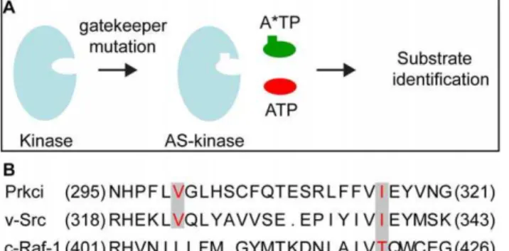
-benzyl ATPcS. The ATP analog-sensitive kinase is generated by replacing a large hydrophobic group within the ATP binding pocket (the ‘‘gatekeeper’’ residue) with a smaller residue thereby enlarging the binding pocket which allows the kinase to accept bulkier ATP analogs (Fig. 1A). The specificity of this approach has been demonstrated for various kinases, e.g. by utilizing a modified v-Src, c-Raf-1, or AMPKa2 for the identification of novel substrates [1,27,28].
To identify an ATP binding site ‘‘gatekeeper’’ residue within the Prkci ATP binding pocket, we selected suitable residues for site-directed mutagenesis based on the high evolutionary conservation of the ATP binding site in different protein kinases, including v-Src [1] and c-Raf-1 [28] that had successfully been engineered to utilizeN6
-benzyl ATP. The comparison of the primary sequence
of Prkci, c-Src, and c-Raf-1 kinase domains suggested Val300and
Ile316 of Prkci to be the most likely ‘‘gatekeeper’’ residues and
therefore to be the most suitable targets for site-directed mutagenesis (Fig. 1B).
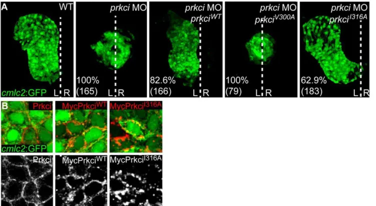
Mutant PrkciI316Ahas Normalin Vivo Biological Activity One stringent requirement for an analog-sensitive kinase is the conservation of its biological properties which includes normalin vivo functionality and substrate specificity. We therefore tested whether the PrkciV300A or PrkciI316Amutants with altered ATP binding pockets had normal biological activity. Loss of Prkci has well-characterized epithelial and organ morphogenesis defects that involve cardiac malformations and a defective neuroepithelium [9,11,12]. In functional rescue experiments, we co-injected at the one-cell stage mRNA encoding either PrkciWTor one of the two mutant Prkci proteins together with an antisense oligonucleotide morpholino (MO) for knockdown of endogenous Prkci. Whereas expression of PrkciI316A allowed normal cardiac development (63% of prkci morphant embryos rescued, n = 183) which was almost as efficient as PrkciWTexpression (83% of prkci morphants rescued, n = 166), the PrkciV300A mutant was not biologically active based on the appearance of prkci morphant cardiac phenotypes among all injected embryos (Fig. 2A) [9].
Because the PrkciI316Amutant was biologically fully functional within the heart, we anticipated that this mutant kinase should correctly localize within myocardial cells. Indeed, expression by injection at the one-cell stage of mRNA encoding Myc-tagged PrkciI316Aresulted in the correct localization of the mutant protein to the cell membrane, similar to Myc-tagged PrkciWT or endogenous Prkci (Fig. 2B). That overexpression of PrkciWT or PrkciI316Adid not cause any ectopic phenotypes strongly suggests substrate specificity (see below). Together, our results indicated that PrkciI316A is biologically functional and predominantly localizes to the correct subcellular compartment at the cell membrane, which makes this mutant kinase a strong candidate for a chemical genetic screen.
Mutant PrkciI316Acan use Bulky ATP Analogs
We next tested whether PrkciI316A could utilize bulky ATP analogs by assaying the phosphorylation efficiency and specificity of several alkylated ATPcS analogs inin vitrokinase assays. ATPcS was utilized instead of regular ATP to ensure a transfer of a phosphorothioate moiety to the phosphoacceptor hydroxyl groups of respective substrates. The substitution of sulfur in place of oxygen generates unique thiophosphorylated epitopes that, when alkylated withp-nitrobenzyl mesylate (PNBM), generate thiopho-sphoester epitopes which can be recognized by a specific monoclonal antibody [29,30]. Using the baculovirus system in Sf9 insect cells, we produced the recombinant kinases and first confirmed that PrkciWT and PrkciI316A could utilize ATPcS to phosphorylate Myelin Basic Protein (MBP) as substrate and that this modification could efficiently be detected with the anti-thiophosphoester antibody (Fig. 3). However, whereas PrkciWT accepted ATPcS, it could not utilize any of the testedN6-alkylated ATPcS analogs (N6-benzyl-, N6-phenethyl-, or N6 -cyclopentyl-ATPcS) [30] as assessed on Western blot upon thein vitrokinase assay. In contrast, PrkciI316A most efficiently utilized N6-benzyl ATPcS (Fig. 3) whereas the other two bulky ATPcS analogs were apparently not efficiently utilized (data not shown). Taken together, PrkciI316A had a normal biological function in the in vivocontext and exerted catalytic activity using a bulkyN6 -benzyl-ATP analog. Hence, PrkciI316A fulfilled the basic requirements required to identify Prkci phosphorylation targets.
Figure 1. Design of the analog-sensitive Prkci. (A) A
space-creating mutation (‘‘gatekeeper mutation’’) is introduced into the kinase ATP binding pocket which allows the analog-sensitive (AS) kinase mutant to accept a bulky ATP analog (A*TP) required for the chemical genetic identification of kinase substrates [modified after [36]]. (B) Alignment of the primary sequence of the ATP binding pocket within the kinase domains of Prkci, v-Src and c-Raf. The residues in red correspond to the amino acids mutated in v-Src [1], c-Raf-1 [28], and Prkci to enlarge the ATP binding pocket.
Evidence for PrkciI316A-mediated Thiophosphorylation in the Zebrafish Embryo
Two principal methods have been used for the enrichment of thiophosphorylated proteins inin vitrophosphorylation assays and in cell culture systems but not yet in any model organism. The
‘‘covalent capture’’ approach is based on the enrichment of thiophosphate-tagged substrates with iodoacetyl-agarose and subsequent analysis by mass spectrometry [31]. An alternative approach is based on ‘‘immunoaffinity purification’’ enrichment using the anti-thiophosphoester antibody, followed by mass spectrometry of enriched peptides [27,30]. Both methods require the utilization of ATPcS analogs for the unique labelling of substrate proteins.
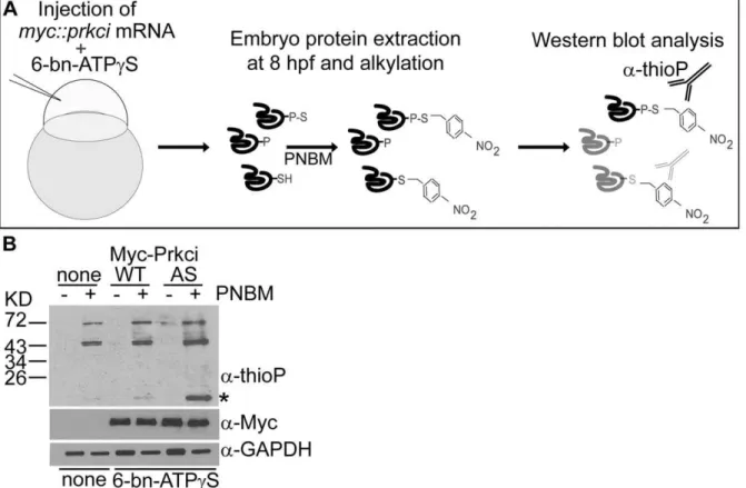
One preeminent challenge of using ATPcS analogs in in vivo
approaches is the potential toxicity of thiophosphates since such protein modifications cannot be removed by phosphatases. We first tested the toxicity of N6-benzyl ATPcS analog by injecting different concentrations (,1 nL injection volume) into wild-type embryos and found that a concentration of 200mM ofN6-benzyl ATPcS was the maximal concentration that could be injected into one-cell stage embryos without affecting development (200mM: 93.3% of embryos developed normally, n = 45; 250mM: 47.8% of embryos developed normally, n = 23; 500mM: 37.2% of embryos developed normally, n = 43). We next tested the feasibility of applying this approach in the zebrafish embryo by optimizing the conditions for introducingN6
-benzyl ATPcS together with mRNA encoding PrkciI316A by microinjection at the one-cell stage (Fig. 4A). With the concentration of 200mM N6-benzyl ATPcS (,1 nL injection volume), we observed that most embryos survived up to 32 hours post fertilization (hpf) (97.2% of PrkciI316A
Figure 2. Mutant PrkciI316Ahas normalin vivobiological activity.(A) Reconstruction of confocal Z-stack sections of embryonic hearts at 28– 30 hpf. Transgenic Tg[cmlc2:GFP]twu34one-cell stage embryos were injected withprkciMO alone or together with mRNA encoding PrkciWTor
analog-sensitive mutant forms of Prkci. Whereas the wild-type heart elongates into a heart tube and towards the left during cardiac jogging, heart development arrests at the cone stage and the heart remains at the embryonic midline inprkcimorphants. In functional rescue experiments, injection ofprkciMO together with mRNA encoding HisMyc-PrkciWTor PrkciI316Arescues heart tube elongation. In comparison, the analog-sensitive mutant
form PrkciV300Afails to rescue heart tube formation inprkcimorphants. Percentiles indicate the occurrence of the most common phenotype as depicted in the images and numbers show the total of embryos tested. White dotted line indicates the embryonic midline. L, left; R, right. (B) Membrane localization of endogenous Prkci detected with an anti-Prkci antibody and exogenous PrkciWTor PrkciI316Ain zebrafish cardiomyocytes detected with an anti-Myc antibody. Images are confocal reconstructions of single Z-stack sections of embryonic hearts marked by the transgenic reporter Tg[cmlc2:GFP]twu34
at 28–30 hpf. Expression of exogenous HisMyc-PrkciWT or HisMyc-PrkciI316A in cardiomyocytes reveals that both recombinant proteins localize to the cell membrane.
doi:10.1371/journal.pone.0040000.g002
Figure 3. Mutant PrkciI316Auses bulkyN6-benzyl ATP
cS.Kinase reaction with Myelin Basic Protein (MBP) and ATPcS orN6-benzyl ATPcS (6-bn- ATPcS), followed by PNBM alkylation. In comparison, only mutant PrkciI316Aefficiently utilizesN6
-benzyl ATPcS to thiophosphorylate MBP. Labeled MBP is detected by Western blot analysis with rabbit monoclonal anti-thiophosphoester antibody (a-thioP).
expressing embryos developed normally, n = 320), which was comparable with embryos co-injected withN6-benzyl ATPcS and mRNA encoding PrkciWT(96.5% of embryos developed normally, n = 261), or with non-injected control embryos (96.7% of embryos developed normally, n = 390). Therefore,N6-benzyl ATPcS in the range of physiological ATP levels is compatible with normal zebrafish embryogenesis.
To assess whether thiophosphorylation had occurred in the developing zebrafish embryo, suchin vivothiophosphorylated 8 hpf extracts were alkylated using PNBM, resolved by SDS-PAGE, and Western blots probed with the anti-thiophosphoester antibody. This analysis revealed that thiophosphorylation of substrates had indeed occurred in the zebrafish embryo and that PrkciI316Ahad catalyzed the selective labelling of at least one putative substrate (Fig. 4B).
Discussion
This study outlines the methodology required for an in vivo
screening approach using an analog-sensitive kinase in a multi-cellular model organism. Our work demonstrates the conditions forin vivothiophosphoester labeling of substrates using an analog-sensitive kinase and bulkyN6-benzyl ATPcS analogs. Injection of
physiological levels of bulkyN6-benzyl ATPcS does not interfere with zebrafish development even though thiophosphorylations are largely irreversible. Viability of zebrafish embryos under such conditions indicates that only a fraction of substrate proteins is modified by the analog-sensitive kinase and suchin vivo
sphorylations are detectable on Western blots. That thiopho-sphorylations are also detected in PrkciWT samples is due to unspecific utilization ofN6-benzyl ATPcS by other enzymes [31]
and to the partial degradation of N6-benzyl ATPcS to ATPcS which can be utilized by other kinases. These contaminations with unspecific thiophosphoylations highlight the need to immunopur-ify embryonic extracts and to perform comparative mass spectrometric analyses for substrate identification.
The method for the enrichment and identification of novel putative kinase targets by immunoaffinity purification with an anti-thiophosphoester antibody is well-established [27,30]. Combining this methodology with the working protocol outlined in our study will soon provide unprecedented insight into kinase signaling in multi-tissue encompassing developmental processes, the regulation of different physiological conditions, or in disease processes involving aberrant kinase signaling. Taken together, our work provides the ground for similar approaches using the analog-sensitive kinase method in this and other multicellular model organisms.
Materials and Methods
Fish Maintenance and Stocks
Zebrafish were maintained at standard conditions [32]. Embryos were kept in egg water (60mg/ml Instant Ocean Sea Salts, Aquarium Systems Inc., USA) and staged at 28.5uC [33]. The following fish strains were used: AB (wild-type), Tg[cmlc2:GFP]twu34[34].
Figure 4. Thiophosphorylation of substrate proteins by PrkciI316in the zebrafish embryo.(A) Schematic diagram of thein vivolabeling method for the selective labeling of PrkciI316Asubstrates during zebrafish development. (B)In vivothiophosphorylation in zebrafish embryos injected
at the one-cell stage with 200mMN6-benzyl-ATPcS (6-bn-ATPcS) and mRNA encoding either PrkciWTor PrkciI316A(AS). Western blot analysis with
rabbit monoclonal anti-thiophosphoester (a-thioP) C51-8 antibody (Epitomics) of 80% epiboly (6–8hpf) samples alkylated with 2.5 mM PNBM reveals a selectively labeled protein in the PrkciI316A(AS) sample (asterisk).
RNA and Antisense Oligonucleotide Morpholino Injections
Constructs were transcribed using the SP6 mMessage mMa-chine kit (Ambion). Tg[cmlc2:GFP]twu34embryos were injected with 2.5 ng of prkci MO [12]. For rescue experiments 100 pg of mRNAs were injected. The heart morphology was assessed at 24hpf. Data presented are the means of at least 2 independent experiments. For in vivo labeling, 200 pg of mRNA encoding HisMyc-PrkciWT or HisMyc-PrkciI316AmRNA were injected at the one-cell stage.
The antisense oligonucleotide morpholino was purchased from Gene Tools, LLC, USA. prkci MO (59R 39): TGTCCCGCAGCGTGGGCATTATGGA [12].
DNA Constructs and Site-directed Mutagenesis
Both constructs encoding wt and mutant forms of Prkci were produced by PCR amplification from a full length cDNA template, pCS2+ HisMyc::prkci [12]. Site directed mutagenesis was performed using the QuickChangeTM XL Site-Directed mutagenesis kit (Stratagene). Primer sequences are available upon request.
Protein Extraction from Zebrafish Embryos
Zebrafish embryos protein extraction was performed as previously described [35]. Embryos were dechorionated with pronase solution in E2 medium in Petri dishes coated with 1% agarose. After washes with E2 medium, embryos were transferred to 1.5 ml tubes. The yolk was disrupted by pipetting with a 1 ml pipet and vortexing for 30 seconds at 1100 rpm in deyolking buffer. Embryos were pelleted several times after washes in washing buffer at 3000 rpm for 30 seconds. Pelleted embryos were homogenized in the appropriated lysis buffer depending on the following experiment.
Recombinant Protein Expression in Insect Cells
To express recombinant PrckiWTor PrkciI316Ain Sf9 insect cells (Sigma), Bac-to-BacHBaculovirus Expression System (Invitrogen) was used according to the manufacturers’ protocol. HisMyc::prk-ciWTandHisMyc::prkciI316cDNAs were cloned into pFastBacTM1. Primer sequences are available upon request.
Whole-mount Immunohistochemistry and Confocal Imaging
Whole-mount antibody stainings were performed as previously described [9]. The following antibodies were used: rabbit anti-aPKC (1:100, Santa Cruz SC-216), mouse anti-Myc (1:200, Invitrogen), goat anti-rabbit RRX (1:250, Jackson
ImmunoRe-search Laboratories), goat anti-mouse Cy5 (1:250, Jackson ImmunoResearch Laboratories). For imaging, samples were embedded in SlowFadeHGold antifade reagent (Invitrogen) under a binocular microscope (Leica). Confocal images were obtained with a Zeiss LSM 510 Meta confocal microscope using 40X or 63X objectives. Zeiss LSM 510 software was used to record images. Images were processed using Photoshop (Adobe).
In vitrokinase Assay
Forin vitrokinase assays 50mg of MBP were incubated at 30uC for 30 minutes with 5mg of recombinant HisMyc-PrckiWT or HisMyc-PrkciI316A and 500mM ATPcS or N6-benzyl ATPcS (BioLog, B072-05) in kinase buffer [25mM Tris-HCl, pH 7.5, 25 mM NaCl, 10 mM MgCl2, 1 mM EGTA, protease inhibitor
cocktail (Roche)]. The other bulky ATP analogs (N6 -phenethyl-ATPcS,N6-cyclopentyl-ATPcS) were generated as described [30]. The kinase reaction was stopped by adding 4x SDS loading buffer and boiling at 95uC for 5 minutes. Samples were analyzed by Western blot using the following antibodies: mouse anti-Myc (1:1000, Invitrogen), anti-thiophosphoester rabbit polyclonal antibody (1:5000 Epitomics), rabbit anti-GAPDH (1:1000), goat mouse HRP (1:5000, Jackson ImmunoResearch), goat anti-rabbit HRP (1:10000, Jackson ImmunoResearch).
In VivoThiophosphorylation in Zebrafish Embryos For in vivo thiophosphorylation 200 pg of mRNA encoding HisMyc-PrkciWTor HisMyc-PrkciI316Awere injected into the yolk at the one-cell stage together with approximately 1 nL of 200mM
N6-benzyl ATPcS. Embryonic protein extracts were prepared at 8 hpf, and pelleted embryos were homogenized in RIPA buffer. Alkylation was perfomed for 2 hours at RT with 2.5 mM PNBM [30]. Samples were analyzed by Western blot using anti-thiopho-sphoester rabbit monoclonal C51-8 antibody (1:5000 Epitomics).
Acknowledgments
We would like to thank R. Fechner, N. Lawson, J. Richter, H.J. Tsai for providing reagents, fish stocks or other support. We would like to thank J. Malkewitz for help with the generation of recombinant baculovirus and protein expression. W. Tegge generously provided thiophosphopeptide. Thanks to members of the Abdelilah-Seyfried and Selbach labs for their comments on the manuscript.
Author Contributions
Conceived and designed the experiments: ECU NH MK MS SAS. Performed the experiments: ECU NH MK SD. Analyzed the data: ECU MK NH MS SAS. Contributed reagents/materials/analysis tools: KS JJA. Wrote the paper: ECU SAS.
References
1. Liu Y, Shah K, Yang F, Witucki L, Shokat KM (1998) Engineering Src family protein kinases with unnatural nucleotide specificity. Chem Biol 5: 91–101. 2. Bakkers J, Verhoeven MC, Abdelilah-Seyfried S (2009) Shaping the zebrafish
heart: from left-right axis specification to epithelial tissue morphogenesis. Dev Biol 330: 213–220.
3. Hirai T, Chida K (2003) Protein kinase Cf(PKCf): activation mechanisms and cellular functions. J Biochem 133: 1–7.
4. St. Johnston D, Sanson B (2011) Epithelial polarity and morphogenesis. Curr Opin Cell Biol 23: 540–546.
5. Suzuki A, Akimoto K, Ohno S (2003) Protein kinase C lambda/iota (PKClambda/iota): a PKC isotype essential for the development of multicellular organisms. J Biochem 133: 9–16.
6. Murray NR, Kalari KR, Fields AP (2011) Protein kinase Ciota expression and oncogenic signaling mechanisms in cancer. J Cell Physiol 226: 879–887. 7. Suzuki A, Ohno S (2006) The PAR-aPKC system: lessons in polarity. J Cell Sci
119: 979–987.
8. Etienne-Manneville S, Hall A (2003) Cell polarity: Par6, aPKC and cytoskeletal crosstalk. Curr Opin Cell Biol 15: 67–72.
9. Horne-Badovinac S, Lin D, Waldron S, Schwarz M, Mbamalu G, et al. (2001) Positional cloning of heart and soul reveals multiple roles for PKC lambda in zebrafish organogenesis. Curr Biol 11: 1492–1502.
10. Peterson RT, Mably JD, Chen JN, Fishman MC (2001) Convergence of distinct pathways to heart patterning revealed by the small molecule concentramide and the mutationheart-and-soul. Curr Biol 11: 1481–1491.
11. Horne-Badovinac S, Rebagliati M, Stainier DY (2003) A cellular framework for gut-looping morphogenesis in zebrafish. Science 302: 662–665.
12. Rohr S, Bit-Avragim N, Abdelilah-Seyfried S (2006) Heart and soul/Prkci and Nagie oko/Mpp5 regulate myocardial coherence and remodeling during cardiac morphogenesis. Development 133: 107–115.
13. Betschinger J, Mechtler K, Knoblich JA (2003) The Par complex directs asymmetric cell division by phosphorylating the cytoskeletal protein Lgl. Nature 422: 326–330.
15. Galli M, Munoz J, Portegijs V, Boxem M, Grill SW, et al. (2011) aPKC phosphorylates NuMA-related LIN-5 to position the mitotic spindle during asymmetric division. Nat Cell Biol 13: 1132–1138.
16. Hutterer A, Betschinger J, Petronczki M, Knoblich JA (2004) Sequential roles of Cdc42, Par-6, aPKC, and Lgl in the establishment of epithelial polarity during Drosophila embryogenesis. Dev Cell 6: 845–854.
17. Morais-de-Sa E, Mirouse V, St. Johnston D (2010) aPKC phosphorylation of Bazooka defines the apical/lateral border in Drosophila epithelial cells. Cell 141: 509–523.
18. Plant PJ, Fawcett JP, Lin DC, Holdorf AD, Binns K, et al. (2003) A polarity complex of mPar-6 and atypical PKC binds, phosphorylates and regulates mammalian Lgl. Nat Cell Biol 5: 301–308.
19. Yamanaka T, Horikoshi Y, Sugiyama Y, Ishiyama C, Suzuki A, et al. (2003) Mammalian Lgl forms a protein complex with PAR-6 and aPKC independently of PAR-3 to regulate epithelial cell polarity. Curr Biol 13: 734–743. 20. Lin D, Edwards AS, Fawcett JP, Mbamalu G, Scott JD, et al. (2000) A
mammalian PAR-3-PAR-6 complex implicated in Cdc42/Rac1 and aPKC signalling and cell polarity. Nat Cell Biol 2: 540–547.
21. Nagai-Tamai Y, Mizuno K, Hirose T, Suzuki A, Ohno S (2002) Regulated protein-protein interaction between aPKC and PAR-3 plays an essential role in the polarization of epithelial cells. Genes Cells 7: 1161–1171.
22. Nishimura T, Kaibuchi K (2007) Numb controls integrin endocytosis for directional cell migration with aPKC and PAR-3. Dev Cell 13: 15–28. 23. Smith CA, Lau KM, Rahmani Z, Dho SE, Brothers G, et al. (2007)
aPKC-mediated phosphorylation regulates asymmetric membrane localization of the cell fate determinant Numb. EMBO J 26: 468–480.
24. Atwood SX, Prehoda KE (2009) aPKC phosphorylates Miranda to polarize fate determinants during neuroblast asymmetric cell division. Curr Biol 19: 723–729.
25. Djiane A, Yogev S, Mlodzik M (2005) The apical determinants aPKC and dPatj regulate Frizzled-dependent planar cell polarity in the Drosophila eye. Cell 121: 621–631.
26. Hao Y, Du Q, Chen X, Zheng Z, Balsbaugh JL, et al. (2010) Par3 controls epithelial spindle orientation by aPKC-mediated phosphorylation of apical Pins. Curr Biol 20: 1809–1818.
27. Banko MR, Allen JJ, Schaffer BE, Wilker EW, Tsou P, et al. (2011) Chemical Genetic Screen for AMPKa2 Substrates Uncovers a Network of Proteins Involved in Mitosis. Mol Cell 44: 878–892.
28. Hindley AD, Park S, Wang L, Shah K, Wang Y, et al. (2004) Engineering the serine/threonine protein kinase Raf-1 to utilise an orthogonal analogue of ATP substituted at the N6 position. FEBS Lett 556: 26–34.
29. Allen JJ, Lazerwith SE, Shokat KM (2005) Bio-orthogonal affinity purification of direct kinase substrates. J Am Chem Soc 127: 5288–5289.
30. Allen JJ, Li M, Brinkworth CS, Paulson JL, Wang D, et al. (2007) A semisynthetic epitope for kinase substrates. Nat Methods 4: 511–516. 31. Blethrow JD, Glavy JS, Morgan DO, Shokat KM (2008) Covalent capture of
kinase-specific phosphopeptides reveals Cdk1-cyclin B substrates. Proc Natl Acad Sci U S A 105: 1442–1447.
32. Westerfield M. (1994) The Zebrafish Book. Eugene: University of Oregon Press. 33. Kimmel CB, Ballard WW, Kimmel SR, Ullmann B, Schilling TF (1995) Stages
of embryonic development of the zebrafish. Dev Dyn 203: 253–310. 34. Huang CJ, Tu CT, Hsiao CD, Hsieh FJ, Tsai HJ (2003) Germ-line transmission
of a myocardium-specific GFP transgene reveals critical regulatory elements in the cardiac myosin light chain 2 promoter of zebrafish. Dev Dyn 228: 30–40. 35. Link V, Shevchenko A, Heisenberg CP (2006) Proteomics of early zebrafish
embryos. BMC Dev Biol 6: 1.