FILIPPE ELIAS DE FREITAS SOARES
PRODUÇÃO, PURIFICAÇÃO E IDENTIFICAÇÃO DE ENZIMAS EXTRACELULARES DE FUNGOS NEMATÓFAGOS E SUAS
ATIVIDADES NEMATICIDAS
Tese apresentada à Universidade Federal de Viçosa, como parte das exigências do Programa de Pós-graduação em Bioquímica Agrícola, para obtenção do título de Doctor Scientiae.
VIÇOSA
Fichi citilográfici prepiridi peli Biblioteci Centril di Universidide Federil de Viçosi - Câmpus Viçosi
T
STares, Filippe Elias de Freitas, 1987-S676p
2014 extracelulares de fungTs nematófagTs e suas atividadesPrTduçãT, purificaçãT e identificaçãT de enzimas nematicidas / Filippe Elias de Freitas STares. - ViçTsa, MG, 2014.
xv, 111f. : il. ; 29 cm.
OrientadTr : JTsé HumbertT de Queiróz.
Tese (dTutTradT) - Universidade Federal de ViçTsa. Inclui bibliTgrafia.
1. FungTs nematófagTs. 2. Enzimas. 3. FungTs -CTntrTle biTlógicT. I. Universidade Federal de ViçTsa. DepartamentT de BiTquímica e BiTlTgia MTlecular. PrTgrama de Pós-graduaçãT em BiTquímica AgrícTla. II. TítulT.
CDD 22. ed. 579.5
"Não há assuntos pouco interessantes; apenas há pessoas pouco interessadas."
AGRADECIMENTOS
À Deus, pelo dom da vida e bençãos ininterruptas.
À Universidade Federal de Viçosa, pela oportunidade e acolhimento, contribuindo de forma satisfatória para meu crescimento profissional e pessoal.
Ao Conselho Nacional de Desenvolvimento Científico e Tecnológico (CNPq) – pela concessão da bolsa de estudo que viabilizou meus estudos e pesquisa e pelo financiamento do projeto de pesquisa derivado dessa tese.
À Fundação de Amparo à Pesquisa do Estado de Minas Gerais (FAPEMIG)– pelo financiamento do projeto de pesquisa derivado dessa tese.
Aos meus orientadores, Professores José Humberto de Queiróz, Fábio Ribeiro Braga e Jackson Victor de Araújo pela orientação, confiança e oportunidade, e, sobretudo pela amizade construída.
Ao Professor Walter dos Santos Lima, do Departamento de Parasitologia da Universidade Federal de Minas Gerais, pela concessão das larvas de Angiostrongylus vasorum.
Aos meus pais, Denise de Freitas Soares e Benedito Elias Soares, pela força, pelo incentivo, pela vida! Vocês são responsáveis por essa conquista.
Ao meu irmão, Denilson Elias de Freitas Soares, pelo carinho, pela amizade.
Aos meus avós, Waldir e Maria Aparecida e aos meus padrinhos Date e Luiz, que sempre torceram por mim!
BIOGRAFIA
FILIPPE ELIAS DE FREITAS SOARES, filho de Benedito Elias Soares e Denise de Freitas Soares, nasceu em Muriaé – Minas Gerais, em 15 de fevereiro de 1987.
Em maio de 2006, iniciou o curso de Bacharelado em Bioquímica na Universidade Federal de Viçosa (UFV) em Viçosa – Minas Gerais, concluindo-o em dezembro de 2010.
Em março de 2011, ingressou no Programa de Mestrado em Bioquímica Agrícola na UFV, concluindo os requisitos necessários para obter o título de Magister Scientiae, defendendo a dissertação em julho de 2012.
SUMÁRIO
LISTA DE TABELAS... vii xi xii xiv 1 4 5 7 8 9 16 23 26 31 32 34 35 37 38 41 45 47 48 49 51 55 58 LISTA DE FIGURAS... RESUMO... ABSTRACT... 1. INTRODUÇÃO GERAL... 2. OBJETIVOS... CAPÍTULO 1 - Nematicidal activity of three novel extracellular proteases of the nematophagous fungus Monacrosporium sinense ...……... Abstract ...……....………...……... Introduction... Materials and methods...………..……..……….... Results... ………....…... Discussion...………....…... References... ………....…... CAPÍTULO 2 - The nematophagous fungus Monacrosporium thaumasium and its nematicidal activity on Angiostrongylus vasorum ………..…….... Abstract... Introduction….……...…...……..………..…...………... Materials and methods …...…...……… Results………....……...………...………... Discussion...………...………...………...
References....………....………...……….….
CAPÍTULO 3 - Proteolytic activity of the nematophagous fungus Arthrobotrys sinensis on Angiostrongylus vasorum larvae……...
Abstract... Introduction.….……...…...……..………..…...………... Materials and methods …...…...……… Results... ………....…... Discussion...………....…...
References………....………...……….………
CAPÍTULO 5 - Statistical screening for the chitinase production by nematophagous fungi from Monacrosporium genus ………... Abstract... Introduction.….……...…...……..………..…...………... Materials and methods …...…...……… Results... ………....…... Discussion...………....…... References………....………...……….…... CAPÍTULO 6 - Nematicidal action of chitinases produced by the fungi Monacrosporium thaumasium under laboratorial conditions ... Abstract... Introduction.….……...…...……..………..…...………... Materials and methods …...…...……… Results... ………....…... Discussion...………....…... References………....………...……….…...
3. REFERÊNCIAS...………...………...
4. CONCLUSÕES GERAIS………...………...
LISTA DE TABELAS CAPÍTULO 1
Table 1 High (+1) and low (-1) levels of the seven analyzed variables (moisture (%), pH, incubation time, temperature, glucose (g/l), yeast extract (g/l) and number of conidia) in the Plackett-Burman statistical design
Pág. 11
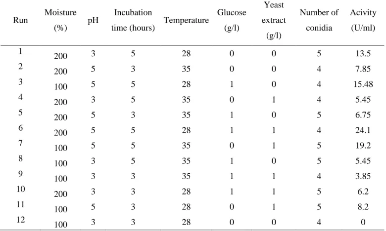
Table 2 Matrix of the Plackett-Burman experimental design of protease production (U/ml) by the nematophagous fungus Monacrosporium sinense (SF53)
Pág. 12
Table 3 Analysis of the studied variables (moisture (%), pH, incubation time, temperature, glucose (g/l), yeast extract (g/l) and number of conidia) in the Plackett-Burman statistical design
Pág. 13
Table 4 Experimental design with 13 runs used for the realization of response surface methodology using two variables (pH and incubation time) each with 5 levels, and their respective values of proteolytic activity
Pág. 17
Table 5 Analysis of variance for the response equation developed in the protease production by the nematophagous fungus Monacrosporium sinense (SF53) in solid-state-fermentation
Pág. 18
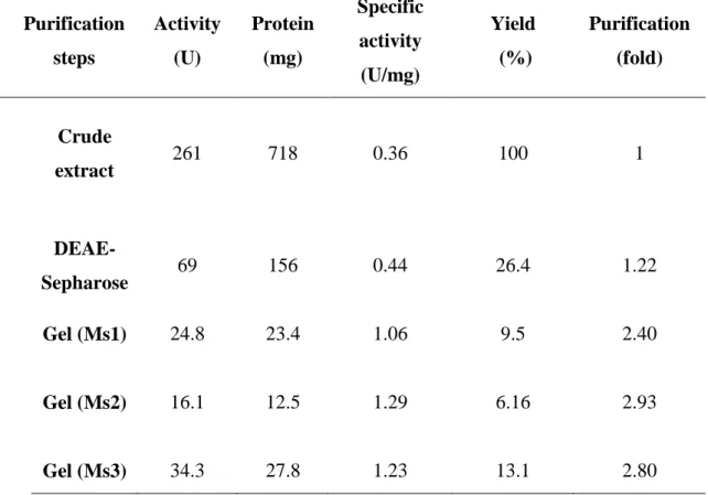
Table 6 Purification procedure of the proteases (Ms1, Ms2 and Ms 3) of Monacrosporium sinense (SF53)
Pág. 21
CAPÍTULO 5
Table 1 High (+1) and low (-1) levels of the five variables (chitin, colloidal chitin, carbon source (glucose), nitrogen source (NaNO3), moisture) in Plackett-Burman statistical design
Pág. 80
Table 2 Matrix of the Plackett-Burman experimental desig chitinase production (U/ml) by the nematophagous fu Monacrosporium thaumasium (NF34)
Pág. 81
Table 3 Matrix of the Plackett-Burman experimental design of chitinase production (U/ml) by the nematophagous fungus Monacrosporium sinense (SF53)
Table 4 Analysis of the factors studied in the Plackett-Burman statistical design of chitinase production by nematophagous fungus Monacrosporium thaumasium (NF34)
Pág. 83
Table 5 Analysis of the factors studied in the Plackett-Burman statistical design of chitinase production by nematophagous fungus Monacrosporium sinense (SF53)
Pág. 85
CAPÍTULO 6
Table 1 Purification process of the chitinases of Monacrosporium thaumasium (NF34)
LISTA DE FIGURAS CAPÍTULO 1
Fig. 1 Response surface curve of protease production by the nematophagous fungus Monacrosporium sinense (SF53) in solid state fermentation
Pág. 19
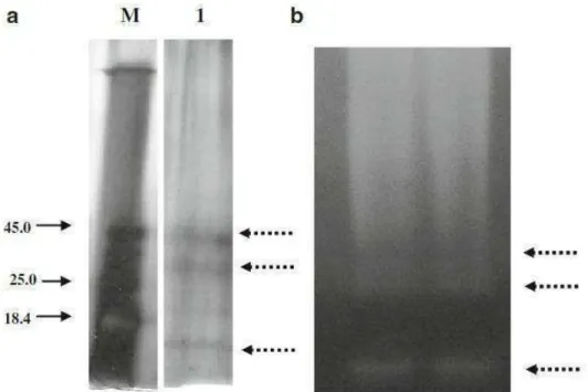
Fig. 2 a- Purification analysis of the proteases produced by Monacrosporium sinense (SF53) in solid-state-fermentation through SDS-PAGE 10% gel b. Zymogram of the crude enzymatic extract produced by M. sinense (SF53) in solid state fermentation which suggests the production of at least three different proteases (Figure 2a,b). Line M: molecular weight marker (kDa); Line 1: Purified proteases. Dashed arrows: a. Proteases produced by Monacrosporium sinense (SF53). b. Action of the enzymes observed by the formation of white halos
Pág. 20
Fig. 3 a. Effect of pH on relative activity (%) of proteases (Ms1, Ms2 and Ms3) of Monacrosporium sinense (SF53), in Tris-HCl 50 mM buffer (pHs 7.0, 8.0 and 9.8). The assay temperature was 60 °C; b. Effect of temperature on relative activity (%) of proteases (Ms1, Ms2 and Ms3) of M. sinense (SF53). Different values of temperature (40 ºC, 50 ºC and 60 ºC) were used at the pH of maximum activity obtained in the previous assay
Pág. 21
CAPÍTULO 2
Fig. 1 Proteolytic profile of the crude extract produced by the fungus Monacrosporium thaumasium (NF34a). White arrows: protease with molecular weight of approximately 40 KDa
Pág. 38
CAPÍTULO 3
Fig. 1 Zymogram of the proteases produced by Arthrobotrys sinensis (SF53) in liquid medium. Through analysis of the zymograms, a single halo of digestion at the beginning of the gel was observed, suggesting that the three proteases of SF53 are produced in an enzymatic complex of large molecular weight.
Pág. 52
Fig. 2 Nematicidal activity of proteases from nematophagous fungus Arthrobotrys sinensis (SF53) on first stage larvae of Angiostrongylus vasorum after 24 hours (treated group (a) and control group (b)).
CAPÍTULO 4
Fig. 1 ab – Specimens of the nematode Dioctophyma renale, male (white arrow) and female (black arrow), recovered during the realization of a total nefrectomia of a young dog parasitized in the sector of clinical and surgery of Department of Veterinary of the University of Viçosa. Bars: A – 4 cm; B – 2.6 cm.
Pág. 63
Fig. 2 Percentages of the ovicidal activity for the effects of types 1, 2 and 3 to 3, 5 and 7 days of interaction of chlamydospores of the fungus Pochonia chlamydosporia (VC4) at different concentrations (500, 1000, 1500 and 2000) on eggs of Dioctophyma renale and the control group. The asterisk denotes a difference (p<0.01)
Pág. 66
Fig. 3 a-f - Hyphae of the fungus Pochonia chlamydosporia, a-d (black arrow), and the eggs of Dioctophyma renale destroyed, e-f (white arrow) at the end of the experiment. Light microscope, 40x objective. Bars: (a) 67 µm. (b) 201 µm; (c) 201 µm; (d) 301 µm; (e) 268 µm and (f) 235 µm.
Pág. 67
Fig. 4 Dioctophyma renale egg destroyed (black arrow) after previous contact with both classes of enzymes (chitinases and proteases) produced by Pochonia chlamydosporia. Intact egg of Dioctophyma renale (white arrow). Light microscope, 10x objective. Bars: 168 µm.
Pág. 68
CAPÍTULO 5
Fig. 1 A–B. Pareto Charts of the studied variables in the Plackett–Burman statistical design.
Pág. 84
Fig. 2 Effect of pH on chitinase activity of the nematophagous fungi Monacrosporium thaumasium (NF34) (black) and M. sinense (SF53) (gray)
Pág. 86
CAPÍTULO 6
Fig. 1 Purification analysis of the chitinases produced by Monacrosporium thaumasium (NF34). Line M: molecular weight marker (kDa); Arrows: Purified chitinases
Pág. 99
Fig. 2 a. Effect of pH on activity (%) of Monacrosporium thaumasium (NF34) chitinases, using the following buffers: 50 mM sodium acetate (pH 3.5, 4.5 and 5.5) and 50 mM Tris-HCl (pH 7.0 and 8.0). The test
RESUMO
SOARES, Filippe Elias de Freitas, D.Sc., Universidade Federal de Viçosa, novembro de 2014. Produção, purificação e identificação de enzimas extracelulares de fungos nematófagos e suas atividades nematicidas. Orientador: José Humberto de Queiróz. Coorientadores: Fábio Ribeiro Braga e Jackson Victor de Araújo.
ABSTRACT
SOARES, Filippe Elias de Freitas, D.Sc., Universidade Federal de Viçosa, November, 2014. Production, purification and identification of extracellular enzymes from nematophagous fungi and their nematicidal activities. Adviser: José Humberto de Queiróz. Co-adviser: Fábio Ribeiro Braga e Jackson Victor de Araújo.
1. INTRODUÇÃO GERAL
O controle biológico é uma das possíveis aplicações biotecnológicas de enzimas fúngicas, evitando o uso de produtos químicos que possam gerar possíveis resíduos. Nesse contexto, o controle biológico realizado com fungos nematófagos é uma alternativa que apresenta grande potencial. Os fungos nematófagos são inimigos naturais de nematoides que podem capturar, matar e digerir os ovos e larvas infectantes de nematoides utilizando hifas especialmente modificadas chamadas armadilhas. Esses fungos produzem enzimas extracelulares envolvidas em diversas etapas da infecção, como liberação de nutrientes para o crescimento do microrganismo, penetração da cutícula pela degradação proteica e digestão do tecido hospedeiro (Mendoza de Gives et al. 2003; Yang et al. 2013; Braga e Araújo, 2014).
Os nematoides do gênero Panagrellus têm distribuição cosmopolita e são utilizados como modelo de experimentação envolvendo sua interação com fungos nematófagos (Gomes et al. 1999; Braga et al 2012a). Outro nematoide de interesse é o Angiostrongylus vasorum (Baillet, 1866) Kamensky, 1905, um protostrongilídeo parasito de cães domésticos e canídeos silvestres. O parasito adulto pode ser encontrado no ventrículo direito, artérias pulmonares e suas ramificações, com conseqüências graves para o hospedeiro definitivo. As larvas desse nematoide podem se encontrar livres no ambiente, levando a possibilidade de infecção humana, uma vez que outros parasitos do gênero Angiostrongylus são comprovadamente zoonóticos (Barçante et al., 2003; Oliveira-Jr et al., 2006). Pode-se destacar ainda o verme renal gigante, Dioctophyma renale, o maior nematoide conhecido. (Bowman et al., 2006). Em seres humanos, a infecção ocorre devido ao hábito de consumir peixe, carne de rã cru, ou anelídeos aquáticos com as larvas infectantes (L3). Nesse sentido, medidas alternativas que possam ser empregadas no combate à disseminação ambiental, destes parasitos e suas formas infectantes são importantes, como o uso de fungos nematófagos.
Braga et al. (2012b) purificaram uma serino protease produzida pelo fungo nematófago Duddingtonia flagrans (AC001) com atividade larvicida sobre larvas infectantes de ciatostomíneos, nematoide parasito gastrintestinal de equinos. Anteriomente, Lopez-Llorca (1990) demonstrou que o fungo nematófago Verticillium suchlasporium produz a protease P32 com ação sobre ovos de fitonematoides. Segers et al. (1994) mostraram que o mesmo fungo secretou outra protease (VCP1) que hidrolisou as proteínas externas da casca do ovo do nematoide parasita de planta Meloidogyne incognita, expondo a camada de quitina. Khan et al. (2004) demonstraram que serino proteases secretadas por Paecilomyces lilacinus foram capazes de interromper o desenvolvimento de ovos de M. javanica e reduzir o número de juveniles. Proteases e quitinases purificadas também demonstraram atividade nematicida sobre Haemonchus contortus, parasita gastrintestinal de ruminantes domésticos (Mansfield et al., 1992). Braga et al. (2010) demonstraram que o extrato bruto enzimático de Pochonia chlamydosporia, fungo nematófago do grupo ovicida, apresentou atividade efetiva sobre larvas e ovos de nematoides.
Os fungos nematófagos podem ser classificados em quatro grupos: endoparasitas, predadores, ovicidas ou oportunistas, e produtores de metabólitos tóxicos, que produzem metabólitos secundários nematicidas (Yang et al. 2013). Pochonia chlamydosporia produz metabólitos do tipo aurovertina, com atividade nematotóxica sobre o fitonematoide Meloidogyne incognita. Os metabólitos secundários produzidos por fungos nematófagos também possuem um grande potencial para serem utilizados como controladores biológicos (Niu et al. 2010).
fungos nematófagos. A ação patogênica desses fungos contra nematoides ainda precisa de maiores estudos para a sua elucidação (Braga e Araújo, 2014; Yang et al., 2013).
Além disso, para que o controle biológico com fungos nematófagos seja incorporado em um sistema industrial de produção, o mesmo deve ser estudado em relação a sua atividade predatória, capacidade de produzir enzimas extracelulares de importância no processo de infecção, bem como a ação destas enzimas sobre nematoides dos mais diversos gêneros (Araújo et al., 2004; Braga et al., 2010a,b; Soares et al., 2012).
2. OBJETIVOS 2.1. Objetivo Geral
Otimizar a produção, purificar e testar a ação nematicida das enzimas extracelulares dos fungos nematófagos Monacrosporium thaumasium (NF34), M. sinense (SF53) e Pochonia chlamydosporia (VC4).
2.2. Objetivos Específicos
Otimizar as condições de cultura dos fungos nematófagos Monacrosporium thaumasium (NF34) e M. sinense (SF53) para uma elevada produção de enzimas utilizando o planejamento fatorial Plackett-Burman e metodologia de superfície de resposta;
Purificar as enzimas produzidas por Monacrosporium thaumasium (NF34), M. sinense (SF53);
CAPÍTULO 1
Nematicidal activity of three novel extracellular proteases of the nematophagous fungus Monacrosporium sinense
Nematicidal activity of three novel extracellular proteases of the nematophagous
fungus Monacrosporium sinense
Filippe E. F. Soares1*; Fabio R. Braga2,3; Jackson V. Araújo2a; Hugo L.A. Geniêr1; Angélica S. Gouveia1; José H. Queiroz1
1 Departamento de Bioquímica e Biologia Molecular. Universidade Federal de Viçosa, Viçosa-MG. Cep: 3657000.
2. Departamento de Veterinária, Universidade Federal de Viçosa, Viçosa-MG. Cep: 3657000.
3 Universidade Vila-Velha, Vila Velha, Espirito Santo. Cep: 29102206. (a) CNPq scholarship. (*) Corresponding author Fax: +55 (31) 3899-3048. E-mail:
Abstract
Extracellular proteases are an important virulence factor for the nematophagous fungi Monacrosporium. The objective of this study was to optimize, purify, partially characterize and to evaluate the nematicidal activity of the proteases produced by the nematophagous fungus Monacrosporium sinense (SF53) by solid state fermentation. Wheat bran was used as substrate for protease production. The variables moisture, pH, incubation time, temperature, glucose, yeast extract and the number of conidia were tested for their influences on protease production by SF53. To determine the optimal level of the selected variables the Central Composite Design (CCD) was applied. The crude extract obtained was purified in two steps, an ion exchange chromatography and a gel excision. SDS-PAGE and zymogram were performed for analysis of the purification process. Proteolytic activity was also tested at different pHs and temperatures. In the in vitro assay, the nematicidal activity of the three proteases was evaluated. pH and
incubation time showed a significant effect (p <0.05) on production of protease. The highest value of activity was 38.0 (U/ml) under the conditions of pH 5.0 and incubation time of 211 hours. SF53 produced three different proteases (Ms1, Ms2 and Ms3) which were directly purified from the zymogram. Ms1, Ms2 and Ms3 showed the following percentage of reduction (p <0.05) on the number of P. redivivus compared to control after 24 hours: 76.8%; 68.1% and 92.1%. This is the first report of the use of proteases of the isolate SF53 on a phytonematode, which may be a research tool in future works.
Keywords: Nematophagous fungi; Monacrosporium sinense; Panagrellus
Introduction
Stock and Nadler (2006) have report that the phytonematode Panagrellus has global distribution. Their species have been described in almost all continents, with the exception of Antarctica and Australia. The species P. redivivus described by Linnaeus (1767), has been used as an experimental model for nematicidal assays with nematophagous fungi, due to their ease of maintenance and use in biological control assays. In this sense, Cardoso et al. (2009) demonstrated the activity of nematophagous fungi against this phytonematode, registering potential effectiveness. However, there are no reports of the enzymatic activity of nematophagous fungi on P. redivivus.
Nematophagous fungi can be exploited for industrial production due to their ability to grow on solid substrates and produce a wide range of extracellular enzymes (Araújo et al. 2000; Sharma et al. 2007). In previous works Soares et al. (2012a, b) showed that nematophagous fungi from the Monacrosporium genus produce proteases with possible nematicidal activity, which may be exploited in the future as biocontroller. However, many factors such as sources of carbon and nitrogen, the quantity of inoculum; initial pH, temperature, moisture and time of fermentation have been reported as influent parameters in the fermentation process and therefore in the production of proteases.
Thus, the objective of this study was to optimize, purify, partially characterize and to evaluate the nematicidal activity of the proteases produced by the nematophagous fungus Monacrosporium sinense (SF53) by solid state fermentation.
Materials and methods
Fungi
An isolate of the nematophagous fungus M. sinense (SF53) originating from soil of Brazil, in the city of Viçosa, Zona da Mata of Minas Gerais, was used. This isolate has been kept in test tubes at 4 °C containing 2% corn-meal-agar (2% CMA) by continuous transfer in the dark for 10 days.
Panagrellus redivivus
The free-living nematode P. redivivus is originated from the Laboratory of Parasitology, Department of Veterinary Medicine, Federal University of Viçosa, Viçosa, Minas Gerais, Brazil. For its maintenance, 1.0 ml of an aqueous suspension with approximately 1000 P. redivivus was inoculated in each sterile polystyrene petri dish containing thin oat flakes medium and water in a 1:1 ratio, according to Araujo et al. (2004).
Production of conidia and culture conditions
Conidia produced by M. sinense (SF53) were placed in sterile saline to prepare
FeSO4 (0.001 g/l) and CuSO4 (0.50 mg/l), according to modified methodology of Braga
et al. (2011).
Solid-state fermentation (SSF)
Wheat bran was used as substrate for protease production. Fermentations were conducted in Erlenmeyer flasks (125 ml) containing 5 g of wheat bran. The moisture was adjusted using a liquid medium, by varying the volume/weight ratio according to the following experimental design. Each flask was covered with hydrophobic cotton and autoclaved at 121 °C for 15 min. After cooling, each flask was inoculated with conidia suspension previously prepared and incubated at 28 °C in BOD for different time periods defined by the experimental design.
Statistical design
Plackett-Burman experimental design (Plackett and Burman 1946) is used to analyze and evaluate the important factors that affect a polynomial first-order response. In the present work, this statistical design was used to scan some variables inherent to the fermentation regarding their influences on protease production by the fungus M. sinense (SF53). The variables moisture, pH, incubation time, temperature, glucose,
Table 1. High (+1) and low (-1) levels of the seven analyzed variables (moisture (%),
pH, incubation time, temperature, glucose (g/l), yeast extract (g/l) and number of conidia) in the Plackett-Burman statistical design
Variables High level (+1) Low level (-1)
Moisture (%) 200 100
pH 5 3
Incubation time (hours) 5 3
Temperature 28 35
Glucose (g/l) 1 0
Yeast extract (g/l) 1 0
Number of conidia 105 104
Table 2. Matrix of the Plackett-Burman experimental design of protease production (U/ml) by the
nematophagous fungus Monacrosporium sinense (SF53)
Run Moisture (%) pH
Incubation
time (hours) Temperature
Glucose (g/l) Yeast extract (g/l) Number of conidia Acivity (U/ml) 1
200 3 5 28 0 0 5 13.5
2
200 5 3 35 0 0 4 7.85
3
100 5 5 28 1 0 4 15.48
4
200 3 5 35 0 1 4 5.45
5
200 5 3 35 1 0 5 6.75
6
200 5 5 28 1 1 4 24.1
7
100 5 5 35 0 1 5 19.2
8
100 3 5 35 1 0 5 5.45
9
100 3 3 35 1 1 4 3.85
10
200 3 3 28 1 1 5 6.2
11
100 5 3 28 0 1 5 8.2
12
100 3 3 28 0 0 4 0
Response surface methodology
To determine the optimal level of the selected variables from the statistical design Plackett-Burman (pH and incubation time) and investigate their interactions, the response surface methodology was applied using the Central Composite Design (CCD). The effect of these significant variables on enzyme activity was studied in 5 experimental levels: -α, -1, 0, 1, + α, where α=2n/4, n is equal to the number of variables and 0 corresponds to the central point.
Table 3. Analysis of the studied variables (moisture (%), pH, incubation time, temperature, glucose
(g/l), yeast extract (g/l) and number of conidia) in the Plackett-Burman statistical design
Effect Coef SE Coef t-test P>|t|
Constant 14.7536 2.1048 7.01 0.002
Moisture (%) 1.945 0.9725 1.1796 0.82 0.456
pH 7.855 3.9275 1.1796 3.33 0.029
Incubation time (hours) 16.7767 8.3883 2.3592 3.56 0.024
Temperature -1.8029 -0.9014 0.6741 -1.34 0.252
Glucose (g/l) 1.2717 0.6358 1.1796 0.54 0.618
Yeast extract (g/l) 2.995 1.4975 1.1796 1.27 0.273 Number of conidia 0.8567 0.4283 2.3592 0.18 0.865 Coef: coefficient; SE: standard deviation; t-test: is the value of the variables determined by Student's t-test at the 5%
probability level P-value.
The relationship between the variables and the response (enzymatic activity) was calculated using the following polynomial equation:
Y = a0 + aixi + aiix2 + aijxixj
Where Y is the response variable, a0 is the coefficient of interception, ai is the
coefficient of the linear effect, aii is the coefficient of the quadratic effect and aij is the
coefficient of interaction effect. xi andxj denote the levels of the coded variables Xi and
Xj in the experiments.
Enzyme extraction
for 30 min at room temperature. The suspension was filtered through cheesecloth. Then all content obtained was centrifuged at 10,000 xg for 10 min at 4 °C to remove insoluble materials. The clear supernatant was used for the assays.
Purification of the proteases
Partial characterization of proteases
The optimum pH for activity of the proteases was determined by incubating the reaction mixture in pH values of 5.0 to 10.0 at 50 °C. The optimum temperature for the activity of the proteases was determined by incubating the reaction mixture at temperatures of 30-70 °C, using the pH of maximum activity obtained in the above assay.
Enzymatic assay
The activity of the proteases of M. sinense (SF53) was measured by the caseinolytic method, according to Soares et al. (2012a). A standard curve of tyrosine was built for the quantification of enzymatic activity. One unit of protease was defined as the amount of enzyme required to liberate 1.0 µg of tyrosine per minute under the assay conditions.
Electrophoretic analysis
Electrophoresis (SDS-PAGE) was performed to proteic analysis, as described by Laemmli (1970) using 10% polyacrylamide gel. The gel was silver stained to allow visualization of proteins. Furthermore, a zymogram was performed with casein as substrate (casein-SDS-PAGE) according Braga et al. (2012a). The enzyme action can be observed by the formation of white halos.
Nematicidal assay
incubated at 28 °C in the dark for 24 hours. Fifty P. redivivus were poured in the sterile tubes. Control group contained only 50 larvae in distilled water. Three repetitions were performed for each group. After 24 hours, the total number of larvae tube present in each of the treated groups and control was counted with the aid of light microscopy. The data obtained in this assay were interpreted by analysis of variance significance levels of 1 and 5% probability. The efficiency of predation on larvae in relation to the control was measured by the Tukey test at 1% probability (Ayres et al., 2003). Subsequently, the mean percent reduction of larvae was calculated according to the following equation:
% = ( larvae recovered from control – larvae recovered from treatment) x 100
larvae recovered from control
3.0. Results
Statistical design
The effects of seven variables (moisture, pH, incubation time, temperature, glucose, yeast extract and number of conidia) on protease production of M. sinense (SF53) by solid state fermentation were analyzed using the statistical design Plackett-Burman (Tables 1 and 2). By analysis of the studied factors, it was found that two of the culture parameters (pH and incubation time), in the assessed levels, showed a significant effect (p <0.05) on protease production (Table 3). The R2 of the model was 87.55% demonstrating the reliability of the model.
Response surface methodology
assays with different combinations of selected factors were performed. The central point was repeated five times to estimate the error. The results for the full factorial experimental design are shown in Table 4.
Table 4. Experimental design with 13 runs used for the realization of response surface
methodology using two variables (pH and incubation time) each with 5 levels, and their respective values of proteolytic activity
Run Order pH
Incubation
time (hours)
Proteolytic activity (U/ml)
1 4.0 96 8.76
2 6.0 96 17.8
3 4.0 192 19.4
4 6.0 192 24.8
5 3.58 144 5.11
6 6.41 144 13.5
7 5.0 76.1 4.02
8 5.0 211 38.0
9 5.0 144 26.3
10 5.0 144 24.4
11 5.0 144 31.1
12 5.0 144 30.7
The final answer which provides proteolytic activity after the removal of terms relating to non-significant variables (p> 0.05) can be obtained by the function:
Y = - 251.94377 + 0.55125 X2 -8.14500 X12
Where Y is the response value (enzymatic activity) and X1 and X2 are coded
levels of pH and incubation time, respectively.
The regression model had its statistical significance tested by the F test, and the variance analysis (ANOVA) was used for the response surface quadratic model (Table 5). The R2 of the model was 0.85 demonstrating the reliability of the quadratic model. In the Figure 1, the three-dimensional response surface generated in accordance with the final model of protease production by the fungus M. sinense (SF53) can be observed.
Table 5. Analysis of variance for the response equation developed in the protease production by
the nematophagous fungus Monacrosporium sinense (SF53) in solid-state-fermentation
Source SS DF MS F-value P > F
pH 0.1181
Incubation time 0.0030
pH*Incubation time 0.7442
pH2 0.0046
Incubation time2 0.2853
Model 1101.967 5 220.3933 8.016033 0.0082
Lack-of-Fit 129.1904 3 43.06348 2.722607 0.1788
Error 63.268 4 15.817
Total 1294.425 12
Fig. 1. Response surface curve of protease production by the nematophagous fungus
Monacrosporium sinense (SF53) in solid state fermentation
Purification of the proteases
Fig. 2a- Purification analysis of the proteases produced by Monacrosporium sinense
Table 6. Purification procedure of the proteases (Ms1, Ms2 and Ms 3) of
Monacrosporium sinense (SF53)
Purification steps
Activity (U)
Protein (mg)
Specific activity (U/mg)
Yield (%)
Purification (fold)
Crude
extract 261 718 0.36 100 1
DEAE-
Sepharose 69 156 0.44 26.4 1.22
Gel (Ms1) 24.8 23.4 1.06 9.5 2.40
Gel (Ms2) 16.1 12.5 1.29 6.16 2.93
Gel (Ms3) 34.3 27.8 1.23 13.1 2.80
Partial characterization of proteases
Fig. 3a. Effect of pH on relative activity (%) of proteases (Ms1, Ms2 and Ms3) of
Monacrosporium sinense (SF53), in Tris-HCl 50 mM buffer (pHs 7.0, 8.0 and 9.8). The
assay temperature was 60 °C; b. Effect of temperature on relative activity (%) of proteases (Ms1, Ms2 and Ms3) of M. sinense (SF53). Different values of temperature (40 ºC, 50 ºC and 60 ºC) were used at the pH of maximum activity obtained in the previous assay.
a
Nematicidal assay
The proteases (Ms1, Ms2 and Ms3) of M. sinense (SF53) showed the following percentage of reduction (p <0.05) on the number of P. redivivus compared to control after 24 hours: 76.8%; 68.1% and 92.1%.
4. Discussion
The growth of nematophagous fungi is related to the carbon: nitrogen (C: N) and their predatory activity is stimulated by the presence of nematodes or substances derived, and the nematode acts as a major source of nitrogen (Araújo et al. 2004; Nguyen et al. 2007; Yang et al. 2011). In this context, the authors of the present work evaluated the influence of C and N sources on protease production by the isolate SF53 of the Monacrosporium genus.
The central composite design was applied to the determination of the optimal levels of the selected variables as significant by the statistical design (pH and incubation time). In the present study, the quadratic model was significant with a value of P>F 0.0082. Moreover, the lack of fit of the model was not significant as observed by the value of P>F 0.1788. The highest activity observed was of 38.0 (U/ml) under the conditions of pH 5.0 and incubation time of 211 hours. These results are in agreement with reports of Braga et al. (2011) about the proteolytic activity of the fungus D. flagrans (AC001).
The results indicated that the linear term of incubation time and the quadratic effect of pH had a significant effect (p <0.01) on protease production by SF53. However, the linear effect of pH, the quadratic effect of incubation time and the interaction effect between pH and incubation time, did not demonstrate significance. In relation to these effects, there is a lack of studies in the literature aiming the optimization of enzyme production by nematophagous fungi. Thus, in recent work Soares et al. (2010) reported that the nematophagous fungus P. marquandii had its protease production by solid state fermentation optimized using central composite designIn that study, the authors also reported that the linear effect of incubation time and the quadratic effect of pH were significant for the process.
Regarding the response surface, figure 1, it was observed that the increase in incubation time and the decrease of pH caused an increase in the studied response (protease production by the isolate SF53). Moreover, some studies have reported that the pH can directly influence enzyme production by nematophagous fungi (Meyer and Wiebe, 2003; Esteves et al., 2009; Yang et al., 2011).
involved in the larvicidal action on nematodes (Khan et al., 2003; Braga et al., 2011; 2012). Further according to Wang et al. (2006), Yang et al. (2008) and Soares et al. (2012a) the Monacrosporium genus produces proteases that play an important role during the infection process of nematode. Similarly in the present study it was observed that the isolate SF53 of Monacrosporium genus produced three proteases (Ms1, Ms2 and MS3), which is an interesting fact and that can be confirmed by zymogram and SDS-PAGE analysis. (Fig. 2a,b). Furthermore, the temperature at which the highest activity value was observed for the proteases (Ms1, Ms2 and Ms3) (60 °C) was identical to the optimum temperatures of serine proteases produced by other isolates of the Monacrosporium genus (Soares et al., 2012a; Wang et al., 2006; Yang et al., 2008).
However, we can observe that the protease Ms3 has remarkable features such as low molecular weight and optimum activity at a very alkaline pH.
Regarding the nematicidal activity, was observed that the three enzymes produced by isolate SF53 demonstrated to be effective in the infection process of nematodes. The evidence of this activity could be observed through the obtained reduction percentages. In this context, as mentioned above, nematophagous fungi can be important biocontrollers of phytonematodes (Cardoso et al., 2009). However, it should be mentioned here that most of the studies use grown fungi, germinative structures (conidia and or chlamydospores) and mycelial mass (Braga et al., 2009; 2012b). However, this is the first report of the use of proteases of the isolate SF53 on a phytonematode, which may be a research tool in future works.
Acknowledgments
References
Araújo JV, Sampaio WM, Vasconcelos RS, Campos AK (2000) Effects of different temperatures and mineral salt on pellets of Monacrosporium thaumasium - a nematode-trapping fungus. Veterinarski Arhiv 80:181-190
Araújo JV, Assis RCL, Campos AK, Mota MA (2004) Atividade in vitro dos fungos nematófagos dos gêneros Arthrobotrys, Duddingtonia e Monacrosporium sobre nematoides trichostrongilídeos (Nematoda: Trichostrongyloidea) parasitos gastrintestinais de bovinos. Rev Bras Parasitol Vet 13:65-71
Ayres M, Ayres JRM, Ayres DL, Santos AS (2003) Aplicações estatísticas nas áreas de ciências biológicas. Sociedade civil mamirauá: Brasília CNPq, Belém, p 290
Bradford MM (1976) A rapid and sensitive method for the quantitation of microgram quantities of protein utilizing the principle of protein-dye binding. Anal Biochem 72:248-254
Braga FR, Araújo JV, Silva AR, Araujo JM, Carvalho RO, Tavela AO, Campos AK, Carvalho GR (2009) Biological control of horse cyathostomin (Nematoda: Cyathostominae) using the nematophagous fungus Duddingtonia flagrans in tropical southeastern Brazil. Vet Parasitol 163:335-340
Braga FR, Araújo JV, Soares FEF, Araujo JM, Ferreira SR, Tavela AO, Silveira WF, Queiroz JH (2012a) Proteolitic action of the crude extract Duddingtonia flagrans on Cyathostomin (Nematoda:Cyathostominae) in coprocultures. Rev Bras Parasitol Vet (in press).
Braga FR, Araújo, J. V. ; Soares, F.E.F ; Araujo, J. M ; Ferreira, S.R. ; Queiroz, J.H (2012b) Use of statistical tools in the study of the conditions of predation of Duddingtonia flagrans versus Panagrellus sp. Bioc Sci Technol 22:559-565
Cardoso ER, Assis LC, Nahas E (2009) Nutrição e crescimento do fungo nematófago Arthrobotrys oligospora. Summa Phytopathol, 35:267-272
Esteves I, Peteira B, Atkins SD, Magan N, Kerry B (2009) Production of extracellular enzymes by different isolates of Pochonia chlamydosporia. Mycol Res 113:867-876 Gupta R, Beg QK, Lorenz P (2002) Bacterial alkaline proteases: molecular approaches and industrial applications. Appl Microbiol Biotechnol 59:15–32
Khan A, Willians K, Molloy MP, Nevalainenc H (2003) Purification and characterization of a serine protease and chitinases from Paecilomyces lilacinus and detection of chitinase activity on 2D gels. Protein Expr Purif 32:210–220
Meyer WJ, Wiebe MG (2003) Enzyme production by the nematode-trapping fungus, Duddingtonia flagrans. Biotechnol Lett 25:791–795
Nagarathnam R, Rengasamy A, Balasubramanian R (2010) Purification and properties of cysteine protease from rhizomes of Curcuma longa (Linn.). J Sci Food Agric 90:97– 105
Nguyen VL, Justin L, Bastow B, Jaffee A, Strong DR (2007) Response of nematode-trapping fungi to organic substrates in a coastal grassland soil. Mycol Res 111:856–862
Pandey A (2003) Solid-state fermentation. Biochem Eng J 13:81-84
Plackett RL, Burman JP (1946) The design of optimum multifactorial experiments. Biometrika 33:305–325
Rai SK, Mukherjee AK (2009) Ecological significance and some biotechnological application of an organic-solvent stable alkaline serine protease from Bacillus subtilis strain DM-04. Bioresour Technol 100:2642–2645
Rao MB, Tankasale AM, Ghatge MS, Desphande VV (1998) Molecular and biotechnological aspects of microbial proteases. Microbiol Mol Biol Rev 62:597–634
production by Paecilomyces marquandii in solid-state-fermentation using response surface methodology. African J Microbiol Res 4:2699-2703
Soares FEF, Braga FR, Araújo JV, Lima WS, Mozer LR, Queiróz JH (2012a) In vitro activity of a serine protease from Monacrosporium thaumasium fungus against first-stage larvae of Angiostrongylus vasorum. Parasitol Res 110:2423-2427
Soares FEF, Braga FR, Araújo JV, Lima WS, Mozer LR, Queiróz JH (2012b) Optimization of protease production by fungus Monacrosporium thaumasium and its action against Angiostrongylus vasorum larvae. Rev Bras Parasitol Vet (in press)
Sharma P, Goel R, Capalash N (2007) Bactecterial laccases. World J Microbiol Biotechnol 23:823-832
Tunga R, Shrivastava B, Banerjee R (2003) Purification and characterization of a protease from solid state cultures of Aspergillus parasiticus. Process Biochem 38:1553– 1558
Yang JK, Ye FP, Mi QL, Tang SQ, Li J, Zhang KQ (2008) Purification and cloning of an extracellular serine protease from the nematodetrapping fungus Monacrosporium cystosporium. J Microbiol Biotechnol 18:852–858
Arthrobotrys oligospora provide insights into nematode-trap formation. PLoS
Pathogens 7:1553-7366
CAPÍTULO 2
The nematophagous fungus Monacrosporium thaumasium and its nematicidal activity on Angiostrongylus vasorum
The nematophagous fungus Monacrosporium thaumasium and its nematicidal
activity on Angiostrongylus vasorum
Filippe Elias de Freitas Soares2b, Fabio Ribeiro Braga1A, Jackson Victor de Araújo1*, Walter dos Santos Lima3, José Humberto de Queiroz2
1. Departamento de Veterinária, Universidade Federal de Viçosa, Viçosa, MG. A. Universidade Vila Velha, ES.
2. Departamento de Bioquímica e Biologia Molecular, Universidade Federal de Viçosa, Viçosa, MG.
3. Departamento de Parasitologia Animal, Universidade Federal de Minas Gerais, Belo Horizonte, MG.
*Scholarship CNPq and b Corresponding author: Mail: filippeufv@yahoo.com.br
Abstract
Background: The dog acts as a reservoir and environmental disseminator of
potentially zoonotic parasites. Aims: The objective of this work was to study the fungus Monacrosporium thaumasium regarding its nematicidal potential in laboratory trials and
its proteolytic profile. Methods: The in vitro test was carried out through two assays (A and B). In assay A, conidia of the fungus N34a were added in positive coprocultures for A. vasorum. In assay B, crude extract (treated group) and distilled water (control group)
produces a protease of approximately 40 kDa. Conclusions: The results of this work confirm that the conidia as well as the crude extract of the fungus M. thaumasium may be used to control A. vasorum L1. The proteolytic profile suggested the presence of one
protease (Mt1) of approximately 40 kDa that in the future may be used in biological control of L1 of this nematode.
Keywords: Nematophagous fungi; Angiostrongylus vasorum; Monacrosporium
thaumasium; protease.
El hongo nematófaga Monacrosporium thaumasium y su actividad nematicida
sobre Angiostrongylus vasorum
Resumen
Antecedentes: El perro actúa como un reservorio y diseminador ambiental de parásitos
potencialmente zoonóticos. Objetivos: El objetivo de este trabajo fue estudiar el hongo Monacrosporium thaumasium con respecto a su potencial nematicida en pruebas de
laboratorio y su perfil proteolítico. Métodos: El ensayo in vitro fue realizado mediante dos ensayos (A y B). En el ensayo A, conidias del hongo N34A se añadieron en coprocultivos positivos para A. vasorum. En el ensayo B, extracto bruto (grupo tratado) y agua destilada (grupo de control) se añadieron a coprocultivos. A continuación, el perfil proteolítico de extracto crudo del hongo nematófaga M. thaumasium (NF34a) se reveló mediante la realización de un zimograma. Resultados: Hubo una reducción (p <0,01) entre las medias de larvas recuperadas de los grupos tratados (conidios y el extracto bruto) en relación con los grupos de control. El zimograma sugirió que el nematófago hongo M. thaumasium produce una proteasa de aproximadamente 40 kDa. Conclusiones: Los resultados de este estudio confirman que las conidias así como el
El perfil proteolítico sugirió la presencia de una proteasa (Mt1) de aproximadamente 40 kDa que en el futuro se pueden utilizar en el control biológico de L1 de este nematodo.
Palabras clave: hongos nematófagos; Angiostrongylus vasorum; Monacrosporium
thaumasium; proteasa.
Introduction
Nematophagous fungi have been successfully used on in vitro studies of biological control. These organisms called "eaters of helminths" stand out for their availability and easy maintenance under laboratory conditions. However, after many years, yet, little is known about the real mechanism of their infection versus nematodes (Larsen, 1999; Araújo et al. 2004). In this context, researches are more and more performed about the production of primary metabolites, such as proteolytic enzymes, which have been studied in assays biological of nematicidal activity (Braga et al., 2012; Soares et al., 2012). On the other hand, nematophagous fungi has been pointed out, especially due to the promising results observed in laboratory conditions through interaction fungi versus first-stage larvae of Angiostrongylus vasorum (Braga et al. 2009).
Species of the genus Angiostrongylus (A. costaricensis, A. cantonensis and A. vasorum) can cause problems for the definitive host (dogs) and eventually parasitize
man (Ash, 1970). The literature has reported some work that have aimed seek alternative measures, such as biological control, to be used in combat of angiostrongyliasis.
poorer regions of Asia, South America, Africa and Australia (Traub et al. 2005). In Brazil, Oliveira-Siqueira et al. (2002) mention that the risk of human infection through contaminated faeces of dogs may be significantly higher than in developed countries where a similar prevalence is observed. In this context, Mota et al. (2003) mention that the faecal environment is the place favorable for the development of helminths that parasitize domestic animals and may also be zoonotic, since in this environment, most genera of helminth passes from the egg stage to infective larvae stage. On the other hand, there is a need for studies aimed at discovering the action of substances produced by these fungi in environments that mimic the natural condition of infection, for example, contaminated faeces, so they can eventually be used in environmental control of larvae of this nematoda. Among those substances, stands out the use of proteases produced by nematophagous fungi (Braga et al. 2011a).
The aim of this work was to study the proteolytic profile of the nematophagous fungus Monacrosporium thaumasium and its activity against A. vasorum larvae.
Materials and methods
Organisms
Positives faeces of dogs were used for A. vasorum previously maintained in the Parasitology Department, Federal University of Minas Gerais (Lima et al. 1985).
To obtain crude extract, it was used an isolate of the nematophagous fungus M. thaumasium (NF34a) originated from soil in Brazil and maintained through continuous
medium. The liquid medium was composed of: glucose (10 g/l), casein (10 g/l), K2HPO4 (5.0 g/l), MgSO4 (0.10 g/l), ZnSO4 (0.0050 g/l); FeSO4 (0.001 g/l) e CuSO4
(0,50 mg/l). Next, the flasks containing the fungal inoculum grown in shaker under agitation of 120 X g and after six days, the supernatant was collected and filtered using Whatman no 1 filter paper at 4 °C. The methodology for production and obtaining of the crude extract was based on the work of Braga et al. (2011a).
Assays
The in vitro test was carried out through two assays (A and B). In assay A, 1000 conidia of the fungus N34a were added in positive coprocultures for A. vasorum, constituting the treated group. The control group was constituted by coprocultures of positive faeces, without fungus. In assay B, 5 ml of crude extract produced by NF34a were added to coprocultures constituting the treated group and coprocultures with only the addition of distilled water (5 ml) constituted the control group. In both assays, coprocultures were incubated at 25 °C in the dark for 8 days. Six replicates were carried for each assay.
At the end of this period were obtained first-stage larvae (L1) using the modified
method of Baermann in both assays (A and B) and the total amount of these was obtained by simple rule of three (Barçante et al. 2003). Then, data were interpreted by analysis of variance and the efficiency of predation of L1 in relation to the control
groups was assessed by the Tukey test at 1% probability (Ayres et al. 2003). Subsequently, the average percentage reduction of larvae was calculated according to the following equation:
%Reduction= ( of control larvae – of treatment larvae) x 100
Proteolytic profile
Next, the proteolytic profile of crude extract of the nematophagous fungus M. thaumasium (NF34a) was revealed by performing a zymogram, using casein as
substrate (Casein-SDS-PAGE), as described by Hummel et al. (1996) with some modifications. The action of the enzyme can be observed by the formation of white halos.
Results
At the end of eight days in assays A and B, it was demonstrated that the conidia and the crude extract of M. thaumasium (NF34a) were effective in reducing the number of A. vasorum L1. In addition, at the end of the assays was noted a statistical difference
Figure 1. Proteolytic profile of the crude extract produced by the fungus
Monacrosporium thaumasium (NF34a). White arrows: protease with molecular weight
of approximately 40 KDa.
Discussion
The soil contaminated with infective stages (eggs and or larvae) has been appointed as one of major source of contamination by geohelminths (Gortari et al. 2007). Fungal structures, such as conidia has proven to be effective in controlled trials against A. vasorum L1 (Braga et al. 2009,2011b). On the other hand, the use of crude
extract is also a artifice, although recent, that has been used to combat eggs and/or larvae of geohelminths (Braga et al 2011b, 2011c). In the present work, the faecal environment was mimetized by use of coprocultures and the results were very promising.
In assay A, conidia of the fungus NF34 poured in the coprocultures decreased the number of L1 recovered (40.0%). This result is interesting and confirms previous
reports of Braga et al. (2009) about the predatory activity of conidia of this fungus after the interaction in culture medium containing agar-water and A. vasorum L1 at the end of
respective works: (1) in this study were utilized coprocultures (poor medium) to mimic the real condition of contamination, while in the work cited above, was used the agar-water which although is also poor in nutrients probably gives a larger pattern of control, especially in relation to contamination by other agents; (2) the agar-water medium routinely used in laboratory tests has not yet presented the results achieved under natural conditions, being perhaps this one important information for the use of faecal material.
In relation to assay B, the results obtained can be compared also with the same report5 of the use of crude extract of the predatory fungus Duddingtonia flagrans on A. vasorum L1. However, even M. thaumasium belonging to the same group of fungi
(predators), this information is important because the concentrated crude extract of D. flagrans was evaluated directly on the L1 in a short period of time, 24 and 48 h,
demonstrating a percentage reduction of 53.5% and 71.3%, respectively. In the present work, the crude extract of NF34 showed the percentage of reduction of 52.0% in recovery of L1 obtained from coprocultures treated at the end of eight days.
The results of this work confirm that the conidia as well as the crude extract of the fungus M. thaumasium may be used to control A. vasorum L1. The proteolytic
profile suggested the presence of one protease (Mt1) of approximately 40 kDa that in the future may be used in biological control of L1 of this nematode.
Authors’ declaration
We are taking the opportunity to declare that: (a) the contents of the article are original and they were not published previously; (b) there is no conflict of interests, related to
financial aspects; (c) all the authors have read and approved this manuscript.
Conflicts of interest
All authors declare no conflict of interest.
Acknowledgements
References
1. Araújo, J.V., Mota, M.A. & Campos, A.K. (2004), Controle biológico de helmintos parasitos de animais por fungos nematófagos. Revista Brasileira de Parasitologia Veterinária 13, 165–170.
2. Ash, L.R. (1970), Diagnostic morphology of the third-stage larvae of Angiostrongylus cantonensis, Angiostrongylus vasorum, Aelurostrongylus
abstrusus, and Anafilaroides rotratus (Nematoda: Metastrongyloidea), Journal
of Parasitology, 56, 249-253.
3. Ayres, M., Ayres, J.R.M., Ayres, D.L., and Santos, A.S. (2003), Aplicações estatísticas nas áreas de ciências biológicas, Belém: Sociedade civil mamirauá:
Brasília CNPq, 290.
4. Barçante, J.M.P., Barçante, T.A., Dias, S.R.C., Vieira, L.Q., Lima, W.S., and Negrão-Corrêa, D. (2003), A method to obtain axenic Angiostrongylus vasorum first-stage larvae from dog feces, Parasitology Research, 89, 89–93.
5. Braga, F.R., Carvalho, R.O., Araujo, J.M., Silva, A.R., Araújo, J.V., Lima, W.S., Tavela, A.O., and Rodrigo, S.F. (2009), Predatory activity of the fungi Duddingtonia flagrans, Monacrosporium thaumasium, Monacrosporium sinense
and Arthrobotrys robusta on Angiostrongylus vasorum first stage larvae,
6. Braga, F.R., Araujo, J.M., Tavela, A.O., Araújo, J.V., Soares, F.E.S., Geniêr, H.L.A., Lima, W.S., Soares, L.R.M., and Queiroz, J.H. (2011a), Atividade larvicida do extrato bruto enzimático do fungo Duddingtonia flagras sobre larvas de primeiro estádio de Angiostrongylus vasorum, Revista da Sociedade Brasileira de Medicina Tropical, 44, 383-385.
7. Braga, F.R., Araújo, J.M., Araújo, J.V., Tavela, A.O., Soares, F.E.F., Frassy, F.N., Lima, W.D.S., and Mozzer, L.R. (2011b), In vitro predatory activity of conidia of isolates fungus of the Duddingtonia flagrans on Angiostrongylus vasorum first stage larvae, Revista da Sociedade Brasileira de Medicina
Tropical, (in press).
8. Braga, F.R., Araújo, J.M., Silva, A.R., Araújo, J.V., Carvalho, R.O., Soares, F.E.S., Queiroz, J.H., and Geniêr, H.L.A. (2011c), Ação ovicida do extrato bruto enzimático do fungo Pochonia chlamydosporia sobre ovos de Ancylostoma sp. Revista da Sociedade Brasileira de Medicina Tropical, 44, 116-118.
9. BRAGA, F.R; Araújo, J. V.; Soares, F.E.F.; Geniêr, H.L.A ; Queiroz, J.H . An extracellular serine protease of an isolate of Duddingtonia flagrans nematophagous fungus. Biocontrol Science and Technology (Print), 1131-1142, 2012.
10. Gortari, C., Cazau, C., and Hours, R., (2007), Hongos nematófagos de huevos de Toxocara canis en un paseo público de La Plata, Argentina, Revista
11. Huang, X.W., Zhao, N.H., and Zhang, K.Q. (2004), Extracellular enzymes
serving as virulence factors in nematophagous fungi involved in infection of the host, Research in Microbiology, 115, 811–816.
12. Hummel, K.M., Penheiter, A.R., Gathman, A.C., and Lilly, W.W. (1996), Anomalous estimation of protease molecular weights using gelatin containing SDS-PAGE, Analitycal Biochemistry, 233, 140–142.
13. Larsen, M. (1999) Biological control of helminths. International Journal for Parasitology 29, 139–146.
14. Lima, W.S., Costa, H.M.A., Guimarães, M.P., and Leite, A.C. (1985), Angiostrongylus vasorum (Baillet, 1866) Nematoda: Prothostrongylidae em cães
de Minas Gerais, Brasil, Memórias do Instituto Oswaldo Cruz, 80, 233-235.
15. Mota, M.A., Campos, A.K., and Araújo, J.V. (2003), Controle biológico de helmintos parasitos de animais: estágio atual e perspectivas futuras. Pesquisa Veterinária Brasileira, 23, 93-100.
16. Oliveira-Sequeira, T.C., Amarante, A.F., Ferrari, T.B., and Nunes, L.C. (2002), Prevalence of intestinal parasites in dogs from São Paulo State, Brazil, Veterinary Parasitology, 103,19-27.
fungus against first-stage larvae of Angiostrongylus vasorum. Parasitology Research 2012, 110: 2423-2427.
CAPÍTULO 3
Proteolytic activity of the nematophagous fungus Arthrobotrys sinensis on Angiostrongylus vasorum larvae
Proteolytic activity of the nematophagous fungus Arthrobotrys sinensis on Angiostrongylus vasorum larvae
Filippe Elias de Freitas Soares1a, José Humberto de Queiroz1, Fabio Ribeiro Braga2*, Walter dos Santos Lima3, Tatiana Tonini Zamprogno*, Jackson Victor
de Araújo2
1. Departamento de Bioquímica e Biologia Molecular, Universidade Federal de Viçosa, Viçosa, MG.
a. corresponding author: filippeufv@yahoo.com.br
2. Departamento de Veterinária, Universidade Federal de Viçosa, Viçosa, MG. * Universidade Vila Velha, Vila Velha-ES, Brasil.
Abstract Abstract
Background: The predatory nematophagous fungus Arthrobotrys sinensis (SF53) produces three proteases with nematicidal activity when grown on solid media culture. However, the proteolytic profile produced by this fungus, when grown in liquid culture medium remains unknown.
Findings: Thus, the objective of this work was to evaluate the production of proteases from nematophagous fungus Arthrobotrys sinensis in liquid medium and its nematicidal activity on first stage larvae of A. vasorum. Proteases were obtained in its crude form, using Whatman no.1 filter paper, followed by centrifugation for 5 min at 10 X g and 4 °C. A zymogram was performed with co-polymerized casein in an acrylamide gel as substrate. An in vitro assay to evaluate the nematicidal action of the proteases of A. sinensis (SF53) produced in liquid medium on A. vasorum L1 was conducted. By the analysis of the zymogram, it was observed a single halo at the beginning of digestion of the gel, suggesting that the three proteases of SF53 are produced in an enzymatic complex of large molecular weight. Regarding nematicidal activity, within 24 hours, the proteases produced in liquid medium of A. sinensis (SF53) showed a percentage reduction of 64% on the number of L1 of A. vasorum.
Conclusion: In the present work, it is suggested that the three proteases of SF53 are produced in an enzymatic complex and was also demonstrated that these enzymes were effective in destroying A. vasorum L1.
Findings
Background
Angiostrongylus vasorum (Baillet, 1866) Kamensky, 1905, is a protostrongylidae parasite nematode of domestic dogs and wild canids, which causes angiostrongylosis, disease important in public health. In this aspect, stands out the presence of free larvae in the environment and thus the possibility of human infection, since other parasites of the genus Angiostrongylus are proven zoonotic. In dogs, the disease is associated with the occurrence of cough, dyspnea, exercise intolerance, weight loss, vomiting, neurological signs, heart failure and death. The infection of dog may occur when ingesting 1) infected paratenic hosts, such as frogs and small mammals, 2) infected intermediate hosts (molluscs) of the genera Biomphalaria, Physa and among others, 3) or food or water contaminated with free infective larvae in the environment [1, 2].
the nematode larvae (production of hydrolytic enzymes, especially proteases) [6].
According to Soares and colleagues [5], one of the promising genera of nematophagous fungi is Monacrosporium. Those authors have developed some works that demonstrate their nematicidal activity. In this context, in a recent study, Soares and colleagues [7] showed that the predatory nematophagous fungus M. sinense (SF53) produces three proteases with nematicidal activity when grown on solid media culture. It is suggested that these extracellular proteases are important at various stages of infection, such as release of nutrients for growth of the microorganism penetration of the cuticle and digestion of the host tissue. However, the production of an enzymatic complex by this fungus remains unclear.
Thus, the objective of this work was to evaluate the production of proteases from nematophagous fungus Arthrobotrys sinensis in liquid medium and its nematicidal activity on first stage larvae of A. vasorum.
Materials
obtained in its crude form, using Whatman no.1 filter paper, followed by centrifugation for 5 min at 10 X g and 4 ° C [8].
In this study, the strain of A. vasorum used has been maintained by the Department of Parasitology, Federal University of Minas Gerais and it is originated from naturally infected dogs, from the city of Caratinga, Minas Gerais [9]. For its obtaining, faeces of infected dogs were collected and placed in a modified Baermann apparatus for the recovery of L1. The faeces remained in the apparatus for 12 hours. After this period, the tube was removed, centrifuged at 200 X g for 2 min, the supernatant was discarded and the pellet containing the A. vasorum L1 was resuspended in 5 ml of 0.85% NaCl. The content present in the tube was homogenized, and from this three aliquots were taken of 10 µL, distributed in glass plate of 7.5 x 2.5 cm. The larvae were counted using a stereomicroscope at increase of 25x [1].
The proteolytic activity was measured by the method of Soares and colleagues [7]. A standard curve of tyrosine was built for the quantification of enzyme activity. One unit of protease was defined as the amount of enzyme required to liberate 1.0 µg of tyrosine per minute under the assay conditions.
A zymogram with co-polymerized casein in an acrylamide gel [10] as substrate (casein-SDS-PAGE) was performed as described by Soares and colleagues [7]. The proteolytic activity was observed by the formation of white halos. The halos of digestion were excised and analyzed by SDS-PAGE (Laemmli, 1970) in order to verify the presence of enzymes.
this experiment were interpreted by analysis of variance in significance levels of 1 and 5% probability. The efficiency of L1 predation compared to control was assessed by the Tukey test at 1% probability [11]. Subsequently, the percentage reduction of average larvae (L1) was calculated according to the following equation:
%= ( L1 recovered from control – L1 recovered from treatment) x 100 L1 recovered from control
Results and Discussion