ORIGINAL ARTICLE
J of Evidence Based Med & Hlthcare, pISSN- 2349-2562, eISSN- 2349-2570/ Vol. 2/Issue 4/Jan 26, 2015 Page 365
PAEDIATRIC HODGKIN’S LYMPHOMA: A CLINICO-PATHOLOGICAL
STUDY
Chandrashekar Thotadamane Nagaraj1, Girish Chandrashekar Jerabandi2
HOW TO CITE THIS ARTICLE:
Chandrashekar Thotadamane Nagaraj, Girish Chandrashekar Jerabandi. ”Paediatric Hodgkin’s Lymphoma: A Clinico-Pathological Study”. Journal of Evidence based Medicine and Healthcare; Volume 2, Issue 4, January 26, 2015; Page: 365-371.
ABSTRACT: Hodgkin's disease (HD) is a lymphoid tumor that forms less than 1% of all de novo neoplasms occurring every year worldwide. In all age groups, Hodgkin lymphoma is highly sensitive to chemotherapy and irradiation. Hence there is need for accurate histopathological reporting in conjugation with ancillary methods. We attempted to study the occurrence of Non Hodgkins lymphoma in paediatric population. The material for present study was obtained from SIMS and referred cases. The histopathology slides and paraffin blocks were reviewed. The gross examination was done carefully noting the size, shape, extent and configuration, nodularity and consistency. Eleven cases were encountered, out of which 5(45.45%) were mixed cellularity, 4(36.36%) were lymphocyte predominant and 2(18.18%) nodular sclerosis. The mean age being 10 yrs 6mts. Case distribution included 1 in 1-5yr, 2 in 5-10yrs and 8 cases in 10-15 yrs age group. It most commonly presented in 10-15yrs age group (72.72%). The younger one was 5 years old. Significant differences exist between lymphocyte predominant HL (LP-HL) and classic HL (CHL) (which includes the lymphocyte rich, nodular sclerosing, mixed cellularity, and lymphocyte depleted subtypes) in terms of natural history, the relation to Epstein-Barr virus, cell morphology, phenotype, molecular characteristics, and clinical behavior. Accurate incidence of data is important in the planning and evaluation of clinical trials. Documentation of cases, advanced diagnostic methods like IHC, cytogenetic studies and treatment modalities with close follow up is needed to achieve better statistical evaluation of the problem.
KEYWORDS: Hodgkin’s Lymphoma, Paediatric.
ORIGINAL ARTICLE
J of Evidence Based Med & Hlthcare, pISSN- 2349-2562, eISSN- 2349-2570/ Vol. 2/Issue 4/Jan 26, 2015 Page 366 METHODOLOGY: This study was under taken to evaluate the incidence and morphological features of hodgkins lymphoma in children of fifteen years and below. The material for present study was obtained from SIMS and referred cases. The clinical history regarding duration of the disease
,
mode of presentation,
symptoms and signs were recorded from the case papers,
request forms,
patient’s history, clinical data along with relevant details obtained from available hospital and departmental records. The histopathology slides and paraffin blocks were reviewed. The gross examination was done carefully noting the size,
shape,
extent and configuration,
nodularity and consistency. A minimum of 4-5 bits were selected from the representative areas of tumor. The tissue for routine microscopy was preserved and fixed in 10% neutral buffered formalin for 24 hours and processed in automatic tissue processor (Histokinette) and embedded in paraffin. The sections 3-5 μ thick, were cut and stained by haematoxylin and eosin in all cases and special stains like and IHC done where ever feasible.OBSERVATIONS: Eleven cases were encountered, out of which 5(45.45%) were mixed cellularity, 4(36.36%) were lymphocyte predominant and 2 (18.18%) nodular sclerosis. The mean age being 10 yrs 6mts. Case distribution included 1 in 1-5yr, 2 in 5-10yrs and 8 cases in 10-15 yrs age group. It most commonly presented in 10-15yrs age group (72.72%).The younger one was 5 years old. Commonest subtype in present study was mixed cellularity constituting about 5 cases
(45.45%) of Hodgkin’s lymphomas and majority of the cases presented in cervical lymphnodes-5 cases (45.45%). The sites of involvement were as follows-cervical lymphadenopathy,generalised lymphadenopathy and axillary lymphadenopathy. Sex ratio of M: F was 1.2:1. The Symptomatology included swelling, pain, fever and weight loss with duration ranging from 1mt-
2yrs. One of the cases showed Hodgkin’s lymphoma with epithelioid cell granulomas. However, no case was reported in infancy in our study. (Table 1)
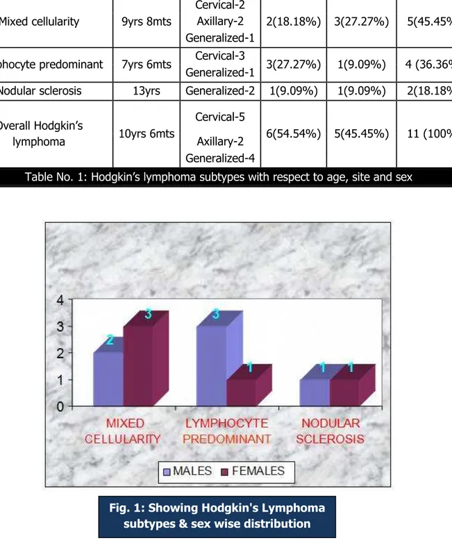
Mixed Cellularity: Five cases (45.45% of total Hodgkin’s lymphoma) were encountered, the mean age being 9 yrs 8mts. The younger one was 5 year old. It most commonly presented in 10-15yrs age group (60%). Case distribution included 2 in 5-10yrs and 3 cases in 10-15 yrs age group. Sex ratio of M: F was 2:3. The sites of involvement were as follows-cervical lymphadenopathy, generalised lymphadenopathy (1) and axillary lymphadenopathy.(2) One of the
cases showed epithelioid cell granulomas. (Table 1)
Lymphocyte predominant: Four cases (36.36% of total Hodgkin’s lymphoma) were encountered, the mean age being 7yrs 6mts. It most commonly presented in 10-15yrs age group (75%). Case distribution included 1 in 5-10yrs and 3 cases in 10-15yrs age group Sex ratio of M:F was 3:1. The sites of involvement were as follows-cervical lymphadenopathy(3) and generalised lymphadenopathy.(1) (Table 1)
ORIGINAL ARTICLE
J of Evidence Based Med & Hlthcare, pISSN- 2349-2562, eISSN- 2349-2570/ Vol. 2/Issue 4/Jan 26, 2015 Page 367
Histological subtypes Mean age Sites Male
(%)
Female (%)
Total No. (%)
Mixed cellularity 9yrs 8mts
Cervical-2
2(18.18%) 3(27.27%) 5(45.45%)
Axillary-2 Generalized-1
Lymphocyte predominant 7yrs 6mts Cervical-3 3(27.27%) 1(9.09%) 4 (36.36%)
Generalized-1
Nodular sclerosis 13yrs Generalized-2 1(9.09%) 1(9.09%) 2(18.18%)
Overall Hodgkin’s
lymphoma 10yrs 6mts
Cervical-5
6(54.54%) 5(45.45%) 11 (100%)
Axillary-2 Generalized-4
Table No. 1: Hodgkin’s lymphoma subtypes with respect to age, site and sex
ORIGINAL ARTICLE
J of Evidence Based Med & Hlthcare, pISSN- 2349-2562, eISSN- 2349-2570/ Vol. 2/Issue 4/Jan 26, 2015 Page 368 DISCUSSION: The age-adjusted standardized rate of Hodgkin’s lymphoma. For children and adolescents younger than 15 years in North America, Western Europe, and Oceania is 5.5 cases per million. For individuals aged 15-20 years, the incidence is 12.1 cases per million populations. These rates are in contrast to those in western Asia (from the Mediterranean to northwest India), where the age-adjusted standardized rate is consistently higher than 7 cases per million. The incidence of Hodgkin lymphoma by age shows a bimodal distribution. In developed nations, the first peak occurs at approximately age 20 years and the second peak is observed in patients aged 55 years or older. Hodgkin lymphoma is uncommon before age 5 years. However, in developing countries, the first peak is shifted into childhood, usually before adolescence.[4]
The REAL/WHO classification recognises a basic distinction between lymphocyte predominance HL (LP-HL) and classic HL (CHL), reflecting the differences in clinical presentation and behaviour, morphology, phenotype, and molecular features. CHL has been classified into four subtypes: lymphocyte rich, nodular sclerosing, with mixed cellularity, and lymphocyte depleted.[5, 3, 6]
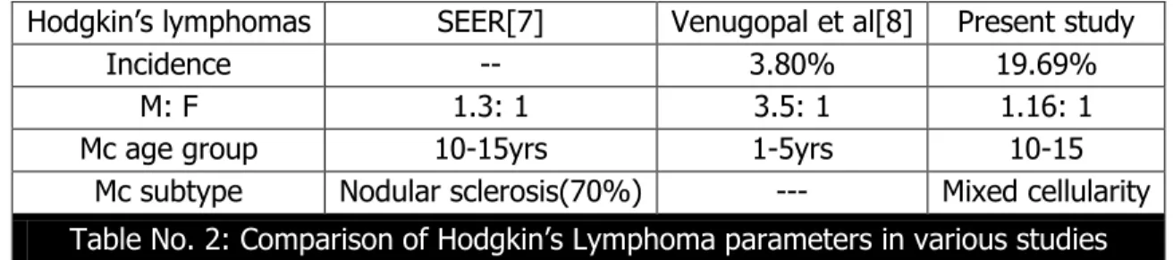
Hodgkin’s Lymphoma: Various parameters like male dominance, sub typing, age group and general incidence in various studies are in conformity with other studies in India and abroad as depicted in the Table No. 2
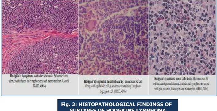
Tumour cells termed Reed-Sternberg (RS) or diagnostic cells—represent the body of the tumour: they measure 20–60 μm in diameter and display a large rim of cytoplasm and at least two nuclei with acidophilic or amphophilic nucleoli, covering more than 50% of the nuclear area. The tumoral population also includes a variable number of mononuclear elements—Hodgkin's cells (HCs)—showing similar cytological features to RS cells and neoplastic cell variants, each
ORIGINAL ARTICLE
J of Evidence Based Med & Hlthcare, pISSN- 2349-2562, eISSN- 2349-2570/ Vol. 2/Issue 4/Jan 26, 2015 Page 369
corresponding to a specific subtype of HD. However nodular sclerosis was the commonest subtype in SEER[7] study and age group was 1-5yrs in Venugopal et al[8] study.
The commonest site of involvement in this study was cervical lymphnodes. This observation correlates with study of Goswamy et al,[9] and Wright and Isscason.[10] This is in contrast to extranodal sites observed in Birch and N. Pratap[11] studies. Mixed cellularity was the
predominant histological subtype in present study and other studies from India and abroad as shown in table no. 2.
Hodgkin’s lymphomas SEER[7] Venugopal et al[8] Present study
Incidence -- 3.80% 19.69%
M: F 1.3: 1 3.5: 1 1.16: 1
Mc age group 10-15yrs 1-5yrs 10-15
Mc subtype Nodular sclerosis(70%) --- Mixed cellularity
Table No. 2: Comparison of Hodgkin’s Lymphoma parameters in various studies
Hodgkin’s lymphoma is rare in children and more frequent in young adults, displaying the histologic types that are associated with a favorable prognosis. The clinical characteristics of this variant of CHL, which accounts for about 6% of all HL cases, has been the object of several studies, including those promoted by the international project on lymphocyte predominant Hodgkin's disease and the German Hodgkin's lymphoma study group. These studies have shown that patients with LR-CHL differ from those with NS-CHL or MC-CHL: they are usually older than 50 and display a higher incidence of stages I–II and a subdiafragmatic location. In contrast, they rarely have bulky disease, B cell symptoms, or mediastinal or extranodal involvement. Thus, the clinical profile of LR-CHL is closer to that of LP-HL, although it has a lower frequency of stages I– II and splenic infiltration is more common. When compared with other types of CHD, LR-CHL gives rise to more frequent late relapses, although these do not behave aggressively.[12]
In underdeveloped countries and populations with poor socioeconomic conditions, the incidence is highest in children and histological types associated with poor prognosis predominate. Nodular sclerosis and lymphocyte predominance, the histological types with a favorable prognosis are significantly more common in younger persons and in women. Lymphocyte predominant is strongly associated with clinical stages I & II, nodular sclerosis predominantly with stage II and lymphocyte depletion primarily with clinical stages III & IV. In our study, male dominance and early occurrence of lymphocyte predominant subtype and stage I presentation is in correlation with above studies.[3, 4, 5]
HISTOLOGICAL SUBTYPES GOSWAMY[9] WRIGHT AND ISSACSON[10] Present study
LYMPHOCYTE PREDOMINANT 4 (4%) 2 (2.85%) 4 (36.36%)
NODULAR SCLEROSIS 15 (15%) 18 (25.71%) 2 (18.18%)
MIXED CELLULARITY 54 (54%) 36 (51.42%) 5 (45.45%)
LYMPHOCYTE DEPLETION 27 (27%) 14 (20%) 0
ORIGINAL ARTICLE
J of Evidence Based Med & Hlthcare, pISSN- 2349-2562, eISSN- 2349-2570/ Vol. 2/Issue 4/Jan 26, 2015 Page 370 CONCLUSION: Despite its well-known histological and clinical features, Hodgkin's lymphoma (HL) has recently been the object of intense research activity, leading to a better understanding of its phenotype, molecular characteristics, histogenesis, and possible mechanisms of lymphomagenesis. The frequency of Hodgkins lymphoma ors and their distribution is comparable to that reported from other studies. Significant differences exist between lymphocyte predominant HL (LP-HL) and classic HL (CHL) (which includes the lymphocyte rich, nodular sclerosing, mixed cellularity, and lymphocyte depleted subtypes) in terms of natural history, the relation to Epstein-Barr virus, cell morphology, phenotype, molecular characteristics, and clinical behavior, accurate incidence of data is important in the planning and evaluation of clinical trials. Documentation of cases, advanced diagnostic methods like IHC, cytogenetic studies and treatment modalities with close follow up is needed to achieve better statistical evaluation of the problem.
REFERENCES:
1.
Arya LS, Dinand V. Current strategies in the treatment of childhood Hodgkins disease. Indian Pediatr. Nov 2005; 42(11): 1115-28.2.
Glaser SL, Clarke CA, Nugent RA, Stearns CB, Dorfman RF. Social class and risk of Hodgkin's disease in young-adult women in 1988-94. Int J Cancer. Mar 1 2002; 98(1): 110-73. Marafioti T, Hummel M, Foss HD, et al. Hodgkin and Reed-Sternberg cells represent an expansion of a single clone originating from a germinal center B-cell with functional immunoglobulin gene rearrangement but defective immunoglobulin transcription. Blood 2000; 95: 1443–50.
4. Swerdlow SH, Campo E, Harris NL, et al. WHO Classification of Tumours of Haematopoietic and Lymphoid Tissues, Fourth Edition. Vol 2. 4th ed. Lyon, France: IARC Press; 2008.
5. Harris NL, Jaffe ES, Diebold J, et al. The World Health Organization classification of neoplastic diseases of the haematopoietic and lymphoid tissues: report of the Clinical Advisory Committee Meeting, Airlie House, Virginia, November 1997. Histopathology 2000; 36: 69–87.
6. Seitz V, Hummel M, Marafioti T, et al. Detection of clonal T-cell receptor gamma-chain gene-rearrangements in Reed-Sternberg cells of classic Hodgkin's disease. Blood 2000; 95: 3020–4.
7. Lyon A. Gloeckner. R, Smith A. M, james G. G, Martha Linet, Thea tamra, John L. Young, Jr. and Greta R. B.: Cancer incidence and survival among children and adolescents: United states SEER programe 1975-1995, NCI: 1-175.
8. Venugopal K.V. T.P. Joseph and K.K. Verma: solid malignant tumor of infancy and childhood
– A clinicopathological study. Indian pediatrics. 1981; Vol. 18: 365-368.
9. Goswamy. K.C and Banerjee CK: Hodgkin’s disease in children. Histopathological classification in relation to age and sex. Ind. J. cancer, 1982: 19: 24-27.
10.Wright DH and Issacson P: follicular center cell lymphoma of childhood: a report of three
ORIGINAL ARTICLE
J of Evidence Based Med & Hlthcare, pISSN- 2349-2562, eISSN- 2349-2570/ Vol. 2/Issue 4/Jan 26, 2015 Page 371
11.Birch J.M, Pratap. Incidence of malignant disease of childhood- a 24 year review of
Manchester children’s tumour registry data. British j. of cancer; 1980; vol. 42: 215-223. 12.Von Wasielewski R, Mengel M, Fischer R, et al. Classical Hodgkin's disease: clinical impact
of the immunophenotype. Am J Pathol 1997; 151: 1123–30.
AUTHORS:
1. Chandrashekar Thotadamane Nagaraj 2. Girish Chandrashekar Jerabandi
PARTICULARS OF CONTRIBUTORS:
1. Assistant Professor, Department of Pathology, Shivamogga Institute of Medical Sciences, Sagar Road, Shivamogga, Karnataka.
2. Associate Professor, Department of Pathology, Shivamogga Institute of Medical Sciences, Sagar Road, Shivamogga, Karnataka.
NAME ADDRESS EMAIL ID OF THE CORRESPONDING AUTHOR:
Dr. Chandrashekar Thotadamane Nagaraj, Assistant Professor, Department of Pathology, Shivamogga Institute of Medical Sciences, Shivamogga, Karnataka.
E-mail: [email protected]