165 165 165 165 165 Mem Inst Oswaldo Cruz, Rio de Janeiro, Vol. 92, Suppl. II: 165-172, 1997
Systemic Modulation of Peripheral Eosinophilia (Air Pouch
Model) in
Schistosoma mansoni
Infection
RG Pacheco, HL Lenzi*
/+Clínica Médica B, Departamento de Medicina Geral, Hospital Universitário Gaffree e Guinle, Universidade do Rio de Janeiro, Rua Mariz e Barros 775, 20270-020 Rio de Janeiro, RJ, Brasil *Departamento de Patologia,
Instituto Oswaldo Cruz, Av. Brasil 4365, 21045-900 Rio de Janeiro, RJ, Brasil
Schistosoma mansoni infection induces in their hosts a marked and sustained eosinophilia, which is influenced or modulated by complex mechanisms, that vary according to the phase of infection. To address this phenomenon, we used the air pouch (AP) model in control and infected Swiss webster mice, analyzing the cellular, tissue response and local expression of adhesion molecules [CD18 (β2-chain), CD44, ICAM-1 (CD54), L-selectin (CD62L), CD49d (α4-chain), LFA1 (CD11a)]. Infected animals were studied at 3 (pre-oviposition phase), 7 (acute phase), and 14 (chronic phase) weeks after infection (5-6 mice/period of infection). Normal mice were age-matched. Results showed that after egg stimula-tion, compared with matched controls, the infected mice, at each point of infecstimula-tion, showed a lower eosinophil response in the acute (7 weeks) and chronic phase (14 weeks) of infection. However, when the infected mice were in pre-oviposition phase (3 weeks) their eosinophil response surpassed the control ones. In the AP wall of infected mice, a significant decrease in the expression of ICAM-1 and CD44 in fibroblastic-like cells and a reduction in the number of CD18 and CD11a in migratory cells were ob-served. The other adhesion molecules were negative or weakly expressed. The results indicated that in the air pouch model, in S. mansoni-infected mice: (1) eosinophil response is strikingly down-regulated, during the acute ovular phase; (2) in the pre-oviposition phase, in contrast, it occurs an up-regulatory modulation of eosinophil response, in which the mechanisms are completely unknown; (3) in the chronic phase of the infection, the down modulation of eosinophil response is less pronounced; 4) Down-regu-lation of adhesion molecules, specially of ICAM-1 appear to be associated with the lower eosinophil response.
Key words: air pouch - adhesion molecules - Schistosoma mansoni egg - eosinophil - inflammation
hanced production of interleukin-5. Other primor-dial precursors stimulating cytokines are also in-volved in the eosinophilic pathway as the stem cell factor, IL-1, IL-3, GM-CSF, and G-CSF (Clutterbuck et al. 1989, Weller 1992).
The eosinophil is one of the most frequent cel-lular component of the most important lesion of schistosomiasis, the granuloma (Warren 1972). Even present in high number, the eosinophil is not a sine qua non condition for granuloma develop-ment (Sher et al. 1990). The acute eosinophil cel-lular response to eggs, and its contribution to the granuloma formation is not a well studied field. The murine experimental Schistosoma mansoni in-fection induces, as the inin-fection ages, a modula-tory mechanism that lower the hypersensitivity re-sponse to eggs (Andrade & Warren 1964, Domingo & Warren 1968, Boros et al. 1975). Schistosomia-sis mansoni has been characterized by a chrono-logical change in cytokine expression of Th1 and Th2 subsets (Pearce et al. 1991, Chensue et al. 1993).These changes are involved in the modula-tory process, and to IL-10 has been attributed a
This work was supported by Fundação Oswaldo Cruz.
+Corresponding author. Fax: +55-21-590.3495
Received 3 September 1997 Accepted 30 September 1997
en-166 166 166 166
166 Systemic Modulation of Peripheral Eosinophilia • RG Pacheco, HL Lenzi
pivotal role in these mechanisms (Stadecker 1994). We have previously shown, in the air pouch model, that S. mansoni egg induces, after 6-8 hr of stimu-lation, an acute intra air pouch eosinophil wave (Pacheco & Lenzi 1993).
In order to get a better insight of the eosinophil participation in schistosomiasis mansoni infection, we performed a sequential study of the eosinophil migration into the air pouch model, after 8 hr of egg stimulation, in mice at 3 (pre-oviposition phase), 7 (acute phase) and 14 (chronic phase) weeks after infection. This study was comple-mented by an immunofluorescence analysis of the expression of adhesion molecules at the air pouch covering, using monoclonal antibodies.
MATERIALS AND METHODS
S. mansoni infection - Five days old Swiss webster mice were infected by percutaneous ex-posure to 70 cercariae of the Belo Horizonte iso-late of S. mansoni (Paraense & Corrêa 1963, 1981). Air pouch - It was prepared as described and reported by Edwards et al. (1981)with modifica-tions. Briefly, one day before the beginning of the air pouch production, the fur on the dorsum of each mouse was shaved with an electric clipper. On day zero, 5 ml of sterile air were injected subcutane-ously into the same region of mice to form the pouches. In order to keep them patent, on day 3, they were replenished with 2 ml of sterile air. All experimental procedures to the air pouch produc-tion were carried out under light ether anesthesia. The sterile air was obtained in a laminar flow station by filtration through a Millipore (0.22 µm) directly into a syringe of 10 ml. On day 8, the pouches were stimulated with eggs or endotoxin-free saline, using individual sterile needles.
Egg purification - Mice with 8-12 weeks of infection, as before, were killed by an overdose of ether inhalation. The intestines were collected, opened, water cleaned, chopped and left to stay in a beaker overnight at room temperature. The ma-terial was processed in a blender, for a few sec-onds, in 1.7% saline, filtered through gradually smaller serial size 48, 80, 100, 150 steel mesh sieves and the eggs retained in a size 325 mesh sieve (Bertel, Caieiras, SP, Brasil). The eggs sedimented for one hour and half at 4°C. Finally, the eggs were collected and centrifuged at 150g for 30 seconds, several times until purification. The pellet containing eggs was kept overnight in an ice bath at 0-4 °C until use.
Injection of eggs - The eggs were adjusted to a 5000/ml concentration in 0.9% endotoxin-free saline and injected by using an insulin needle and syringe. The same number of matched control ani-mals received endotoxin-free saline.
Cell harvesting - The animals were killed by an overdose of ether inhalation, and 1 ml of phos-phate-buffer saline (PBS) was injected into the air pouches. The pouches were gently massaged and opened, at their caudal portion. The material was collected with a Pasteur pipette and maintained in ice bath until cell counting and cytocentrifugation.
Cell counting - Total cell count was done in Neubauer chamber. The absolute eosinophil num-ber was obtained from the percentage of eosino-phil in the centrifuged slide, and the total cell count.
Cytospin preparation - The collected materials from the air pouches were cytocentrifuged (Cito-spin, Incibras, Brasil) during 10 min, at 640 rpm. The slides were stained by May-Grünwald method, and 200-400 viable cells per slide were counted.
Experimental design - Infected Swiss webster mice of both sexes, after 2 (pre-oviposition phase), 6 (acute phase) and 13 (chronic phase) weeks of infection (5-6 mice/group) were picked randomly for the air pouch production. After 8 days, the air pouches were stimulated with 2,500 eggs diluted in endotoxin-free saline; 8 hr later, mice were killed and intrabursal cells were collected. The same num-ber of matched controls which received only en-dotoxin-free saline were also analyzed.
Histopathology - The whole air pouch were fixed in Formalin Millonig (Carson et al. 1973), at pH 7.0-7.4. The samples were embedded in par-affin and the slides stained with hematoxylin/eosin, PAS-Alcian blue pH 1.0 and 2.5, Lennert‘s Giemsa, Evans blue for eosinophils detection using confo-cal microscopy (Lenzi et al. 1996) , in LSM-410 (Zeiss).
Direct immunofluorescence - Adhesion mol-ecules - A small piece of the cephalic portion of the air pouch attached to the skin, was immediately frozen in liquid nitrogen and maintained at -20ºC until the cryostat slides preparation. The slides were fixed with cold acetone and conserved at -20ºC until the immunofluorescence studies. Mono-clonal antibodies (PharMingen) diluted in a 1:15 ratio to ICAM-1 (CD54), CD44, CD18 (β2 chain), LFA1 (CD11a), L-Selectin (CD62L), LEPAM (CD49d) were used. Briefly after rehydration with PBS, the slides were blocked with a solution of 1% BSA and 3 % powder skimmed milk. The di-luted mAb was maintained in contact with slides during 45 min at 37ºC. The slides were stained, mounted in glycerol with p-phenylenediamine and cover glass and examined in the same day in a microscope for immunofluorescence (Zeiss-Ger-many).
167 167167 167167 Mem Inst Oswaldo Cruz, Rio de Janeiro, Vol. 92, Supp. II, 1997
Statistical significance was determined by using unpaired t-test and significance was determined with the use of p values < 0.05.
RESULTS
Absolute eosinophil number - Saline stimula-tion elicited the lowest eosinophil responses, even in infected mice. After egg stimulation, compared with matched controls, the infected mice, in each point of infection, showed a lower eosinophil re-sponse in the acute (7 weeks) and chronic phase (14 weeks) of infection. However, when the in-fected mice were in pre-oviposition phase (3 weeks), their eosinophil response surpassed the control ones (Fig. 1). Although the eosinophil lev-els decreased in normal and infected mice, at the 7th week of age/infection, the normal mice showed a continuous increase of total cell count during the experiment. Otherwise, the infected ones presented always lower total cell count than the controls, specially at the 7th week of infection (Fig. 2)
Eosinophil percentage - Differently from the absolute number, the eosinophil percentage, as a rule, was higher with saline than with egg stimula-tion. The infected mice always showed a higher eosinophil percentage than control mice when the same stimulus (saline or egg) and the same age/ time of infection were compared. After saline stimulation, the eosinophil percentage enhanced gradually as the mice were aging (Fig. 3).
Histopathology - After egg stimulation, the neu-trophil infiltration, in the air pouch wall, was higher in normal mice than in infected ones, in all studied points. Otherwise, the eosinophil infiltration was much higher in normal mice at the 3rd week. At the
7th and 14th weeks of infection, the eosinophil in-filtration was similar to the matched controls. The normal mice had few eosinophil cells in the bone marrow, even when the eosinophil infiltration in the air pouch wall was pronounced (Fig. 4A, B). The infected mice on the contrary, mainly in the acute phase (7 weeks) showed a striking increase in the bone marrow eosinophil lineage, which was not accompanied by proportional eosinophil infiltration in the air pouch wall (Fig. 4C, D).
Immunohistology - The expression of adhesion molecules, in different structural components of the air pouch, was always compared between con-trol and infected mice with 7 and 14 weeks of in-fection.
Fig. 1: absolute eosinophil number, 8 hr after air pouch stimu-lation with LPS-free saline 0.9% or 2,500 Schistosoma mansoni
eggs in infected mice at the 3rd, 7th and 14th weeks of infec-tion and in matched controls. Mean of 5-6 mice after counting 200-400 cells in centrifuged slides stained with Mäy-Grünwald stain. Nor: normal mice; Inf: infected mice.
Fig. 2: total cell count (line) and absolute (bar) counts in the air pouches of normal (N) and Schistosoma mansoni-infected-mice (I), 8 hr after S. mansoni egg stimulation, during different times of age/infection.
168 168 168 168
168 Systemic Modulation of Peripheral Eosinophilia • RG Pacheco, HL Lenzi
L-selectin (CD62L), CD49d (integrin α4-chain) and CD11a (LFA-1) did not show significative dif-ference between infected and control mice, and no one correlation could be drawn relating to the eosi-nophil migration.
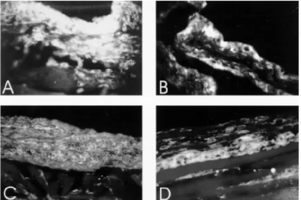
CD18 labeled weakly fibroblasts of the air pouch wall of all groups, and was more intense on migrating cells, which were more numerous in air pouch of control mice stimulated with eggs (Fig. 5A). CD54 (ICAM-1), after egg stimulation, marked diffuse and intensely all the air pouch wall layers of the control mice (Fig. 5B), showing im-pressive decrease in infected ones. This different behavior of CD54 expression was also detected in vascular endothelium of arteries, veins and
capil-laries. CD44 showed similar staining characteris-tics as CD54 (ICAM-1), except that it was more intense on fibroblasts and in the innermost layers of the air pouch wall (Fig. 5C, D). Frequently, CD54 and CD44 presented a cadherin-like pattern of immunofluorescence (Table). The lowest expres-sion of CD18, CD54, and CD44 was coincident with the decrease in the number of eosinophils in-side the air pouch.
DISCUSSION
169 169169 169169 Mem Inst Oswaldo Cruz, Rio de Janeiro, Vol. 92, Supp. II, 1997
pre-oviposition phase of the infection occurred an up-modulation, while in the acute (7 weeks) and chronic (14 weeks) phases, the eosinophil response was down-modulated. The literature has empha-sized multiple putative immunoregulatory mecha-nisms, during the course of S. mansoni infection, such as suppressor T cell activity (Colley et al. 1978), adherent phagocytic suppressor cells (Todd et al. 1979), serum-mediated suppression (Colley
et al. 1977, Ottesen & Poindexter 1980), circulat-ing immune complexes (Goes et al. 1991) and idiotypic/anti-idiotypic interactions, involving also anti-idiotypic T lymphocytes (Lima et al. 1986, Powell & Colley 1987, Parra et al. 1988, 1991). However, all these mechanisms are related to the pos-oviposition phases of the infection, and prob-ably some of them interfere with eosinophil re-sponse. Authors (Colley 1972, Mahmoud et al. Fig. 5: adhesion molecules expression, in air pouch wall of normal and infected mice, after 8 hr of stimulation. Air pouch of normal mouse (seven weeks old) with intense CD18+ cells (A). Expression of ICAM-1 in the air pouch linning of normal mouse (seven
weeks old) (B). Diffuse cadherin-like pattern of CD44 in normal (C), compared with weak distribution and fusiform pattern in fibroblasts of the air pouch wall of 14 week-infected mice (D).
TABLE
Expression of adhesion molecules in air pouch wall of infected compared with control mice, 8 hr after
Schistosoma mansoni egg stimulation
Time of Infection
Adhesion molecules a 7 weeks 14 weeks
CD62L (L-selectin) -
-CD49d (integrin α4-chain) (migrating cells) ↑ ↑
CD11a (LFA-1) (mast cells) = nd
CD18 (migrating cells) ↓↓↓ ↓↓↓
CD54 (ICAM-1) ↓↓↓ ↓↓↓
CD44 ↓↓ ↓↓
170 170 170 170
170 Systemic Modulation of Peripheral Eosinophilia • RG Pacheco, HL Lenzi
1975, Lenzi & Lenzi 1990) have shown that the eosinophil number fluctuate, during the S. mansoni
infection, in distinct compartments (bone marrow, blood, peritoneal cavity, milky spots and organs with egg embolism), reaching higher number in the acute phase, with high levels persisting even in chronic phase of infection. Our results showed that the down modulation of the intrabursal eosinophil response was coincident with the largest systemic and peri-ovular eosinophilia (around 7 weeks), in-dicating, two important aspects: (1) The down-modulation is independent of eosinophil produc-tion and circulaproduc-tion, not being due to a lack of eosi-nophils supply; (2) It is a striking modulation be-cause it occurs when the peripheral and central (bone marrow) eosinophil levels are very high (Fig. 4C ).
The data also suggest that the modulatory mechanisms act, at least, on two levels: endothe-lium, and air pouch wall. This hypothesis was based on the decrease of the following adhesion molecules: (a) ICAM-1 in vascular endothelium of arteries, veins and capillaries that are adjacent to the air pouch wall and (b) ICAM-1 and CD44 in the air pouch wall. These events could interfere with leukocyte-endothelial recognition phase of the acute inflammatory response, decreasing the num-ber of migrating cells, detected by anti-CD18. Other additional mechanisms can contribute to explain the down-modulation in the acute phase: (1) massive and compartmentalized mobilization of blood eosinophils to organs that suffer egg em-bolism; (2) large release of soluble adhesion mol-ecules to the circulation, blocking leukocyte-en-dothelial interactions (Evan Secor et al. 1994); (3) compartmentalized response of the air pouch to egg stimulation. Using protein A-stimulated air pouch, Teixeira et al. (1994) showed a decrease of polimorphonuclear migration in mice infected with
S. mansoni, mainly in the acute phase of the infec-tion. The same phenomenon was observed by Abath et al. (1988), in acute Trypanosoma cruzi
infected mice; and (4) alterations of eosinophil ca-pabilities by soluble factors. Dessein et al. (1984) showed that the “Eosinophil Cytotoxicity Enhanc-ing Activity” (ECEA) released by blood mono-nuclear cells was suppressed in most patients, ex-cept in those with heavy Schistosoma infections, including patients with hepatosplenomegaly.
As is shown in Fig. 1, when the intrabursal eosi-nophilia of control versus infected mice was com-pared in post-oviposition phases, always the in-fected groups attained lower number of eosinophils by cytological analysis. However, when the eosi-nophil levels from animals with 7 weeks were com-pared with ones of 14 weeks of infection, there was an absolute and relative increase in the later
ones (Figs 1 and 3). This event suggest the exist-ence of different and unknown control mechanisms on the inflammatory reaction in the acute and chronic phases of the schistosomal infection. Lenzi (1991) has shown that in the chronic phase of in-fection, when compared with the acute one, oc-curred a decrease of eosinopoiesis, mega-karyopoiesis, and lymphopoiesis, and an increase in the tissue mast and plasma cells, indicating changes in growth factors and cytokines networks during the course of the infection. Our data also showed an enhance of the eosinophil migratory capability to the air pouch, with advancing age, better observed in control group, by the increase of the eosinophil percentage, after saline stimula-tion (Fig. 3). The age and chronic-infected-related changes do not involve defects in the functions of T cells, including cytokine production (Engwerda et al. 1996). Actually, the studied animals were in the range of young adults (Hobbs et al. 1993). It is interesting to note that, in the infected mice, the eosinophil response inside the air pouch was in-versely related to the systemic eosinophil supply.
The up-modulation of the intrabursal eosino-philia detected in the pre-oviposition phase of S. mansoni infection, could be due to lack of immune modulation that is part of the acute syndrome (Cheever 1992). Indeed, the first peak of blood eosinophilia that occurs in the pre-oviposition phase is not affected by T-lymphocyte depletion (Colley et al. 1973, Fine et al. 1973, Lenzi et al 1987). .
In conclusion, the results of this study, using the air pouch model, showed that the acute eosi-nophil response is differently modulated, accord-ing to the time of S. mansoni infection. Bursal (air pouch) and endothelial cell-derived adhesion mol-ecules, such as ICAM-1 (and CD44 ?) appear to play a prominent role. The egg is the main factor that switch, not only the immune, but also the acute inflammatory response. Further comprehensive studies using intercrines a (C-X-C), and β (C-C) subfamilies (Oppenheim et al. 1991), affiliated with the recruitment phase of the inflammatory response should be applied to a better elucidation of the acute tissue eosinophilia.
REFERENCES
Abath F, Montenegro S, Carvalho A 1988. In vivo leukocyte chemotaxis during the development of acute experimental Trypanosoma cruzi infection.
Braz J Medical Biol Res21: 1013-1014.
Andrade Z, Warren K 1964. Mild prolonged schistoso-miasis in mice: alterations in host response with time and the development of portal fibrosis. Trans R Soc Trop Med Hyg58: 53-57.
Schisto-171 171171 171171 Mem Inst Oswaldo Cruz, Rio de Janeiro, Vol. 92, Supp. II, 1997
somiasis mansoni. J Immunol114: 1437-1441. Capron M 1992. Dual function of eosinophils in
patho-genesis and protective immunity against parasites.
Mem Inst Oswaldo Cruz 87 (Suppl V): 83-89. Carson FL, Martin JH, Lynn JA 1973. Formalin
fixa-tion for electron microscopy. A re-evaluafixa-tion. Am J Clin Pathol59: 365-373.
Cheever A 1992. Pathogenesis of Schistosoma mansoni infection. Mem Inst Oswaldo Cruz87 (Suppl IV): 337-340.
Chensue S, Warmington K, Hershey S, Terebuh P, Othman M, Kunkel S 1993. Evolving T cell re-sponses in murine schistosomiasis. Th2 cells medi-ate secondary granulomatous hypersensitivity and are regulated by CD8+ T cells in vivo. J mmunol 151: 1391-1400.
Clutterbuck E, Hirst E, Sanderson C 1989. Human interleukin-5 (IL-5) regulates the production of eosi-nophils in human bone marrow cultures: compari-son and interaction with IL-1, IL-3, IL-6, and GMCSF. Blood73: 1504-1512.
Colley DG 1972. Intradermal immune response to a schistosomal egg antigen during experimental mu-rine Schistosoma mansoni infection. Proc Soc Exp Biol Med140: 772-775.
Colley DG, Hieny SE, Bartholomew RK, Cook JA 1977. Immune responses during human schistosomiasis mansoni. III. Regulatory effect of patients sera on human lymphocyte blastogenic responses to schis-tosomal antigen preparations. Am J Trop Med Hyg 26: 917-925.
Colley DG, Katz SP, Wikel SK 1973. Schistosomiais: An experimental model for the study of immuno-pathologic mechanisms which involve eosinophils.
Adv Biosciences12: 653-665.
Colley DG, Lewis FA, Goodname RW 1978. Immune responses during human schistosomiasis mansoni. IV. Induction of suppressor cell activity by schisto-some antigen preparations and Concavalin A. J Immunol120: 1225-1232.
Dessein AJ, Lenzi HL, Bina JC, Carvalho EM, Weiser WY, Andrade ZA, David JR 1984. Modulation of eosinophil cytotoxicity by blood mononuclear cells from healthy subjects and patients with chronic schis-tosomiasis mansoni. Cell Immunol85: 100-113. Domingo E, Warren K 1968. Endogenous
desensitiza-tion: changing host granulomatous response to schis-tosome eggs at different stages of infection with Schistosoma mansoni. Am J Pathol52: 369-377. Edwards J, Sedgwick A, Willoughby D 1981. The
for-mation of a structure with the features of synovial lining by subcutaneous injection of air: an in vivo tissue culture system. J Pathol134: 147-156. Engwerda C, Fox B, Handwerger B 1996. Cytokine
production by T lymphocytes from young and aged mice. J Immunol156: 3621-3630.
Evan Secor W, dos Reis MG, Ramos EAG, Peixoto Matos E, Reis EAG, Carmo TMA, Harn jr., DA 1994. Soluble intercellular adhesion molecules in human schistosomiasis: correlation with disease se-verity and immunoregulated responses to egg anti-gens. Infec Immun62: 2695-2701.
Fine DP, Buchanan RD, Colley DG 1973. Schistosoma mansoni infection in mice depleted of thymus-de-pendent lymphocytes. I. Eosinophilia and immuno-logic responses to a schistosomal egg preparation.
Am J Pathol71: 193-206.
Goes AM, Gazzinelli G, Rocha R, Katz N, Doughty BL 1991. Granulomatous hypersensitivity to Schistosoma mansoni egg antigens in human schistosomiasis. III.
In vitro granuloma modulation induced by immune complexes. Am J Trop Med Hyg44: 434-443. Hobbs MV, Weigle WD, Nooman DJ, Torbett BE,
McEvilly RJ, Kock RJ, Cardenas GJ, Ernst DN 1993. Pattern of cytokine gene expression by CD4+ T cells from young and old mice. J Immunol150: 3602-3614.
Lenzi HL 1991. A dinâmica da resposta hematológica e celular na esquistossomose mansônica, com ênfase nas séries eosinofílica e mastocitária. Thesis. Universidade Federal do Minas Gerais, 580 pp. Lenzi H, Lenzi J 1990. Comparative distribution of
eosi-nophils in bone marrow, blood and peripheral cav-ity in murine schistosomiasis. Braz J Med Biol Res 23: 989-994.
Lenzi HL, Pelajo-Machado M, Caputo LFG, Vale BS 1996. Microscopia de varredura laser confocal: 2 -Recursos técnicos e aplicações biomédicas. Newslab 18: 106-122.
Lenzi HL, Sobral ACL, Lenzi JA 1987. “In vivo” kinet-ics of eosinophils and mast cells in experimental murine schistosomiasis. Mem Inst Oswaldo Cruz 82
(Suppl IV): 67-76.
Lima MS, Gazzinelli G. Nascimento E, Parra JC, Montesano MA, Colley DG 1986. Immune responses during human schistosomiasis mansoni. Evidence for antiidiotypic T lymphocyte responsiveness. J Clin Invest78: 983-988.
Mahmoud A, Warren K, Graham Jr R 1975. Antieosinophil serum and the kinetics of eosinophilia in schistosomiasis mansoni. J Exp Med142: 560-574.
Oppenheim JJ, Zacharae COC, Matsushima K 1991. Properties of the novel proinflammatory supergene “intercrine” cytokine family. Ann Rev Immunol9: 617-648.
Ottesen EA, Poindexter RW 1980. Modulation of the host response in human schistosomiasis. II. Humoral factors which inhibit lymphocyte proliferative re-sponses to parasite antigens. Am J Trop Med Hyg 29: 592-597.
Pacheco RG, Lenzi HL 1993. Air pouch kinetics after challenging with Schistosoma mansoni eggs. Abstr 153, in Abstracts of 4° International Symposium on Schistossomiasis, Rio de Janeiro.
Paraense WL, Corrêa LR 1963. Susceptibility of
Australorbis tenagophilus to infection with Schisto-soma mansoni. Rev Inst Med Trop São Paulo 5: 23-29.
Paraense WL, Corrêa LR 1981. Observations on two biological races of Schistosoma mansoni. Mem Inst Oswaldo Cruz 76: 287-291.
hy-172 172 172 172
172 Systemic Modulation of Peripheral Eosinophilia • RG Pacheco, HL Lenzi
persensitivity to Schistosoma mansoni egg antigens in human schistosomiasis. II. In vitro granuloma modulation by polyclonal idiotypic antibodies. J Immunol147: 3949-3954.
Parra JC, Lima MS, Gazzinelli G, Colley DG 1988. Immune responses during human schistosomiasis mansoni. XV. Anti-idiotypic T cells can recognize and respond to anti-SEA idiotypes directly. J Immunol140: 2401-2405.
Pearce E, Caspar P, Grzych J, Lewis F, Sher A 1991. Downregulation of Th1 cytokine production accom-panies induction of Th2 response by a parasitic helm-inth, Schistosoma mansoni. J Exp Med173: 159-166. Powell MR, Colley DG 1987. Anti-idiotypic T lympho-cyte responsiveness in murine schistosomiasis mansoni. Cell Immunol104: 377-385.
Race G, Michaels R, Larsh J, Matthews J 1969. Schis-tosoma mansoni eggs: an electron microscopic study of shell pores and microbarbs. Proc Soc Exp Biol Med130: 990-992.
Sher A, Coffman RL, Hieny S, Scott P, Cheever AW 1990. Interleukin 5 is required for the blood and tis-sue eosinophilia but not granuloma formation in-duced by infection with Schistosoma mansoni. Proc
Natl Acad Sci USA87: 61-65.
Stadecker M 1994. The shrinking schistosomal egg granuloma: how accessory cells control T cell-me-diated pathology. Exp Parasitol79: 198-201. Tagboto S 1996. Interleukin-5, eosinophils and the
con-trol of helminth infections in man and laboratory animals. J Helminthol69: 271-278.
Tood CW, Goodname RW, Colley DG 1979. Immune responses during human schistosomiasis mansoni. V. Suppression of schistosome antigen-specific lym-phocyte blastogenesis by adherent/phagocytic cells.
J Immunol122: 1440-1446.
Teixeira K , Coutinho A, Montenegro S 1994. “In vivo” leukocyte chemotaxis in experimental mice Schis-tosoma mansoni infection. Rev Inst Med Trop S Paulo36: 283-285.
Wardlaw A, Moqbel R, Barry Kay A 1995. Eosino-phils : biology and role in disease. Adv Immunol60: 151-266.
Warren KS 1972. The immunopathogenesis of schisto-somiasis: a multidisciplinary approach. Trans R Soc Trop Med Hyg66: 417-432.