Karina Eiras Dela Coleta Pizzol
ALTERAÇÕES DO TECIDO MOLE,
ESPAÇO FARÍNGEO E
ESTABILIDADE APÓS AVANÇO
MAXILO-MANDIBULAR COM ROTAÇÃO
ANTI-HORÁRIA E PRÓTESE TOTAL
DE ATM
Karina Eiras Dela Coleta Pizzol
ALTERAÇÕES DO TECIDO MOLE, ESPAÇO
FARÍNGEO E ESTABILIDADE APÓS AVANÇO
MAXILO-MANDIBULAR COM ROTAÇÃO
ANTI-HORÁRIA E PRÓTESE TOTAL DE ATM
Araraquara 2008
Tese apresentada ao Programa de
Pós-graduação em Ciências Odontológicas - Área
de Ortodontia, da Faculdade de Odontologia
de Araraquara, Universidade Estadual
Paulista, para obtenção do título de Doutor
em Ortodontia.
Orientador: Prof. Dr. João Roberto
Pizzol, Karina Eiras Dela Coleta.
Alterações do tecido mole, espaço faringeano e estabilidade após avanço maxilo-mandibular com rotação anti-horária e prótese total de ATM / Karina Eiras Dela Coleta Pizzol. – Araraquara: [s.n.], 2008.
141 f. ; 30 cm.
Tese (Doutorado) – Universidade Estadual Paulista, Faculdade de Odontologia
Orientador : Prof. Dr. João Roberto Gonçalves
1. Cirurgia 2. Prótese articular 3. Avaliação I. Título
Karina Eiras Dela Coleta Pizzol
Alterações do tecido mole, espaço
faríngeo e estabilidade após avanço
maxilo-mandibular com rotação
anti-horária e prótese total de ATM
Comissão Julgadora
Tese para obtenção do grau de Doutor
Presidente e Orientador: Prof. Dr. João Roberto
Gonçalves
2º Examinador: Prof. Dr. Ary dos Santos-Pinto
3º Examinador: Prof. Dr. Roberto Henrique Barbeiro
4º Examinador: Prof. Dr. Darceny Zanetta Barbosa
5º Examinador: Profa. Dra. Terumi Okada
Araraquara, 23 de setembro de 2008
Dados Curriculares
Karina Eiras Dela Coleta Pizzol
Nascimento: 09/02/76- Torrinha_S.P.
Filiação: Roberto Dela Coleta
Laura Helena Eiras Dela Coleta
1995/1998: Curso de Graduação
Faculdade de Odontologia de
Araraquara “Júlio de Mesquita
Filho”(UNESP)
1999/2002: Curso de Pós-Graduação em
Ortodontia, nível Mestrado, no
Centro de Pesquisas Odontológicas
São Leopoldo Mandic
Dedicatória
A De us,
Agradeço todas as dificuldades que enfrentei; não fosse por elas, eu não teria saído do lugar.
"Minhas imperfeições e fracassos são como uma bênção de Deus, assim como meus sucessos e meus talentos, e eu coloco ambos a seus pés."
( Mahatma Gandhi )
Aos meus Pais,
Robe rt o e La ura, por terem proporcionado o suporte necessário para que eu pudesse vencer todas as etapas da minha vida, por me ensinaram que caráter e
honestidade valem mais do que qualquer bem material. Pelo amor incondicional, pela dedicação e pela vida.
Ao meu amado Marido N ilt on,
Por ser meu chão, minha luz e meu porto seguro. Pelo amor incondicional, respeito, companheirismo e
cumplicidade em todos os dias de nossas vidas.
“Se um dia tiver que escolher entre o mundo e o amor...
Lembre-se:
Se escolher o mundo ficará sem o amor, mas se
escolher o amor, com ele conquistará o mundo.”
(Albert Einsten)
“O verdadeiro amor é exigente, implacável, e, ao
mesmo tempo, infinitamente delicado. "
À minha filha Le t íc ia,
Que me ensinou a lutar por um sonho, a doar amor sem esperar nada em troca e perceber que a vida pode ser tão simples e bela quanto um olhar ou um sorriso.
"O dia mais importante não é o dia em que conhecemos uma pessoa e sim quando ela passa a existir dentro de
nós."
Aos meus irmãos,
Flá via e T hia go, pela amizade, amor, carinho e compreensão que sempre dedicaram a mim.
Aos meus avós,
Apa re c ida, Alic e e Be ne dit o por serem minha referência de superação, de luta e de dedicação à família.
"Há quem diga que todas as noites são de sonhos. Más há também quem garanta que nem todas, só as de verão.
No fundo, isso não tem importância. O que interessa
mesmo não é a noite em si, são os sonhos. Sonhos que
o homem sonha sempre, em todos os lugares, em todas
as épocas do ano, dormindo ou acordado."
( William Shakespeare )
Agra de c im e nt o Espe c ia l
Ao meu Mestre e Orientador,
João Roberto Gonçalves, que foi acima de tudo um grande amigo. Obrigada pela confiança, pela contribuição em minha formação profissional, pelo exemplo de dedicação e competência, pelo respeito e por todas as oportunidades concedidas durante o período de Pós-Graduação. Meu muito obrigado por tornar este sonho realidade.
"Não há nada como o sonho para criar o futuro.
Utopia hoje, carne e osso amanhã."
( Victor Hugo )
Ao professor,
Ary dos Sa nt os Pint o pela confiança, disponibilidade, pelo respeito, pela dedicação, esmero e satisfação em ensinar, pela valiosa contribuição científica na execução deste trabalho.
"Grande professor é aquele que realiza o que ensina."
( Columbano )
Ao professor,
La ry M . Wolford meu muito obrigado por permitir a elaboração deste trabalho, por sua dedicação e pela valiosa contribuição científica na minha formação profissional.
"Do mesmo modo que o campo, por mais fértil que seja, sem
cultivo não pode dar frutos, assim é o espírito sem
estudo."
( Cícero )
Meu muito Obrigado Aos Professores,
Dirc e u Ba rna bé Ra ve li, Luiz Gonza ga Ga ndini J r., Lídia
Pa rze k ia n M a rt ins, M a uríc io T . Sa k im a , Rit a Corde iro e
Lourde s dos Sa nt os-Pint o pela disponibilidade e
ensinamentos que contribuíram fundamentalmente para a minha formação e crescimento profissional.
“Nunca um desejo lhe é dado sem que também lhe seja dado o
poder de realizá-lo. Entretanto, você pode ter que se esforçar
por ele."
( Richard Bach )
Élc io M a rc a nt onio e Robe rt o H e nrique Ba rbe iro pelo
incentivo hoje e sempre, por despertarem meu
"A vida é em parte o que nós fazemos dela, e em parte
o que é feito pelos amigos que nós escolhemos."
( Tennessee Williams )
Aos amigos de Turma,
Pa ulo (Beca), M a rc us V iníc ius, Ric a rdo e Re na t opelo
incentivo, amizade, e por todos os momentos que passamos juntos.
"Seja cortês com todos, mas íntimo de poucos, e deixe estes poucos serem bem testados antes que você dê a eles a sua
confiança. A verdadeira amizade é uma planta de crescimento
lento, e deve experiementar e resistir os choques da
adversidade antes de ser receber o nome de amizade."
( George Washington )
Aos amigos conquistados na Pós-Graduação,
Adria no, André, Lua na, Sa va na , Am a nda , Ce c ília , Ra fa e l,
Luiz Guilhe rm e , H e lde r, N a nc y, Luc ia na e M ic he le pela
amizade e apoio durante todos os momentos.
"Amizade, palavra que designa vários sentimentos, que
não pode ser trocada por meras coisas materiais. Deve
ser guardada e conservada no coração."
Os m e us sinc e ros a gra de c im e nt os...
À Faculdade de Odontologia de Araraquara na pessoa do
Prof. Dr. J osé Clá udio M a rt ins Se ga la , pela oportunidade
de crescimento profissional junto à UNESP.
Aos Funcionários do Departamento de Clínica Infantil, secretaria de Pós-graduação e secretaria da
Diretoria, pela disponibilidade, respeito e dedicação sempre.
Aos funcionários da biblioteca, em especial à M a ria
H e le na e à Ce re s, que muito colaboraram para a
Sumário
Resumo 15
Abstract 17
1 Introdução 19
2 Proposição 23
3 Capítulos 25
3.1 Capítulo 1 27
3.2 Capítulo 2 62
3.3 Capítulo 3 89
4 Considerações finais 120
5 Referências 129
Pizzol KEDC. Alterações do tecido mole, espaço faríngeo e estabilidade
após avanço maxilo-mandibular com rotação anti-horária e prótese total
de ATM [tese doutorado]. Araraquara: Faculdade de Odontologia da
UNESP; 2008
Resumo
Este estudo avaliou a resposta do tecido mole, do espaço faríngeo e a
estabilidade após avanço maxilo-mandibular com rotação anti-horária e
reconstrução da ATM com próteses totais articulares do tipo TMJ
Concepts system®. As mudanças cirúrgicas e pós-cirúrgicas foram
analisadas utilizando-se telerradiografias laterais. Com o movimento
cirúrgico, houve redução do ângulo do plano oclusal (14,9 ± 8,0°) e
aumento do espaço aéreo faríngeo - PASnar (4,9mm). A região anterior
da maxila moveu-se para a frente e para cima enquanto a porção
posterior, para a frente e para baixo. A mandíbula avançou, e sofreu
rotação no sentido anti-horário. No período pós-cirúrgico, a maxila
apresentou alterações mínimas no plano horizontal, enquanto todas as
medidas mandibulares permaneceram estáveis. A postura da cabeça
(OPT/NS) mostrou flexão imediatamente após a cirurgia e extensão em
longo prazo, enquanto a curvatura cervical (OPT/CVT) não apresentou
mudanças. Os resultados cirúrgicos mostraram ainda aumento das
o hióide, permanecendo estáveis durante o período de observação. A
distância entre o osso hióide e o plano mandibular reduziu durante e
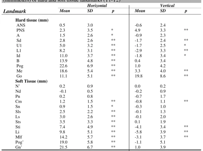
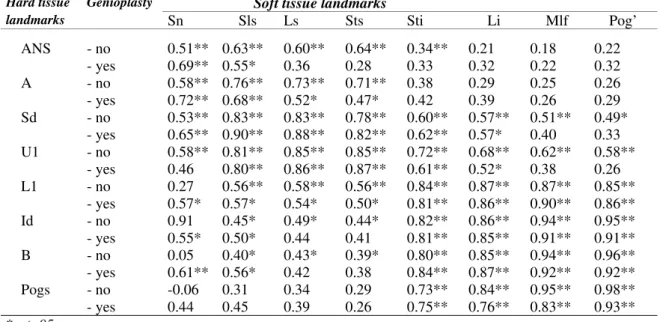
após a cirurgia. Já a resposta do tecido mole evidenciou diferentes
razões entre tecido duro/mole nos pacientes com e sem genioplastia. As
mudanças horizontais na morfologia do lábio superior após avanço,
impacção da maxila, sutura em VY e sutura da base alar mostraram
maior movimento do que as mudanças observadas em tecido duro. O
avanço maxilo-mandibular com rotação anti-horária do plano oclusal
associado a próteses totais de ATM (TMJ Concepts system®)
mostrou-se estável durante o período de obmostrou-servação. O espaço aéreo faríngeo
aumentou significativamente, tendo sido influenciado pela posição da
cabeça após a cirurgia. A resposta dos tecidos moles ante os
movimentos esqueléticos realizados mostrou-se previsível.
Pizzol KEDC. Stability, soft tissue response and oropharyngeal airway
space changes after maxillo-mandibular advancement and
counter-clockwise rotation with total joint TMJ prostheses [tese doutorado].
Araraquara: Faculdade de Odontologia da UNESP; 2008
Abstract
This study evaluated stability, soft tissue response and oropharyngeal
airway space changes after maxillo-mandibular advancement and
counter-clockwise rotation with TMJ reconstruction using TMJ Concepts
system® total joint prostheses. Lateral cephalograms were analyzed to
estimate surgical and post surgical changes. During surgery, the
occlusal plane angle decreased 14.9 ± 8.0° and the retroglossal airway
space (PASnar) increased 4.9mm. The anterior region of maxilla moved
forward and upward while the posterior nasal spine moved downward
and forward. The mandible changed forward and rotated in a
counter-clockwise direction. At long-term follow-up evaluation the maxilla
showed minor horizontal changes, while all mandibular measurements
remained stable. Head posture (OPT/NS) showed flexure immediately
after surgery and extension long-term post surgery, while cervical
curvature (OPT/CVT) had no significant changes. Surgery increased the
distances between the third cervical vertebrae (C3) and menton, and C3
and hyoid, remaining stable afterwards. The distance from the hyoid to
follow-up. Soft tissue response indicated different hard/soft tissue ratios
between patients with or without genioplasties. Horizontal changes in
upper lip morphology after maxillary advancement/impaction, VY
closure, and alar base cinch sutures showed greater movement, than
observed in hard tissue. TMJ Concepts total joint prostheses associated
with maxillo-mandibular advancement and counter-clockwise rotation
showed to be stable during the follow-up observation period. Immediate
increase in oropharyngeal airway dimension, was influenced by
post-surgical changes in head posture but remained stable over the follow-up
period. Soft tissue changes showed a known predictable response.
1 Introdução
Certas patologias associadas à articulação
temporo-mandibular podem gerar alterações clínicas envolvendo oclusão,
músculos, respiração e estética facial18. Sintomas comuns da disfunção
temporo-mandibular incluem sons/estalidos na ATM, dores, limitação de
movimentos mandibulares, mudanças na oclusão, dificuldade
mastigatória entre outros. Embora a grande maioria dessas disfunções
articulares possa ser tratada com terapias não invasivas, existe um
grupo restrito de pacientes com degenerações articulares irreversíveis
que requerem reparo ou reconstrução cirúrgica, tradicionalmente
realizada com tecido autógeno. Entretanto, quando esses enxertos
autógenos são associados a avanços mandibulares de grande
amplitude, observam-se resultados pouco previsíveis com freqüente
reabsorção e recidiva. Algumas condições articulares específicas
podem predispor a comportamento semelhante. Exemplos destas
condições são: 1) ATMs previamente operadas (duas ou mais cirurgias
anteriores); 2) colocação prévia de implantes aloplásticos de ATM
contendo Proplast/Teflon, Silastic, acrílico ou cimentos ósseos; 3)
patologias inflamatórias, infecciosas ou reabsorções condilares em
anquilose; 6) ausência de ATM decorrente de patologia, trauma ou
deformidade congênita e 7) tumores envolvendo a região da fossa e/ou
côndilo e ramo mandibular33. Nesses casos, as próteses totais de ATM
produzidas pelo processo de prototipagem são a melhor opção. Por
meio da tecnologia CAD/CAM (computer assisted design/computer
assisted manufacture) é possível obter detalhes anatômicos que
permitem uma adaptação precisa para cada caso em particular.
As patologias de ATM podem afetar pacientes de qualquer
idade e de ambos os gêneros. Quando essas condições (ex: artrite
reumatóide, lupus, doenças auto-imunes do tecido conjuntivo,
reabsorção condilar idiopática entre outros) ocorrem em pacientes
jovens, podem ocasionar alterações no crescimento maxilo-mandibular,
deformidades dentofaciais, além de distúrbios respiratórios. Em
pacientes adultos, processos degenerativos da ATM podem também
alterar a morfologia crânio-facial com restrição importante do espaço
aéreo faríngeo, exigindo reconstrução articular associada à cirurgia
ortognática com finalidade de otimizar os resultados estéticos e
funcionais5,25.
Quando a cirurgia ortognática é realizada em associação à
colocação de próteses articulares, é possível obter-se resultados mais
estáveis31 para a correção da deformidade facial do que quando essa
cirurgia é realizada sem levar em consideração o estado das ATMs20,26.
Entretanto, a estabilidade dos resultados não se restringe somente aos
fatores relacionados ao movimento cirúrgico ou à intervenção ou não
nas ATMs, mas também a fatores que contribuem para o sucesso ou
falha das próteses articulares como: hipersensibilidade ao metal32,
micromovimento da prótese, perda de componentes da prótese, fratura
ou corrosão do material2,4,10, biocompatibilidade2-4, contaminação por
bactéria11,12 e desenvolvimento de osso heterotópico ao redor da
prótese30.
Não existem trabalhos na literatura que se propõem avaliar
a estabilidade do movimento ortodôntico-cirúrgico, bem como as
mudanças respiratórias e de tecido mole, quando próteses totais
articulares são colocadas concomitantemente ao ato cirúrgico. A grande
maioria das pesquisas restringe-se a relatos da colocação isolada de
próteses articulares sem correção da má oclusão esquelética associada
ou propõe-se a avaliar a sintomatologia e os índices de qualidade de
2 Proposição
2.1 Proposição geral
O objetivo deste estudo foi avaliar a estabilidade do
avanço maxilo-mandibular com rotação anti-horária do plano oclusal,
associado à prótese total de ATM (TMJ Concepts system®) sobre o
comportamento esquelético, do tecido mole e do espaço aéreo faríngeo.
2.2 Proposições específicas
Este estudo tem como propósitos:
1. Avaliar a estabilidade do avanço com rotação
anti-horária maxilo-mandibular associada à colocação de prótese total de
ATM (TMJ Concepts system®);
2. Avaliar as mudanças e estabilidade no espaço aéreo
faríngeo promovidas pela técnica cirúrgica;
3. Determinar se existe correlação entre a quantidade de
movimento cirúrgico e as mudanças no tecido mole e determinar sua
razão de correspondência;
4. Avaliar a influência da genioplastia nas mudanças do
Esta tese de Doutoramento foi redigida em capítulos
correspondentes a artigos de periódicos para publicação.
Capítulo 1 Maxillo-Mandibular Counter-Clockwise Rotation and
Mandibular Advancement with TMJ Concepts® Total Joint Prostheses:
Part I - Skeletal and Dental Stability
Karina E. Dela Coleta, Larry M. Wolford, João Roberto Gonçalves, Ary dos Santos Pinto, Lécio Pitombeira Pinto, Daniel Serra Cassano
Artigo submetido à publicação no periódico International Journal of Oral
Maxillofacial Surgery.
Capítulo 2 Maxillo-Mandibular Counter-Clockwise Rotation and
Mandibular Advancement with TMJ Concepts® Total Joint Prostheses:
Part II – Airway Changes and Stability
Karina E. Dela Coleta, Larry M. Wolford, João Roberto Gonçalves, Ary dos Santos Pinto, Daniel Serra Cassano, Daniela A. Godoy Gonçalves
Artigo submetido à publicação no periódico International Journal of Oral
Maxillofacial Surgery.
Capítulo 3 Maxillo-Mandibular Counter-Clockwise Rotation and
Mandibular Advancement with TMJ Concepts® Total Joint Prostheses:
Part IV - Soft Tissue Response
Karina E. Dela Coleta, Larry M. Wolford, João Roberto Gonçalves, Ary dos Santos Pinto, Daniel Serra Cassano, Daniela A. Godoy Gonçalves
Artigo submetido à publicação no periódico International Journal of Oral
Advancement with TMJ Concepts
®Total Joint Prostheses:
Part I - Skeletal and Dental Stability
1
Karina E. Dela Coleta,
2
Larry M. Wolford,
1
Joao Roberto Gonçalves,
1
Ary dos Santos Pinto,
3
Lécio Pitombeira Pinto,
2
Daniel Serra Cassano,
1
Pediatric Dentistry Department - Araraquara Dental School, Sao Paulo State University, Brazil
2
Department of Oral and Maxillofacial Surgery, Texas A&M University Health Science Center, Baylor College of Dentistry, Baylor University Medical Center, Dallas, TX, USA
3
Department of Restorative Dentistry, Pharmacology, Dental and Nursing School, Federal University of Ceará, Fortaleza, Brazil
Address correspondence and reprint requests to: Larry M. Wolford, DMD:
3409 Worth St, Suite 400 Dallas, TX 75246
Abstract
The purpose of this study was to evaluate skeletal and dental stability in patients that
had TMJ reconstruction and mandibular counter-clockwise advancement using TMJ
Concepts total joint prostheses (TMJ Concepts Inc. Ventura, CA) with maxillary
osteotomies being performed at the same operation. Forty-seven females (14 to 57
years old) met the criteria for inclusion with an average post surgical follow-up of 40.6
months (range 12 to 143 months). Lateral cephalograms were analyzed to estimate
surgical and post surgical changes. During surgery, the occlusal plane angle decreased
14.9 ± 8.0°. The maxilla moved forward at ANS 1.3 ± 2.4mm, point A 2.5 ± 2.2mm,
and upper incisor tip (U1T) 5.6 ± 3.0mm and upward at ANS -0.6 ± 1.9mm, point A
-1 ± 1.9mm, and U1T -1.3 ± 1.9mm. The posterior nasal spine moved downward and
forward 5.5 ± 4.2 mm and 2.9 ± 3.1 mm respectively. The mandible advanced 7.9 ± 3.5
mm at the lower incisor tips, 12.4 ± 5.4 mm at point B, 17.3 ± 7.0 mm at menton, 18.4
±8.5 mm at pogonion, and 11.0 ± 5.3 mm at gonion. Vertically the lower incisors
moved upward -2.9 ± 4.0 mm, B point and Pog remained unchanged but Me moved
downward 2.6 ± 3.9 mm and Go 18.4 ± 9.2 mm. At longest follow-up post surgery, the
maxilla showed minor horizontal changes while all mandibular measurements remained
stable. TMJ reconstruction and mandibular advancement with TMJ Concepts total joint
prosthesis in conjunction with maxillary osteotomies for counter-clockwise rotation of
the maxillo-mandibular complex was a stable procedure for these patients at the longest
follow-up.
Introduction
Temporomandibular joint (TMJ) pathology can create clinical problems
including masticatory musculature, jaws, occlusion, and other associated structures
resulting in pain and jaw dysfunction. Although many cases of TMJ dysfunction and
symptoms can usually be managed with non-surgical therapies, there remains a group
of patients with irreversible TMJ damage, requiring surgical repair or reconstruction;
traditionally with autogenous tissues. However, certain specific TMJ conditions and
pathology can have adverse outcomes using autogenous grafts, producing a significant
failure rate with their use7,23. These conditions include: 1) multiply operated TMJs (2 or
more previous operations); 2) previous TMJ alloplastic implants containing
Proplast/Teflon (PT, Vitek Inc., Houston, TX), Silastic (Dow Corning Inc, Midland,
MO), acrylic, bone cements, metal-on-metal articulations or failed prostheses; 3)
inflammatory, infective, reactive, or resorptive TMJ pathologies; 4) connective tissue
and autoimmune diseases; 5) fibrous and bony ankylosis; 6) absence of TMJ structures
due to pathology, trauma, or congenital deformity; and 7) tumors involving the fossa
and/or condyle and mandibular ramus region28. In these cases a custom-made total joint
prostheses may be the best option. Using CAD/CAM (computer assisted
design/computer assisted manufacture) technology, prostheses are designed and
manufactured to fit the specific anatomical requirements for each patient.
TMJ pathology can affect patients of any age and both genders but, when these
conditions occur in young patients, maxillo-mandibular growth alterations commonly
occur resulting in dentofacial deformities and associated malocclusions. In adults, TMJ
pathology (i.e., rheumatoid arthritis, psoriatic arthritis, reactive arthritis, condylar
processes of the condyles may require TMJ reconstruction and orthognathic surgery to
achieve optimal functional and esthetic results.
Consideration should be given for surgical correction of co-existing TMJ
pathology as part of the orthognathic surgical correction plan. Wolford et al.27,
routinely perform concomitant TMJ and orthognathic surgery for correction of patients
with co-existing dentofacial deformities and TMJ internal derangement, with a high
success rate. There have been variable success rates reported for TMJ prostheses,
ranging from 60% to 100%16. There are risks and complications that can occur with the
use of TMJ total joint prostheses. A common problem in patients with previous PT and
Silastic implants as well as bone cements, acrylic, or metal-on-metal articulations, is the
recurrent development of foreign body giant-cell reaction (FBGCR) and reactive bone
that can cause limited jaw function as well as pain, fibrous and/or bony ankylosis.
When reconstructing these patients with the TMJ Concepts total joint prostheses,
packing autologous fat grafts around the articulating area of the prostheses has been
shown to decrease the FBGCR and minimize the occurrence of excessive joint fibrosis
and heterotopic calcification, consequently providing improved range of motion in
prosthetic TMJ reconstruction and decreased pain25.
There are many known factors which influence the success or failure of the total
joint prostheses. The challenge is to minimize these factors such as metal
hypersensitivity26, prosthesis micro movement, loosening of the prosthetic components,
material wear, break-down, and corrosion7, biocompatible and functionally compatible
materials2, FBGCR7,11, prosthesis failure11,19, bacterial contamination, and development
It is considered a surgical success at long-term follow-up when the total joint
prostheses provide TMJ and occlusal stability, improve function, decrease pain, and a
long functional lifetime. Previous studies9-13,23,24,28,29 have showed that TMJ
reconstruction with total joint prostheses resulted in a significant decrease in pain, and
improvement in jaw function, diet and maximal interincisal opening. The present study
has the specific purpose of evaluating skeletal and dental stability of TMJ
reconstruction and mandibular advancement in a counter-clockwise direction using
TMJ total joint prostheses with maxillary osteotomies being performed at the same
operation.
Patients and Methods
This retrospective study evaluated records of 50 consecutive patients from a
single private practice, from 1990 through 2003, who underwent TMJ reconstruction
and counter-clockwise rotation of the maxillo-mandibular complex. Criteria for study
inclusion were: 1. End-stage bilateral or unilateral TMJ reconstruction and mandibular
advancement using custom-made TMJ total joint prostheses (TMJ Concepts system®),
and maxillary osteotomies for counter-clockwise rotation of the maxillo-mandibular
complex and occlusal plane angle; 2. All surgical procedures performed by one surgeon
(L.M.W.) at Baylor University Medical Center, Dallas, TX, USA; 3. Use of maxillary
and mandibular rigid fixation; 4. Females at least 14 years of age and males at least 17
years of age; 5. Absence of post surgical trauma; and 6. Minimum of 12-month post
surgery follow-up. Patients were rejected based on the following criteria: 1.
Craniofacial syndromes; and 2. Records (radiographs) inadequate or poor quality.
patient excluded because of less than 12 month follow-up. The two males were
excluded from the study to make a homogeneous sample of 47 females (Table 1).
The custom-made total joint prostheses used in this study, were originally
developed in 1989 by Techmedica Inc., Camarillo, CA, USA, and since 1997, have
been manufactured by TMJ Concepts, Inc., Ventura, CA, USA. These prostheses are
CAD/CAM devices (computer assisted design/computer assisted manufacture),
designed to fit the specific anatomical requirements for each patient.
Forty-three patients had bilateral TMJ reconstruction and 4 patients had a
unilateral prosthesis with a sagittal split osteotomy on the contra-lateral side. All
patients had Le Fort I maxillary osteotomies, most with segmentation. All patients had
coronoidectomies on the prosthesis side(s) at the reconstruction surgery or at a previous
surgery. Mean patient age at the time of surgery was 34.5 years (range, 14 years to 57
years). Presurgical (T1) records were taken 1 day (range, 1 to 6 days) before surgery;
immediate post surgical (T2) records were taken 5 days (range, 2 to 16 days) after
surgery; and longest follow-up (T3) records were taken at a mean of 40.6 months
(range, 12 to 143 months) after surgery.
Imaging evaluation
Two examiners were calibrated by repetition of the process until the method
was considered adequate by a third examiner. Standardized lateral cephalometric
radiographs (Quint Sectograph; American Dental Co, Hawthorne, CA) were randomly
apart. Average values between the 2 replicates were used to decrease landmark
technical errors.
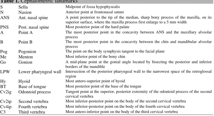
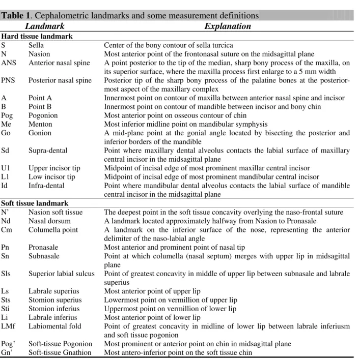
There were 16 landmarks identified by one examiner and digitized using
DFPlus software (Dentofacial Software Inc, Toronto, Canada). The following
landmarks were used to compute 25 measurements (Tab 2, Fig 1): Nasion, Sella, Point
A, Anterior nasal spine, Posterior nasal spine, Point B, Pogonion, Menton, Gonion, and
dental points. S-N minus 7° was used as the horizontal reference plane (HRP) and a
line perpendicular through sella as the vertical reference plane (VRP). Horizontal and
vertical changes for each landmark were evaluated. Surgical changes were computed as
the differences between T2 and T1 values and post surgical changes between T3 and T2
values.
Null hypothesis
Mandibular advancement with counter-clockwise rotation of the occlusal plane
with total joint TMJ prostheses is an unstable procedure.
Statistical method
All data were transferred to SPSS (release 9.0; SPSS Chicago, IL) for statistical
analysis. The skewness and kurtosis statistics showed normal distributions for all
variables. Paired t tests were performed to evaluate the surgical (T2-T1) and post
surgical changes (T3-T2). A significance level of p < .05 was applied. The reliability of
tracing, landmark identification, and analytical measurements had an intraclass
Patients who received bilateral TMJ prostheses (n=43) and unilateral (n=4) were
compared as separate groups. Because there were no statistically significant differences
between those groups in post surgical changes, all the patients were analyzed as a single
group. Patients were also divided into two groups with group 1 having 12 to 24 months
post surgical follow-up (n = 18) and group 2 having 25 to 143 months follow-up (n =
29). There were no statistically significant differences in any of the parameters
evaluated between the two groups. Therefore, all 47 patients were analyzed as a single
group.
Surgical Technique
Seven patients required a preliminary surgical stage to remove previously
placed, failed total joint prostheses that contained metal (i.e. Vitek total joint
prostheses, Vitek Inc., Houston, TX; Christensen total joint prostheses, TMJ Implants
Inc., Golden, CO) , so that an accurate CT scan could be taken. Metal in the TMJ
and/or ramus can interfere with the CT scan imaging data, and significantly distort the
3-dimensional (3-D) plastic model on which the custom-made total joint prostheses are
made. CT scans were taken on all patients extending from supero-posterior to the TMJ
to anterior to the chin, maxilla and nasal bones. The 3-D plastic model was then
created using stereolithography technology (Figure 3 A). A surgical prediction tracing
was developed from a presurgical lateral cephalometric radiograph to determine the
desired final position of the maxilla and mandible. The 3-D model was mounted on an
anatomical articulator and precise model surgery performed to reposition the mandible
to the desired post surgical position relative to the maxilla that remained in its original
position on the model. Once the mandibular position was achieved, the mandible was
position of the mandible. The condyles were cut off and if indicated, bony
recontouring of the fosse and lateral aspect of the rami was completed. Any
recontouring on the 3-D model had to be accurately duplicated on the patient at the time
of surgery.
The custom-made total joint prostheses were then manufactured using
CAD/CAM technology on the 3-D model to fit the patient’s specific anatomical
requirements (Figure 3 B). Immediately prior to surgery, the mandibular movements
done on the 3-D model were accurately duplicated on anatomically mounted dental
models, and an intermediate splint constructed to aid in repositioning the mandible.
The maxillary model was then repositioned and sectioned if necessary to achieve the
best occlusal relationship. A final splint was constructed when indicated.
At surgery, an endaural or preauricular approach was used to perform the
condylectomy, joint debridement, coronoidectomy to release the temporalis muscle,
and if indicated, accurate bony recontouring of the fossa as dictated by the recontouring
done on the 3-D model. Through a submandibular approach, the medial pterygoid and
masseter muscles were reflected off the mandibular ramus and lateral recontouring
completed as indicated from the 3-D model. The mandible was then mobilized and
repositioned using the intermediate splint and inter-maxillary fixation applied. The
fossa component was inserted through the endaural / preauricular incision and
stabilized to the zygomatic arch with 3 to 4, 2 mm diameter bone screws. The
mandibular prosthetic component was inserted through the submandibular incision and
stabilized to the ramus with 8 to 12, 2 mm diameter bone screws. Following
abdomen) packed around the articulating area of the prostheses to help prevent fibrosis
and heterotopic/reactive bone formation post surgery. The incisions were closed.
Multiple maxillary osteotomies were then performed to establish the best
possible functional and esthetic result, since presurgery the maxilla was usually AP
retruded as well as had anterior vertical maxillary excess and/or posterior vertical
maxillary deficiency with a high occlusal plane angulation.The maxilla was stabilized
with bone plates and porous block hydroxyapatite grafts (PBHA, Interpore 200,
Interpore Inc., Irvine, CA).
When indicated, genioplasty, turbinectomies, nasoseptoplasty, rhinoplasty, etc.,
were performed at the same surgery. Many of these patients, particularly those with
significant retrognathia, had moderate to severe presurgical sleep apnea symptoms
because of the decreased oropharyngeal airway. The suprahyoid muscles were not
deliberately detached from the genial tubercles in any of the cases. Alloplastic
materials such as PBHA or HTR (Hard Tissue Replacement, Walter Lorenz Inc,
Jacksonville, FL) were used for augmentation genioplasties although some patients had
osseous genioplasties.
Post surgery, no maxillo-mandibular fixation was used in any cases, but light
interarch elastics were routinely applied to help support the mandible, since the muscles
of mastication were reflected from the mandible and were initially non-functional. Post
operative elastics were generally discontinued following adequate functional return of
the pterygomasseteric musculature (usually 2 to 4 weeks post surgery), unless required
beginning approximately 6 to 8 weeks post surgery. Patients were instructed to open
and close their jaws and begin shifting their jaws from side to side 4 to 5 sessions per
day for 10 to 15 minutes each session. Patients were maintained on a puree to soft diet
for 4 months post surgery to allow the maxilla to complete the initial bone healing
phase. Patients were then encouraged to begin working up to a normal diet.
Orthodontic appliances were usually maintained for at least 6 months post surgery and
then removed at the discretion of the orthodontist.
Results
Surgical changes (T2-T1)
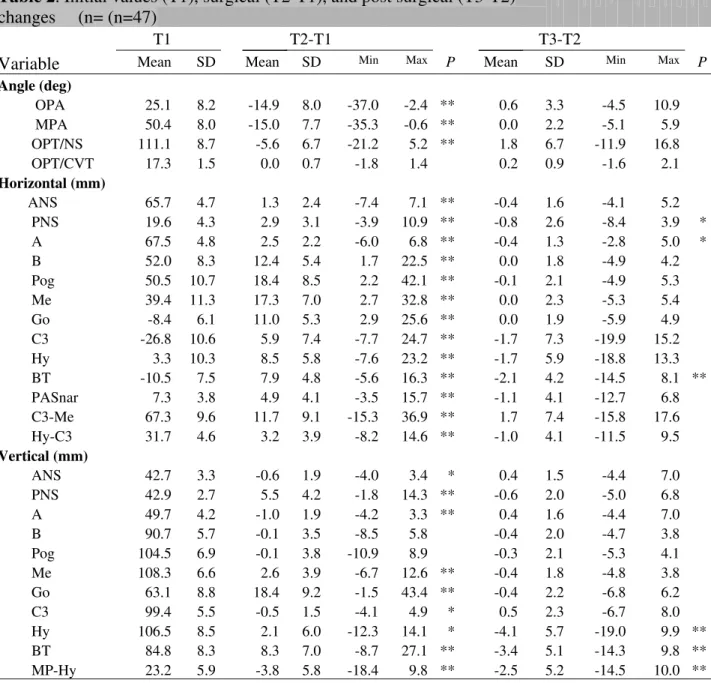
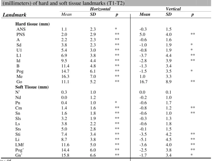
Initial values, surgical and longest follow-up changes are listed in Table 3. The
mean surgical changes showed upward and forward movement of the maxillary anterior
region (Fig 2). The horizontal movement of anterior nasal spine (ANS) was 1.3 mm
(range -7.3 to 7.1mm), and point A was 2.5 mm (range -6.0 to 6.8 mm). In the
horizontal direction, positive values mean forward movement, negative values mean
posterior movement. The vertical movement (positive values mean downward
movement, negative values mean upward movement) of the ANS was 0.6 mm (range
-4.0 to 3.4 mm) and point A was -1.0 mm (range -4.2 to 3.3 mm). Posterior nasal spine
(PNS) was displaced downward 5.5 mm (range 1.8 to 14.3 mm) and forward 2.9 mm
(range 3.9 to 10.9 mm). The upper incisor tip (U1T) moved forward 5.6mm (range
-0.6 to 11.3 mm) and vertically -1.3 mm (range -5.7 to 2.6 mm).
All the anterior mandibular measurements were advanced in a horizontal
direction with lower incisor tip (L1T) 7.9 mm (range 0.9 to 14.3 mm), point B 12.4 mm
(Me) 17.3 mm (range 2.6 to 32.8 mm). In the vertical direction, L1T showed a superior
movement of -2.9 mm (range -16.5 to 2.3 mm), while B point and Pog showed no
movement. However, Me showed an inferior movement of 2.6 mm (range -6.7 to 12.5
mm). Gonion (Go) moved downward 18.4 mm (range -1.5 to 43.4 mm) and forward
11.0 mm (range 2.8 to 25.6 mm). The occlusal plane angle (OPA) relative to HRP
decreased a mean -14.9º (range -37.0 to -2.3º) and SNPog angle increased 9.1º (range
1.0 to 20.1º).
There was a surgical increase in the SNA angle of 2.3º (range -6.5 to 6.4º) and
SNB of 6.9º (range 1.0 to 12.8º). TheANB angle decreased -4.6º (range -10.5 to 2.2º)
because of the greater increase of the SNB value compared to SNA. Overjet (OJ)
decreased -2.2 mm (range -7.4 to 1.8 mm). Overbite (OB) increased 1.6 mm (range -14.7
to 3.9 mm).
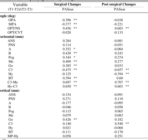
Post surgical stability (T3-T2)
Point A and posterior nasal spine (PNS), in their horizontal direction only,
showed a change backwards of -0.4 mm (range -2.8 to 5.0 mm) and -0.8 mm (range
-8.4 to 3.9 mm) respectively, that were considered statistically significant (p<0.05). The
remaining maxillary landmarks remained stable. All the anterior mandible
measurements (L1T, B, Pog, Me) showed no statistically significant change at
long-term follow-up (p<0.05). Neither OPA nor SNPog angles had significant changes long
term post surgery. Therefore, all horizontal and vertical mandibular measurements
remained stable during the follow-up period (Table 3). There was a mean decrease in
no statistically significant change (T3-T2). Overbite (OB) increased 0.7 mm (range -0.7
to 2.1 mm).
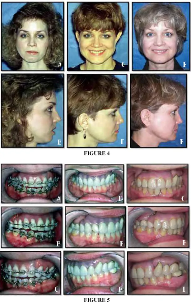
Case 1: (CT patient #41) This 28 year old female presented 4 years post trauma
that involved multiple mandibular fractures including bilateral subcondylar fractures,
comminution of the right condyle, symphysis fracture with loss of a central incisor, as
well as fracture of the anterior maxilla resulting in the loss of 7 teeth from the left
lateral incisor through the right 2nd bicuspid. The missing teeth were previously
replaced with 5 osseo-integrated dental implants and a prosthesis. She had one previous
surgery on her right TMJ with no improvement. Her diagnoses included: 1) Right
TMJ severe arthritis, 2) Anterior open bite, 3) Transverse facial asymmetry, and 4)
Retruded maxilla and mandible (Fig 4 A and B; 5 A-C; 6 A). She had severe right TMJ
pain, headaches, myofascial pain, and difficulty eating and chewing.
Following orthodontic preparation, surgery was performed (Figure 6 B) in one
operation including: 1) Right TMJ reconstruction and mandibular counter-clockwise
advancement (right ramus was lengthened and advanced 26 mm) with a custom made
TMJ total joint prosthesis (TMJ Concepts system®), 2) Right coronoidectomy, 3) Left
mandibular ramus sagittal split osteotomy, and 4) Multiple maxillary osteotomies to
down graft the posterior aspect, advance it, and transversely level the occlusal plane.
The A-P occlusal plane was decreased 16 degrees. The patient’s longest follow-up at
completion of the study was 79 months post surgery showing good stability (Figures 4
C and D; 5 D-F), with elimination of TMJ pain, headaches, and myofascial pain;
improved jaw function, occlusion, and facial esthetics. The patient recently returned
remains pain free with good jaw stability (Figure 6 D), esthetics, and function with an
incisal opening of 42 mm.
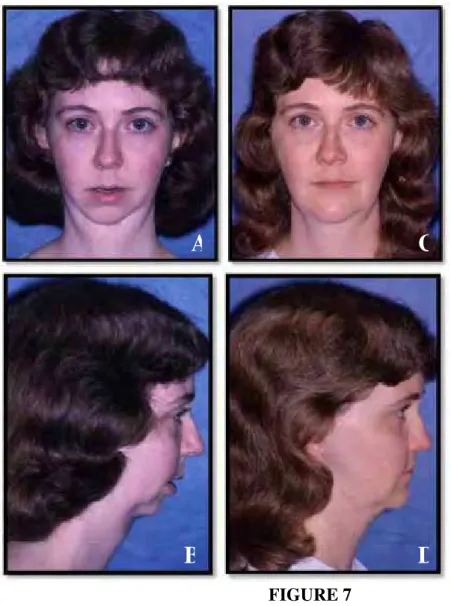
Case 2: (TW, patient # 47) This 25 year old female was referred after failed
previous bilateral TMJ surgery, maxillary osteotomies, and genioplasty (Fig. 7 A and
B; 8 A-C; 9 A). She reported problems with other joints and a rheumatology evaluation
diagnosed a non-specific CTAD. MRI showed severe condylar resorption and a
reactive pannus surrounding the TMJ articular discs. Her surgical diagnoses included:
1) Severe bilateral condylar resorption, 2) Maxillary A-P and posterior vertical
hypoplasia, 3) Severe A-P mandibular hypoplasia, 4) Class II occlusion with severe
apertognathia (7 mm), 5) Decreased oropharyngeal airway (A-P dimension of 2 mm,
where normal is 11 + 2 mm) causing severe sleep apnea, 6) Severe masticatory
dysfunction, and 7) Severe TMJ and myofascial pain.
The surgical procedures performed (Figure 9 B) included: 1) Bilateral TMJ
reconstruction and mandibular advancement in a counter-clockwise direction utilizing
the TMJ Concepts/Techmedica custom-made total joint prostheses with the rami
lengthened 17 mm and the chin (pogonion) advanced 24 mm, 2) Bilateral
coronoidectomies, 3) Multiple maxillary osteotomies with the maxillary incisor tips
advanced 7 mm and the posterior maxilla inferiorly positioned 5 mm stabilized with
bone plates and PBHA grafts, and 4) Osseous chin augmentation (Figure 9 B). The
mandibular occlusal plane was surgically decreased 19 degrees. At 6 years post
surgery, the patient maintained a stable facial balance and occlusion (Figure 7 C and D,
surgery. Pain levels decreased from 9 at T1 to 1 at T3. The sleep apnea was resolved,
and the patient could eat relatively normally.
Discussion
TMJ reconstruction with total joint prostheses is indicated in specific TMJ
conditions and pathology with irreversible joint damage. Some of those progressive
TMJ disease conditions (i.e., rheumatoid arthritis, psoriatic arthritis, reactive arthritis,
idiopathic condylar resorption, etc.) are predominantly found in females and can result
in malocclusion, facial disfigurement, TMJ dysfunction, and pain7,9-13,23,29.
The demographic data from our study revealed that the need for
maxillo-mandibular surgery with total joint TMJ prostheses reconstruction involves a relatively
younger patient population (many under the age of 40 years including teenagers), which
means that the longevity of the prosthesis is an important variable. Longevity and
stability of any implanted joint prosthesis is based on the proper indication for its use,
correct placement and maintenance of the prosthesis, the properties and
biocompatibility of the materials used, recipient’s biological acceptance of the device,
the implant's stability in situ, and the ability of the recipient to understand the
limitations involved with having a prostheses in place. TMJ Concepts custom-made
total joint prosthesis system was designed with these factors in mind9.
Previous studies have shown that TMJ reconstruction with this specific total
joint prosthesis system resulted in a significant improvement in pain, function, diet and
increase in maximum interincisal opening9-13,23,28,29. There are only a few studies
associated with total joint prostheses23,29. This present study evaluated this aspect of
TMJ prostheses using the TMJ Concepts custom-made total joint prostheses.
In this study, surgical changes showed upward and forward movement of the
anterior region of the maxilla, while the posterior region was displaced downward and
forward; thus the palatal plane angle also rotated in a counter-clockwise direction. The
amount and direction of the surgical movement of the maxilla was directly related to
the mandibular movement.
The post surgical stability of upward maxillary repositioning by Le Fort I
osteotomy, was shown to be relatively stable by many authors3,17,18. According to the
literature the stability of the surgical movement of the maxilla (in the vertical and
horizontal planes) was stable with counter-clockwise rotation of the
maxillo-mandibular complex in the presence of healthy TMJs4,22. In our study, point A showed
a post surgical mean change of -0.4mm in the horizontal plane, and although clinically
insignificant, it was statistically significant. This alteration can be explained in part by
post surgical bone remodeling or by post surgical orthodontic movement. Point A is
considered a dento-alveolar point, being subject to alteration of incisor position. With
retraction of maxillary incisors, Point A can move posteriorly, and soft tissue tension
created by maxillary advancement can also cause remodeling of point A. The posterior
nasal spine (PNS) showed a clinically minimal post surgical mean horizontal movement
of -0.8 mm, but statistically significant. This change can be associated with bone
remodeling also. Most of the cases studied received three pieces maxillary
segmentation with a midline split in the palate that could affect the posterior nasal spine
In reference to the counter-clockwise rotation and advancement of the mandible,
all of the anterior points of the mandible remained stable in the post surgical long-term
follow-up period. The mean mandibular advancement at the incisor tips was 7.9 mm,
Point B 12.4 mm, and pogonion substantially greater with 18.4 mm as a result of the
counter-clockwise rotation of the maxillo-mandibular complex. The counter-clockwise
rotation resulted in pogonion advancing 6.0 mm more than point B and 10.5 mm more
than the lower incisor tips. This demonstrates the advantage of counter-clockwise
rotation in advancing the mandible and chin in the high occlusal plane angle facial type
patient. Decrease in the occlusal plane and mandibular plane angles were directly
correlated with the anterior movement of the mandible. Our clinical results, confirm
Wolford et al.22 previous supportive research and philosophy that maxillo-mandibular
counter-clockwise rotation, in high occlusal plane facial types, may improve function
and esthetics with a stable occlusion4,22.
In the vertical plane, the counter-clockwise rotation of the mandible resulted in
an upward movement of L1T and no movement at B point and Pog. The mean Menton
surgical movement was in a downward direction as a result on geometrically
up-righting the anterior aspect of the mandible (L1T to Me) causing Menton to rotate
downward and forward compared to the lower incisor tips. Gonion showed a major
downward surgical movement, due to reorientation of the mandibular ramus in that
direction.
The long-term post surgical mandibular stability in this study was found to be
maxillo-mandibular complex and occlusal plane in patients with healthy TMJs. These
results are significantly better than those found by other authors such as Moore et al.14;
Arnett & Tamborello1, in which mandibular surgical advancement (without
counter-clockwise rotation) was performed on patients without regard to the presence or
absence of TMJ pathology, nor was any appropriate TMJ surgical intervention provided
for any of the patients with TMJ pathology in those studies. This allowed post surgical
relapse related to condylar remodeling and resorption to occur is some of their patients.
One of biggest changes with the surgery in our study occurred in the occlusal
plane angle. According to Ricketts15, the normal occlusal plane angle is 8 + 4 degrees
and is defined as: A line tangent to the lower bicuspid cusp tips through the second
molar buccal groove and the angle formed with the Frankfort horizontal plane. In our
study, the T1 occlusal plane angle was a mean of 25.1o and was surgically decreased at
T2 to a mean of 10.2o with a mean change of 14.9o. The alteration of this angle is
significantly influential on the horizontal and vertical menton position. With a decrease
in the occlusal plane angulation, there is an increase of the horizontal projection of
Menton compared to the lower incisor tips. Counter-clockwise rotation of
maxillo-mandibular complex with maxillo-mandibular advancement has inherent risks to the healthy as
well as the TMJ with untreated pathology. The mandibular lever arm is lengthened so
the soft tissues including skin, muscles, periostium, etc., are stretched increasing the
load to the TMJs. This can create or exacerbate TMJ problems.
Post surgical increased loading of the joints occurs until the TMJs, soft tissues,
muscles, skeletal structures, and occlusion reach a state of equilibrium and adaptation
maxillo-mandibular complex in a counter-clockwise direction may further increase the
loading of the TMJ by stretching the associated soft tissues, it is a very stable procedure
in the presence of healthy TMJs4,6,22,27. According to Wolford et al.6,30,31, patients with
co-existing TMJ dysfunction undergoing mandibular advancement without surgical
correction of the TMJ pathology, are likely to have significantly increased signs and
symptoms of TMJ dysfunction and pain.
Several studies have noted that at some period after surgery, the condyles tend
to move posteriorly and superior1y in the fossa following mandibular advancement20,21.
Van Sickels et al.20 noted this phenomenon with both wire osteosynthesis and rigid
fixation from 8 weeks to 2 years after surgery. This posterior movement may be an
adaptive response to mandibular advancement and change in the fulcrum arm length of
the mandible, related to TMJ disc position change pre and post surgery, and/or soft
tissue tension related to the advancement.
In our study, the joints were replaced by TMJ total joint prostheses (TMJ
Concepts system), making it possible to get highly predictable functional, esthetic and
stable results, since the TMJ prostheses are not affected by muscle adaptation, disc
position, or physiological loading of the joints.
The Techmedica custom-made total joint prostheses (now manufactured by
TMJ Concepts) were previously evaluated by Henry &Wolford7, to determine the
outcomes in patients with a history of Proplast-Teflon (PT) TMJ implants. Twenty-six
patients (43 joints) were evaluated, with a follow-up from 4 to 24 months. The
of residual pain rated as good in 46%, fair 38%, and poor in 16% of the patients. The
residual pain for the most part was related to the multiply operated patients, pre surgical
irreversible pain, and continued foreign body giant cell reaction from failed previous
TMJ alloplastic implants and prostheses containing PT.
The main problems associated with TMJ total joint reconstruction is related to
wear at the articular surfaces, foreign body reaction, mobility of the implant with
displacement, and implant fracture, caused by the use of inappropriate alloplastic
materials5. Wolford & Karras25 conducted a comparative study on patients who had
Techmedica total joint prostheses placed. A total of 22 patients had fat grafts placed
and were compared with 37 patients without fat grafts. Statistically significant
improvement was found for MIO and excursive movements in the fat-grafted joints
compared with the non-grafted joints. In addition, 35% of the non-grafted joints
required additional surgery for the removal of heterotopic/reactive bone or severe
fibrosis, whereas none of the fat-grafted joints required secondary joint surgery.
Because TMJ patients are often relatively young (mean age in this study was 35
years), a total TMJ prosthesis must have a very long lifetime and once the prosthesis is
implanted, there is no way to go back to the previous anatomy19. Our follow-up period
ranged from 12 to 143 months, with a median of 40.6 months. Only 10 patients had
been followed for five years or more. It will be important to continue to monitor
groups of patients such as ours over the coming years, particularly the younger patients.
Speculand et al.16 stated that it is not possible to determine the lifetime of this type of
TMJ prostheses. Wolford24, demonstrated that custom-made total joint prostheses,
very well for TMJ reconstruction. Total joint prostheses with use of appropriate
materials are the only predictable alternative for many patients. During the past 19
years that these prostheses have been available, the senior author (LMW) has placed
over 540 prostheses and has not replaced any because of wearing out. The longevity of
the prostheses remains unknown.
The current study demonstrates that the TMJ Concepts total joint prostheses
work well with good stability at longest follow-up (12 to 143 months), and is a viable
technique for TMJ reconstruction, with mandibular advancement and
counter-clockwise rotation of the maxillo-mandibular complex and occlusal plane, when
indicated for patients with irreversible end-stage TMJ pathology and co-existing
dentofacial deformity.
References
1. Arnett GW, Tamborello JA. Progressive Class II development. Female idiopathic
condylar resorption. Oral Maxillofac Surg Clin North Am 1990; 2:699-716.
2. Baird DN, Rea WJ. The temporomandibular joint implant controversy: A review of
autogenous/alloplastic materials and their complications. J Nutr Environ Med 1998;
8:289-300.
3. Brammer J, Finn R, Bell WH, Sinn D, Reisch J, Dana K. Stability after bimaxillary
surgery to correct vertical maxillary excess and mandibular deficiency. J Oral Surg
1980; 38:664-670.
4. Chemello PD, Wolford LM, Buschang MS. Occlusal plane alteration in orthognathic
surgery, Part II: Long-term stability of results. Am J Orthod Dentofacial Orthop
5. Fontenot MG. Temporomandibular joint devices: past present and future. In: Sessle
BI, Bryant PS, Dionne RA, eds. Temporomandibular Disorders and Related Pain
Conditions. Seattle: IASP Press, 1995: 309–322.
6. Goncalves JR, Cassano DS, Wolford LM, Santos-Pinto A, Marquez IM. Postsurgical
Stability of Counterclockwise Maxillomandibular Advancement Surgery: Affect of
Articular Disc Repositioning. J Oral Maxillofac Surg 2008;66:724-738.
7. Henry CH, Wolford LM. Treatment outcomes for temporomandibular joint
reconstruction after Proplast-Teflon implant failure. J Oral Maxillofac Surg 1993;
51:352-360.
8. Henry CH, Hughes CV, Gerard HC, Hudson AP, Wolford LM. Reactive arthritis:
Preliminary microbiologic analysis of the human temporomandibular joint. J Oral
Maxillofac Surg 2000; 58:1137-1144.
9. Mercuri LG, Wolford LM, Sanders B, White RD, Hurder A, Henderson W. Custom
CAD/CAM Total Temporomandibular Joint Reconstruction System: Preliminary
Multicenter Report. J Oral Maxillofac Surg 1995;53:106-115.
10. Mercuri, LG. Subjective and objective outcomes in patients reconstructed with a
custom-fitted alloplastic temporomandibular joint prosthesis. J Oral Maxillofac
Surg 1999;57:1427-1430.
11. Mercuri LG, Giobbie-Hurder A. Long-term outcomes after total alloplastic
temporomandibular joint reconstruction following exposure to failed materials. J
Oral Maxillofac Surg 2004;62:1088-1096.
12. Mercuri LG, Wolford LM, Sanders B, White RD, Giobbie-Hurder A. Long-term
follow-up of the CAD/CAM patient fitted total temporomandibular joint
13. Mercuri LG. The use of alloplastic prostheses for tmeporomandibular joint
reconstruction. J Oral Maxillofac Surg 2000;58:70-75.
14. Moore KE, Gooris PJJ, Stoelinga PJ. The contributing role of condylar resorption to
skeletal relapse following mandibular advancement surgery. Report of five cases. J
Oral Maxillofac Surg 1991; 49:448-460.
15. Ricketts, RM. Cephalometric analysis synthesis. Angle Orthod 1961; 31:141-156.
16. Speculand B, Hensher R, Powell D. Total prosthetic replacement of the TMJ:
experience with two systems 1988–1997. Br J Oral Maxillofac Surg2000; 38:360–
369.
17. Stoker NG, Epker BN. The posterior maxillary osteotomy: A retrospective study of
treatment results. Int J Oral Surg 1974; 3:153-157.
18. Turvey TA, Phillips C, Zaytoun HSJr, Profitt WR. Simultaneous superior
repositioning of the maxilla and mandibular advancement. Am J Orthod Dentofacial
Orthop 1988; 94:372-381.
19. Van Loon JP, De Bont L, Boering G. Evaluation of temporomandibular joint
prostheses: Review of literature. J Oral Maxillofac Surg 1995;53:984-997.
20. Van Sickels JE, Tiner BD, Keeling S, Clark G, Bays R, Rugh J. Condylar position
with rigid fixation versus wire osteosynthesis of a sagittal split osteotomy. J Oral
Maxillofac Surg 1996; 54 (Suppl 3):105-106.
21. Will LA, Joondeph DR, Hohl TH, West RA. Condylar position following
mandibular advancement. J Oral Maxillofac Surg 1984; 47:578-588.
22. Wolford LM, Chemello PD, Hilliard F. Occlusal plane alteration in orthognathic
surgery- Part I: Effects on function and esthetics. Am J Orthod Dentofacial Orthop
23. Wolford LM, Cottrell DA, Henry CH. Temporomandibular joint reconstruction of
the complex patient with the Techmedica custom-made total joint prosthesis. J Oral
Maxillofac Surg 1994; 52:2-10.
24. Wolford LM. Temporomandibular joint devices: Treatment factors and Outcomes.
Oral Surg Oral Med Oral Pathol Oral Radiol Endod 1997; 83:143-149.
25. Wolford LM, Karras SC. Autologous fat transplantation around temporomandibular
joint total joint prostheses: Preliminary treatment outcomes. J Oral Maxillofac Surg
1997; 55:245-251.
26. Wolford LM, Mehra P, Rea W. Metal hypersensitivity in patients with total joint
prosthesis. J Oral Maxillofac Surg 2000; 58:29 (suppl 1) (abstr).
27. Wolford LM, Karras SC, Mehra P. Concomitant temporomandibular joint and
orthognathic surgery: A preliminary report. J Oral Maxillofac Surg 2002;
60:356-363.
28. Wolford LM, Dingwerth DJ, Talwar RM, Pitta MC. Comparison of 2
Temporomandibular Joint Total Joint Prosthesis Systems. J Oral Maxillofac Surg
2003; 61:685-690.
29. Wolford LM, Pitta MC, Reiche-Fischel O, Franco PF. TMJ Concepts/Techmedica
custom-made TMJ total joint prosthesis: 5-year follow-up study. Int J Oral
Maxillofac Surg 2003; 32: 268–274.
30. Wolford LM, Reiche-Fischel O, Mehra P. Changes in temporomandibular joint
dysfunction after orthognathic surgery. J Oral Maxillofac Surg 2003; 61:655-660. 31. Wolford LM, Reich-Fischel O, Mehra P. Changes in TMJ Dysfunction after
Legends
Table 1. Demographics of the 47 female patients included in the study.
Table 2. Cephalometric landmarks used for analysis.
Table 3. Initial values, surgical and post surgical changes.
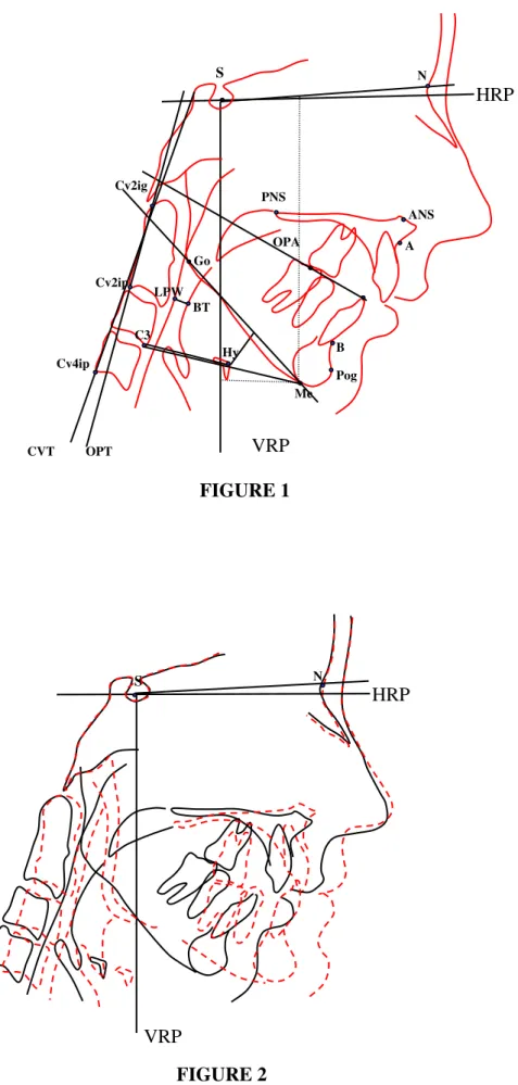
Figure 1. Reference landmarks and lines measured on a lateral cephalogram. The horizontal reference plane (HRP) was constructed at 7o to the SN plane through
sella (S). The vertical reference plane (VRP) was constructed perpendicular to HRP
through sella (S).
Figure 2. Superimposition of pre and post surgical lateral cephalograms demonstrate the surgical changes achieved.
Figure 3: A, A 3-D stereolithography model of the patient’s jaws and jaw joints was constructed from CT scan data. The mandible was repositioned on the model. The red
marks indicate areas of bony recontouring to facilitate the fit of the prosthesis. B,
Custom-fitted total joint prostheses are constructed to fit the specific anatomical
requirements for each patient.
Figure 4: Case 1 (CT, patient # 41) A, B, This 28 year old female is seen 4 years post
trauma with multiple mandibular fractures and loss of 8 maxillary teeth. She presents
with right TMJ severe arthritis and pain. The mandible and maxilla are significantly
seen 79 months post surgery following right TMJ reconstruction and mandibular
advancement with custom-made TMJ total joint prostheses, left mandibular ramus
sagittal split for advancement, and maxillary osteotomies. E, F, The patient was recently evaluated at 18 years post surgery showing the maintenance of good facial
balance.
Figure 5: Case 1 – A-C, the presurgical occlusion demonstrates anterior open bite and
the replacement of 7 teeth (left lateral incisor through the right 2nd bicuspid) with
osseo-integrated implants and prosthesis. D-F, the occlusion remained stable 79 months post
surgery. G-I, at 18 years post surgery, she maintained a stable occlusion.
Figure 6: Case 1 – A, the pretreatment cephalometric analysis shows a retruded
maxilla and mandible, anterior open bite, steep occlusal and mandibular plane angles,
vertical facial asymmetry, and significant degenerative changes in the right condyle . B,
the STO (prediction tracing) demonstrates the TMJ and orthognathic procedures
required to achieve a good functional and esthetic result including right TMJ
reconstruction and mandibular advancement with custom made TMJ total joint
prostheses, left mandibular ramus sagittal osteotomy, right coronoidectomy, and
maxillary osteotomies for counter-clockwise rotation and transverse leveling of the
maxillo-mandibular complex. C, Cephalometric analysis at 18 years post surgery demonstrates good facial balance. D, Superimposition of the immediate post surgery (red lines) and 18 year follow-up (black lines) cephalometric tracings demonstrate the
Figure 7: Case 2 – A, B, this 25 year old female presented with severe TMJ arthritis, significantly retruded maxilla and mandible, high occlusal plane angle, severe pain, and
severe sleep apnea. C, D, the patient is seen 6 years post surgery following bilateral TMJ reconstruction and mandibular advancement with custom-made TMJ total joint
prostheses (TMJ Concepts system®), bilateral coronoidectomies, simultaneous
maxillary osteotomies, and genioplasty demonstrating a good stable, functional and
esthetic outcome. She had a significant decrease in pain and elimination of the sleep
apnea.
Figure 8: Case 2 – A-C, the presurgical occlusion demonstrated an anterior open bite
(7 mm) and Class II end-on cuspid relationship. D-F, the occlusion remained stable 6 years post surgery.
Figure 9: Case 2 – A, the pretreatment cephalometric analysis shows a retruded
maxilla and mandible, anterior open bite, steep occlusal and mandibular plane angles,
and severely decreased oropharyngeal airway. B, the STO (prediction tracing) demonstrates the TMJ and orthognathic procedures required to achieve a good
functional and esthetic result including bilateral TMJ reconstruction and mandibular
advancement with custom-made TMJ total joint prostheses (TMJ Concepts system®),
bilateral coronoidectomies, maxillary osteotomies for counter-clockwise rotation of the
maxillo-mandibular complex and occlusal plane angle, and osseous genioplasty. C, Cephalometric analysis at 6 years post surgery demonstrates good facial balance. D,
Superimposition of the immediate presurgery (red lines) and 6 year post surgery
follow-up (black lines) cephalometric tracings demonstrate the treatment changes
Age Previous TMJ Surg Follow-up
n
Name
Gender (years)
TMJ Diagnoses
Right Left (months)
1 PA F 29 (B) A, (B) FA, (B) SI, (B) FBGCR 3 3 16
2 JA F 34 (B) A, (B) PTI, (B) FBGCR 1 2 25
3 JB F 50 (B) A, (B) PTI, (B) FBGCR 3 3 61
4 GB F 38 (B) A 0 0 46
5 SB F 21 (B) A 0 0 37
6 SBu F 52 (B) A 0 0 26
7 JD F 38 (B) A 0 0 37
8 CE F 14 (B) A 0 0 13
9 DE F 42 (B) A 0 0 32
10 MF F 36 (B) A 1 1 48
11 JF F 42 (B) A 0 0 12
12 KG F 39 (B) A 0 0 14
13 CG F 27 (B) A, (B) FA, (B) PTI, (B) FBGCR 3 3 32
14 EG F 43 (B) PsA 1 1 22
15 CGa F 44 (L) A 0 0 51
16 PG F 32 (B) BA, (B) SI, (B) FBGCR 2 2 12
17 SG F 45 (B) A, (B) FA, (B) F Vitek TJP, (B) FBGCR 6 3 14
18 KH F 42 (B) BA, (B) PTI, (B) FRG, (B) FBGCR 4 5 46
19 VH F 37 (B) A 0 0 22
20 KHo F 20 (B) ICR 0 0 12
21 KHa F 36 (B) A, (B) PTI, (B) SI, (B) FBGCR 3 3 35
22 CH F 57 (B) A 0 0 14
23 SJ F 18 (B) ICR 0 0 19
24 SL F 17 (B) JRA 0 0 25
25 EL F 42 (B) F Christ TJP, (B) FBGCR 1 1 14
26 NL F 46 (B) A 3 3 14
27 LL F 26 (L) A, (L) PTI, (L) FBGCR 0 1 62
28 EM F 44 (L) A 0 4 53
29 LM F 21 (B) ICR 0 0 13
30 MM F 34 (B) A, (B) PTI, (B) FBGCR 2 2 24
31 CM F 22 (B) ICR 2 2 51
32 RN F 28 (B) A 0 4 35
33 BO F 43 (B) A, (B) PTI, (B) FBGCR 1 1 49
34 DP F 30 (B) A, (B) PTI, (B) F Vitek TJP, (B) FBGCR 6 6 108 35 BP F 41 (B) PTI, (B) SI, (B) F Christ TJP, (B) FBGCR 8 8 16
36 LR F 21 (B) A 2 2 43
37 DS F 37 (B) A, (B) PTI, (B) FBGCR 2 2 91
38 LS F 41 (B) A 0 0 99
39 KS F 15 (B) JRA 0 0 86
40 ES F 35 (B) F Vitek TJP, (B) FBGCR 3 3 56
41 CT F 28 (R) A, (R) SbCoFx 0 0 79
42 GT F 21 (B) ICR 2 1 18
43 CTu F 51 (B) F Vitek TJP, (B) FBGCR 2 2 35
44 KV F 37 (B) SI, (B) F Vitek TJP, (B) FBGCR 5 4 143
45 CW F 36 (B) PTI, (B) FRG, (B) BA 4 4 51
46 CWe F 46 (B) A 1 0 24
47 TW F 25 (B) RA 1 1 72
Abreviations
(B) - Bilateral (L) - Left side (R) - Right side A - Arthritis
RA - Rheumatoid Arthritis JRA - Juvenal Rheumatoid Arthritis PsA - Psoriatic Arthritis ICR - Idiopatic Condylar Resorption PTI - Proplast/Teflon Implant SI - Silastic Implant FRG - Failed Rib Graft BA - Bony Ankylosis FA - Fibrous Ankylosis SbCoFx - Sub-condylar Fracture
F Vitek TJP - Failed Vitek-Kent Total Joint Prosthesis F Christ TJP - Failed Christensen Total Joint Prosthesis FBGCR - Foreign Body Giant Cell Reaction