Cellular Defense and Sensory Cell Survival
Require Distinct Functions of
ebi
in
Drosophila
Young-Mi Lim1, Yoshimasa Yagi2, Leo Tsuda1*
1Animal Models of Aging Project Team, Center for Development of Advanced Medicine for Dementia (CAMD), National Center for Geriatrics and Gerontology (NCGG), Obu, Aichi, Japan,2Division of Biological Science, Graduate School of Science, Nagoya University, Nagoya, Aichi, 464–8602, Japan
Abstract
The innate immune response and stress-induced apoptosis are well-established signaling pathways related to cellular defense. NF-κB and AP-1 are redox-sensitive transcription fac-tors that play important roles in those pathways. Here we show that Ebi, aDrosophila homo-log of the mammalian co-repressor molecule transducinβ-like 1 (TBL1), variously regulates the expression of specific genes that are targets of redox-sensitive transcription factors. In response to different stimuli, Ebi activated gene expression to support the acute immune response in fat bodies, whereas Ebi repressed genes that are involved in apoptosis in pho-toreceptor cells. Thus, Ebi seems to act as a regulatory switch for genes that are activated or repressed in response to different external stimuli. Our results offer clearin vivoevidence that the Ebi-containing co-repressor complex acts in a distinct manner to regulate transcrip-tion that is required for modulating the output of various processes duringDrosophila
development.
Introduction
The innate immune response is a cellular defense system that protects animals from threats such as external infections and is required for maintenance of normal physiology [1]. The innate immune system seems to be evolutionarily conserved; in fact,Drosophila melanogaster
and humans share conserved signaling machinery including receptors, signaling mediators, and transcription factors that govern the response [2]. In particular, nuclear factor-kappa B (NF-κB) plays a key role in regulating transcriptional events of the response [2,3].Drosophila
contains three types of NF-κB-like transcription factors, namely Dorsal, Dorsal-related immu-nity factor, and Relish (Rel) [2]. A large body of evidence suggests that NF-κB works together with several distinct transcription factors such as the antioxidant response element—binding protein activator protein-1 (AP-1) [4,5]. NF-κB and AP-1, which are called redox-sensitive transcription factors, participate not only in the innate immune response but also in inflamma-tion, cancer formainflamma-tion, and stress-induced apoptotic processes in cells [6,7].
a11111
OPEN ACCESS
Citation:Lim Y-M, Yagi Y, Tsuda L (2015) Cellular
Defense and Sensory Cell Survival Require Distinct Functions ofebiinDrosophila. PLoS ONE 10(11): e0141457. doi:10.1371/journal.pone.0141457
Editor:Barbara Jennings, Oxford Brookes University,
UNITED KINGDOM
Received:April 9, 2015
Accepted:October 8, 2015
Published:November 2, 2015
Copyright:© 2015 Lim et al. This is an open access
article distributed under the terms of theCreative Commons Attribution License, which permits unrestricted use, distribution, and reproduction in any medium, provided the original author and source are credited.
Data Availability Statement:All relevant data are
within the paper and its Supporting Information files.
Funding:The authors received no specific funding
for this work.
Competing Interests:The authors have declared
AP-1 and NF-κB can act as both transcriptional activators and repressors of specific target genes [8,9,10,11]. A growing number of studies have shown that many kinds of cofactors are involved in these events [12,13]. Among the cofactors, co-repressor complexes, such as nuclear receptor co-repressor (N-CoR) and the silencing mediator for retinoid and thyroid hormone receptor (SMRT) complex, appear to play important roles in reactive oxygen species—induced apoptotic signaling pathways and the innate immune response, which are both regulated by AP-1 and NF-κB [14,15,16,17]. Moreover, the N-CoR/SMRT repressor complex regulates the expression of many types of genes by associating with transducinβ-like protein 1 (TBL1) and TBL1-related protein (TBLR1), two highly related F-box/WD-40–containing factors [18, 19,20,21]. Notably, TBL1 and TBLR1 act as exchange factors that facilitate the addition and/ or extraction of factors of the transcriptional repressor complex to yield a transcriptionally active complex at the genomic target sites of various nuclear hormone receptors [16,22]. How-ever, Yoonet al. failed to observe transcriptional activation mediated by TBL1/TBLR1, and therefore the precise activities of these factors as transcriptional activators remain enigmatic [23].
Ebi, theDrosophilahomolog of TBL1/TBLR1, is also a transcriptional co-repressor when present in a complex with SMRTER, theDrosophilacounterpart of N-CoR/SMRT [24,25,26]. A recent study showed that Ebi can associate with AP-1 and that the repressor activity of Ebi is required for long-term survival of sensory photoreceptor cells [27]. AP-1/Ebi activity is required for suppression of pro-apoptotic genes (PAGs) such ashid,grim,reaper, andsickle. Loss of function ofebicauses late-onset photoreceptor cell degeneration that is AP-1 depen-dent [27].
Herein we analyzed the function of Ebi in the cellular defense response using RNA interfer-ence (RNAi)-mediated knockdown. Our work clearly shows that Ebi acts as a positive regulator for the transcriptional regulation mediated by Rel (NF-κB) and AP-1 in fat bodies. Conversely, Ebi represses Rel and AP-1 target genes in other organs including the eye disc. These results indicate that Ebi has two distinct functions with respect to regulating the expression of Rel and AP-1 target genes. The distinct function of Ebi might be required for maintaining appropriate activation of specific pathways in different tissues. Hence, Ebi seems to act as a molecular switch that modulates the output of genes that are regulated by redox-sensitive transcription factors.
Results
ebi
is required for the innate immune response in the presence of
bacterial infection
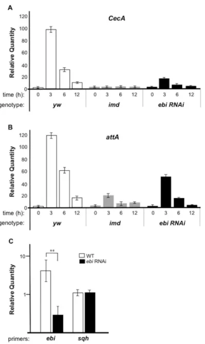
Our previous study revealed that Ebi associates with and regulates the transcriptional activity of AP-1 [27]. It has been shown that AP-1–mediated transcriptional activity regulates the innate immune response [28,29]. Thus, we investigated whether Ebi is also involved in the cel-lular defense response inDrosophila. In theDrosophilainnate immune response, the expres-sion of anti-microbial peptide genes (AMPs) is regulated by Rel together with AP-1 [29]. Therefore, we examined the function of Ebi in the regulation of AMP expression. In the pres-ence of a Gram-negative bacterial challenge, the expression of AMPs such asCecropin A
Mutation ofimdprohibits AMP induction during bacterial infection [30] (Fig 1A and 1B). Using fat bodies and the hemocyteGal4line (Cg-Gal4), we introduced double-stranded RNA (dsRNA) targetingebi(ebiHMS01390, for the purpose of RNAi) into fat bodies and observed
>70% reduction inebimRNA level in this organ (Fig 1C). Under this condition, we found that
reduced Ebi activity in fat bodies led to decreased expression of AMPs, suggesting that Ebi pos-itively regulates Rel target genes (Fig 1A and 1B).
Fig 1.ebiis required for Rel target gene expression during bacterial infection.qPCR analysis of mRNA from larvae ofyw(n = 30, each time point),Cg-Gal4>ebi RNAi(n = 30, each time point), orimdmutant larvae (n = 30, each time point). The experiment was performed two times. Expression ofCecA(A) andattA(B) was assessed at different times after bacterial challenge (0, 3, 6, 12 h). The experiment was performed three times. (C) qPCR analysis of mRNA from wild-type (WT) (n = 25) or afterCg-Gal4>ebi RNAi(n = 28).Sqh
(spaghetti squash)was used as a control for mRNA level. The data represent the mean±SD.**p<0.01,vs. mock withebiRNAi. The data was plotted by a log scale.
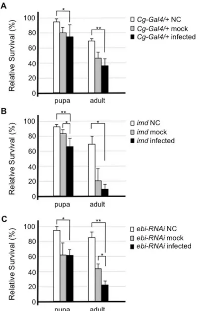
Our data thus far indicated that Ebi may mediate cellular defense signaling against Gram-negative bacterial infection. It has been shown that the innate immune system inDrosophila
supports the survival of each animal upon bacterial infection [2]. Thus, we assessed the survival of both larvae and adults after bacterial infection.Imdmutants had a markedly decreased sur-vival rate at both the pupal and adult stages upon infection (Fig 2B, compare toFig 2A). Larvae in whichebiRNAi was introduced in fat bodies also had significantly reduced survival of adult flies upon bacterial challenge (Fig 2C). These results suggested that Ebi activity in fat body cells is required for the innate immune response in the presence of infectious stimulation.
Fig 2. Ebi is involved in the cellular defense response against bacterial infection.Susceptibility to
Enterobacter cloacaein flies of each genotype was assessed. Flies were infected withE.cloacaeby pricking, and the percentage of surviving flies 2 days after infection was calculated. Error bars denote standard error. The experiment was performed four times. BothCg-Gal4/+(control) flies (A) andImdmutant flies (B) showed increased sensitivity to bacterial challenge.Cg-Gal4>ebi RNAi, in whichebiexpression was inhibited, showed enhanced lethality to bacterial challenge (C). Data represent the mean±SD.*p<0.05;**p<0.01. NC: not challenged (not pricked); mock: mock infection (pricked with a clean needle); infected: infected by pricking the insect with a pin inoculated with bacteria.
Ebi is a transcriptional activator in the fat bodies
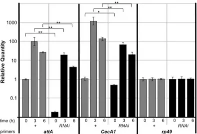
We found that treatment of fat bodies withebi-specific RNAi decreased the expression of AMPs as assessed with mRNA isolated from whole larvae. To investigate whether the regula-tion of Ebi against AMPs are cell autonomous, we analyzed AMP expression to be limited in fat bodies. We checked up the reduction in expression of AMPs when Ebi was inhibited in fat bodies (Fig 3). We noticed thatattAandCecA1were expressed at their basal levels duringebi
RNAi, suggesting that cell-autonomous Ebi function is necessary for AMP expression. We con-firmed these results using an independentebiRNAi line (ebiGLC01413), which indicated that the results were probably not a consequence of off-target effects (S2 Fig).
We previously reported that Ebi represses PAGs, many of which are targets of AP-1, in pho-toreceptor cells and S2 cells, and that loss ofebifunction results in upregulation of PAGs [27]. We thus monitored the expression ofhid,grim,reaper, andsicklebefore and after bacterial challenge. Although the basal expression of these genes was relatively low, the expression of all the PAGs did not increase withebiRNAi, and rather it seemed that expression was reduced whenebiwas inhibited (Fig 4). We also confirmed that expression ofpuckered(puc), which is another AP-1 target gene, was also inhibited byebiRNAi (Fig 4) [31]. These results indicated that, in fat bodies, Ebi itself functions as an activator rather than a repressor.
Ebi downregulates Rel target genes in non
—
fat body tissues
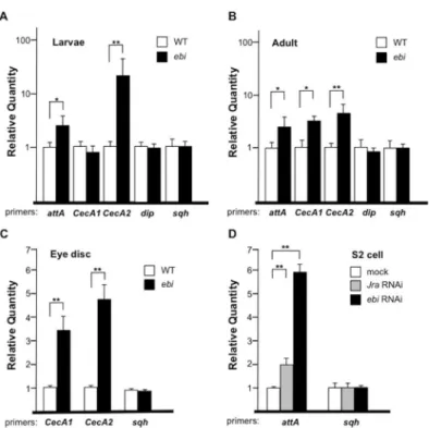
To examine whether the Ebi-mediated upregulation of AMP expression is fat-body specific, we assessed the effect of Ebi on AMP expression in non—fat body tissues. First, we monitored AMP expression inebimutant larvae and adult flies. For theebimutant analysis, we used sev-eral different mutant strains; the combination ofebiPandebi4has been shown to yield a severe loss-of-function phenotype that results in lethality at approximately the first or second instar larval stage, and the combination of strainsebi7andebi90yields adult escapers [24,26]. In this experiment, we found that some, if not all, AMPs such asCecAandattAwere ectopically induced inebimutant larvae and adult flies in the absence of bacterial infection (Fig 5A and 5B). To clarify the tissue specificity of Ebi activity with respect to Rel target genes, we assessed
Fig 3. Ebi positively regulates Rel target genes in fat bodies.Real-time qPCR analysis of mRNA from fat bodies isolated from larvae ofCg-Gal4/+(+) orCg-Gal4>ebi RNAi(RNAi) (n = 30 for each time point). Time: time elapsed after bacterial infection Specific primers for each gene were analyzed. Data represent the mean±SD.*p<0.05;**p<0.01. The experiment was performed three times. The data was plotted by a log scale.
Fig 4. Ebi positively regulates AP-1 target genes in fat bodies.qPCR analysis of mRNA from larvae ofyw
(+) orCg-Gal4>ebi RNAi(RNAi) (n = 30, each time point). Time: time elapsed after bacterial infection.hid:
head involution defective(also know asW);puc:puckerd.Rp49was used as a control. Data represent the mean±SD.*p<0.05;**p<0.01. The experiment was performed three times.
doi:10.1371/journal.pone.0141457.g004
Fig 5. Ebi and AP-1 antagonizeIMDsignaling.(A–D) qPCR analysis of mRNA from wild-type (WT) orebi
mutant (ebiP/ebi4) first-instar larvae (n = 36 and 25, respectively) (A), from WT orebimutant escaper (ebi7/ ebi90) adult flies (n = 30 and 21, respectively) 1 day after eclosion (B), from WT orebimutant escaper (ebi11/ ebi4) third-instar larvae eye-antennal discs (n = 80 and 65, respectively) (C), and from S2 cells treated with mock dsRNA, dsRNA againstJra(RNAi), orebiRNAi (D).attA:attacinA;sqh:spaghetti squash. Data represent the mean±SD.*p<0.05;**p<0.01. (A) and (B) were platted by a log scale.
CecAexpression in eye-antenna discs. In this case we used the mutant combinationebi11and
ebi4, which is lethal at the pupal stage [26]. We observed increased expression ofCecA1and
CecA2inebimutant eye-antenna discs (Fig 5C). To assess the cell autonomy of the effect of Ebi in the negative regulation of AMP expression, we tested the role of Ebi in the expression of endogenousattAin S2 cells. As with a previous study that showed thatattAexpression is repressed by AP-1 [29], RNAi directed towardsJra(Drosophilac-jun) caused a 2-fold increase inattAexpression (Fig 5D). We also found that reducedebiexpression greatly increasedattA
expression (Fig 5D). These results supported the idea that Ebi may play a role as a repressor of AMP transcription in non—fat body tissues such as eye-antenna discs.
Ebi regulates AMP expression through the promoter region
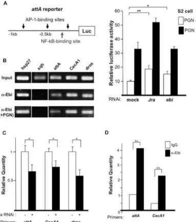
Next, we performed a reporter gene assay using anattAreporter containing binding sites for NF-κB and AP-1 upstream of the luciferase gene (Fig 6A) [32]. Peptidoglycan stimulated IMD signaling andattAexpression by more than 3-fold (Fig 6A) [33]. The basal and peptidoglycan-induced activities of the reporter were increased following treatment withJradsRNA (i.e.,
Fig 6. Ebi directly regulates the expression of AMPs.(A) Left: Reporter analysis using theattApromoter region; right: dsRNA-mediated knockdown ofJraorebiin the absence or presence of peptidoglycan (PGN). Data represent the mean±SD.*p<0.05;**p<0.01. The experiment was performed three times. (B) ChIP
analysis using different AMPs as a probe. The positive control washsp27. The experiment was performed two times. (C) ChIP-qPCR results obtained with primers specific to the promoter regions of AMPs with anti-Ebi in the absence (white bars) or presence (black bars) ofJra-specific dsRNA in S2 cells. Amplification was normalized to the control without dsRNA treatment. In all cases, the enrichment of each promoter region was inhibited by dsRNA againstJra. Data represent the mean±SD.*p<0.05;**p<0.01. The experiment was performed three times. (D) ChIP-qPCR results obtained with primers specific to the promoter regions of AMPs (attAandCecA1) with anti-Ebi (black bar) or IgG (white bar) in fat bodies (n = 100). Amplification was normalized to the internal control and calculated for each input.**p<0.01.
RNAi) (Fig 6A) [29]. Althoughebidownregulation did not enhance the reporter activity upon stimulation with peptidoglycan (Fig 6A, right), RNAi-induced reduction inebiexpression induced a small but significant elevation in the basal reporter activity. These results suggested that Ebi represses AMP expression mainly through AP-1.
Direct recruitment of Ebi to the promoter region of AMPs
To investigate how Ebi regulates AMP expression at the molecular level in S2 cells, we per-formed chromatin immunoprecipitation (ChIP). Ebi was found to be associated with the pro-moter regions ofattA,CecA1,and dros(Fig 6B). Because many AMP promoter regions contain binding sites for both AP-1 and NF-κB, we tested whether Ebi recruitment to promoters is AP-1 dependent [32]. We introduced dsRNA againstJraand found that recruitment of Ebi to the promoter regions of AMPs was decreased (Fig 6C), which is consistent with a previous report showing that Ebi associates with AP-1 and represses AP-1 target genes [27]. The observation that stimulation with peptidoglycan did not change the recruitment of Ebi to the promoter regions of those genes (Fig 6B) suggested that Ebi is continuously recruited to AMP promoter regions via AP-1. To elucidate thein vivofunction of Ebi, we performed ChIP using fat-body extract, which revealed that Ebi associated with theattA,CecA1andhidpromoter regions in fat bodies (Fig 6D;S3 Fig). These results suggested that Ebi is directly involved in the transcrip-tional regulation of AMPs and PAGs in fat bodies.
Discussion
Ebi plays distinct roles in the expression of target genes in different tissues [34]. When flies are challenged by external stimuli such as bacterial infection, cells in the fat body require high expression of AMPs to mount an adequate defense response [3]. Under such conditions, the co-repressor system may be an obstacle for the organism. Therefore, Ebi-containing co-repres-sors may be converted to activator complexes to allow efficient expression of AMPs. In non—
fat bodies such as photoreceptor cells, high levels of AMP expression are not required, and cells must be protected from apoptotic induction during stress signaling. Under these conditions, Ebi seems to act as part of a co-repressor complex to repress any excess expression of PAGs.
It has been shown that most of the promoter regions of AMPs contain consensus sequences for binding NF-κB and AP-1 [32]. There seems to be a balance for the utilization of these two transcription factors. In cultured S2 cells, AP-1 acts as a negative regulator of AMP transcrip-tion, whereas AP-1 is required for AMP activation in fat bodies. In support of this notranscrip-tion,Jra
knockdown did not impair AMP expression in S2 cells (Fig 6A), whereas it impairs PAGs expression in S2 cells [27].
Actually, the diverse function of Ebi against target gene expression seems to be evolution-arily conserved. TBL1 is involved in transcriptional activation as well as repression [16]. TBL1/ TBLR1 seems to act as a specific adaptor molecule that mediates the exchange of co-repressors for co-activators [16]. This TBL1/TBLR1-based molecular switch may contribute to the effi-cient response to external signaling [16].
switching transcriptional repression to activation by regulating the turnover of particular tran-scriptional cofactors.
External signaling pathways are thought to interact with co-repressors and co-activators [12]. Notably, Ebi has been shown to act under several distinct signaling pathways, such as those governed byEGF receptor,Notch, orwingless[24,25,37].
A recent study indicated that the opposing functions of the TBL1/TBLR1 complex are regu-lated by the Toll-like receptor signaling pathway through phosphorylation of N-CoR [38]. This suggests that extracellular signaling is closely related to the regulation of the opposing activities of TBL1/Ebi family molecules in transcriptional regulation. Thus, we expect that cellular sig-naling, such as that mediated by the Toll pathway, may contribute to Ebi activity as a transcrip-tional activator of AMPs in fat bodies.
Materials and Methods
Drosophila
stocks
The following stocks were used in this study: Oregon-R as a wild type,ebi4,ebi90,ebiP,ebip7, andebi11[24,25];y1w1118;imd1[39];GMR-Gal4[40];UAS-imd,UAS-RelN,Cg-Gal4[41],
ebiHMS01390(ebi RNAi) andebiGLC01413(ebi RNAi) was obtained from the Bloomington Dro-sophila Stock Center, USA.egrGS1226was obtained from the Kyoto Stock Center, Japan.
Bacterial infection of larvae
The bacterial infection assay was performed as described [41]. Briefly, wandering third instar larvae were washed with distilled water before infection. Bacterial infection was carried out by puncturing larvae with a needle that was previously dipped in a solution ofEnterobacter cloa-cae. After infection, the larvae were placed on wet filter paper inside a moist chamber and col-lected into tubes 0, 3, 6, or 12 h after infection and stored at−80°C until RNA extraction.
For the survival assay, wandering third instar larvae (n = 40, each) were infected and placed on wet filter paper inside a moist chamber for 1 h after infection. Larvae that did not die because of injury were moved to a culture tube, and surviving pupae or adult flies were counted.
Peptidoglycan stimulation in S2 cells
Lipopolysaccharide (Sigma) was added to the medium of S2 cells (1 × 10⁶cells) to a final con-centration of 1μg/ml. After 1.5 h, cells were collected and mRNA isolation or ChIP analysis
performed.
Primer information
All the primer information is described inS1 Methods.
Real-time quantitative PCR (qPCR)
Most qPCR procedures were performed as described [27]. Briefly, RNA was extracted using an RNA purification kit (QIAGEN), and each cDNA was synthesized with Primescript RT reagent (TAKARA). The mRNA level was quantified using a Thermal Cycler Dice Real Time System with SYBR Premix Ex Taq (TAKARA). Data were normalized torp49orsqhmRNA. The ther-mal cycling parameters were 40 cycles of 95°C for 10 s and 60°C for 30 s.
Luciferase assay
A reporter construct forattA-lucwas obtained from Dr. Jean-Luc Imler. The reporter con-structs were transfected into S2 cells (1 × 10⁶cells) using Effectene Reagent (QIAGEN), together withpActin-RLin the presence or absence of dsRNA. After 48 h, the cells were lysed, and firefly luciferase activity was analyzed with the dual luciferase reporter assay system (Pro-mega). Luciferase activity in each sample was normalized toRenillaluciferase activity.
ChIP analysis
S2 cells (1 × 10⁷) were collected in phosphate-buffered saline, fixed with 1% formaldehyde for 15 min at room temperature, and subjected to ChIP as described [42]. Cross-linked adducts were resuspended and sonicated, resulting in DNA fragments of 500–1000 bp. Immunoprecip-itation was performed using an rabbit polyclonal antibody against Ebi [24]. Protein-bound, immunoprecipitated DNA was dissolved in Tris/EDTA buffer (pH 7.8) and incubated at 65°C for 6 h. Digestion buffer (10 mM Tris-HCl, 100 mM NaCl, 25 mM EDTA, pH 8.0) was added to the sample and incubated for 1 h at 45°C with 0.1 mg/ml proteinase K (Sigma). DNA was purified using the PureLink Plasmid purification kit (Invitrogen) and used as a template for qPCR. The primer set forhsp27, which is a target site for ecdysone receptor (EcR), was used as a positive control [26]. Oligonucleotides for ChIP analysis, real-time qPCR, and dsRNA for ChIP analysis are described in the supplemental information.
Acridine orange staining
Acridine orange staining was performed as described [40]. Eye-antennal discs were dissected in phosphate-buffered saline and incubated with 1.6μM acridine orange solution inDrosophila
Ringer [43] and then mounted in Vectashield (Vector Laboratories).
Histochemistry
Sectioning of eyes and epon embedding were carried out as described [24]. Briefly, dissected eyes were fixed in 2.5% glutaraldehyde, dehydrated, and embedded in epon plastic. Thin sec-tions were stained with toluidine blue for light microscopy.
Additional Materials and Methods are described inS1 Methods.
Supporting Information
S1 Fig.sqhgene expression during bacterial infection.qPCR analysis of mRNA from larvae ofyw,Cg-Gal4>ebi RNAi, orimdmutant larvae.sqhexpression was observed at different
times after bacterial challenge (0, 3, 6, 12 h). Data represent the mean ± SD. (TIF)
S2 Fig. Ebi positively regulates Rel target genes in fat bodies.qPCR analysis of mRNA from larvae ofCg-Gal4/+(+) orCg-Gal4>ebi RNAi(ebiGLC01413) (n = 30 for each). The word“time”
implies the time after bacteria infection. Specific primers for each gene were analyzed. Data represent the mean ± SD.p<0.05;p<0.01. n = 4. The experiment was performed three
times. (TIF)
input. Data represent the mean ± SD.p<0.01. The experiment was performed two times.
(TIF)
S1 Methods. Oligonucleotides for ChIP analysis, real-time PCR, and dsRNA for ChIP.
(DOC)
Acknowledgments
We are grateful to S. Hayashi for critical discussion and support.
We also thank J. Imler for providing theattA-lucconstruct and our lab members for techni-cal assistance and discussion.
Author Contributions
Conceived and designed the experiments: LT Y-ML. Performed the experiments: LT Y-ML YY. Analyzed the data: LT Y-ML. Contributed reagents/materials/analysis tools: LT Y-ML. Wrote the paper: LT Y-ML.
References
1. Pott J, Hornef M (2012) Innate immune signaling at the intestinal epithelium in homeostasis and dis-ease. EMBO Rep 13: 684–698. doi:10.1038/embor.2012.96PMID:22801555
2. Ganesan S, Aggarwal K, Paquette N, and Silverman N (2011) NF-κB/Rel proteins and the humoral immune responses of Drosophila melanogaster. Curr Top Microbiol Immunol 349: 25–60. doi:10.
1007/82_2010_107PMID:20852987
3. Herbein G, Varin A, Fulop T (2006) NF-κB, AP-1, Zinc-deficiency and aging. Biogerontol 7: 409–419.
4. Gerald D, Berra E, Frapart YM, Chan DA, Giaccia AJ, Mansuy D, et al. (2004) JunD reduces tumor angiogenesis by protecting cells from oxidative stress. Cell 118: 781–794. PMID:15369676
5. Piette J, Piret B, Bonizzi G, Schoonbroodt S, Merville MP, Legrand-Poels S, et al. (1997) Multiple redox regulation in NF-kappaB transcription factor activation. Biol Chem 378: 1237–1245. PMID:9426183
6. Aggarwal BB (2000) Tumor necrosis factors receptor associated signaling molecules and their role in activation of apoptosis, JNK and NF-κB. Ann Rheum 59 (suppl I): i6–i16.
7. Garg AK, Aggarwal BB (2002) Reactive oxygen intermediates in TNF signaling. Mol Immunol 39: 509–
517. PMID:12431383
8. Hsu JC, Cressman DE, Taub R (1993) Promoter-specific trans-activation and inhibition mediated by JunB. Cancer Res 53: 3789–3794. PMID:8339292
9. Berger I, Shaul Y (1998) c-Fos antagonizes the junD gene positive autoregulatory loop: a novel c-Fos role in promoter switching. Gene 211: 375–382. PMID:9602173
10. Baek SH, Ohgi KA, Rose DW, Koo EH, Glass CK, Rosenfeld MG (2002) Exchange of N-CoR corepres-sor and Tip60 coactivator complexes links gene expression by NF-kappaB and beta-amyloid precurcorepres-sor protein. Cell 110: 55–67. PMID:12150997
11. Espinosa L, Ingles-Esteve, Robert-Moreno A, Bigas A (2003) IkappaBalpha and p65 regulate the cyto-plasmic shuttling of nuclear corepressors: cross-talk between Notch and NFkappaB pathways. Mol Biol Cell 14: 491–502. PMID:12589049
12. Rosenfeld MG, Lunyak VV, Glass CK (2006) Sensors and signals: a coactivator/corepressor/epige-netic code for integrating signal-dependent programs of transcriptional response. Genes Dev 20: 1405–1428. PMID:16751179
13. Payankaulam S, Li LM, Arnosti DN (2010) Transcriptional repression: conserved and evolved features. Curr Biol 20: R764–R771. doi:10.1016/j.cub.2010.06.037PMID:20833321
14. Lee SK, Kim JH, Lee YC, Cheong J, Lee JW (2000) Silencing mediator of retinoic acid and thyroid hor-mone receptors, as a novel transcriptional corepressor molecules of activating protein-1, nuclear fac-tor-kappaB, and serum response factor. J Biol Chem 275: 12470–12474. PMID:10777532
15. Zhang J, Kalkum M, Chait BT, Roeder RG (2002) The N-CoR-HDAC3 nuclear receptor corepressor complex inhibits the JNK pathway through the integral subunit GPS2. Mol Cell 9: 611–623. PMID:
16. Perissi V, Aggarwal A, Glass CK, Rose DW, Rosenfeld MG (2004) A corepressor/coactivator exchange complex required for transcriptional activation by nuclear receptors and other regulated transcription factors. Cell 116: 511–526. PMID:14980219
17. Yan J, Gao Z, He Q, Weng J, Ye J (2007) Nuclear corepressor is required for inhibition of phosphoenol-pyruvate carboxykinase expression by tumor necrosis factor-alpha. Mol Endocrinol 21: 1630–1641.
PMID:17456789
18. Guenther MG, Lane WS, Fischle W, Verdin E, Lazar MA, Shekhattar R. (2000) A core SMRT corepres-sor complex containing HDAC3 and TBL1, a WD40-repeat protein linked to deafness. Genes Dev 14: 1048–1057. PMID:10809664
19. Tomita A, Buchholz DR, Shi YB (2004) Recruitment of N-CoR/SMRT-TBLR1 corepressor complex by unliganded thyroid hormone receptor for gene repression during frog development. Mol Cell Biol 24: 3337–3346. PMID:15060155
20. Yoon HG, Chan DW, Huang ZQ, Li J, Fondell JD, Qin J, et al. (2003) Purification and functional charac-terization of the human N-CoR complex: the roles of HDAC3, TBL1 and TBLR1. EMBO J 17: 1336–
1346.
21. Ogawa S, Lozach J, Jepsen K, Sawka-Verhelle D, Perissi V, Sasik R, et al. (2004) A nuclear receptor corepressor transcriptional checkpoint controlling activator protein 1-dependent gene networks required for macrophage activation. Proc Natl Acad Sci, U S A 101: 14461–14466. PMID:15452344
22. Perissi V, Scafoglio C, Zhang J, Ohgi KA, Rose DW, Glass CK, et al. (2008) TBL1 and TBLR1 phos-phorylation on regulated gene promoters overcomes dual CtBP and NCoR/SMRT transcriptional repression checkpoints. Mol Cell 29: 755–766. doi:10.1016/j.molcel.2008.01.020PMID:18374649
23. Yoon HG, Choi Y, Cole PA, Wong J (2005). Reading and function of a histone code involved in target-ing corepressor complexes for repression. Mol Cell Biol 25: 324–335. PMID:15601853
24. Dong X, Tsuda L, Zavitz KH, Lin M, Li S, Carthew RW, et al. (1999)ebiregulates epidermal growth fac-tor recepfac-tor signalling pathways in Drosophila. Genes Dev 13: 954–965. PMID:10215623
25. Tsuda L, Nagaraj R, Zipursky SL, Banerjee U (2002) An EGFR/Ebi/Sno pathway promotes delta expression by inactivating Su(H)/SMRTER repression during inductive notch signalling. Cell 110: 625–
637. PMID:12230979
26. Tsuda L, Kaido M, Lim YM, Kato K, Aigaki T, Hayashi S. (2006) An NRSF/REST-like repressor down-stream of Ebi/SMRTER/Su(H) regulates eye development in Drosophila. EMBO J 25: 3191–3202.
PMID:16763555
27. Lim YM, Hayashi S and Tsuda L (2012) Ebi/AP-1 suppresses pro-apoptotic gene expression and per-mits long-term survival ofDrosophilasensory neurons. PLOS ONE 7: e37028. doi:10.1371/journal. pone.0037028PMID:22666340
28. Boutros M, Agaisse H, Perimmon N (2002) Sequential Activaton of Signaling Pathways during innate immune responses in Drosophila. Dev Cell 3: 711–722. PMID:12431377
29. Kim T, Yoon J, Cho H, Lee WB, Kim J, Yoon JH, et al. (2005) Downregulation of lipopolysaccharide response in Drosophila by negative crosstalk between the AP1 and NF-kappaB signalling modules. Nat Immunol 6: 211–218. PMID:15640802
30. Georgel P, Naitza S, Kappler C, Ferrandon D, Zachary D, Swimmer C, et al. (2001) Drosophila immune deficiency (IMD) is a death domain protein that activates antibacterial defense and can promote apopto-sis. Dev Cell 1: 503–514. PMID:11703941
31. Martin-Blanco E, Gampel A, Ring J, Virdee K, Kirov N, Tolkovsky AM, et al. (1998) Puckered encodes a phosphatase that mediates a feedback loop regulating JNK activity during dorsal closure in Drosophila. Genes Dev 12: 557–570. PMID:9472024
32. Kim LK, Choi UY, Cho HS, Lee JS, Lee WB, Kim J, et al. (2007) Down-regulation of NF-kappaB target genes by the AP-1 and STAT complex during the innate immune response in Drosophila. PLOS Biol 5: e238. PMID:17803358
33. Kappler C, Meister M, Lagueux M, Gateff E, Hoffmann JA, Reichhart JM. (1993) Insect immunity. Two 17 bp repeats nesting a kappa B-related sequence confer inducibility to the diptericin gene and bind a polypeptide in bacteria-challenged Drosophila. EMBO J 12: 1561–1568. PMID:8467806
34. Lim YM, Yamasaki Y, and Tsuda L (2013) Ebi alleviates excessive growth signaling through multiple epigenetic functions in Drosophila. Genes to Cell 18: 909–920.
35. Boulton SJ, Brook A, Staehling-Hampton K, Heitzler P, Dyson N (2000) A role for Ebi in neuronal cell cycle control. EMBO J 19: 5376–8386. PMID:11032805
37. Li J, Wang CY (2008) TBL1-TBLR1 and beta-catenin recruit each other to Wnt target-gene promoter for transcription activation and oncogenesis. Nat Cell Biol 10: 160–169. doi:10.1038/ncb1684PMID:
18193033
38. Huang W, Ghisletti S, Perissi V, Rosenfeld MG, Glass CK (2009) Transcriptional integration of TLR2 and TLR4 signaling at the NCoR derepression checkpoint. Moll Cell 35: 48–57.
39. Yagi Y, Lim YM, Tsuda L, and Nishida Y (2013)fat facetsinduces polyubiquitination of Imd and inhibits the innate immune response in Drosophila. Genes to Cell 18: 934–945.
40. Hay BA, Wolff T, Rubin GM (1994) Expression of baculovirus P35 prevents cell death in Drosophila. Development 120: 2121–2129. PMID:7925015
41. Yagi Y, Ip YT (2005) Helicase89B is a Mot1p/BTAF1 homologue that mediates an antimicrobial response in Drosophila. EMBO reports 6: 1088–1094. PMID:16200050
42. Hecht A, Grunstein M (1999) Mapping DNA interaction sites of chromosomal proteins using immuno-precipitation and polymerase chain reaction. Methods Enzymol 304: 399–414. PMID:10372373
43. Robb JA (1969) Maintenance of imaginal discs of Drosophila melanogaster in chemically defined media. J Cell Biol 41: 876–885. PMID:5768877