High Intensity Exercise in Multiple Sclerosis:
Effects on Muscle Contractile Characteristics
and Exercise Capacity, a Randomised
Controlled Trial
Inez Wens1☯*, Ulrik Dalgas2☯, Frank Vandenabeele1‡
, Lotte Grevendonk1‡, Kenneth Verboven1‡, Dominique Hansen1‡, Bert O. Eijnde1☯
1REVAL Rehabilitation Research Center, BIOMED Biomedical Research Institute, Faculty of Medicine and Life Sciences, Hasselt University, Agoralaan Building A, Diepenbeek, Belgium,2Section of Sport Science, Dep. Public Health, Aarhus University, Dalgas Avenue 4, 8000, Aarhus, C, Denmark
☯These authors contributed equally to this work. ‡These authors also contributed equally to this work. *[email protected]
Abstract
Introduction
Low-to-moderate intensity exercise improves muscle contractile properties and endurance capacity in multiple sclerosis (MS). The impact of high intensity exercise remains unknown.
Methods
Thirty-four MS patients were randomized into a sedentary control group (SED, n = 11) and 2 exercise groups that performed 12 weeks of a high intensity interval (HITR, n = 12) or high intensity continuous cardiovascular training (HCTR, n = 11), both in combination with resis-tance training. M.vastus lateralis fiber cross sectional area (CSA) and proportion, knee-flexor/extensor strength, body composition, maximal endurance capacity and self-reported physical activity levels were assessed before and after 12 weeks.
Results
Compared to SED, 12 weeks of high intensity exercise increased mean fiber CSA (HITR: +21±7%, HCTR: +23±5%). Furthermore, fiber type I CSA increased in HCTR (+29±6%), whereas type II (+23±7%) and IIa (+23±6%,) CSA increased in HITR. Muscle strength improved in HITR and HCTR (between +13±7% and +45±20%) and body fat percentage tended to decrease (HITR: -3.9±2.0% and HCTR: -2.5±1.2%). Furthermore, endurance capacity (Wmax+21±4%, time to exhaustion +24±5%, VO2max+17±5%) and lean tissue mass (+1.4±0.5%) only increased in HITR. Finally self-reported physical activity levels increased 73±19% and 86±27% in HCTR and HITR, respectively.
OPEN ACCESS
Citation:Wens I, Dalgas U, Vandenabeele F, Grevendonk L, Verboven K, Hansen D, et al. (2015) High Intensity Exercise in Multiple Sclerosis: Effects on Muscle Contractile Characteristics and Exercise Capacity, a Randomised Controlled Trial. PLoS ONE 10(9): e0133697. doi:10.1371/journal.pone.0133697
Editor:Conrad P. Earnest, Texas A&M University, UNITED STATES
Received:November 13, 2014
Accepted:June 30, 2015
Published:September 29, 2015
Copyright:© 2015 Wens et al. This is an open access article distributed under the terms of the Creative Commons Attribution License, which permits unrestricted use, distribution, and reproduction in any medium, provided the original author and source are credited.
Data Availability Statement:All relevant data are within the paper and its Supporting Information files.
Conclusion
High intensity cardiovascular exercise combined with resistance training was safe, well tol-erated and improved muscle contractile characteristics and endurance capacity in MS.
Trial Registration
ClinicalTrials.govNCT01845896
Introduction
The heterogeneous symptoms of multiple sclerosis (MS) often lead to a more sedentary lifestyle [1]. This may result in disuse-related loss of exercise capacity and muscle strength, which in turn can affect quality of life [2]. Increasing evidence favors exercise therapy as a method for overall symptom management [3]. Observational [4,5] as well as interventional studies [6–9] have reported improvements in exercise tolerance, muscle strength, functional capacity and health-related quality of life after low-to-moderate intensity cardiovascular or resistance train-ing. Although combined cardiovascular and resistance training could, from a theoretical point of view, positively affect both the cardiovascular system and muscle strength/activation[10], this type of rehabilitation/exercise therapy has not been investigated extensively [11–15].
Several authors already suggested that MS patients could benefit more from higher training intensities [10,16,17], but so far, no studies on combined exercise have evaluated high intensity training in MS. In healthy controls (HC) and in other populations, high intensity exercise and high intensity interval training (HIT) have previously been investigated, showing profound
improvements in endurance performance and muscle strength [18,19], reduced subcutaneous and abdominal fat [20], improved functional recovery (after stroke) [21] and beneficial effects to the heart [22], emphasising the need to investigate this in MS.
To date the impact of MS on skeletal muscle characteristics, such as muscle fiber cross sec-tional area (CSA) and proportion remains unclear. Recently, we reported reduced muscle fiber CSA and changed fiber proportions in MS patients, compared to HC [23]. The impact of exer-cise on muscle contractile properties in MS has only been investigated by Dalgas and co-work-ers [24]. They reported increased m.vastus lateralis mean fiber CSA combined with improved muscle strength following 12 weeks of progressive resistance training. Despite the importance of understanding the effects of exercise on muscle fiber characteristics to optimize exercise and rehabilitations programs in MS, the impact of other training modalities and intensities on mus-cle fiber CSA and fiber type proportion in MS, has not been investigated yet.
To determine the effects of high intensity exercise in MS, this study aimed to investigate the impact of high intensity interval or continuous cardiovascular exercise, both in combination with resistance training, on muscle contractile characteristics, in terms of muscle fiber CSA/ proportion, muscle strength and muscle mass and on endurance capacity in MS. It was hypoth-esized that the applied intense programs could improve mean muscle fiber CSA and propor-tion as well as muscle strength and endurance capacity.
Methods
Participants
Thirty-four MS patients diagnosed according to McDonald criteria (EDSS range 1–5), aged >18 years, were included following written informed consent (Fig 1). Subjects were excluded if
analysis, decision to publish, or preparation of the manuscript.
they had other disorders (cancer, cardiovascular, pulmonary and/or renal), were pregnant, par-ticipated in another study, were already physical active, had an acute MS-exacerbation 6 months prior to the start of the study or contra-indications to perform physical exercise.
The study was approved by the ethical committee of Jessa Hospital Hasselt (S1 Protocol) and Hasselt University (12/02/2013), whereupon the preparation of the study started in March 2013 (to order the appropriate equipment, to organise info sessions etc.). Next, this study was registered at ClinicalTrials.gov (NCT01845896, initial release 30/04/2013), at the beginning of patient recruitment (April-June). Furthermore, the authors confirm that all on-going and related trials for this intervention are registered. Finally, all tests were performed in accordance with the Declaration of Helsinki.
Study design overview
All MS patients were randomized, by means of sealed envelopes, into a sedentary control group (SED, n = 11) and 2 exercise groups that performed 12 weeks of a high intensity interval + resistance training (HITR, n = 12) or high intensity continuous endurance + resistance
train-ing (HCTR, n = 11). M.vastus lateralis fiber CSA and proportion, knee flexor and extensor
strength, body composition, maximal endurance capacity and self-reported physical activity levels were assessed before and after the intervention. Neither the patients nor the researchers involved in the project were blinded to group allocation. SED remained physical inactive ing the study course and were instructed to continue their current level of physical activity dur-ing the period of the study (S1 CONSORT Checklist).
Exercise intervention program
After the baseline measurements, the subjects were enrolled in a well-controlled and supervised training program, to increase cardiorespiratory fitness, as well as strength of the major periph-eral muscle groups. Subjects participated in 5 sessions per 2 weeks. Training sessions were interspersed by at least one day of rest, to ensure adequate recovery. Each session started with endurance training, followed by resistance training, interspersed by a short resting period.
HITR program: Each session started with a 5min warm-up on a cycle ergometer. Hereafter, high intensity cycle interval training was performed. During the first 6 weeks exercise duration gradually increased from 5x1min interspersed by 1min rest intervals to 5x2min and 1min rest
Fig 1. Consort flow diagram for participants’inclusion.
intervals. Exercise intensity was defined as the heart rate, corresponding to 100% of the maxi-mal workload (which was comparable to approximately 80–90% of the maximal heart rate). During the second 6 weeks, duration remained stable at 5x2min and the heart rate increased to reach a level corresponding to 100–120% of the maximal work load (which was comparable to approximately 90–100% of the initial maximal heart rate). The second part consisted of moder-ate-to-high intensity resistance training (leg press, leg curl, leg extension, vertical traction, arm curl and chest press, Technogym). In order to exercise at similar relative workload, resistance training of the lower limb was performed unilaterally, due to the frequent bilateral strength dif-ferences seen between the legs of MS patients.[25] Training intensity and volume were adjusted from 1x10 repetitions to 2x20 repetitions at maximal attainable load. Maximal attainable load was expressed as the maximal load that the subject was able to manage, under guidance and consequent encouragement. By applying the same standardised encouragements in all groups, subjects were stimulated to perform at their personal maximal ability.
HCTR program: Each session started with a cardiovascular part, consisting of cycling and treadmill walking/running (Technogym). Session duration and exercise intensity increased as the intervention progressed, starting from 1x6min/session to 2x10min/session, at a high work-load, corresponding to 80–90% of maximal heart rate and according to individual capabilities. The second part of the training session comprised similar resistance training, as described in the HITR program.
All exercises were performed at a high workload corresponding to 14–16 ratings of per-ceived exertion on 20-point Borg scale (RPE) and were adjusted to individual disability level. The Borg Rating of Perceiver Exertion Scale measures perceived exertion and is used to docu-ment the person’s exertion during a test or to assess the intensity of training and rehabilitation. The scale ranges from 6 to 20, where 6 means“no exertion at all”and 20 means“maximal exer-tion”. Continuous encouragement by the instructors led to a systematic increase of the training load over the 12-week training period. All sessions were ended by stretching of the extremities, and RPE-level was recorded.
Primary outcome measure
1. Muscle fiber CSA and proportion. To investigate muscle fiber CSA and proportion, muscle biopsies form the middle part of the m.vastus lateralis (Bergström needle technique) of the weakest leg (see isometric muscle strength measurements) were collected by an experienced medical doctor. The second biopsy, following 12 weeks of exercise or usual care, was taken 2-3cm proximal to the biopsy taken at baseline. Muscle samples were immediately mounted with Tissue-Tek, frozen in isopentane cooled with liquid nitrogen and stored at -80°C, until further analysis. The cross-sections of the biopsies, collected at baseline and after 12 weeks, were pro-cessed simultaneously.
Serial transverse sections (9μm) from the obtained muscle samples were cut at -20°C and
Calculation of the fiber CSA was performed for the major fiber types (I, IIa and IIx) and for the mean fiber CSA, since the number of type IIax and IIc fibers was too small for statistical comparison and CSA calculation.
Secondary outcome measures
Approximately 1–2 weeks before the muscle biopsy was performed secondary outcome mea-sures were assessed from all subjects.
1. Isometric muscle strength. After 5min of warming-up on a cycle ergometer and follow-ing habitation, the maximal voluntary isometric muscle strength of the knee extensors and flex-ors (45° and 90° knee angle) were measured, as described elsewhere [27], using an isokinetic dynamometer (System 3, Biodex, ENRAF-NONIUS, New York, USA). Two maximal isometric extensions (4s) and flexions (4s), followed by a 30s rest interval, were performed. The highest iso-metric extension and flexion peak torques (Nm) were selected as the maximal isoiso-metric strength. Baseline results were used to classify the legs of each patient as weakest or strongest leg. This sub-division was maintained in further analysis, replacing a conventional left-right classification.
2. Endurance capacity. During the exercise test to volitional fatigue, an electronically braked cycle ergometer (eBike Basic, General Electric GmbH, Bitz, Germany) with pulmonary gas exchange analysis (Jaeger Oxycon, Erich Jaeger GmbH, Germany) was used (cycling fre-quency: 70 rpm). Jaeger calibration (ambient conditions, volume calibration and O2/CO2
cali-bration) was performed at the start of each test day. This test was performed at least 48 hours separated from the isometric muscle strength test to exclude interference of muscle fatigue. Female and male MS patients started at 20W and 30W, respectively, during the first minute. Hereafter, workloads increased, respectively, 10W and 15W per minute. Oxygen uptake (VO2),
expiratory volume (VE), and respiratory exchange ratio (RER) were collected breath-by-breath and averaged every 10 seconds. Using a 12-lead ECG device, heart rate (HR) was monitored every minute. At the end of the test RER values were evaluated to verify that the test was maxi-mal (RER1.15) [28]. In addition, maximal cycling resistance (Wmax), maximal heart rate
(HRmax), test duration and VO2max, defined as the corresponding load, heart rate, amount of
minutes and oxygen uptake measured at the level of exhaustion, were reported.
3. Body composition. A Dual Energy X-ray Absorptiometry scan (Hologic Series Delphi-A Fan Beam X-ray Bone Densitometer, Vilvoorde, Belgium) was performed pre- en post-inter-vention. Fat and lean tissue mass were obtained for whole body, legs, trunk, gynoid and android region. Waist-to-hip fat mass ratio (android fat (g)/gynoid fat (g) ratio) and fat mass of the trunk/fat mass of the limbs ratio were calculated.
4. Physical activity level. Before and after the intervention, patients were asked to report their physical activity level by using the Physical Activity Scale for Individuals with Physical Disabilities (PASIPD) [29]. Respondents were asked to report the number of days and average hours in a day spent engaging in 13 activities (including recreational, household, and occupa-tional activities) over the last 7 days. Frequency responses range from 1 (never) to 4 (often), and duration responses range from 1 (less than 1 hour) to 4 (more than 4 hours). Total scores were calculated as the product of the average hours spent in an activity daily and the metabolic equivalents (MET) summed over each item. Scores range from 0 (no activity) to over 100 METh/week (very high). At baseline all patients needed to be physical inactive, to be included
in the study. Physical inactivity was defined as<30 METh/week.
Statistical analysis
(SED, HCTR and HITR) were analysed by a one-way ANOVA, whereas within group
differ-ences (post minus pre) were analysed with a paired student’s t-test. Relative changes due to the intervention were calculated as the mean of the individual changes and expressed as a percent-age. Correlations between changes of the primary and changes of the secondary outcome mea-sures on grouped data from all groups were analysed by means of Pearson’s correlation analysis. Multiple comparison was corrected by means of Bonferroni correction. All data are presented as mean±SE. P<0.01 represents the threshold for statistical significance.
Results
Baseline subject characteristics and adherence to the intervention
At baseline, no differences in general subject and disease characteristics (Table 1) as well as out-come measures were found between groups. Approximately 90% of the 30 supervised training sessions were attended in both exercise groups and no severe symptoms exacerbations and/or adverse events were reported. Furthermore, no patient drop out was noted.
Primary outcome measure
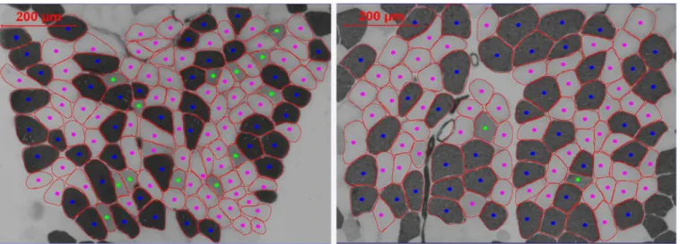
1. Muscle fiber CSA and proportion. Fig 2shows a representative image of muscle fiber types before and after high intensity exercise. In SED muscle fiber CSA and proportion did not change (p>0.05). Mean CSA significantly increased in HITR and HCTR following 12 weeks of exercise (p = 0.009 and p = 0.002, respectively). Furthermore, muscle fiber type I CSA increased in HCTR (p = 0.003), whereas muscle fiber type II and IIa increased in HITR (p = 0.007 and
p = 0.002, respectively). Fiber type IIx CSA did not change (p>0.05). In general, no changes in fiber type proportion were observed in any exercise group after 12 weeks of exercise. However, within group effects were observed on type IIx of HCTR (p = 0.001), after comparison of the
pre- and post-intervention fiber type proportion values (Table 2).
Secondary outcome measures
1. Isometric muscle strength. Muscle strength of SED remained stable during 12 weeks of usual care (p>0.05,Fig 3). Compared to SED, knee flexion and knee extension strength of the weakest leg of HITR improved by 24±13 to 44±20% (p values between 0.01 and 0.006), whereas
only hamstring strength of the strongest leg of HITR improved by 13±7 to 20±7% (p = 0.006). Table 1. Baseline subject and disease characteristics.Data is presented as mean±SE. Differences between groups (SED, HCTR and HITR) were ana-lysed by a one-way ANOVA. Abbreviations used: MS, multiple sclerosis; SED, sedentary group; HCTR, intense continuous endurance + resistance training;
HITR, high intensity interval training + resistance training, BMI, body mass index; RR, relapsing remitting; CP, chronic progressive; EDSS, expanded disability
status scale; immunomodulatory: interferonβ, glatiramer acetate, fingolimod, natalizumab.
SED (n = 11) HCTR (n = 11) HITR (n = 12) p-value
age (y) 47±3 47±3 43±3 0.22
height (m) 1.67±0.02 1.69±0.02 1.7±0.02 0.32
weight (kg) 75.8±3.6 70.2±3.7 75.9±4.1 0.17
BMI (kg/m2) 27.0±1.4 24.4±1.2 26.1±1.14 0.11
Lean tissue mass (kg) 43.2±2.1 45.4±2.6 48.5±3.1 0.11
Fat percentage (%) 38.2±2.1 33.6±2.8 36.2±1.9 0.20
gender (m/f) 2/9 5/6 5/7 0.12
type MS (RR/CP) 8/3 8/3 10/2 0.8
EDSS 2.5±0.3 2.7±0.3 2.3±0.3 0.41
Immunomodulatory MS treatment 72% 80% 80% 0.23
Furthermore, HCTR flexion and extension strength improved, from pre- to post trial, in the
weakest leg by 19±9 to 33±17% (p values between 0.01 and 0.006), whereas muscle strength of the strongest leg remained stable (p>0.05).
2. Endurance capacity. After 12 weeks, endurance capacity variables remained stable in SED and HCTR. Compared to SED and HCTR, Wmax(+21±4%, p = 0.0001), test duration (+24
±5%, p = 0.00008) and VO2max(+17±5%, p = 0.001) significantly improved in HITR (Table 3). Fig 2. Representative image of fiber type analysis before (left) and after (right) high intensity exercise.Different fiber types are distinguished by color (dark blue: type I, pink: type IIa, green: type IIx, light blue: type IIc). Calculation of the fiber CSA was performed for the major fiber types (I, IIa and IIx) and for the mean fiber CSA, since the number of fibers expressing the minor fiber types (IIax and IIc) was too small for statistical comparison and CSA calculation.
doi:10.1371/journal.pone.0133697.g002
Table 2. Muscle fiber type proportion and cross sectional area (CSA) at baseline and after 12 weeks of usual care or high intensity aerobic exercise in combination with resistance training.Data are reported as mean±SE. Differences between groups (SED, HCTR and HITR) were analysed by a one-way ANOVA, whereas within group differences (post minus pre) were analysed with a paired student’s t-test. Relative changes due to the intervention were calculated as the mean of the individual changes and expressed as a percentage. Abbreviations used: SED, sedentary (usual care); HCTR, high intensity
con-tinuous exercise + resistance training; HITR, high intensity interval training + resistance training.
SED HCTR HITR
Pre Post % Pre Post % Pre Post %
Fiber type proportion (%)
Type I 44.2±3.9 47.5±2.9 7.9±7.5 40.1±4.7 46.9±4.7b 26.8±11.3 41.3±3.0 46.3±2.6b 21.7±10.1 Type IIa 34.2±3.9 34.2±2.3 5.1±13.1 34.1±2.9 38.9±4.6 6.6±7.5 40.9±3.8 44.5±2.4 6.9±8.1 Type IIx 21.2±4.5 17.7±2.0 19.2±12.6 24.3±2.7 13.5±2.6a -46.0
±7.6c 18.5±2.8 10.1±2.8 -20.1±25.4 Fiber CSA (μm2)
Mean 3738±267 3740±431 3.5±4.3 3551±351 3905±408a 23.3
±4.9c 4038±321 4892±379a 21.1 ±7.3d Type I 4078±384 4050±531 4.0±5.5 3630±443 4071±470a 29.8
±5.5c 4410±188 4916±399 12.1±8.7 Type II 3487±265 3478±334 6.9±5.8 3285±321 3622±398b 20.8
±7.9 3612±429 4551±462a 22.7 ±6.8 Type IIa 3703±306 3729±402 3.6±3.1 3719±366 4014±522b 15.1
±5.3 4037±444 5034±447a 22.8 ±6.2d
Type IIx 3446±305 3191±318 5.4±8.2 2771±277 2955±258 14.5±8.9 3187±438 3920±519b 23.6 ±8.8
ap<0.01
bp0.05, compared with pre-intervention value, within group.
cp<0.01
dp0.05, pre to post change compared with change from pre to post in SED.
3. Body composition. Following 12 weeks of exercise, body weight remained stable in all groups (p>0.05). Within HITR and HCTR, body fat percentage tended to decrease by 3.9±2.0% (p = 0.04) and 2.5±1.2% (p = 0.02), respectively. Furthermore, lean tissue mass significantly increased 1.4±0.5% within HITR (p = 0.01), whereas it remained stable in HCTR and SED Fig 3. Percentage change of knee extension and flexion after 12 weeks of physical inactive living (usual care, SED), high intensity continuous training + resistance training (HCTR) and high intensity interval training + resistance training (HITR).Data are reported as mean±SE.*p<0.05, compared with pre-intervention value, within group.ˠp<0.05, pre to post change compared with change from pre to post in SED. Abbreviations used: KF, knee flexion; KE, knee extension.
doi:10.1371/journal.pone.0133697.g003
Table 3. Exercise capacity, body composition and physical activity level after 12 weeks of usual care or high intensity aerobic exercise in combi-nation with resistance training.Data are reported as mean±SE. Differences between groups (SED, HCTR and HITR) were analysed by a one-way
ANOVA, whereas within group differences (post minus pre) were analysed with a paired student’s t-test. Relative changes due to the intervention were calcu-lated as the mean of the individual changes and expressed as a percentage. Abbreviations used: SED, sedentary (usual care); HCTR, high intensity
continu-ous exercise + resistance training; HITR, high intensity interval training + resistance training; MET, metabolic equivalent.
SED HCTR HITR
Pre Post % Pre Post % Pre Post %
Exercise capacity:
Maximal cycling resistance (watt) 121±8 115±11 -4.6±2.7 131±18 133±18 3.6±2.8 158±15 188±15a 21.2 ±3.9c Maximal cycling resistance (watt/kg) 1.6±0.12 1.6±0.15 -4.6±2.7 1.85±0.24 1.9±0.23 3.6±2.8 2.0±0.17 2.4±0.16a 21.2
±3.9c
Test duration (min) 10.4±0.8 9.9±0.8 -3.1±2.9 9.5±1.0 9.8±0.9 5.2±3.1 12.1±0.9 14.5±0.9a 24.7 ±4.6c
VO2max (ml/min) 1647±133 1645±160 2.5±4.1 1870±238 1969±230 7.5±5.8 2031±186 2379±197a 17.8±4.6c VO2max (ml/min/kg) 21.9±1.8 23.6±2.1 2.5±4.1 26.3±3.1 28.2±3.0 7.5±5.8 26.6±2.2 30.7±2.1a 17.8±4.6c Minute Ventilation (l/min) 57±4 62±7 9.9±6.5 70±11 76±11b 13.3
±7.7 76±7 96±6a 32.7 ±8.7 Breathing frequency 32±2 39±3a 25.7
±5.5 32±2 37±2a 14.3
±4.6 32±2 41±3a 39.6 ±16.8 Tidal Volume (ml) 1789±138 1617±154 -11.2±6.2 2155±241 2086±287 -1.2±4.6 2394±190 2425±189 -0.5±5.2 RER max 1.18±0.04 1.17±0.03 -3.2±2.8 1.3±0.03 1.2±0.02 -2.2±2.9 1.2±0.03 1.2±0.02 1.3±2.5 HR rest (beats/min) 75±4 87±4a 14.3
±3.8 76±3 80±4 7.0±5.8 75±3 84±3 12.5±4.6 HR max (beats/min) 142±7 153±5 6.5±2.3 154±6 162±6b 3.7
±1.5 160±6 168±5a 6.2 ±2.2 Body composition:
Lean tissue mass (kg) 43.2±2.1 43.5±2.1 0.6±0.6 45.4±2.6 46.2±2.5 0.9±0.9 48.5±3.1 49.9±3.1a 1.4 ±0.5 Fat percentage (%) 38.2±2.1 37.3±2.2 -2.8±1.6 33.6±2.8 32.6±2.8b -2.5
±1.2 36.2±1.9 34.3±2.0b -3.9 ±2.0 Physical activity level: (MET*h/week) 16±2.6 15.8±3.7 2.9±13 14.7±2.7 23.9±4.4a 73±19c 25.8±6.6 37.6±7.2a 86±27c
a p<0.01,
b p<0.05, compared with pre-intervention value, within group.
c p<0.01, pre to post change compared with change from pre to post in SED.
(p>0.05,Table 3). Finally, other adipose and lean tissue mass indices remained stable in all groups (p>0.05).
4. Physical activity level. Compared to SED, the physical activity level of HITR and HCTR
significantly increased by 86±27% (p = 0.004) and 73±19% (p = 0.003), respectively, following 12 weeks of exercise. In SED the physical activity level remained stable (Table 3).
Correlations
Overall, no significant correlations were found between the change of the primary and second-ary outcome measures on pooled data.
Discussion
This study is the first to investigate the impact of high intensity cardiovascular exercise com-bined with resistance training on muscle contractile characteristics and endurance capacity in MS. Moreover, 12 weeks of the applied high intensity programs were safe, well tolerated and induced beneficial adaptations in MS patients. In particular, muscle fiber CSA, muscle strength of the weaker legs and self-reported physical activity levels improved following both HITR and
HCTR. In addition, further improvements of the endurance capacity, muscle flexion strength of
the stronger legs and lean tissue mass were only seen in HITR. These results are clinically
rele-vant, due to the need for exercise programs that are able to counteract reduced endurance capacity, muscle strength and muscle mass of particularly the lower limbs, enhancing physical function in MS patients.
Safety and tolerability
Several studies have already demonstrated the benefits of resistance training [6] or endurance training [7–9] in MS. The effect of combined training has only been sparsely explored [11–14] and the impact of high intensity combined exercise has never been investigated before. The lat-ter could be explained by safety concerns regarding the symptom instability of MS patients often seen during/after high intensity exercise, which is frequently caused by the exercise-induced increase in body temperature [30]. Interestingly, no dropout or adverse events were reported during and after 12 weeks of HITR and HCTR, demonstrating that mild-to-moderately
impaired MS patients tolerate intense exercise programs.
Continuous vs. interval training
The present study showed an improvement of the endurance capacity, muscle flexion strength of the stronger legs and lean tissue mass in HITR, and improved muscle strength of the weaker
leg and self-reported physical activity levels in HITR and HCTR, suggesting that exercise
effi-ciency is even higher in HITR. This is in line with literature in other patient populations,
inves-tigating the difference between continue and interval training, stating that exercise intensity is an important factor to improve, amongst others, cardiorespiratory fitness [31–33], but also arterial stiffness [34] and hypertension [35]. In general, the magnitude of improvements was greater after high intensity interval training. Importantly, and as already suggested by others [10], the observed training improvements in the present study were often larger compared to those reported after mild-to-moderate combined exercise programs in MS patients [11–15]. This suggests that higher training intensities are more effective and that training adaptations are intensity related in MS.
Interestingly, the maximal heart rate changed from baseline to post training in HITR. This
regulation at baseline, which can broadly be defined as the inability of the heart to increase its rate commensurate with increased activity or demand, which might be induced by cardiac autonomic dysfunction, as already reported by our research group [36,37]. In other popula-tions, exercise is able to increase peak heart rate and to reverse, at least partially, impaired chronotropic regulation [38–42], which contribute to the exercise-induced increase in exercise capacity and other outcome measures. Since this was only seen in HITR and not in HCTR, it
suggests again that higher training intensities might be more effective in MS. Nevertheless, impaired chronotropic regulation was never investigated into depth in MS patients and war-rants further research in the future.
Muscular effects
Recently, we reported that MS affects muscle fiber CSA and proportion [23]. To our knowl-edge, only Dalgas et al. investigated the effects of exercise (progressive resistance training) on muscle fiber CSA in MS [24], reporting increased mean muscle fiber CSA (8±15%), predomi-nantly in type II muscle fiber CSA (14±19%) and a tendency towards increased type I CSA [24]. In the present study, mean muscle fiber CSA (HITR: 21±7%, HCTR: 23±5%) and lean
mus-cle mass further increased, suggesting an additional value of the high intensity aerobic exercise. This is, partly, in accordance with results reported in sedentary HC, demonstrating a signifi-cant increase of the area of type I and IIx fibers after high intensity interval training [43]. In addition, high intensity aerobic exercise induced an increased CSA of both type IIa and IIx fibers and no changes in type I fiber size in elite ice hockey players [44].
Based on an often more inactive lifestyle of MS patients, Dalgas et al. expected an inactivity-related higher proportion of type IIx fibers and a possibility to transform type IIx to IIa fibers after progressive resistance training [45,46]. However, they were not able to report any changes in the proportion of fiber types. In the present study, type IIx proportions decreased after 12 weeks of HCTR, whereas the type I proportion tended to increase in HCTR and HITR. These
results are comparable with data reported in healthy elderly populations, reporting a reduction of the type IIx proportion and an increase of the proportion of the type IIa fibers [47,48]. Inter-estingly, these studies used higher training frequencies [47] or longer training periods [48], compared to the work of Dalgas et al. [24], suggesting that a higher training volume and inten-sity is required to induce fiber type changes than to induce changes in fiber type CSA.
Limitations
Since this is the first study that investigated the effects of high intensity exercise on muscle fiber CSA and proportion in MS, we were not able to perform a pre-trial power analysis, due to the absence of a defined effect size. Nevertheless, a post-hoc power analysis (R 2.15.2 software) on mean muscle fiber CSA and based on the present results, demonstrated that 5 persons in each group would be sufficient to provide a>80% power to detect a 20% increase of mean muscle fiber CSA after 12 weeks of high intensity exercise (p = 0.05,σ= 7%), demonstrating a
Conclusion
The present study showed that 12 weeks of high intensity cardiovascular exercise in combina-tion with resistance training was safe, well tolerated and improved muscle contractile charac-teristics and endurance capacity, with interval training seemingly superior to continuous training.
Supporting Information
S1 CONSORT Checklist. CONSORT Checklist.
(DOC)
S1 Protocol. Trial Protocol.
(DOCX)
Acknowledgments
We thank all MS patients for participating in this study. Our gratitude goes to prof. dr. Niel Hens (Interuniversity Institute for Biostatistics and Statistical Bioinformatics, Hasselt Univer-sity, Belgium and Centre for Health Economics Research & Modelling Infectious Diseases, Vaccine and Infectious Disease Institute, University of Antwerp, Belgium) for statistical advise and discussion, to prof. dr. Bart Van Wijmeersch (Rehabilitation and MS Center, Overpelt, Bel-gium) for the recruitment and medical examination of all patients and to Devid Muys, without whose help and support this study would not have been possible.
Author Contributions
Conceived and designed the experiments: IW UD BOE. Performed the experiments: IW FV LG KV DH. Analyzed the data: IW UD BOE. Contributed reagents/materials/analysis tools: IW UD BOE. Wrote the paper: IW UD FV LG KV DH BOE.
References
1. Stuifbergen AK (1997) Physical activity and perceived health status in persons with multiple sclerosis. J Neurosci Nurs 29: 238–243. PMID:9307926
2. Compston A, Coles A (2002) Multiple sclerosis. Lancet 359: 1221–1231. PMID:11955556
3. Motl RW, Gosney JL (2008) Effect of exercise training on quality of life in multiple sclerosis: a meta-analysis. Mult Scler 14: 129–135. PMID:17881388
4. Motl RW, Snook EM, Wynn DR, Vollmer T (2008) Physical activity correlates with neurological impairment and disability in multiple sclerosis. J Nerv Ment Dis 196: 492–495. doi:10.1097/NMD. 0b013e318177351bPMID:18552627
5. Stuifbergen AK, Blozis SA, Harrison TC, Becker HA (2006) Exercise, Functional Limitations, and Qual-ity of Life: A Longitudinal Study of Persons With Multiple Sclerosis. Archives of Physical Medicine and Rehabilitation 87: 935–943. PMID:16813781
6. Kjolhede T, Vissing K, Dalgas U (2012) Multiple sclerosis and progressive resistance training: a sys-tematic review. Mult Scler 18: 1215–1228. 1352458512437418 [pii];doi:10.1177/1352458512437418
PMID:22760230
7. Petajan JH, Gappmaier E, White AT, Spencer MK, Mino L, Hicks RW (1996) Impact of aerobic training on fitness and quality of life in multiple sclerosis. Ann Neurol 39: 432–441. doi:10.1002/ana. 410390405PMID:8619521
8. Schulz KH, Gold SM, Witte J, Bartsch K, Lang UE, Hellweg R, et al. (2004) Impact of aerobic training on immune-endocrine parameters, neurotrophic factors, quality of life and coordinative function in multiple sclerosis. J Neurol Sci 225: 11–18. PMID:15465080
10. Dalgas U, Stenager E, Ingemann-Hansen T (2008) Multiple sclerosis and physical exercise: recom-mendations for the application of resistance-, endurance- and combined training. Mult Scler 14: 35–53. PMID:17881393
11. Wens I, Hansen D, Eijnde BO (2012) The impact of 24 weeks of combined cardiovascular and strength training on glucose tolerance, muscle strength and aerobic capacity in persons with multiple sclerosis. Mult Scler 18: S35–S37.
12. Romberg A, Virtanen A, Ruutiainen J, Aunola S, Karppi SL, Vaara M, et al. (2004) Effects of a 6-month exercise program on patients with multiple sclerosis: A randomized study. Neurology 63: 2034–2038. PMID:15596746
13. Surakka J, Romberg A, Ruutiainen J, Aunola S, Virtanen A, Karppi SL, et al. (2004) Effects of aerobic and strength exercise on motor fatigue in men and women with multiple sclerosis: a randomized con-trolled trial. Clin Rehabil 18: 737–746. PMID:15573829
14. Motl RW, Smith DC, Elliott J, Weikert M, Dlugonski D, Sosnoff JJ (2012) Combined training improves walking mobility in persons with significant disability from multiple sclerosis: a pilot study. J Neurol Phys Ther 36: 32–37. doi:10.1097/NPT.0b013e3182477c92PMID:22333922
15. Wens I, Hansen D, Verboven K, Deckx N, Kosten L, Stevens A, et al. (2014) The impact of 24 weeks resistance and endurance exercise on glucose tolerance in persons with multiple sclerosis. Am J Phys Med Rehabil epub ahead of print.
16. Dalgas U, Ingemann-Hansen T, Stenager E (2009) Physical Exercise and MS Recommendations. Int MS J 16: 5–11. PMID:19413920
17. Collett J, Dawes H, Meaney A, Sackley C, Barker K, Wade D, et al. (2011) Exercise for multiple sclero-sis: a single-blind randomized trial comparing three exercise intensities. Mult Scler.
18. Sloth M, Sloth D, Overgaard K, Dalgas U (2013) Effects of sprint interval training on VO and aerobic exercise performance: A systematic review and meta-analysis. Scand J Med Sci Sports 23: e341– e352. doi:10.1111/sms.12092PMID:23889316
19. Raymond MJ, Bramley-Tzerefos RE, Jeffs KJ, Winter A, Holland AE (2013) Systematic review of high-intensity progressive resistance strength training of the lower limb compared with other intensities of strength training in older adults. Arch Phys Med Rehabil 94: 1458–1472. S0003-9993(13)00201-3 [pii]; doi:10.1016/j.apmr.2013.02.022PMID:23473702
20. Boutcher SH (2011) High-intensity intermittent exercise and fat loss. J Obes 2011: 868305. doi:10. 1155/2011/868305PMID:21113312
21. Boyne P, Dunning K, Carl D, Gerson M, Khoury J, Kissela B (2013) High-intensity interval training in stroke rehabilitation. Top Stroke Rehabil 20: 317–330. 84K65632217XP8K4 [pii];doi: 10.1310/tsr2004-317PMID:23893831
22. Kemi OJ, Wisloff U (2010) High-intensity aerobic exercise training improves the heart in health and dis-ease. J Cardiopulm Rehabil Prev 30: 2–11. doi:10.1097/HCR.0b013e3181c56b89PMID:20040880
23. Wens I, Dalgas U, Vandenabeele F, Krekels M, Grevendonk L, Eijnde BO (2014) Multiple sclerosis affects skeletal muscle characteristics. PLoS One 9: e108158. doi:10.1371/journal.pone.0108158; PONE-D-14-19881 [pii]. PMID:25264868
24. Dalgas U, Stenager E, Jakobsen J, Petersen T, Overgaard K, Ingemann-Hansen T (2010) Muscle fiber size increases following resistance training in multiple sclerosis. Mult Scler.
25. Thoumie P, Lamotte D, Cantalloube S, Faucher M, Amarenco G (2005) Motor determinants of gait in 100 ambulatory patients with multiple sclerosis. Multiple Sclerosis 11: 485–491. doi:10.1191/ 1352458505ms1176oaPMID:16042234
26. Brooke MH, Kaiser KK (1970) Muscle fiber types: how many and what kind? Arch Neurol 23: 369–379. PMID:4248905
27. Broekmans T, Roelants M, Feys P, Alders G, Gijbels D, Hanssen I, et al. (2011) Effects of long-term resistance training and simultaneous electro-stimulation on muscle strength and functional mobility in multiple sclerosis. Mult Scler 17: 468–477. doi:10.1177/1352458510391339PMID:21148266
28. Langeskov-Christensen M, Langeskov-Christensen D, Overgaard K, Moller AB, Dalgas U (2014) Valid-ity and reliabilValid-ity of VO(2)-max measurements in persons with multiple sclerosis. J Neurol Sci 342: 79– 87. S0022-510X(14)00249-4 [pii];doi:10.1016/j.jns.2014.04.028PMID:24825731
29. Washburn RA, Zhu W, McAuley E, Frogley M, Figoni SF (2002) The physical activity scale for individu-als with physical disabilities: development and evaluation. Arch Phys Med Rehabil 83: 193–200. S0003999302323773 [pii]. PMID:11833022
30. Smith RM, Adeney-Steel M, Fulcher G, Longley WA (2006) Symptom change with exercise is a tempo-rary phenomenon for people with multiple sclerosis. Arch Phys Med Rehabil 87: 723–727. PMID:
31. Mitranun W, Deerochanawong C, Tanaka H, Suksom D (2013) Continuous vs interval training on glyce-mic control and macro- and glyce-microvascular reactivity in type 2 diabetic patients. Scand J Med Sci Sports. doi:10.1111/sms.12112PMID:24102912
32. Tjonna AE, Lee SJ, Rognmo O, Stolen TO, Bye A, Haram PM, et al. (2008) Aerobic interval training ver-sus continuous moderate exercise as a treatment for the metabolic syndrome: a pilot study. Circulation 118: 346–354. CIRCULATIONAHA.108.772822 [pii];doi:10.1161/CIRCULATIONAHA.108.772822
PMID:18606913
33. Ciolac EG, Bocchi EA, Bortolotto LA, Carvalho VO, Greve JM, Guimaraes GV (2010) Effects of high-intensity aerobic interval training vs. moderate exercise on hemodynamic, metabolic and neuro-humoral abnormalities of young normotensive women at high familial risk for hypertension. Hypertens Res 33: 836–843. hr201072 [pii];doi:10.1038/hr.2010.72PMID:20448634
34. Guimaraes GV, Ciolac EG, Carvalho VO, D'Avila VM, Bortolotto LA, Bocchi EA (2010) Effects of contin-uous vs. interval exercise training on blood pressure and arterial stiffness in treated hypertension. Hypertens Res 33: 627–632. hr201042 [pii];doi:10.1038/hr.2010.42PMID:20379194
35. Ciolac EG (2012) High-intensity interval training and hypertension: maximizing the benefits of exercise? Am J Cardiovasc Dis 2: 102–110. PMID:22720199
36. Hansen D, Wens I, Dendale P, Eijnde BO (2013) Exercise-onset heart rate increase is slowed in multi-ple sclerosis patients: does a disturbed cardiac autonomic control affect exercise tolerance? NeuroR-ehabilitation 33: 139–146. 40N14553GG00TK54 [pii];doi:10.3233/NRE-130938PMID:23949040
37. Hansen D, Wens I, Keytsman C, Eijnde BO, Dendale P (2014) Is long-term exercise intervention effec-tive to improve cardiac autonomic control during exercise in subjects with multiple sclerosis? A random-ized controlled trial. Eur J Phys Rehabil Med. R33Y9999N00A140300 [pii].
38. Brubaker PH, Kitzman DW (2011) Chronotropic incompetence: causes, consequences, and manage-ment. Circulation 123: 1010–1020. 123/9/1010 [pii];doi:10.1161/CIRCULATIONAHA.110.940577
PMID:21382903
39. Brubaker PH, Kitzman DW (2007) Prevalence and management of chronotropic incompetence in heart failure. Curr Cardiol Rep 9: 229–235. PMID:17470336
40. Keteyian SJ, Brawner CA, Schairer JR, Levine TB, Levine AB, Rogers FJ, et al. (1999) Effects of exer-cise training on chronotropic incompetence in patients with heart failure. Am Heart J 138: 233–240. S0002870399000101 [pii]. PMID:10426833
41. Miossi R, Benatti FB, Luciade de Sa PA, Lima FR, Borba EF, Prado DM, et al. (2012) Using exercise training to counterbalance chronotropic incompetence and delayed heart rate recovery in systemic lupus erythematosus: a randomized trial. Arthritis Care Res (Hoboken) 64: 1159–1166. doi:10.1002/ acr.21678
42. Morton RD, West DJ, Stephens JW, Bain SC, Bracken RM (2010) Heart rate prescribed walking train-ing improves cardiorespiratory fitness but not glycaemic control in people with type 2 diabetes. J Sports Sci 28: 93–99. 918540682 [pii];doi:10.1080/02640410903365685PMID:20391086
43. Simoneau JA, Lortie G, Boulay MR, Marcotte M, Thibault MC, Bouchard C (1985) Human skeletal mus-cle fiber type alteration with high-intensity intermittent training. Eur J Appl Physiol Occup Physiol 54: 250–253. PMID:4065109
44. Green HJ, Thomson JA, Daub WD, Houston ME, Ranney DA (1979) Fiber composition, fiber size and enzyme activities in vastus lateralis of elite athletes involved in high intensity exercise. Eur J Appl Phy-siol Occup PhyPhy-siol 41: 109–117. PMID:157274
45. Dalgas U, Stenager E, Jakobsen J, Petersen T, Hansen HJ, Knudsen C, et al. (2009) Resistance train-ing improves muscle strength and functional capacity in multiple sclerosis. Neurology 73: 1478–1484. doi:10.1212/WNL.0b013e3181bf98b4PMID:19884575
46. Terzis G, Stratakos G, Manta P, Georgiadis G (2008) Throwing performance after resistance training and detraining. J Strength Cond Res 22: 1198–1204. doi:10.1519/JSC.0b013e31816d5c97PMID:
18545188
47. Hakkinen K, Newton RU, Gordon SE, McCormick M, Volek JS, Nindl BC, et al. (1998) Changes in mus-cle morphology, electromyographic activity, and force production characteristics during progressive strength training in young and older men. J Gerontol A Biol Sci Med Sci 53: B415–B423. PMID:
9823737
48. Hikida RS, Staron RS, Hagerman FC, Walsh S, Kaiser E, Shell S, et al. (2000) Effects of high-intensity resistance training on untrained older men. II. Muscle fiber characteristics and nucleo-cytoplasmic rela-tionships. J Gerontol A Biol Sci Med Sci 55: B347–B354. PMID:10898248