57
Comparative morphometric study of shank bone in the tom
(
Meleagris gallopavo
) and local cock (
Gallus banikaval
)
S. Al-Sadi
Department of Anatomy, College Veterinary Medicine, University of Mosul, Mosul, Iraq
(Received May 29, 2011; Accepted April 1, 2012)
Abstract
The study was carried out on 20 legs of ten adult clinically and healthy local and Tom were obtained from Mosul local market, were divided into three groups; the first and second groups were subjected to gross feature regarding to shape, position, relationship of tibiotarsal and fibula in both birds also the length and diameters of shank bone while third group study morphological of muscles, blood and nerve supply of leg. The purpose of this study, this part of the limb is popularly known as the (drum stick), the bird in lowering its body flexes knee and hock joints and this passively tenses these tendons of leg which clamp the digits about the perch, and that is the much longer than the femur and, in spite of importance study to parameters of leg are more economic to choose breed of fertilization depend on the measurement, the outcome of this investigation may served as a guide for successful study of domestic birds in Iraq. The results include in both birds, the leg is consist of tibia fuses with tarsal element, forming tibiotarsus and fiblula articulates with the femur that in contrast to mammals. In Tom the tibia has two cnemial crest in proximal extremity and the distal extremity has tendinal groove, but in local cock it has one cnemial crest of proximal extremity, and it has two tendinal groove in the distal extremity, while hock joint in the Tom and local cock is an intertarsal joint that unites the tibiotarsus with the tarsometatarsus but the stiff joint is similar to that seen in mammals. The mean length of tibiotarsal in Tom 17.99±0.44 cm and the mean length of tibiotarsal in local coke 11.74±0.31 cm, the mean diameter of tibiotarsal in Tom 3.02±0.0021 cm proximal part, 2.21±0.005 cm middle part, 1.94±0.0021 cm distal part, but the mean diameter of tibiotarsal in local coke 2.86±0.048 cm proximal part, 2.02±0.067 cm middle part, 1.51±0.0022 cm distal part. While the mean length of fibula in Tom 11.62±0.21 cm and the mean length of fibula in local coke 7.27±0.32 cm, the mean diameter of fibula in Tom 1.51±0.0021 cm proximal part, 0.81±0.0033 cm middle part, and 0.33±0.0043 cm the distal part,also the mean diameter of fibula in local coke 1.12±0.0025 cm proximal part, 0.51±0.007 cm middle part, and 0.23±0.0054 cm distal part. Tendon of muscles of shank bone in Tom generally ossification but remain that tendon in local cock. Also cranialis tibialis muscle has two head, femoral head is usually smaller than the tibial head and gastrocnimeus muscles is composed of three part into two birds which passes through the planter aspect of the tarsometatarsal joint, as soon as flexor digitorium muscle of both species can be grouped into three morphological level (superficial intermediate and deep), the muscles in turkey are very clearly distinguished are read deep color than it is rose color in local cock, blood, nerve supply and venous drainages of the shank bone in both birds by cranial tibial artery is passage with cranial tibial vein and common fibular nerve.
Keywords: shank bone. Tom; Meleagris gallopavo; local cock; Gallus banikaval. Available online at http://www.vetmedmosul.org/ijvs
يمورلا
كيدلا
يف
قاسلل
ةنراقم
ةيسايق
ةيحيرشت
ةسار
لا
كيدلاو
م
يلح
يدعاسلا
ةيمس
حيرش لا
رف
،
رطي لا
بطلا
ي ك
،
لصو لا
عم ج
ا
،لصو لا
،
ارعل
ةصا لا
ع
ثح لا
رجا
٢٠
تع ج
س
نم
رشع
ي
و
يقارع
يمو و
ي ح لا
وسلا
نم
خا
لصو لا
يد ل
Iraqi Journal of Veterinary Sciences, Vol. 26, No. 2, 2012 (57-64)
58
عيم جم
ثاث
لا
:
لواا
عوج لا
تس
ين لاو
ا
م ع
ت او
عقومو
ل ش
ي شلاو
ل
م ع
س يق
تس
و
نيريطلا
اك
يف
سلا
)
ي شلاو
لا
(
سلا
ا ع
ت او
عقومو
ل ش
تس
ل لا
عو ج لاو
يقارعلاو
يمورلا
كيد ل
ھ عب
عم
د لا
كل كو
ومدلا
او
ي علاو
دي ولا
فير ل
ا ع ل
.
و
نم
فدھلا
سا دلا
دعي
ثيح
سلا
لح لاو
لا
نيب
طبارلا
يسيئرلا
ا اا
و ع
سيو
سلا
م ع
)
ل طلا
ع
ر لا
ع
(
و
او
نجاودلا
يف
يفرطلا
ل يھلا
و ت
ي لا
ھ لا
علا
نم
يس ي لا
سا دلا
ي ھا
لصا
سلا
م عل
نا ب
رش م
قاع
هل
يف
م ع
و ا
سلا
م ع
او
س ي لا
ھ
سا
ع
حي لا
يف
ديجلا
جاا
ب ت
ثيح
نييريطلا
ا ل
يضوحلا
ئ لا
,
سا د ل
ليل
ه سا
دعتو
ارعلا
يف
ي ح لا
ويطلا
وح
حالا
.
ثح لا
جئ ن
ت يبو
ي ي م
:
م ع
ا
كيد ل
سلا
سا
ر كاو
رعاو
و ا
يمورلا
لا
نم
سلا
م ع
و يو
ي ح لا
كيدلا
يف
هي ع
وھ
م
م
غسرلا
عم
جمد ت
ي لا
ئ لا
يف
وجوم
ريغ
ا ھو
نيريطلا
اك
يف
لا
عم
ل
ي
لا
ي شلا
م عو
ي
لا
يغس الا
م علا
و ل
ن
د ع
سلا
م علو
رطلا
يف
رتو
ا يمو
ينادلا
فرطلا
يف
ني ق
نيديح
يمورلا
كيدلا
يص لا
ف
نييرتو
نيبا يمو
ي ق
ديح
هل
ي ح لا
كيدلا
د ع
يب
وھ
ل
هھب شم
نيريطلا
يف
لا
ل م
ن لو
يطش لا
يغسرلاو
يغسرلا
ي
لا
حتاا
نم
و ي
ك
ل م
ھل
نيريطلا
اك
او
نئ لا
يف
هي ع
.
دعم
او
يمورلا
كيدلا
يف
لا
و
ي
ل
17.99±0.44
مس
دلا
يفو
ي ح لا
كي
ي
ل
11.74±0.31مس
ھرطق
دعمو
يف
يمورلا
كيدلا
د ع
ينادلا
ء جلا
3.02±0.0021
مس
و
ء جلا
د ع
طسواا
2.21±0.005
مس
و
ء جلا
د ع
يص لا
ل ي
1.94±0.0021
مس
ل ي
ينادلا
ء جلا
د ع
ي ح لا
كيدلا
يف
ھرطق
دعم
ما
2.86±0.048
مس
ل ي
طسواا
ء جلا
د عو
2.02±0.067
مس
يص لا
ء جلا
د عو
ل ي
1.51±0.0022
مس
ل ي
يمورلا
كيدلا
يف
ي شلا
و
دعم
يب
11.62±0.21
مس
ل ي
ي ح لا
كيدلا
د عو
7.27±0.32
مس
ما
ل ي
ينادلا
ء جلا
د ع
يمورلا
كيدلا
يف
ھرطق
دعم
1.51±0.0021
مس
ل ي
طسواا
ء جلا
د عو
0.81±0.0033
مس
ء جلا
د عو
ل ي
يص لا
0.33±0.0043
مس
ل ي
ينادلا
ء جلا
د ع
ي ح لا
كيدلا
يف
ھرطق
دعم
يب
1.12±0.0025
مس
ل ي
طسواا
ء جلا
د عو
0.51±0.007
مس
ل ي
يص لا
ء جلا
د عو
0.23±0.0054
مس
.
يف
ھل ح
ع
تو
يمورلا
كيدلا
يف
ا علا
غدنا
توا
م ع تو
ي ح لا
كيدلا
ماا
ي
لا
ع لو
ءا جا
ثاث
نم
سلا
نطب
ع
و تو
ي
لا
نم
رغصا
ع
لا
نييسا
يم
ع
ريستو
يو سم
اث
لا
نييريطلا
اك
يف
ع صال
ب لا
ا علا
مس تو
نئ لا
يف
وجوم
ع
ف م
وھو
يطش لا
يغسرلا
ل
لا
يحيرشت
)
طسوو
يحطس
ي عو
.(
م
يمورلا
كيدلا
ا ع
و تو
كيدلا
يف
و لا
ي و
و تو
قم غلا
ر حاا
ھنو ب
ادج
ي
ي ح لا
ي علاو
ين يرشلا
ھج لاو
ل
دي ولا
فير لاو
نييريطلا
اك
د ع
س
و شلا
ب علاو
يم ماا
ي
لا
يرشلا
اخ
نم
يم ماا
ي
لا
دي ولا
عم
اريسيو
علا
.
Introduction
Chicken is primary source of protein, and turkey is special poultry species that deserves it is own approach, and hatching to growing and processing (1,2), the out come of this investigation many served as a quid for successful study of the previous (3,4) but avian leg has received very little attention when compared with other species studies on shank, also Tom and local cock legs provide base support for food in addition birds are different to most of mammals in a number of immediately obvious ways, most of the possible causes of poultry problems causes leg defect (2,5) the leg plays an important role in determination the degree of the movement of the limbs in birds, certain avian tendons are known to mineralize normally in a gender related manner and large amounts of myoglobin and more mitochondria as well as greater vascularity, the red fibers and white (6,7).
The aim of this investigation is to study in detail the different part of the local cock leg and Tom leg.
Materials and methods
Five healthy adult Tom (meleagris gallopavo) as well as five healthy adult local cock (gallus banikaval) were obtained from Mosul local market, the age of these birds an
average from (2-3) year old with ranging body weight of (2-3) kg were used in order to expose the pelvic limb, feathers of the leg were verified, the specimens were refrigerated, then by dissection the muscles were separated very carefully from each other and cleaned than the specimens were studied anatomically various measurements of shank bone of different part of bone were attempted length of tibiotarsal bone was taken from the distance between the cranial point of the proximal extremities to the caudal point to the distal extremities as well as to the length fibula, the diameter of tibiotarsal bone and fibula it was measured from three level of bones (cranial, middle and caudal) the distance between the transverse line pass on the medial margins to the lateral margins of the bone, the techniques the same as that used by (8) are using digital verner caliper the readings were represented in centimeter these measurements included (Table 1-6) twenty measurement were taken to give full description of the shape of the shank bone.
Results
59
lateral part of the upper shaft exhibits a ridge for attachment of the fibula, in Tom the proximal extremity has two cnemial crest, cranial usually larger than caudal crest while in local cock it has one large cranial cnemial crest (Fig. 1, 2) also tibiotarsus is characterized proximally by the cranial auricular area and caudal auricular area.In Tom the distal extremity of the tibiotarsaus presents one tendinal groove but two tendinal groove in local cock (Fig. 1, 2) and that bone has prominent condoyle for articulation with tarsometatarsus depression for the attachment of the collateral ligament of the tibiotarsus - tarsometarsus joint, there are present of either side of the distal extremity while some of the proximal tarsal bones fused with the distal end of the tibia.
Table 1: show the length and diameter of tibiotarsal in Tom.
Parameters (cm)
Diameter Tom
No. Lengths
Proximal Middle Distal Right tibiotarsal 17.6 3.1 2.3 2.1 1
Left tibiotarsal 17.7 3.1 2.2 2.1 Right tibiotarsal 17.6 2.9 2.1 1.9 2
Left tibiotarsal 17.6 2.8 2.1 1.9 Right tibiotarsal 18.2 3.2 2.3 1.9 3
Left tibiotarsal 18.1 3.1 2.3 1.8 Right tibiotarsal 18.2 3.1 2.1 1.9 4
Left tibiotarsal 18.2 3.1 2.2 1.8 Right tibiotarsal 18.3 2.9 2.3 2.0 5
Left tibiotarsal 18.4 2.9 2.3 2.0
Table 2: show the length and diameter of tibiotarsal in local coke.
Parameters (cm)
Diameter Local
cock
No. Lengths Proximal Middle Distal Right tibiotarsal 11.8 2.9 2.0 1.6 1
Left tibiotarsal 11.8 2.8 2.1 1.5 Right tibiotarsal 11.6 2.8 2.0 1.5 2
Left tibiotarsal 11.7 2.8 2.1 1.5 Right tibiotarsal 11.6 2.9 1.9 1.4 3
Left tibiotarsal 11.6 2.9 2.0 1.5 Right tibiotarsal 11.8 2.8 2.0 1.5 4
Left tibiotarsal 11.8 2.9 2.0 1.5 Right tibiotarsal 11.9 2.9 2.0 1.5 5
Left tibiotarsal 11.8 2.9 2.1 1.6
The fibula is reduced long bone in Tom and local cock. It has two extremities and shaft, the proximal extremity is enlarged part of the fibula, that extremity is articulates with the lateral facet of tibiotarsus. But in two birds the distal extremity is fused with tibiotarsus, and the shaft has two
surface cranial and caudal, in addition two border medial and lateral (Fig. 3).
Muscles of the curs (shank) divide into the craniolateral and caudomedial.
Fibularis longus
The origins mainly fleshy,but often having small aponeurotic connection with end of the tibiotarsus proximally insertion to the tibial cartilage, schematics (1,2).
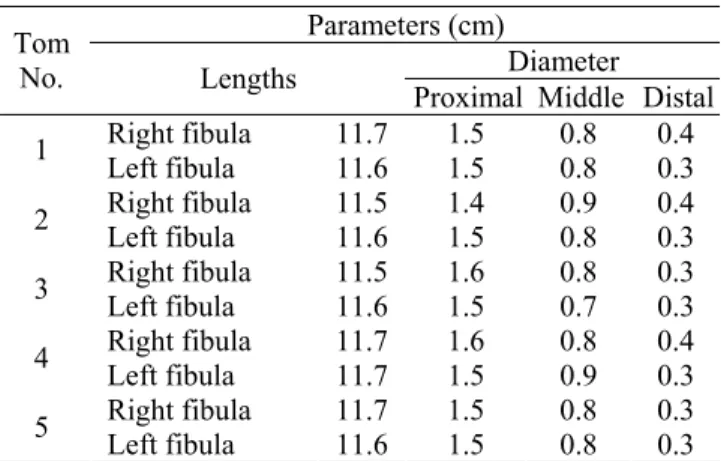
Table 3: show the length and diameter of fibula in Tom.
Parameters (cm)
Diameter Tom
No. Lengths
Proximal Middle Distal Right fibula 11.7 1.5 0.8 0.4 1
Left fibula 11.6 1.5 0.8 0.3 Right fibula 11.5 1.4 0.9 0.4 2
Left fibula 11.6 1.5 0.8 0.3 Right fibula 11.5 1.6 0.8 0.3 3
Left fibula 11.6 1.5 0.7 0.3 Right fibula 11.7 1.6 0.8 0.4 4
Left fibula 11.7 1.5 0.9 0.3 Right fibula 11.7 1.5 0.8 0.3 5
Left fibula 11.6 1.5 0.8 0.3
Table 4: show the length and diameter of fibula in local coke.
Parameters (cm)
Diameter Local
cock
No. Lengths Proximal Middle Distal Right fibula 7.3 1.1 0.5 0.1 1
Left fibula 7.2 1.2 0.4 0.2 Right fibula 7.1 1.0 0.5 0.1 2
Left fibula 7.2 1.1 0.6 0.2 Right fibula 7.3 1.2 0.5 0.1 3
Left fibula 7.3 1.2 0.5 0.2 Right fibula 7.4 1.1 0.6 0.1 4
Left fibula 7.3 1.0 0.5 0.1 Right fibula 7.3 1.2 0.5 0.2 5
Left fibula 7.3 1.2 0.5 0.1
Table 5: show the mean ± ES of length and diameter of tibiotarsal in Tom and local coke.
Parameters (cm) Diameter tibiotarsal in
Lengths
Proximal Middle Distal
Tom 17.99±
0.44 3.02± 0.021 2.21± 0.005 1.94± 0.0021
Iraqi Journal of Veterinary Sciences, Vol. 26, No. 2, 2012 (57-64)
60
Table 6: show the mean ± ES of length and diameter of Fibula in Tom and local coke.
Parameters (cm) Diameter Fibula in
Lengths
Proximal Middle Distal
Tom 11.62±
0.21
1.51± 0.008
0.81± 0.0033
0.33± 0.0043
Local cock 7.27± 0.32
1.12± 0.0052
0.51± 0.007
0.23± 0.0054
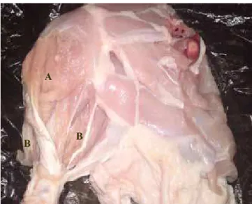
Schematic 1: Showing superficial muscles of the left leg in Tom 1-fibulars longus 2-tibialis cranialis 3-fibulars brevis 4-external digitorium longus 5-gastrocnemius 6-flexor digitorum longus.
Fibularis brevis
Usually smaller and shorter muscle in local cock while larger muscle in turkey, origin mainly aponeurotic from the lateral surface of the tibia distal to the fibular crest insertion proximal to the lateral condoyle of the tibotarsus. (Fig. 4), schematics (1,2).
Tibialis cranialis
It is located on the cranial surface of the tibia and deep to the fibularis longus it is readily separated into two heads femoral and tibial (Fig. 4,5) femoral head, is usually some smaller than the tibial head, origin femoral head by a tendon from the condoyle of femur, tibial head fleshy to partially aponeurotic from the cnemial crests, the two muscular bellies fuse distally to from a single strong tendon in the tendinal groove of the tibiotarsus.
Schematic 2: Showing second and third layers muscles of the left leg in local cock 1 – Ì- tibial and femoral head, 2- fibulars longus 3-fibulars brevis 4- plantaris A,B,C- pars externa, media and interna 5- extensor digitorium longus 6-flexor digitorum longus.
Extensor digitorium longus
Is located deep to the cranialis tibialis origin usually from the shaft of tibia and deep to the origin of the tibial head of the tibials cranialis, insertion by the tendon passes beneath the common retinaculum which retains the tendon of the tibialis cranialis (Fig. 5,6).
Gastrocnemius muscle
Is the largest and strongest of the crural muscles and it consist of three heads (external, internal and middle) usually the external and internal heads are larger than middle head (Fig. 7,8) origin, the external head (pars externa) by aponeurotic connections with the lateral femoral arm of the lop, the middle head (pars media) mostly fleshy from the medial aspect of the popliteal area, the internal head (pars interna) mainly fleshy from the cnemial crest and head of tibia insertion by common tendon contain sheath like aponeurotic, in turkey it form more or less separate tendon and becomes ossified as along bony ridge forming of a fibro- osseous compartment sheath for the flexor tendon of the toes (Fig. 6)
Plantaris
61
Fig. 1: The tibiotarsal bone of A- local cock and B- In Tom showing cranial and caudal view.
Fig. 2: The tibotarsal and fibula of Tom showing cranial and caudal view of tibia and fibula (A) cranial cnemial crest in proximal extremity.
Caudomedial muscle of the shank bone
The digital flexor muscle. It can be grouped into three morphological units based on there respective levels of insertion, superficial digit flexors (flexor perforates digit 11,111 and 1v) intermediate digit flexor (flexor perforates digit 11,111) and deep flexors (flexor digitorum longus) in Tom and local cock (Fig. 7,8).
Superficial digital flexor muscle (flexor perforates digit 11,111 and 1v), each of three muscles typically has a lateral and medial head (Fig. 7,8) arise from the caudal surface of the tibiatarsus and proximal extremity of fibula,
intermediate digital flexor (flexor perforates digit 11,111) origin of one or two aponeurotic from the caudal cnemial crest in Tom and and local cock, deep digital flexor (flexor digitorum longus) originated by two or three bellies in both Tom and local cock from the caudal surface of tibia tarsus and Most of the digital flexor muscles arise by extensive aponeurotic interconnections with each other (Fig. 8,9) and all the insertion tendon of digital flexor muscle from the tibial cartilage bundle of three tendons is exposed the broad and more deeply situated tendon of the of the flexor perforates 111 forms a sheath around the flexor perforates1v on the lateral side and flexor perforates 111on the medial side.
Fig. 3: A- fibula in local cock and B- In Tom showing the cranial and caudal view.
Fig. 4: lateral view of left leg showing superficial muscle of Tom A- Fibularis longus. B- Fibularis brevis. C-Cranilis tibialis. D-Femoral head. E-Tibial head.
A
B
A
B
A
A
B
C
D
Iraqi Journal of Veterinary Sciences, Vol. 26, No. 2, 2012 (57-64)
62
Fig. 5: left leg of local cock showing A- The first and B- Second layer of muscles.
Fig. 6: shows the insertion ossify tendon of gastrocnemus and external digitorum longus muscle in shank bone of Tom after boiling of muscle.
Popliteus. Deep small muscle origin from caudal surface of tibia and inserts in the cranial surfuse of fibula (Fig. 9).
Blood supply to the shank bone in Tom and local cock.
At the distal region of femur the ischiatic artery becomes the popliteal artery releases the medial tibial artery enters medial head of the gastrocniemius then curves distal with in the muscle to the mid-leg level, accompanies the popliteal vein and the medial tibial nerve reaching the interossous plane between the proximal tibia and fibula (Fig. 9,10) the popliteal artery turns courses distally in the limb and divided into cranial and caudal tibia arteries, also is distributed to the deeper level of the flexer compartment of the leg and distally part the tarsometatarseal joint.
Fig. 7: The right leg of local cock showing third layer of muscles:A- three parts of gastrocnemus muscle and B- three parts of digit flexor muscle.
Fig. 8: the left leg of Tom showing third layer of muscles: A- three parts of gastrocnemus muscle,B- Cranial tibial artery C - Common fibular nerve and D- three parts of digit flexor muscle.
Innervations of the shank in Tom and local cock. Nerves supply of shank are tibial nerve is larger division of the ischiatic nerve, it supply most of the flexor muscles of shank is divided into medial tibial and lateral tibial nerves, medial tibia] nerve continues with the popliteal artery in
A B
C
D
B
B A
A
63
proximal region of extensor and flexor muscle of the shank bone they are supplies by fibular nerves is divided into three nerves, superficial nerve gives muscular rams to the flbularis longus and fibularis brevis and metatarsal cutaneoul rams, deep fibular nerve pass under the tibalis cranialis gives muscular ramus to the flexor digitorium and divided in to digital nerve, common fibular nerve, crosses lateral to the shaft of fibula, related to the fibular vessels gives.
Fig. 9: the right leg of Tom showing fourth layer of muscles: A- plantars muscle, B- three parts of digit flexor muscle and C- popliteus.
Discussion
The legs variation of shape and sizes in both birds tibiotarsus and fibula in Tom are longer, wider and more striated than in local cock were not reported by any of the above mentioned on authors in any of the birds due to bipeds and man, has aspecific longer and wider bones for the leg these characteristic measurements are attributed of compression of the body weight and the demands of the erect posture (9).
Shank bone consist of tibiotarsal and fibula, our results for both species were agreement with (6) while some of the proximal tarsal bones are fused with the distal end of tibia to increase in length and the whole skeletal.
The fibula is reduced long bone in Tom and local cock. It has two extremities and shaft, (6) report that the fibula in Tom and local cock showed the typical avian condition of having agreatly reduce fibula, is greater size and extend, the present study demonstrated that there were hock joint in
Tom and local cock that unites the tibiotarsal with the tarsometatarsus which also included the fused metatarsals second,third and fourth many workers' obtain this result because differential growth in birds (7).
Tendon of limb muscles in Tom is ossification because these tendons have unique properties as well as several features common to calcifying cartilage and bone in large birds (10). In Tom the dark and red muscles are very clearly distinguished because contain larger amount of myoglobin,and more heavily vascularized (10).
Fig. 10: the left leg of local cock showing:A- Cranial tibial artery B- Common fibular nerve.Muscular branches to the both heads of the tibialis crailis and extensor digiterium longus.
In our result show the gastrocnemius muscle was largest and has three head in Tom and in local cock because gastrocnemius are sufficiently well developed in the gallinaceous birds to bo however the relative development of each of them may show some variation in birds generally often more or less correlated with the relative length of the leg is strongest and largest of crural muscles (11,12).
The present result were the tibialis cranialis muscles consist of two head and location directly under the fibularis longus in Tom and local but in mammals this muscles small and it has one head, our result agreement with (6) fibularis longs muscles showed considerable variation in its relative development in birds. It may extentded over most of the crus concealing the tibialis cranialis, as in the turkey, or it may be some what reduced in size and not concealing that muscle as (in local bread, there is very little variation in this
A B
C A
Iraqi Journal of Veterinary Sciences, Vol. 26, No. 2, 2012 (57-64)
64
muscle among birds except for the length of the fleshy belly, the present study showed that flexor digital muscle has three level superficial, intermediat and deep, the result agreement with (6) most birds have three heads of origin, or fourth head.
According to the fibularis brevis in our result that muscles smaller and shorter is relatively larger and long muscle in some birds e. g vultures, falcons and parrots, the muscles is absent or reduced in some species of bird, lucluding stroks and falmingoes (6).
References
1. Gineke M. Certified production of commercial turkey. Would poultry. Magazine on production processing and marketing. 2010;40(26):4. 2. French N. Managing turkey poultry quality in the hatchery. Would
poultry. Magazine on production processing and marketing Turkey special 10. 2008;11(24):7.
3. Johnson P. Effect of the unilateral weight bearing on pelvic limb development in broiler vascular studies. Res Vet Sci. 1988;44(2):164. 4. Getty R. The anatomy of the domestic animals. volume 1. W.B.
Saunders comp,.UK. London. 1975. p.35-37.
5. Dyce KM, Sack WO, Wensing CJG. Textbook of veterinary anatomy. 3ed. Ed. Sunders. Philadelphia. London. 2002. p.779 -780
6. Vander B J. Avian mycology.in Sisson and Grossman.The anatomy of domestic animals Rev by Getty. R.W.B. Saunders comp. Philadelphia USA. 1975. p.1837- 1839.
7. Oliver U. Vascular mineral spatial correlation in the calcifying turkey leg tendon. Connect Tissue Res. 2002;43(4):595-605.
8. Saleh MN, Mohamed MA, Galal AT. Parametric studies on the hip bone of some mammals. Assiut Vet Med J. 1986;17(34):67.
9. Arsenault Al. Crystal collagen relation ships in calcified turkey leg tendon s visualized by selected area dark field electron microscopy. JAVA. 2003;3(50):161.
10.Vukovic S. Development of glycogen body in Turkey (Meleagvis gallopavo) embryo. Vet Skiarhv. 2005;75(2):101.
11.Dickson JG. The wild Turkey biology and arrangement. published by stack pole books in mechanic Sbury.. 2003. p.412 -413.