Universidade Federal de São Carlos
Centro de Ciências Biológicas e Saúde
Departamento de Fisioterapia
Programa de Pós Graduação em Fisioterapia
Laboratório de Cinesiologia Clínica Ocupacional
Bruno Leonardo da Silva Grüninger
COMO OCORREM OS MOVIMENTOS DA MANDÍBULA E DA CABEÇA EM SUJEITOS COM DISFUNÇÃO TEMPOROMANDIBULAR E EM SUJEITOS SAUDÁVEIS?
Orientadora: Profª Drª Ana Beatriz de Oliveira
Universidade Federal de São Carlos
Centro de Ciências Biológicas e Saúde
Departamento de Fisioterapia
Programa de Pós Graduação em Fisioterapia
Laboratório de Cinesiologia Clínica Ocupacional
Bruno Leonardo da Silva Grüninger
COMO OCORREM OS MOVIMENTOS DA MANDÍBULA E DA CABEÇA EM SUJEITOS COM DISFUNÇÃO TEMPOROMANDIBULAR E EM SUJEITOS SAUDÁVEIS?
Dissertação apresentada ao Programa de Pós-graduação em Fisioterapia, do Centro de Ciências Biológicas e da Saúde da Universidade Federal de São Carlos como parte dos requisitos para obtenção do título de Mestre em Fisioterapia. Área de concentração: Processo de Avaliação e Intervenção em Fisioterapia
Orientadora: Profª Drª Ana Beatriz de Oliveira
AGRADECIMENTOS
“E aprendi que se depende sempre, de tanta, muita, diferente gente (...)
É tão bonito quando a gente sente que nunca está sozinho, por mais que pense estar (...)”
À Deus,
À minha orientadora, Bia, por ser mais que uma orientadora. Por desde a iniciação cientifica até o momento, conseguir me guiar, incentivar e apoiar em todas as iniciativas. Muito obrigado pela amizade, paciência, ensinamentos e apoio irrestrito. Sei
que fiz sua vida de orientadora a distância ser mais difícil que o normal, mas sua forma de lidar com tudo me faz ainda querer seguir esse caminho.
À todos amigos do laboratório, Helen, Chico, Dechris, Lê Bergamin, Marininha. Sempre tornaram os momentos juntos em risadas, cafezinhos e bolachas. Obrigado
pelos ensinamentos e pelo incentivo de sempre.
À Lianna, pela ajuda irrestrita nas coletas e no dia-a-dia. Dispôs-se a fazer de tudo para que esse projeto funcionasse. Obrigado pelos conselhos, suporte e prontidão.
À Lê Calixtre, que desde o começo de tudo me abraçou e carregou junto. Se hoje termino essa fase, devo muito, nem sei quanto, a você! Foi minha co-orientadora, conselheira, escritora, revisora. Muito obrigado por dividir minhas dúvidas, por entrar
de cabeça nessa, pelo entrosamento. Acho difícil encontrar outra companheira de pesquisa como você! Obrigado pelas risadas e todos os momentos ao seu lado, bons e
ruins. Sem você, nada disso seria possível.
Aos voluntários que, faça chuva faça sol, seja noite ou bem cedo, estavam presente, com um sorriso no rosto. Sem cada um de vocês, nada disso aconteceria.
Aos amigos e companheiros de trabalho, Leandro e Maurício pelos ensinamentos e risadas diárias.
Aos amigos da Fisio 07. Os presentes e os não tão presentes, que sempre trouxeram mais brilho aos olhos em nossos encontros. Obrigado pelo carinho de sempre!
Aos bons, velhos e inseparáveis amigos de Pirassununga e de São Carlos. Mesmo à distância, mesmo o tempo sem falar, mesmo os finais de semana que “tinha coisas de
mestrado pra fazer”, vocês estavam lá, perguntando, incentivando e participando. Obrigado por ouvir minhas lamentações e reclamações, me apoiar irrestritamente.
À minha família, a de Pirassununga e a mais nova, de Tatuí. Que sempre me esperam aos finais de semana com muito carinho e alegria. Obrigado pela força, pelos abraços
apertados e almoços de domingo.
Ao meu pai, guerreiro, um leão gigante que sempre está ao meu lado, seja pra me defender, me abraçar ou me mostrar o caminho a ser seguido. Provavelmente o que mais
sofreu com minha ausência, mesmo que em nenhum momento tenha deixado isso aparecer. Obrigado pelo amor, por ser um cara determinado, perseguidor do que deseja. Se eu for 1% pro meu filho do que o sr. foi pra mim, ele terá um pai fantástico. Se tenho
caráter, personalidade, profissionalismo e paixão pelo que faço, devo isso aos seus ensinamentos transmitidos. Muito obrigado.
À minha esposa, Marina. Amiga, professora, fisioterapeuta, pesquisadora, companheira. Você que me apoiou em todos os momentos possíveis, desde o princípio. O motivo que me faz querer todos os dias ser melhor. O motivo de, lá atrás, eu não ter deixado a Fisio
de lado. Obrigado por não desistir de mim. Obrigado por me inspirar e me ensinar. Obrigado por, em cada passo, me ajudar, mostrar, voltar atrás. Muito amor para nós e
RESUMO
Considerando a carência de estudos que avaliam a relação entre articulação temporomandibular e componentes cervicais em atividades dinâmicas, este estudo objetivou investigar a cinemática e o controle motor da cabeça e da mandíbula e avaliar a performance dos músculos cervicais em pacientes com disfunção temporomandibular comparado com sujeitos saudáveis. Foram coletadas cinemática da cabeça e mandíbula em 71 mulheres (Grupo saudável – HG: N=33; Grupo Disfunção temporomandibular – TMDG: N=38) durante movimentos cervicais (flexão – extensão, rotação e inclinação), e durante atividades funcionais (abertura máxima da boca (MMO), fala, mastigação e uso do computador). Frequência mediana foi calculada para as posições da cabeça nas atividades funcionais. Foi analisada a correlação cruzada para séries de tempo da mandíbula e cabeça durante atividades funcionais. O controle motor da cabeça (Senso de posição articular – JPS) e performance dos músculos cervicais (teste de flexão crânio cervical – CCFT) também forem testados. Os grupos foram comparados usando Teste-T para amostras independentes ou seu correspondente não-paramétrico, Mann-Whitney. Não foram encontradas diferenças para o JPS e CCFT, enquanto que a abertura máxima da boca foi maior para o grupo saudável. Frequência mediana foi maior para a posição da cabeça do grupo saudável, no plano sagital durante o uso do computador em comparação com o TMDG. Isso pode indicar uma maior variação e melhor estratégias motoras para o HG. Correlação cruzada mostrou correlação negativa entre cabeça e mandíbula durante a MMO. No entanto, a consistência dessa correlação foi fraca em outras atividades. Parece que quanto maior a exigência biomecânica, mais forte é a correlação. Estudos mais aprofundados devem focar-se em tarefas mais complexas.
ABSTRACT
Considering the lack of studies assessing the relationship between temporomandibular joint and neck components in a dynamic approach, this study aimed to investigate kinematics and motor control of the head and jaw in functional and isolated activities, and to evaluate cervical muscle performance and motor control of the head in temporomandibular disorder (TMD) patients compared to healthy subjects. Kinematics of head and jaw were recorded from 71 women (Healthy Group – HG: N= 33; Temporomandibular Disorder Group – TMDG: N=38) during neck movements (flexion-extension, rotation and lateral bending), and functional tasks (maximal mouth opening (MMO), speaking, chewing and computer use). Median frequency was calculated for head positions in functional tasks. Cross-correlation analysis was performed for the time series of jaw and head recorded in functional tasks. Motor control of the head (joint position sense - JPS), and cervical muscle performance (craniocervical flexion test - CCFT) were also tested. TMDG and HG were compared using independent t-test or Mann-Whitney test. No difference was observed in CCFT and JPS, while jaw depression was larger in HG. Median frequency was higher for head position of HG in sagittal plane during computer use in relation to TMDG. It may indicate more variation and better motor strategies in HG. Cross-correlation analysis showed a clear negative correlation between head and jaw in MMO. However, the consistency of this correlation was weak in other tasks. It seems that the higher the biomechanical requirement the stronger the correlation. Further studies should focus on more complex tasks
.
LISTA DE FIGURAS
Figura 1. Posicionamento do lasersobre cabeça do voluntário no teste “Joint
Position Sense” e posicionamento do voluntário no “Teste de flexão Crânio-cervical”.
24
Figura 2. Posição dos marcadores e posição do sujeito na avaliação cinemática durante o uso do computador.
25
Figura 3. Posição dos sujeitos durante o uso do computador. 26 Figura 4. Série temporal da relação de movimentos entre cabeça e mandíbula
durante as atividades funcionais no plano sagital.
LISTA DE TABELAS
Tabela 1. Testes especiais (JPS e CCFT) para Grupo DTM e saudáveis 28 Tabela 2. Amplitude máxima durante movimentos de abertura da mandíbula e
movimentos da cabeça e posição da cabeça durante a abertura máxima da mandíbula
28
Tabela 3. Movimentos tridimensionais durante atividades funcionais nos planos sagital, frontal e transversal
30
Tabela 4. Frequência mediana para os movimentos funcionais nos planos sagital, frontal e transversal
31
Tabela 5. Correlação cruzada dos movimentos da cabeça e da mandíbula durante atividades funcionais no plano sagital
LISTA DE APÊNDICES
LISTA DE ANEXOS
Anexo I Confirmação de Submissão do manuscrito no periódico “Journal of Biomechanics”
LISTA DE ABREVIATURAS
TMD Temporomandibular Disorder – Disfunção Temporomandibular
TMJ Temporomandibular Joint –Articulação Temporomandibular BMI Body Mass Index –Índice de Massa Corpórea
HG Health group –Grupo Saudável
TMDG Temporomandibular Disorders Group – Grupo Disfunção Temporomandibular
FAI Fonseca Anamnesic Index ndice Anamnésico de Fonseca NDI Neck Disability Index
RDC/TMD Critérios de Diagnóstico de Pesquisa/ Disfunção Temporomandibular MMO Maximal Mouth opening –Abertura Máxima da Boca
JPS Joint Position Sense –Senso de posição articular
CCFT Crânio cervical flexion test - Teste de Flexão Crânio-Cervical CE-R Constant Error – Right -Erro Constante Direita
CE-L Constant Error–Left - Erro Constante Esquerda AE-R Absolut Error – Right - Erro Absoluto Direita AE-L Absolut Error – Left - Erro Absoluto Esquerda VE-R Variable Error – Right - Erro Variável Direita VE-L Variable Error – Left - Erro Variável Esquerda
RMSE - R Root Mean Square Error – Right Erro Quadrático Médio Direita RMSE - L Root Mean Square Error – Left - Erro Quadrático Médio Esquerda ROM
SD
Range of motion – Amplitude de Movimento
Desvio padrão
MF Frequência mediana
Sumário
CONTEXTUALIZAÇÃO ... 15
Introduction ... 20
Methods ... 22
Participants ... 22
Assessment protocol ... 23
Special tests ... 23
Kinematics ... 25
Statistical Analysis ... 28
Results ... 29
Discussion ... 34
Conclusion ... 37
References... 38
CONSIDERAÇÕES FINAIS E DESDOBRAMENTOS FUTUROS ... 42
Apêndice I ... 43
15
CONTEXTUALIZAÇÃO
A articulação temporomandibular (ATM) é importante em diversas atividades essenciais do ser humano, como alimentação, comunicação e expressão facial (Calixtre et al, 2014). Portanto, trata-se de uma das articulações do corpo humano que mais se movimenta. Em situações normais, os côndilos direito e esquerdo se movimentam em conjunto a fim de executar depressão, elevação, lateralização e protrusão da mandíbula – movimentos fundamentais para realização das atividades funcionais (Armijo-Olivo 2012). A abertura da boca ou depressão da mandíbula depende do rolamento posterior de ambos os côndilos mandibulares sob os discos articulares e translação anterior do complexo côndilo-disco na fossa mandibular do osso temporal. Esses movimentos são controlados pelos músculos mastigatórios e qualquer alteração nessas estruturas pode causar discinesia uni ou bilateral e disfunção do sistema mastigatório (Neumann et al, 2006). A disfunção temporomandibular (DTM) é definida como um grupo de alterações do sistema estomatognático, como dos músculos mastigatórios, articulação temporomandibular (ATM) e estruturas associadas, as quais podem estar relacionadas com dores de cabeça, pescoço, ombro e coluna cervical (Bender, 2014).
Pacientes com DTM frequentemente apresentam alterações na coluna cervical, assim, essa relação tem sido investigada. Estudos mostraram movimentos coordenados entre mandíbula, cabeça e pescoço para realizar atividades de abertura e fechamento da boca, conhecido como “integração funcional”: durante o movimento de abertura máxima da boca, há a necessidade de uma extensão da coluna cervical alta. (Eriksson et al., 2000, Zafar et al., 2000).
Existem três diferentes teorias que tentam elucidar essa relação funcional: a neuroanatômica, a biomecânica e a muscular.
16
A relação biomecânica apresenta sua explicação baseada na movimentação do crânio, mandíbula e coluna cervical. No inicio do movimento de abertura da boca, o crânio precisa deslizar anteriormente, assim ocorre o movimento de extensão na articulação atlânto-occpital, e ao mesmo tempo, os dentes superiores devem deslizar para a frente, uma vez que estão ligados ao crânio e, consequentemente, a posição dos dentes de contato desloca-se posteriormente até a posição de intercuspidação. Quando o crânio desliza para trás, ocorre a situação inversa. Portanto, os movimentos craniocervicais causam movimentos adaptativos na mandíbula e em estruturas associadas (Armijo-Olivo et al., 2006). Uma vez que esses movimentos adaptativos estejam restritos, tanto a mandíbula quanto estruturas associadas podem estar alteradas (Armijo-Olivo et al., 2006). Previamente, já foi descrito na literatura que o controle integrado entre a cabeça e a mandíbula pode ser afetado através de restrição mecânica da cabeça (Häggman-Henrikson et al., 2006).
Do ponto de vista apenas muscular, estudos que induziram dor no masseter demonstraram aumento da contribuição de estruturas cervicais durante movimentos de abertura-fechamento da boca. (Wiesinger et al., 2013) observaram que, mesmo que a amplitude de abertura tenha permanecido semelhante pré e pós injeção de solução salina, a estratégia do controle do posicionamento da cabeça e da mandíbula foi altera da após a estimualção nocioceptiva na região trigeminal. Essa estratégia de controle motor diz respeito às informações sensoriais enviadas pelos neurônios aferentes para as estruturas responsáveis pela execução do movimento da cabeça, no caso.
Como a coluna cervical é em grande parte dependente do apoio dos músculos cervicais, eles acabam sendo altamente propensos a fadiga, o que reduz o seu desempenho e, consequentemente, o equilíbrio entre músculos flexores e músculos extensores do pescoço se apresenta comprometido.
Armijo-Olivo et al. (2010) também identificaram um aumento na atividade eletromiográfica dos músculos flexores cervicais superficiais em indivíduos com DTM, observado através do teste de flexão crânio cervical, indicando que essa hiperativação pode representar uma diminuição da atividade dos músculos cervicais profundos.
17
que, já foi apresentado que patologias cervicais não específicas alteram o controle motor cervical (Revel et ai. 1991). A influência aferente dos músculos cervicais é conhecida como tendo o papel principal do controle da postura da cabeça (Pinsault et al., 2008). Desta forma, o mau funcionamento destes músculos provoca um alinhamento inadequado das estruturas e, portanto, disfunção cervical.
Para esse componente de controle motor, Revel e colaboradores (1991) desenvolveram um teste clínico de fácil aplicação para verificar a sensação de posicionamento da cabeça durante a sua rotação. Acredita-se que se a musculatura cervical está sofrendo de algum fator inerente (isto é, sentido alterado da posição), todo o complexo funcional (cabeça-mandíbula) poderia ser alterado. Isto acontece porque a cabeça teria uma má orientação espacial ou porque mudanças na força muscular, resistência ou qualidade da ativação muscular aumentaria a irritação dos tecidos sensíveis do pescoço, causando dor e consequentemente perpetuando dor nas regiões cervical e orofacial (Armijo-Olivo e Magee, 2012).
Armijo-Olivo et al. (2011a) identificaram diversos estudos que tentaram demonstrar alguma relação entre o movimento da cabeça, coluna cervical e o sistema estomatognático. No entanto, as informações fornecidas pelos estudos incluídos são baseadas em dados descritivos e apresentam baixa qualidade metodológica (Chaves et al., (2014). Além disso, poucos estudos fornecem informações relevantes sobre movimentos funcionais e mais complexos. A maioria dos estudos está focada na avaliação da abertura e fechamento da boca, protrusão e lateralização da mandíbula. Visscher et al (1991), por exemplo avaliou a abertura da boca a partir de uma posição pré-estabelecida fixa da cabeça. Estudos que e exploram e comparam o comportamento da cabeça e mandíbula entre indivíduos saudáveis e com DTM são ainda mais raros.
Embora haja estudos que analisam e confirmam essa integração funcional (Häggman-Henrikson et al, 2002; Eriksson, et al., 2004) ainda há necessidade de se correlacione mecanismos integrativos durante atividades dinâmicas e funcionais.
18
19
ESTUDO
How do jaw and head movements occur in TMD patients and healthy
subjects?
20
Introduction
Temporomandibular disorder (TMD) has been related to dysfunction and pain of the masticatory muscles (Ferreira et al., 2014), and represents approximately 40% of all chronic pain problems (Mongini et al., 2007). TMD is also associated with symptoms affecting head and neck such as headache, neck dysfunction and altered posture (Silveira et al., 2014). Literature supports the interdependence between temporomandibular joint (TMJ) and neck structures, since the disease or injury in one area may induce pain and/or dysfunction in other area (La Touche et al., 2015).
The combination of jaw and head movements has been addressed as functional
integration (Nordh, 1998), and it seems to be task-dependent (Eriksson et al., 2000). Functional integration depends on the coordination of jaw and neck muscles, providing simultaneous movements of TMJ, atlanto-occipital joint and cervical spine (Zafar et al., 2000). For example, chewing is associated to an extension of the upper cervical, identified from the movement of the head. Moreover, during elevation of the jaw, head returns to its initial position or does a small flexion. Thus, considering this relationship between neck and TMJ regions, an alteration in one of the regions can possibly influence the other, and vice versa (Visscher et al., 2000b; Eriksson et al., 2007; La Touche et al., 2015).
Three theories are described to elucidate this relationship. The neuroanatomic theory is supported by the convergence of nociceptive neurons receiving trigeminal and cervical sensory inputs due to the topographical arrangement. Thus, the stimulation of structures innervated by the trigeminal nerve may causes a painful sensation in the cervical region and in the neck (Leandri et al., 2001).
Another theory addresses biomechanics to explain that when the head slides anteriorly on the atlas, there is extension in the atlanto-occipital joint; at the same time, the upper teeth must slide forward since they are connected to the skull, and, consequently, the position of the contact teeth moves posteriorly to intercuspidal position. When skull slides backwards, opposite situation occurs. Therefore, craniocervical movements cause adaptive changes in the jaw and associated structures. However, the ranges for such adaptive movements are diminished. Thus, any decrease in jaw and associated structures may lead to dysfunction (Armijo Olivo et al., 2006).
21
during mouth opening-closing movements (La Touche et al., 2015). The injection of saline solution in the masseter region produced a painful stimulus, but the opening width of the mouth remained similar before and after injection (Wiesinger et al., 2013). The motor control strategy between the jaw and the head was altered after stimulation of the trigeminal region.
It has been previously described that the integrated control between head and jaw can be affected by mechanical restriction of the head (Häggman-Henrikson et al., 2006). Cervical spine is highly dependent on the support of the neck muscles, exposing them to fatigue, and, consequently, to altered performance (Olivo et al., 2010). Armijo-Olivo et al. (2010) have also identified an increase in electromyographic activity of the superficial cervical flexor muscles in subjects with TMD, while there was a decrease in strength and resistance of deep cervical flexor and extensor muscles. Furthermore, subjects with neck disability were induced to pain in the masticatory system, and showed an increase in fatigue and pain in the region of the cervical musculature (La Touche et al., 2015).
Proprioceptive inputs of neck region have an important contribution to the positioning of the head and its spatial orientation (Pinsault et al., 2008). Neural control system of the head and neck depends on the convergence of proprioceptive and visual signals (Lakie and Loram, 2006). Thus, Revel et al. (1991) considered that non-specific cervical pathologies alter cervical motor control and afferent input from the cervical muscles (Pinsault et al., 2008). Therefore, malfunctioning of these muscles causes an inappropriate alignment of the structures and neck disability. Thus, if cervical musculature is suffering from some inherent factor (i.e, altered sense of position), the entire functional complex (head-jaw) could be altered.
22
Therefore, the objective of the this study was to investigate kinematics and motor control of the head and jaw in functional and isolated activities of both segments, as well as to evaluate cervical muscle performance and motor control of the head in individuals with TMD when compared to healthy subjects. We hypothesized that TMD patients will present decreased motor control of the head, and reduced cervical musculature function. Moreover, we expect functional integration of jaw and head to be impaired in individuals with TMD compared to healthy subjects.
Methods
Participants
Seventy-one women with mean age of 26.86 (±7.76) years, and body mass index (BMI) of 22.26 (±2.86) kg participated in the study. According to inclusion and exclusion criteria (see below), subjects were divided into two groups: HG (Healthy Group; N= 33 subjects; mean age of 28.2 ±10.15 years, BMI of 22.08±2.77 kg/m²) and TMDG (Temporomandibular Disorder Group; N=38 subjects; mean age of 25.63±4.41 years; BMI of 22.42±2.96 kg/m²). All subjects signed an informed consent form approved by the local ethics committee (Process #1.790.335/2016).
To be included in the HG, subjects had to present less than 3 points according to the Numerical Pain Rating Scale (NPRS) applied for both neck and TMJ regions; less than 20 points to Fonseca Anamnestic Index (FAI); less than 4 points in the Neck Disability Index (NDI). In order to be included in the TMDG, subjects had to be diagnosed with myogenic or mixed TMD, according to Research Diagnostic Criteria for Temporomandibular Disorder (RDC/TMD) (Dworking, 1992).
Subjects were excluded if present history of jaw fracture, TMJ luxation, systemic ligament laxity, pregnancy, rheumatic or neurologic system diseases, orthodontics or physical therapy treatment in the last six months, and previous cervical or oral surgery.
TMD subjects, according to RDC/TMD, presented the following diagnoses: - 10 subjects – Ia (Myofascial pain without limited mouth opening); - 28 subjects – Ib (Myofascial pain with limited mouth opening); - 24 subjects – IIa (disc displacement with reduction);
- 1 subject IIb (disc displacement without reduction with limited mouth opening);
23
- 11 subjects IIIa (Arthalgia);
- 1 subject IIIc (Osteoarthrosis with joint pain).
Considering a statistical power of 80%, and alpha level of 0.5, the estimated sample size was 16 participants in each group (G*Power, version 3.0.10). Maximum mouth opening (MMO) was considered as the main outcome. Data for TMD subjects were estimated from literature (Felicio et al., 2013) as 22.86±4.2 mm. We considered healthy subjects with MMO of 27.9±4.2 mm, according to data from a pilot study. Minimal clinically important difference was set at 9 mm (Kropmans, 2000). Once this study is part of larger research, we decided to include more subjects in the sample since data were available.
Assessment protocol
Special tests and kinematic evaluation were performed by a blind rater, in pre-set order.
Special tests
Joint Position Sense test – JPS: Applied to evaluate head motor control from the ability to reposition the head to reach a target (Johnston et al., 2008; Jull et al., 2008). The subject was seated in front of a target (90 cm away), with a laser pointer fixed on the top of the head, and pointing to the center of the target (Figure 1A). She was instructed to perform 10 movements of maximal rotation of the head to each side (Figure 1B). The order of movements (to the right or left) was random. When she positioned her head back to the supposed initial position, a mark was made on the target where the laser was pointing. At the end, a picture of the target was recorded, from a distance of 2 m, to be later analyzed (CorelDraw X5). We calculated, for both right and left directions:
- CE (constant error): mean of the raw error over the ten trials to each side incorporating the positive and negative values in each trial, i.e. ∑10𝑛=1𝑟𝑎𝑤 𝑒𝑟𝑟𝑜𝑟 𝑛
10 (Lee et al., 2006; Hill et al., 2009);
- AE (absolute error): the mean of the total deviation from the starting point over the ten trials to each side, ignoring positive (overshoot) and negative (undershoot) values – absolute raw error, i.e. ∑10𝑛=1𝑎𝑏𝑠𝑜𝑙𝑢𝑡𝑒 𝑟𝑎𝑤 𝑒𝑟𝑟𝑜𝑟 𝑛
24
- VE (variable error): root mean square of the difference between the raw error and the calculated CE, i.e. √∑10𝑛=1(𝑟𝑎𝑤 𝑒𝑟𝑟𝑜𝑟 𝑛−𝐶𝐸 𝑛)2
10 ;
- RMSE (root mean square error): square root of the sum of the CE squared and the VE squared, i.e. √𝐶𝐸2+ 𝑉𝐸2 .
Craniocervical Flexion Test - CCFT: Applied to evaluate the performance of deep cervical flexor stabilizing muscles in the cervical spine (Chiu, 2005; Jull et al., 2008). CCFT is an indirect assessment of activation (observed as activation scores - AS), and resistance (observed as cumulative performance indexes - CPI) of deep cervical flexors, as proposed by Jull et al., (2008). Test is performed using a pressure biofeedback device located in the neck region (Stabilizer, Chattanooga Group Inc.), and a visual aid positioned in front of the subject, with the subject lying supine, keeping the legs flexed (Figure 1C). Each participant attempted to reach 5 different levels. The device has a graduation with marks corresponding to increments of 2mmHg, ranging from 20mmHg to 30mmHg. The level of effort required in the test is progressive, and observed according to what the subject can perform. In order to move to the next level, the subject must be able to complete ten 10-second repetitions on each possible level.
25
Figure 1. (A) Starting position in JPS test, with laser pointer on the top of the head pointing to the center of the target; (B) Subject performing left head rotation during JPS test; (C) Positioning of subject and
evaluator during CCFT.
Kinematics
A six-camera Qualisys motion capture system (Qualisys, Inc., Gothenburg, Sweden) was used to perform kinematic analysis of head and jaw movements. Data was recorded at 120 Hz. Fifteen markers and one cluster device were used (Figure 2). Trunk movements were reconstructed from six (20 mm) markers positioned on the spinous processes of C7 and T10, right and left acromion, sternal notch and xiphoid process. The head plan was formed by a marker on the glabella (12 mm), and three markers (15mm) fixed on a plastic tiara: two were placed one centimeter above each ear (temporal region) and the third at the midpoint of these points, at the top of the head
A
B
26
(Figures 2A and 2B). Three markers (12 mm) were positioned at the corners of an equilateral triangular stainless steel extraoral cluster (size 40 mm; weight 2 g). The cluster was fixed on the mandibular anterior gingiva using a surgical adhesive to provide a mandibular reference system, together with markers (12 mm) on the TMJs and mandibular angles (Mapelli et al., 2009; Sforza et al., 2008).In the static trial, an additional marker was located on the midline incisal edge to identify a virtual dental landmark relative to the cluster. The 3D-reconstruction of the jaw was then obtained.
Figure 2. (A and B) Position of the markers on head, trunk and jaw.
For organizational reasons and better presentation of the results, we divided the kinematics into two phases: isolated and functional movements.
Isolated head movements: In order to record head position in neck movements, subject was instructed to perform a series of 3 movements: maximal neck flexion-extension, right and left rotation, right and left lateral neck bending. All movements were performed three times for each side. The movements were always initiated from the neutral position of the neck, and performed at free speed.
Functional movements: head and jaw movements were recorded with the subject seated on a regular non-adjustable chair. She was asked to perform:
1) Maximal mouth opening (MMO) at free velocity, starting from the mandibular intercuspidal position;
B
27
2) Reading out loud a sequence of 11 predetermined words, chosen for presenting several phonemes of the Brazilian Portuguese language, in her habitual speed and tone of voice. This task was labeled as speaking;
3) Chewing for 20 seconds on the side of her preference. A standardized piece of flexible, odorless, colorless film was used (Biasoto-González, 2010), and the chewing was performed at free speed.
4) A laptop computer was positioned on a regular office table, and each subject selected a text of preference to read. The recording was performed during 600 seconds, and the volunteer was instructed to sit on a regular non-adjustable chair to perform computer use (Figure 3A e 3B).
During the recording of all functional movements, some markers were lost. Therefore, we proceed with a careful data control quality, and exclusion of subjects for specific recordings. Therefore, the number of subjects in each analysis is presented with the results.
Figure 3. (A and B) Markers and data collection during computer use.
Data processing: Qualisys Track Manager (Qualisys AB Packhusgatan, Gothenburg, Sweden), and Visual 3D (C-Motion, Inc., Germantown, MD, USA) software were used to track the position of the markers and to reconstruct 3-D movements of each segment, respectively. Rotational movements were identified and Cardan Angles x, y e z were obtained (Mapelli et al., 2009).
Tridimensional displacement of the head in relation to the trunk was subtracted from movements of the jaw in order to isolate any movement of the head occurred during functional and isolated tasks (Mapelli et al., 2009).
28
The 3D angular displacement of the jaw and head was processed using MatLab (v7.0.1, MathWorks Inc., Natick, MD, USA).
For the isolated movements, we extracted the maximal position of jaw depression, head flexion, extension, bending and rotation. Head position during maximal jaw depression was also obtained. During functional tasks, jaw and head position in frontal, sagittal and transverse planes were analyzed. Positive and negative values refer to the movement direction in each plane - sagittal (+: jaw elevation and head extension), frontal (+: jaw lateral deviation right and right head bending), and transversal (+: jaw protrusion and left head rotation). In order to extract information about motor control of the head, median frequency of 3-D head position was calculated in MMO, speaking, chewing and computer use through the Fast Fourier Transformer (FFT).
A cross-correlation analysis was performed between the time series of head and jaw angular positions in the sagittal plane recorded during MMO, speaking, chewing and computer use. Cross-correlation coefficients (r2) were calculated for zero-lag (CC zero-lag) between time series. Maximal absolute cross-correlation coefficients (CC max), for all possible time lags, were also calculated. Was considered negative and positive values of CC max separately, in order to verify any specific behavior in HG and TMDG. Considering that craniocervical movements cause adaptive changes in the jaw and associated structures (Armijo Olivo et al., 2006), we expected to find negative correlation coefficients, showing that head extension was combined to jaw depression during functional tasks. Coefficients between 0 and 0.19 were considered as "little if any correlation"; from 0.20 to 0.39 as weak correlation; between 0.40 and 0.69 as moderate correlation; from 0.70 and 0.89 as strong correlation; values higher than 0.9 as very high correlation (Munro, 2005).
Statistical Analysis
29
Results
Results of special tests, CCFT and JPS, are shown in Table 1. No significant difference was observed. However, mean value of CCFT was higher for HG compared to TMDG. HG also had higher variability in this test compared to TMDG.
Table 1: Mean and standard deviation (SD) values of results of special tests. Statistical results (P-value) for the comparison between TMD and Healthy subjects are also presented.
TMD Group Healthy Group P-value
mean (SD) mean (SD)
CCFT 49.47 (34.00) 68.24 (62.40) 0.268 JPS
CE-R 4.91 (1.82) 4.86 (2.09) 0.927
CE-L 4.93 (2.26) 4.57 (2.37) 0.593
AE-R 4.91 (1.82) 4.86 (2.09) 0.927
AE-L 4.93 (2.26) 4.57 (2.37) 0.377
VE-R 22.76 (11.51) 22.57 (9.14) 0.840
VE-L 23.67 (15.16) 26.29 (12.05) 0.377
RMSE-R 655.52 (650.58) 612.87 (537.18) 0.849
RMSE-L 795.20 (1022.70) 864.13 (765.81) 0.177
TMD: temporomandibular disorder; CCFT: craniocervical flexion test; JPS: joint position sense; CE-R: constant error right; CE-L: constant error left; AE-R: absolut error right; AE-L: absolut error left; VE-R: variable error right; VE-L: variable error left; RMSE: root mean square error; -R: right side; -L: left side.
Isolated movements were performed in similar and maximal amplitude by subjects from both groups (Table 2). Therefore, jaw depression was larger in healthy subjects (P<0.05). Head extension was also larger in HG, but no significant difference was identified.
Table 2: Mean and standard deviation (SD) of maximal range (in degrees) recorded during isolated movements, and head position during maximal jaw depression (in degrees). Statistical results for the comparison between temporomandibular disorders (TMD) and Healthy groups are also presented. Positive or negative values are associated to the direction of movement - head extension, right bending, and left rotation have positive values, while jaw depression, head flexion, left bending and right rotation have negative ones.
TMD Group Healthy Group P-value
mean (SD) mean (SD)
Jaw depression -20.85 (6.87) -26.91 (4.17) 0.000*
Head position at jaw depression 4.23 (3.46) 4.95 (2.59) 0.306
Head flexion -66.20 (10.12) -65.32 (11.17) 0.876
Head extension 56.37 (11.59) 62.21 (15.18) 0.053
Head bending - R 39.05 (6.36) 38.43 (7.55) 0.648
Head bending - L -39.99 (6.27) -39.11 (7.40) 0.377
Head rotation - R 44.74 (6.76) 68.64 (9.97) 0.473
Head rotation - L -66.31 (9.18) -70.14 (10.42) 0.117
30
31
Table 3: Mean and standard deviation values (in degrees) of tridimensional movements recorded for jaw and head during speaking, chewing and computer use. Data are reported as mean and standard deviation (SD). Statistical results (P-value) for the comparison between TMDG and HG are also presented. Number of subjects included in each group is presented (N). Positive or negative values are associated to the direction of movement – jaw elevation, head extension, right bending, and left rotation have positive values, while jaw depression, head flexion, left bending and right rotation have negative ones.
Speaking Chewing Computer Use
TMDG (N=36) HG (N=33) P-value TMDG (N=37) HG (N=33) P-value TMDG (N=29) HG (N=33) P-value
mean (SD) mean (SD) mean (SD) mean (SD) mean (SD) mean (SD)
Jaw - sagittal plane
Mean -0.90 (1.24) -0.65 (1.32) 0.414 -0.31 (1.25) -0.37 (1.72) 0.915 7.16 (8.51) 5.75 (6.15) 0.456
Standard Deviation 1.80 (0.36) 1.72 (0.44) 0.402 2.13 (0.86) 1.95 (0.92) 0.346 2.22 (0.96) 2.58 (2.83) 0.516
Jaw - frontal plane
Mean -0.07 (0.34) -0.07 (0.50) 0.838 -0.04 (0.34) -0.06 (0.45) 0.636 1.10 (2.26) -9.22 (59.92) 0.313
Standard Deviation 0.22 (0.10) 0.26 (0.14) 0.285 0.41 (0.18) 0.42 (0.28) 0.686 1.17 (0.79) 1.77 (4.15) 0.877
Jaw - transversal plane
Mean -0.01 (0.46) 0.01 (0.35) 0.973 0.02 (0.37) 0.06 (0.43) 0.717 -0.17 (6.88) 1.24 (3.76) 0.358
Standard Deviation 0.23 (0.10) 0.20 (0.07) 0.180 0.42 (0.18) 0.47 (0.66) 0.954 1.09 (0.79) 1.06 (1.02) 0.448
Head - sagittal plane
Mean 1.60 (1.78) 1.82 (1.49) 0.577 0.43 (1.05) 0.53 (1.09) 0.583 -3.65 (6.55) -3.73 (5.01) 0.960
Standard Deviation 1.02 (0.50) 0.97 (0.46) 0.686 0.57 (0.24) 0.68 (0.39) 0.175 2.42 (1.18) 2.48 (1.27) 0.852
Head - frontal plane
Mean 0.06 (1.06) 0.17 (0.98) 0.532 -0.29 (0.76) -0.01 (0.63) 0.264 -1.22 (4.94) 1.12 (3.76) 0.375
Standard Deviation 0.53 (0.30) 0.58 (0.38) 0.087 0.37 (0.26) 0.34 (0.38) 0.359 2.61 (1.62) 2.50 (1.54) 0.725
Head - transversal plane
Mean -0.23 (0.73) -0.37 (1.10) 0.661 0.08 (0.79) -0.15 (0.67) 0.114 2.71 (3.38) 2.03 (2.63) 0.039*
32
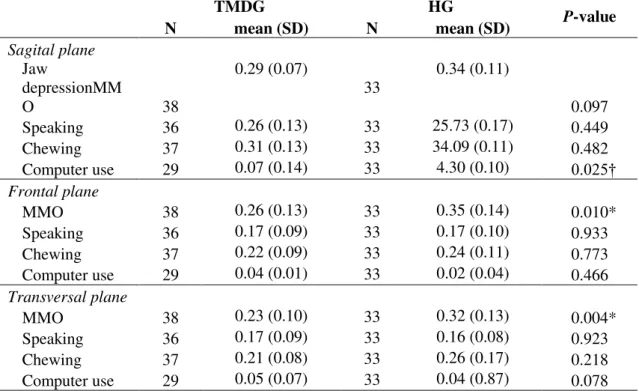
Table 4 shows data of median frequency (MF) of head movements during functional tasks and MMO. Significant differences were observed in frontal and transversal planes for MMO and in sagittal plane for computer use. In all situations healthy subjects had higher median frequency than TMD patients. Variability across subjects was quite similar in both groups.
Table 4: Mean and standard deviation (SD) values of median frequency (Hz) of the head during MMO, speaking, chewing and computer use. Number of subjects (N) in each condition and group, and statistical results for the comparison between TMD and Healthy subjects are also presented.
TMDG HG P-value
N mean (SD) N mean (SD)
Sagital plane
Jaw
depressionMM
O 38
0.29 (0.07)
33
0.34 (0.11)
0.097
Speaking 36 0.26 (0.13) 33 25.73 (0.17) 0.449
Chewing 37 0.31 (0.13) 33 34.09 (0.11) 0.482
Computer use 29 0.07 (0.14) 33 4.30 (0.10) 0.025†
Frontal plane
MMO 38 0.26 (0.13) 33 0.35 (0.14) 0.010*
Speaking 36 0.17 (0.09) 33 0.17 (0.10) 0.933
Chewing 37 0.22 (0.09) 33 0.24 (0.11) 0.773
Computer use 29 0.04 (0.01) 33 0.02 (0.04) 0.466
Transversal plane
MMO 38 0.23 (0.10) 33 0.32 (0.13) 0.004*
Speaking 36 0.17 (0.09) 33 0.16 (0.08) 0.923
Chewing 37 0.21 (0.08) 33 0.26 (0.17) 0.218
Computer use 29 0.05 (0.07) 33 0.04 (0.87) 0.078
* P<0.05, t-test for independent samples; † P<0.05, Mann-Whitney test. TMD: Temporomandibular disorders; MMO: maximal mouth opening.
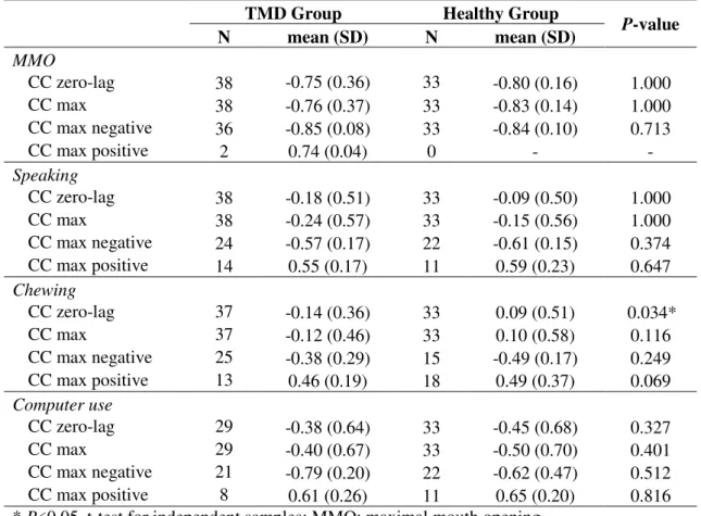
Results of cross-correlation analysis are presented in Table 5. In general, the correlation between head and jaw movements on the sagittal plane was mostly negative. It means that jaw depression was combined with head extension. A significant difference happened for zero-lag cross-correlation in the chewing time series. High or very high correlation was observed during MMO. However, no consistent difference was observed between groups. Therefore, variability across subjects was higher among TMD patients than among healthy subjects.
33
correlation during speaking in healthy subjects. The only statistical significant difference between TMDG and HG was found for CC zero-lag in chewing.
Table 5: Results of the cross-correlation (CC) analysis (r2) performed between jaw and head
movements in the sagittal plane during MMO, speaking, chewing and computer use. The analysis was performed for zero-lag between the time series (CC zero-lag). Maximal cross correlation (CC max) was also calculated, for all lag combinations. Positive and negative CC max results are shown separately. Data are reported as mean and standard deviation (SD). Statistical results for the comparison between TMD and Healthy subjects are also presented.
TMD Group Healthy Group P-value
N mean (SD) N mean (SD)
MMO
CC zero-lag 38 -0.75 (0.36) 33 -0.80 (0.16) 1.000
CC max 38 -0.76 (0.37) 33 -0.83 (0.14) 1.000
CC max negative 36 -0.85 (0.08) 33 -0.84 (0.10) 0.713
CC max positive 2 0.74 (0.04) 0 - -
Speaking
CC zero-lag 38 -0.18 (0.51) 33 -0.09 (0.50) 1.000
CC max 38 -0.24 (0.57) 33 -0.15 (0.56) 1.000
CC max negative 24 -0.57 (0.17) 22 -0.61 (0.15) 0.374
CC max positive 14 0.55 (0.17) 11 0.59 (0.23) 0.647
Chewing
CC zero-lag 37 -0.14 (0.36) 33 0.09 (0.51) 0.034*
CC max 37 -0.12 (0.46) 33 0.10 (0.58) 0.116
CC max negative 25 -0.38 (0.29) 15 -0.49 (0.17) 0.249
CC max positive 13 0.46 (0.19) 18 0.49 (0.37) 0.069
Computer use
CC zero-lag 29 -0.38 (0.64) 33 -0.45 (0.68) 0.327
CC max 29 -0.40 (0.67) 33 -0.50 (0.70) 0.401
CC max negative 21 -0.79 (0.20) 22 -0.62 (0.47) 0.512
CC max positive 8 0.61 (0.26) 11 0.65 (0.20) 0.816
* P<0.05, t-test for independent samples; MMO: maximal mouth opening.
34
Figure 4. Time series of the jaw (gray line) and head (black line) movements in sagittal plane for one representative. Maximal mouth opening (MMO), speaking, chewing and computer use data are presented. Axes X representing time (s) and Y representing Range of Motion (ROM), in degrees.
Discussion
According to our findings, jaw and head movements occur in a similar way in TMD patients and healthy subjects. However, TMD patients have limited MMO. We could show the combination of head and jaw movements, called functional integration, happens in a very clear way during MMO. On the other hand, this combination was not
35
consistent across subjects during speaking, chewing and computer use. No differences in the performance of JPS and CCFT were observed between groups.
Observing the maximal range recorded during isolated movements, only the MMO had significant difference between groups. Mapelli et al (2016) found a decrease in MMO in individuals with chronic TMD, however, with no statistical difference. TMDG may present a protection mechanism to avoid pain and, therefore, reduction in MMO. Considering that maximal ROM can be achieved in simple and involuntary activities, such defense may play an important role in protecting TMJ.
We have not found any difference on JPS among healthy and TMD subjects. However, studies using similar methodology in subjects with craniocervical dysfunction (DCC) found significant differences compared to healthy subjects (Pinsault et al., 2010; Sarig et al., 2014). Considering the muscular relationship between craniocervical and TMD dysfunctions (Armijo-Olivo et al., 2015), we hypothesized that subjects with TMD would present alteration in motor control of the head, what has not happened. We believe that difference on methodology can explain these findings. JPS test has been performed using sophisticated computer-based software (de Vries et al., 2015), but we decided to perform a clinically feasible test, as proposed by Roren et al ( 2009).
CCFT has not shown difference between groups. According to the literature, reduced cervical flexor endurance was expected in TMD subjects (Armijo-Olivo and Magee, 2012; Jull and Falla, 2016). However, these studies usually evaluate the electromyography activity of the superficial flexors during CCFT and hardly take into account the number of repetitions and the load made on the test.
Heterogeneity of TMD diagnosis can explain the lack of difference when comparing TMD patients to healthy subjects. The inclusion of subjects with mixed TMD brings components that influence both the articular discs and TMJ articular surface. Only 10 subjects had only myofascial components, where relations based on changes in muscular system could, maybe, be better observed. In addition, our sample may not have several mandibular functional impairment.
36
However, we found higher MF for HG, particularly for head position in sagittal plane during computer use. It may indicate a better use of motor strategies resulting in greater motor variability when compared to TMDG (Srinivasan, 2015).
Currently, motor variability has been reported in studies as a positive factor (Chau et al., 2005). Such variability may indeed have an important functional role in skill acquisition and prevention of overuse injuries (Srinivasan and Mathiassen, 2012). Motor variability refers to the intrinsic variability naturally present in the motor control system. It occurs even in the simplest movements, and is usually manifested as a difference in joint movements, joint coordination and/or muscle activities between successive repeats of a task (Srinivasan, 2012). Therefore, we have to assume that MF was the only metrics used, and that we are speculating that it may say something about motor variability. The fact that the main difference has appeared in sagittal plane during computer use reinforces this idea since it was recorded in a long period of time if compared to other tasks. Further studies may consider the use of other metrics. Another important aspect is that few information about physiologically or clinically interesting effect sizes in motor variability is available making it difficult to compare data (Côté et al., 2005).
Still addressing motor variability, it is important to consider that extrinsic factors such as pace, accuracy demands and workstation configuration are expected to affect motor variability (Srinivasan et al., 2015). In our study, subjects performed all tasks in a free pace. It might be that the control of some parameters would result in different behavior of each group, not only in computer use, but also in MMO, speaking and chewing tasks. Moreover, the extent of variability in a single movement component (the head, in our case) from one body segment may not be sufficient to evaluate the entire information present in motor variability. Functional movements like chewing or speaking require coordination of multiple muscles, joints and body parts, such as jaw movement, masseter muscle activation, and also head stabilization through the coordinated action of neck muscles. We have shown a small piece of information, and this component of motor variability should be deeply investigated in further studies, including other functional activities relevant to TMD patients.
37
subjects (expect two of TMDG) presented high or very high negative correlation between head and jaw. For those subjects, head extension was strongly combined to jaw depression, as described in the literature (Häggman-Henrikson et al., 2006; Eriksson et al., 2007). During the other tasks, we also observed correlation between jaw depression and head extension, but the behavior was very variable across subjects. However, most of the subjects always presented negative correlation between head and jaw movements, varying from strong and moderate correlation. We believe that the consistence observed in MMO is related to the larger range of motion performed in this task, that require the subject to perform maximal jaw depression. Other studies have already reported that the type of food used to evaluate chewing may influence the behavior of the masticatory system (Eriksson et al., 2007). Therefore, we do believe that other tasks involving more biomechanical demand, such as biting an apple or chewing resistant food, could rise up interesting information. The challenge on doing that is the use of precise kinematics recording since it always requires intraoral apparatus or markers.
This study has some limitations. The subjects of this study present heterogeneity of TMD classification. Moreover, our patients did not really present severe dysfunction and it’s demonstrated by the low functional impairment and the lack of difference between TMDG and HG in CCFT and JPS. Possibly, the assessment of patients with more severe symptoms would present different results. Therefore, our results are valid to an important part of the TMD patient population, comprising people that show limitations in jaw depression and pain. On the other hand, to our knowledge, this is the first study investigating dynamic patterns of head and jaw movements and its combination in subjects with TMD.
Conclusion
38
References
Armijo-Olivo, S., Magee, D., 2012. Cervical musculoskeletal impairments and temporomandibular disorders. J. oral Maxillofac. Res. 3, e4. doi:10.5037/jomr.2012.3404
Armijo-Olivo, S.L., Fuentes, J.P., Major, P.W., Warren, S., Thie, N.M., Magee, D.J., 2010. Is maximal strength of the cervical flexor muscles reduced in patients with temporomandibular disorders? Arch. Phys. Med. Rehabil. 91, 1236–42. doi:10.1016/j.apmr.2010.05.003
Armijo Olivo, S., Magee, D.J., Parfitt, M., Major, P., Thie, N.M.R., 2006. The association between the cervical spine, the stomatognathic system, and craniofacial pain: a critical review. J. Orofac. Pain 20, 271–87.
Bogduk, N., Mercer, S., 2000. Biomechanics of the cervical spine . I : Normal kinematics. Clin. Biomech. (Bristol, Avon) 15, 633–648.
Campos, J. a D.B., Carrascosa, a C., Maroco, J., 2012. Validity and reliability of the Portuguese version of Mandibular Function Impairment Questionnaire. J. Oral Rehabil. 39, 377–83. doi:10.1111/j.1365-2842.2011.02276.x
Chaves, T.C., Turci, A.M., Pinheiro, C.F., Sousa, L.M., Grossi, D.B., 2014. Static body postural misalignment in individuals with temporomandibular disorders : a systematic review. Brazilian J. Phys. Ther.
Chiu, thomas T.L.T.-H.H.A.J., 2005. Correlations among physical impairments, pain, disability and patient satisfaction in patients With Chronic Neck Pain. Arch phys Med rehabil 86, 534–540. doi:10.1016/j.apmr.2004.02.030
Côté, J.N., Aymond, D., Mathieu, P.A., Feldman, A.G., Levin, M.F., 2005. Differences in multi-joint kinematic patterns of repetitive hammering in healthy, fatigued and shoulder-injured individuals. Clin. Biomech. 20, 581–590. doi:10.1016/j.clinbiomech.2005.02.012
Cuccia, a M., Caradonna, C., Annunziata, V., Caradonna, D., 2010. Osteopathic manual therapy versus conventional conservative therapy in the treatment of temporomandibular disorders: a randomized controlled trial. J. Bodyw. Mov. Ther. 14, 179–84. doi:10.1016/j.jbmt.2009.08.002
De Felício, C.M., Mapelli, A., Sidequersky, F.V., Tartaglia, G.M., Sforza, C., 2013. Mandibular kinematics and masticatory muscles EMG in patients with short lasting TMD of mild-moderate severity. J. Electromyogr. Kinesiol. 23, 627–33. doi:10.1016/j.jelekin.2013.01.016
de Vries, J., Ischebeck, B.K., Voogt, L.P., van der Geest, J.N., Janssen, M., Frens, M.A., Kleinrensink, G.J., 2015. Joint position sense error in people with neck pain: A systematic review. Man. Ther. 20, 736–744. doi:10.1016/j.math.2015.04.015
Eriksson, P.-O., Häggman-Henrikson, B., Zafar, H., 2007. Jaw-neck dysfunction in whiplash-associated disorders. Arch. Oral Biol. 52, 404–8. doi:10.1016/j.archoralbio.2006.12.016
39 Faul, F., Erdfelder, E., Buchner, A., Lang, A.-G., 2009. Statistical power analyses using G*Power 3.1: tests for correlation and regression analyses. Behav. Res. Methods 41, 1149–60. doi:10.3758/BRM.41.4.1149
Ferreira, C.L.P., Machado, B.C.Z., Borges, C.G.P., Rodrigues Da Silva, M.A.M., Sforza, C., De Felício, C.M., 2014. Impaired orofacial motor functions on chronic temporomandibular disorders. J. Electromyogr. Kinesiol. 24, 565–571. doi:10.1016/j.jelekin.2014.04.005
Häggman-Henrikson, B., Nordh, E., Zafar, H., Eriksson, P.-O., 2006. Head immobilization can impair jaw function. J. Dent. Res. 85, 1001–5.
Hill, R., Jensen, P., Baardsen, T., Kulvik, K., Jull, G., Treleaven, J., 2009. Head repositioning accuracy to neutral: a comparative study of error calculation. Man. Ther. 14, 110–4. doi:10.1016/j.math.2008.02.008
Iunes, DH; Carvalho, L.B.-G.D., 2009. Craniocervical posture analysis in patients with temporomandibular disorder. Rev. Bras. Fisioter. 13, 89–95.
Johnston, V., Hons, B., Jull, G., Souvlis, T., Jimmieson, N.L., 2008. Neck Movement and Muscle Activity Characteristics in Female Office Workers With Neck Pain. Spine (Phila. Pa. 1976). 33, 555–563.
Jull, G. a, O’Leary, S.P., Falla, D.L., 2008. Clinical assessment of the deep cervical flexor muscles: the craniocervical flexion test. J. Manipulative Physiol. Ther. 31, 525– 33. doi:10.1016/j.jmpt.2008.08.003
Jull, G., Falla, D., 2016. Does increased superficial neck flexor activity in the craniocervical flexion test reflect reduced deep flexor activity in people with neck pain? Man. Ther. 25, 43–47. doi:10.1016/j.math.2016.05.336
La Touche, R., Paris-Alemany, A., Gil-Martínez, A., Pardo-Montero, J., Angulo-Díaz-Parreño, S., Fernández-Carnero, J., 2015. Masticatory sensory-motor changes after an experimental chewing test influenced by pain catastrophizing and neck-pain-related disability in patients with headache attributed to temporomandibular disorders. J. Headache Pain 16, 1–14. doi:10.1186/s10194-015-0500-1
Lakie, M., Loram, I.D., 2006. Manually controlled human balancing using visual, vestibular and proprioceptive senses involves a common, low frequency neural process. J. Physiol. 577, 403–16. doi:10.1113/jphysiol.2006.116772
Leandri, M., Gottlieb, A., Cruccu, G., 2001. Head extensor reflex evoked by trigeminal stimulation in humans. Clin. Neurophysiol. 112, 1828–32.
Lee, H.-Y., Teng, C.-C., Chai, H.-M., Wang, S.-F., 2006. Test-retest reliability of cervicocephalic kinesthetic sensibility in three cardinal planes. Man. Ther. 11, 61–8. doi:10.1016/j.math.2005.03.008
MacDermid, J.C., Walton, D.M., Avery, S., Blanchard, A., Etruw, E., McAlpine, C., Goldsmith, C.H., 2009. Measurement properties of the neck disability index: a systematic review. J. Orthop. Sports Phys. Ther. 39, 400–17. doi:10.2519/jospt.2009.2930
Mapelli, A., Galante, D., Lovecchio, N., Sforza, C., Ferrario, V.F., 2009. Translation and rotation movements of the mandible during mouth opening and closing. Clin. Anat. 22, 311–8. doi:10.1002/ca.20756
40 in different types of facial pain and its relation to anxiety and depression: A cross-sectional study on 649 patients. Pain 131, 106–111. doi:10.1016/j.pain.2006.12.017 Munro, B.H., 2005. Statistical methods for healthcare research.
Nordh, P.O.E.H.Z.E., 1998. Concomitant mandibular and head-neck movements during jaw opening – closing in man. J. Oral Rehabil.
Pereira, F., Favilla, E., Dworkin, S., Huggins, K., 2009. Critérios de diagnóstico para pesquisa das desordens temporomandibulares RDC/TMD. line 27.
Pinsault, N., Anxionnaz, M., Vuillerme, N., 2010. Cervical joint position sense in rugby players versus non-rugby players. Phys. Ther. Sport 11, 66–70. doi:10.1016/j.ptsp.2010.02.004
Pinsault, N., Fleury, A., Virone, G., Bouvier, B., Vaillant, J., Vuillerme, N., 2008. Test-retest reliability of cervicocephalic relocation test to neutral head position. Physiother. Theory Pract. 24, 380–91. doi:10.1080/09593980701884824
Revel, M., Andre-Deshays, C., Minguet, M., 1991. Cervicocephalic kinesthetic sensibility in patients with cervical pain. Arch. Phys. Med. Rehabil. 72, 288–91.
Roren, A., Mayoux-Benhamou, M.-A., Fayad, F., Poiraudeau, S., Lantz, D., Revel, M., 2009. Comparison of visual and ultrasound based techniques to measure head repositioning in healthy and neck-pain subjects. Man. Ther. 14, 270–7. doi:10.1016/j.math.2008.03.002
Sarig, H., Tamar, P.L., Sprecher, E., Krasovsky, A., 2014. Do neck kinematics correlate with pain intensity , neck disability or with fear of motion ? Man. Ther. 19, 252–258. doi:10.1016/j.math.2013.10.006
Sforza, C., Peretta, R., Grandi, G., Ferronato, G., Ferrario, V.F., 2008. Soft tissue facial planes and masticatory muscle function in skeletal Class III patients before and after orthognathic surgery treatment. J. Oral Maxillofac. Surg. 66, 691–8. doi:10.1016/j.joms.2007.06.645
Silveira, A., Armijo-Olivo, S., Gadotti, I.C., Magee, D., 2014. Masticatory and Cervical Muscle Tenderness and Pain Sensitivity in a Remote Area in Subjects with a Temporomandibular Disorder and Neck Disability. J. oral facial pain headache 28, 138– 147. doi:10.11607/ofph.1112
Srinivasan, D., Mathiassen, S.E., 2012. Motor variability in occupational health and performance. Clin. Biomech. 27, 979–993. doi:10.1016/j.clinbiomech.2012.08.007 Srinivasan, D., Mathiassen, S.E., Samani, A., Madeleine, P., 2015. The combined influence of task accuracy and pace on motor variability in a standardised repetitive precision task. Ergonomics 58, 1–10. doi:10.1080/00140139.2015.1005174
Vernon, H., 2008. The Neck Disability Index: State-of-the-Art, 1991-2008. J. Manipulative Physiol. Ther. 491–502. doi:10.1016/j.jmpt.2008.08.006
Vernon, H., Mior, S., 1991. The Neck Disability Index: a study of reliability and validity. J. Manipulative Physiol. Ther. 14, 409–15.
Visscher, C.M., Huddleston Slater, J.J., Lobbezoo, F., Naeije, M., 2000a. Kinematics of the human mandible for different head postures. J. Oral Rehabil. 27, 299–305.
41 craniomandibular or cervical spinal pain complaints. Eur. J. Oral Sci. 108, 475–83. Wiesinger, B., Häggman-Henrikson, B., Hellström, F., Wänman, a, 2013. Experimental masseter muscle pain alters jaw-neck motor strategy. Eur. J. Pain 17, 995–1004. doi:10.1002/j.1532-2149.2012.00263.x
42 CONSIDERAÇÕES FINAIS E DESDOBRAMENTOS FUTUROS
A realização do presente estudo possibilitou verificar o comportamento similar que ocorreu para as variáveis cinemática, independente da condição clínica do indivíduo em relação à disfunção temporomandibular. A verificação de como ocorre o mecanismo de integração funcional entre cabeça e mandíbula também foi importante.
Embora não tenha havido, como esperado, diferença entre os grupos para as variáveis estudadas, foi possível aplicar, de forma inédita na literatura, outros tipos de análises em condições funcionais diversas. Essa metodologia pode auxiliar na compreensão do tema em futuros estudos. A correlação entre a cabeça e mandíbula em movimentos funcionais pode ser verificada e, embora não tenha sido identificada diferença estatística entre os grupos, compreende um tema para novas investigações.
Futuramente é importante pensar em avaliar atividades biomecanicamente mais desafiadoras, como mastigar alimentos mais resistentes e maiores. Também é importante entender o mecanismo antecipatório que o individuo realiza logo no começo de todas as atividades, onde parece haver uma grande oscilação da coordenação dos movimentos da cabeça. Análises que envolvam medidas clinicamente reprodutíveis também são importantes de serem consideradas.
43 Apêndice I
46 Anexo I