Os testes de hipersensibilidade cutânea contra alérgenos inalantes são
marcadores de gravidade da ceratoconjuntivite vernal?
1Post-graduate student, Eye Institute, Department of Ophthalmology Universidade Federal de São Paulo -UNIFESP - São Paulo (SP) - Brazil.
2Assistant Professor of ABC School of Medicine and Associate Researcher of Department of Pediatrics -UNIFESP - São Paulo (SP) - Brazil.
3Professor of Allergy, Immunology, and Rheumatology Discipline, Department of Pediatrics - UNIFESP - São Paulo (SP) - Brazil.
4Head of External Disease and Cornea Sector, Department of Ophthalmology - UNIFESP - São Paulo (SP) - Brazil. 5Assistant Professor of External Diseases and Cornea Sector, Department of Ophthalmology - UNIFESP - São Paulo (SP) - Brazil.
6Professor of Ophthalmology, Department of Ophthalmology & Vision Science, University of California, Davis - USA.
Corresponding author: Lauro Augusto de Oliveira. Rua Botucatu, 822 - São Paulo (SP)
Zip Code: 04023-062 E-mail: [email protected] Recebido para publicação em 20.09.2006 Última versão recebida em 02.07.2007 Aprovação em 19.09.2007
Nota Editorial: Depois de concluída a análise do arti-go sob sigilo editorial e com a anuência dos Drs. Bruno Machado Fontes e Maria Cristina Nishiwaki-Dantas sobre a divulgação de seus nomes como revisores, agradecemos suas participações neste processo. Lauro Augusto de Oliveira1 Marcia Carvalho Mallozi2 Dirceu Sole3
Denise de Freitas4
Luciene Barbosa de Sousa5 Mark J. Mannis6
allergens a severity marker for vernal
keratoconjunctivitis?
INTRODUCTION
Vernal keratoconjunctivitis (VKC) is a common form of ocular allergy that affects mainly children and teenagers. The diagnosis is based on cli-nical signs and symptoms including ocular itching, photophobia, mucous discharge, giant papillary reaction in the upper tarsal conjunctiva or limbus, superficial keratitis, and shield ulcers(1). Clinical features can vary
from mild to extremely severe, occasionally resulting in significant ocular morbidity. Severe cases of VKC may be chronic and symptomatic, re-quiring aggressive therapy with corticosteroids. Steroids may predispose patients to infection, increased intraocular pressure, cataract formation, and even blindness(2).
Great effort has been expended to determine the role of cutaneous hypersensitivity tests against inhalant allergens as a complementary tool Keywords: Hypersensitivity; Conjunctivitis, allergic; Conjunctiva/immunology; Skin tests
Purpose: The purpose of this study was to analyze the cutaneous
sensi-tivity to a variety of allergens in patients with vernal keratoconjunctivitis (VKC) and to demonstrate the relation between skin response and clinical aspects of the disease. Methods: Twenty patients with vernal kerato-conjunctivitis were randomly chosen from the External Disease and Cornea Sector. They were clinically evaluated, and a score ranging from 0 to 20 was applied based on signs and symptoms on ophthalmic examination. All subjects underwent a skin prick test against standardized allergens, such as house dust mites D. pteronyssinus, D. farinae, and Blomia tropicalis, as well as allergens from cat, dog, fungi and feather.
Results: Seventy-five per cent of patients were positive for at least one of the allergens tested. House dust mites were responsible for the majority of the cases (75%). There was a poor correlation between the clinical score and sensitivity to allergens (r= - 0.119 for fungi; r= - 0.174 for dog; r= - 0.243 for house dust mites; r= - 0.090 for feather). A significant correlation was found only for cat allergen extract (r = - 0.510; p=0.024).
Conclusion: Our study demonstrated poor correlation between cutaneous
hypersensitivity tests and clinical findings in patients with vernal keratoconjuntivitis. We concluded that skin response to inhalant aller-gens is not a useful test to identify clinical severity and chronicity of inflammatory process in this disease.
to identify the etiology of ocular allergy, as well as to esta-blish a clinical-laboratory correlation that will predict which patients are predisposed to develop chronic inflammation(3-5).
The role of immunologic investigation has been questio-ned in patients with VKC. It was reported that a high percen-tage of patients with ocular allergies showed negative or insignificant cutaneous hypersensitivity test results and se-rologic levels of total and specific IgE(6). Other studies
de-monstrated variable degrees of correlation between those parameters(7-10).
The objective of the present study was to evaluate pa-tients with VKC in the External Diseases and Cornea Service, Department of Ophthalmology, Universidade Federal de São Paulo (UNIFESP/EPM). We analyzed their cutaneous hyper-sensitivity to inhalant allergens and correlated the testing results with severity of ocular inflammation.
METHODS
Twenty patients with VKC, regularly seen in the External Diseases and Cornea Service, Department of Ophthalmology, Universidade Federal de São Paulo (UNIFESP/EPM) were randomly invited to volunteer in this prospective study. In-formed consent was obtained for all participants from their legally authorized representatives. The study was reviewed and approved by the Research Ethics Committee of the Uni-versidade Federal de São Paulo. Subjects were asked about their symptoms with special attention to age at onset of symptoms, seasonal exacerbation, and personal and family history of allergic diseases, especially atopic disorders such as rhinitis, asthma, food allergies, and allergies caused by medication. All patients were clinically evaluated and re-ceived a score based on severity of their ocular inflammation. Primary signs and symptoms of VKC were considered: eyelid edema, conjunctival hyperemia, tearing, photophobia, giant papillary reaction, Trantas’ nodules, pannus, punctate epi-thelial keratitis, and shield ulcers. The score for each of these criteria varied from 0 to 2.5, with a maximum total score of 20. Ophthalmologic evaluations were performed by the same ophthalmologist (LAO).
Cutaneous hypersensitivity tests were performed using the puncture technique(11) with standardized allergens such
as house dust mites D. pteronyssinus, D. farinae, and Blomia tropicalis as well as allergens from cat, dog, fungus and feather. These tests were performed and supervised by the Allergy, Immunology and Rheumatology Sector, Depart-ment of Pediatrics, Universidade Federal de São Paulo (UNI-FESP/EPM). We used histamine solution (1 mg/ml) as a po-sitive control, and the vehicle of the same solution as ne-gative control. As exclusion criteria, we considered all si-tuations that might interfere with cutaneous hypersensiti-vity, such as the use of anti-histamines, tricyclic anti-depres-sants, and systemic corticosteroids.
Drops of the allergenic extracts and controls were applied
to the volar surface of the forearm at 5 cm intervals. They were then transfixed with special needles to cause mild skin scarification. The reading and measurement of a hardening papule was performed 15 minutes later. The largest diameter and its orthogonal measurement, passing through its cen-termost point were measured. The presence of at least one positive test, a papule diameter of ≥3 mm, characterized the patient as allergic. To evaluate the intensity, we multiplied both diameters, and considered the number of positive tests of each patient.
For statistical analysis, we applied the Mann-Whitney test, Spearman’s correlation coefficient, and chi-square test. P≤0.05 was considered significant.
RESULTS
Twenty patients volunteered for this study. The mean age of our patients was 11.3 years, varying from 4 to 19. The ratio of males to females was 3:1. The age at onset of symptoms varied from 2 to 14 years (mean= 5.5). Seventeen out of 20 (85%) subjects reported worsening of the symptoms during the warmest months (Summer in São Paulo - Brazil; December to February), 2/20 (10%) reported chronic inflammation (itching and red eye) all year, and 1 (5%) during the coldest months (Winter; June to August). Ten of twenty (50%) repor-ted a family history of allergic disease. Fifteen of twenty (75%) reported other personal associated atopic diseases, and 10/20 (50%) reported both conditions (Table 1).
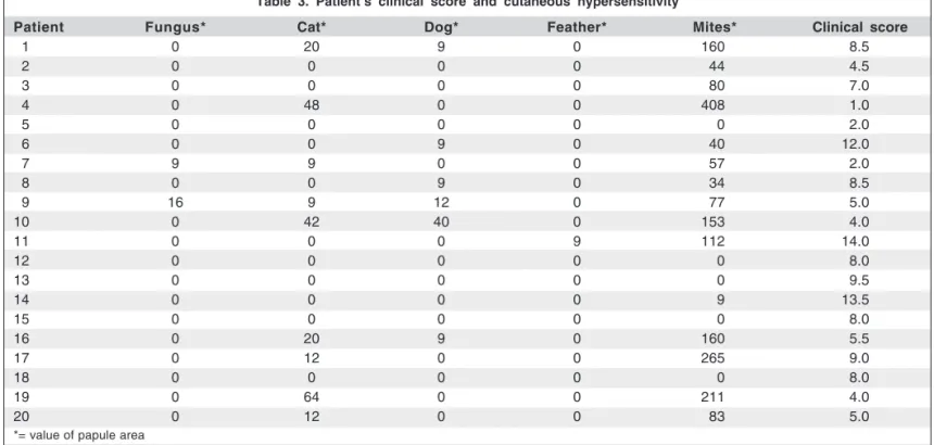
the relation between the number of positive tests and the clinical severity score was not significant (rs= -0.206; p=0.384).
DISCUSSION
The demographic and epidemiological characteristics of patients in the present study are very similar to those reported in other studies on VKC(2,12).
It is generally believed that 15-20% of the world popu-lation is atopic(13). Atopic profiles are well documented for
both personal cases and family histories of allergic diseases. In this study, we verified that 50% of subjects reported a family history of atopy, and 75% referred other atopic asso-ciated diseases. Allergic rhinitis (70%) and asthma (35%) were the most predominant.
Immediate cutaneous hypersensitivity testing has been an important tool in identifying allergens that can cause allergic diseases(14). However, distinct conditions, such as
dermographism(15), anti-histamines and tricyclic
anti-depres-sant administration can interfere in the results. These pro-blems can be obviated by applying tests with positive and negative controls. None of our patients tested negative to histamine or positive to the vehicle of the solution. Seventy-five percent of patients showed susceptibility to inhalant allergens. The same susceptibility to this kind of allergens was described in patients with asthma or rhinitis in our environment(13,16). Some authors found 15% sensitization in a
non-allergic children population against the same inhalant allergens used in this study(16).
VKC is mediated primarily by IgE, with active partici-pation of inflammatory cells in the conjunctiva. Activated eosinophils, soluble mediators, and the expression of adhe-sion molecules have been recognized as being involved in the ocular surface inflammation and responsible for tissue damage in corneal epithelium(17-20).
The etiologic evaluation of VKC has demonstrated new findings and new questions about its pathophysiology. Leo-nardi et al. demonstrated a poor correlation between systemic sensitivity tests (IgE) and ocular sensitivity (IgE from the tear Table 1. Clinical-epidemiological aspects of subjects
Patient Gender Age Age at symptom onset Season of worse symptoms Presence of atopy Familial history of atopy
01 F 05 03 Warm -
-02 M 17 07 Warm -
-03 M 04 02 Warm +
-04 M 15 09 Warm + +
05 M 11 06 Warm + +
06 M 07 04 Warm + +
07 M 10 04 Warm + +
08 M 10 04 Warm + +
09 M 18 14 No preference +
-10 M 12 06 No preference +
-11 M 11 05 Warm -
-12 M 05 05 Warm + +
13 F 10 03 Warm +
-14 F 11 07 Warm + +
15 F 06 04 Warm +
-16 F 12 07 Cold + +
17 M 17 06 Warm + +
18 M 08 03 Warm -
-19 M 17 06 Warm + +
20 M 19 05 Warm -
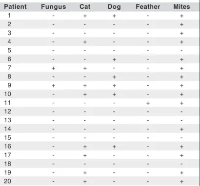
-Table 2. Cutaneous immediate hypersensitivity tests with various inhalant allergens
Patient Fungus Cat Dog Feather Mites
01 - + + - +
02 - - - - +
03 - - - - +
04 - + - - +
05 - - - -
-06 - - + - +
07 + + - - +
08 - - + - +
09 + + + - +
10 - + + - +
11 - - - + +
12 - - - -
-13 - - - -
-14 - - - - +
15 - - - -
-16 - + + - +
17 - + - - +
18 - - - -
-19 - + - - +
film), when they observed that 35% of their patients with VKC presented specific IgE only in tear samples. It suggests that the conjunctiva may be the only target tissue in some patients with allergic disease(6). Some studies demonstrated
that 50% of VKC patients showed negative cutaneous hyper-sensitivity testing as well as serologic IgE levels. They sug-gested that an aberration on Th2 lymphocyte line might be evolved in the pathogenesis of VKC(17). This is reinforced by
the finding of T cell clones, mainly Th2, in conjunctival tissue biopsies from patients with VKC. So far, by the description of increased in situ hybridization in areas where TCD4+ lym-phocytes responsible for the signal to IL-5 were predominant, and associated with high levels of IL-5, instead of IL-2 in the tears from specimens biopsed from VKC patients(21-25).
In the present study, we observed that 75% of patients were sensitive to at least one inhalant allergen. However, when we compare the clinical severity score of patients with positive and negative hypersensitivity tests, there was no significance. When we analyze the number of allergens to which the patients were sensitive and compared to intensity of cutaneous reaction (papule), there was also no significant correlation. Surprisingly, there was a negative and signi-ficant correlation with the allergens derived from cat, sugges-ting that some cat allergens could be protective: that is, the larger the papule area, the lower the clinical score. However, the present study is based on a small population, and this might not prove true in a larger cohort.
Given the poor correlation between clinical features and immediate cutaneous hypersensitivity, we believe that tests against the inhalant allergens used in this study were not helpful to identify patients with VKC who developed severe
and chronic ocular inflammation. Moreover, we do not be-lieve that these cutaneous tests proved to be of benefit in determining therapy against specific allergens, since the ocu-lar inflammation does not correlate with the skin reaction. Perhaps the unique benefit of these tests is to identify the allergens to which the patient is sensitive. Based on that testing, we may be able to advise patients which allergens to avoid. However, testing is not predictive of either intensity or severity of ocular inflammation once the patient is exposed to that specific antigen.
This poor correlation also reinforces the idea that other mechanisms are involved in the pathophysiology of VKC. As mentioned above, the conjunctiva can be an isolated and independent source of inflammation in patients with allergic diseases. It is a disease with a multifactorial pathogenesis, mediated by Th2 lymphocytes, eosinophils, mast cells, and a complex array of cytokines and chemical mediators.
Additional studies with larger numbers and using con-trol subjects without VKC may be helpful to determine the estimated risk relative to severity of cutaneous hypersen-sitivity.
CONCLUSION
This study demonstrated poor correlation between im-mediate cutaneous hypersensitivity testing and the clinical features in patients with vernal keratoconjunctivitis. It de-monstrated that cutaneous testing to inhalant allergens was not beneficial in predicting the potential for developing se-vere and/or chronic ocular surface inflammation.
Table 3. Patient’s clinical score and cutaneous hypersensitivity
Patient Fungus* Cat* Dog* Feather* Mites* Clinical score
01 0 20 9 0 160 8.5
02 0 0 0 0 44 4.5
03 0 0 0 0 80 7.0
04 0 48 0 0 408 1.0
05 0 0 0 0 0 2.0
06 0 0 9 0 40 12.0
07 9 9 0 0 57 2.0
08 0 0 9 0 34 8.5
09 16 9 12 0 77 5.0
10 0 42 40 0 153 4.0
11 0 0 0 9 112 14.0
12 0 0 0 0 0 8.0
13 0 0 0 0 0 9.5
14 0 0 0 0 9 13.5
15 0 0 0 0 0 8.0
16 0 20 9 0 160 5.5
17 0 12 0 0 265 9.0
18 0 0 0 0 0 8.0
19 0 64 0 0 211 4.0
20 0 12 0 0 83 5.0
RESUMO
Objetivo: Avaliar o papel da sensibilização cutânea a
dife-rentes aeroalérgenos em pacientes com ceratoconjuntivite vernal e a correlação entre esta e os aspectos clínicos da doença. Métodos: Vinte pacientes do setor de doenças exter-nas e córnea foram aleatoriamente convidados para participar deste estudo. Os pacientes foram avaliados e a eles foi atri-buído um escore clínico variando de 0 a 20 de acordo com sinais e sintomas presentes no exame oftalmológico. Todos os pacientes foram submetidos a testes cutâneos de hipersen-sibilidade imediata contra aeroalérgenos padronizados como os ácaros domiciliares D. pteronyssinus, D. farinae e Blomia tropicalis, assim como também a alérgenos de epitélio de gato, epitélio de cão, mistura de fungos e mistura de penas.
Resultados: Setenta e cinco por cento dos pacientes tiveram
teste de hipersensibilidade imediata positivo contra pelo menos um dos antígenos testados. Os ácaros domiciliares foram responsáveis pela maioria destes casos (75%). Houve uma pobre correlação entre o escore clínico e a hipersen-sibilidade cutânea aos alérgenos (r= -0,119 para fungos; r= -0,174 para epitélio de cão; r= -0,243 para ácaros domi-ciliares; r= -0,090 para mistura de penas). Houve correlação significativa apenas contra epitélio de gatos (r= -0,510; p=0,024). Conclusão: O estudo demonstrou uma pobre cor-relação entre os testes cutâneos de hipersensibilidade ime-diata e os achados clínicos nos pacientes com ceratoconjun-tivite vernal. Os testes cutâneos de hipersensibilidade ime-diata contra aeroalérgenos não foi parâmetro eficaz na identi-ficação dos casos de maior gravidade e cronicidade de cerato-conjuntivite vernal.
Descritores: Hipersensibilidade; Conjuntivite alérgica;
Con-juntiva/imunologia; Testes cutâneos
REFERENCES
1. Bonini S, Bonini S, Lambiase A, Marchi S, Pasqualetti P, Zuccaro O, et al. Vernal keratoconjunctivitis revisited: a case series of 195 patients with long-term follow-up. Ophthalmology. 2000;107(6):1157-63.
2. Tabbara KF. Ocular complications of vernal keratoconjunctivitis. Can J Oph-thalmol. 1999;34(2):88-92.
3. Pastorello EA, Incorvaia C, Ortolani C, Bonini S, Canonica GW, Romagnani S, et al. Studies on the relationship between the level of specific IgE antibodies and the clinical expression of allergy: I. Definition of levels distinguishing patients with symptomatic from patients with asymptomatic allergy to common aeroallergens. J Allergy Clin Immunol. 1995;96(5 Pt 1):580-7.
4. Ownby DR. Clinical significance of IgE. In: Middleton E Jr, Reed CE, Ellis EF, Adkinson NF Jr, Yunginger JW, Busse WW, editors. Allergy: principles and practice. 4th ed. St. Louis: Mosby; 1993. p.1059-76.
5. Norman PS, Lichtenstein LM, Ishizaka K, Comparison of specific IgE anti-bodies, leukocyte sensitivity by histamine release, direct skin tests, and symptoms in hay fever. In: Goodfriend L, Sehon A, Orange R, editors. Mechanisms in allergy: reagin-mediated hipersensitivity. New York: Marcel Dekker; 1973. p.151-62.
6. Leonardi A, Fregona IA, Gismondi M, Daniotti E, Carniel G, Secchi AG. Correlation between conjunctival provocation test (CPT) and systemic aller-gometric tests in allergic conjunctivitis. Eye. 1990;4(Pt 5):760-4.
7. Orgel HA, Kemp JP, Meltzer ED, Hamburger RN. Atopy and IgE in a pediatric allergy practice. Ann Allergy.1977;39(3):161-8.
8. Pucci N, Novembre E, Lombardi E, Cianferoni A, Bernardini R, Massai C, et al. Atopy and serum eosinophil cationic protein in 110 white children with vernal keratoconjunctivitis: differences between tarsal and limbal forms. Clin Exp Allergy. 2003;33(3):325-30. Comment in: Clin Exp Allergy. 2003;33(3); 279-81.
9. Soothill JF, Stokes CR, Turner MW, Norman AP, Taylor B. Predisposing factors and the development of reaginic allergy in infancy. Clin Allergy. 1976;6(4):305-19.
10. Foucard T. A follow-up study of children with asthmatoid bronchitis. I. Skin test reactions and IgE antibodies to common allergens. Acta Paediatr Scand. 1973;62(6):633-44.
11. Pepys J. Skin tests for immediate, type, allergic reactions. Proc R Soc Med. 1972;65(3):271-2.
12. Van Asperen PP, Kemp AS, Mellis CM. Skin test reactivity and clinical allergen sensitivity in infancy. J Allergy Clin Immunol. 1984;73(3):381-6. 13. Arruda LK, Rizzo MC, Chapman MD, Fernandez-Caldas E, Baggio D,
Platts-Mills TA, et al. Exposure and sensitization to dust mite allergens among asthmatic children in São Paulo, Brazil. Clin Exp Allergy. 1991;21 (4):433-9.
14. JinY, Xu Y, Xue S, Liu H, Zhao J, Xu M. Predicting the development of early skin test sensitization in offspring of parents with asthma. Eur J Clin Invest. 2007;37(6):522-7.
15. Volonakis MK, Tsaptsinos NJ, Kontou-Fili K. The diagnostic value of skin-prick tests in dermographic individuals. Allergy Proc. 1991;12(2):103-6. 16. Esteves PC, Rosário Filho NA, Trippia SG, Caleffe LG. Sensibilização
atópica em escolares e adultos de Curitiba, Paraná. Rev Bras Alergia Imu-nopatol. 1999;22(5):156-60.
17. Bonini S, Bonini S. IgE and non IgE mechanisms in ocular allergy. Ann Allergy. 1993;71(3):296-9.
18. Trocme SD, Kephart GM, Allansmith MR, Bourne WM, Gleich GJ. Con-junctival deposition of eosinophil granule major basic protein in vernal keratoconjunctivitis and contact lens-associated giant papillary conjunctivitis. Am J Ophthalmol. 1989;108(1):57-63.
19. Trocme SD, Kephart GM, Bourne WM, Buckley RJ, Gleich GJ. Eosinophil granule major basic protein deposition in corneal ulcers associated with vernal keratoconjunctivitis. Am J Ophthalmol. 1993;115(5):640-3.
20. Bonini S, Tomassini M, Bonini S, Capron M, Balsano F. The eosinophil has a pivotal role in allergic inflammation of the eye. Int Arch Allergy Appl Immunol. 1992;99:354-8.
21. Maggi E, Biswas P, Del Prete G, Parronchi P, Macchia D, Simonelli C, et al. Accumulation of Th2-like helper T cells in the conjunctiva of patients with vernal conjunctivitis. J Immunol. 1991;146(4):1169-74.
22. Van Leeuwen BH, Martinson ME, Webb GC, Young IG. Molecular orga-nization of the cytokine gene cluster, involving the human IL-3, IL-4, IL-5, and GM-CSF genes, on human chromosome 5. Blood. 1989;73(5): 1142-8.
23. Trocme SD, Aldave AJ. The eye and the eosinophil. Surv Ophthalmol. 1994; 39(3):241-52. Review.
24. Bonini S, Bonini S, Lambiase A, Magrini L, Rumi C, Del Prete Ge, et al. Vernal keratoconjunctivitis: a model of 5q cytokine gene cluster disease. Int Arch Allergy Immunol. 1995;107(1-3):95-8.