Disorders
Nicole R. Zu¨rcher1,2, Ophe´lie Rogier1, Jasmine Boshyan2, Loyse Hippolyte1, Britt Russo1, Nanna Gillberg3, Adam Helles3, Torsten Ruest1, Eric Lemonnier4, Christopher Gillberg3, Nouchine Hadjikhani1,2,3* 1Brain Mind Institute, EPFL, Lausanne, Switzerland,2Athinoula A. Martinos Center for Biomedical Imaging, Harvard Medical School, Massachusetts General Hospital, Charlestown, Massachusetts, United States of America,3Gillberg Centrum, University of Gothenburg, Gothenburg, Sweden,4Laboratoire de Neurosciences, Universite´ de Brest, Brest, France
Abstract
Intuitive grasping of the meaning of subtle social cues is particularly affected in autism spectrum disorders (ASD). Despite their relevance in social communication, the effect of averted gaze in fearful faces in conveying a signal of environmental threat has not been investigated using real face stimuli in adults with ASD. Here, using functional MRI, we show that briefly presented fearful faces with averted gaze, previously shown to be a strong communicative signal of environmental danger, produce different patterns of brain activation than fearful faces with direct gaze in a group of 26 normally intelligent adults with ASD compared with 26 matched controls. While implicit cue of threat produces brain activation in attention, emotion processing and mental state attribution networks in controls, this effect is absent in individuals with ASD. Instead, individuals with ASD show activation in the subcortical face-processing system in response to direct eye contact. An effect of differences in looking behavior was excluded in a separate eye tracking experiment. Our data suggest that individuals with ASD are more sensitive to direct eye contact than to social signals of danger conveyed by averted fearful gaze.
Citation:Zu¨rcher NR, Rogier O, Boshyan J, Hippolyte L, Russo B, et al. (2013) Perception of Social Cues of Danger in Autism Spectrum Disorders. PLoS ONE 8(12): e81206. doi:10.1371/journal.pone.0081206
Editor:Atsushi Senju, Birkbeck, University of London, United Kingdom
ReceivedAugust 22, 2013;AcceptedOctober 20, 2013;PublishedDecember 4, 2013
Copyright:ß2013 Zu¨rcher et al. This is an open-access article distributed under the terms of the Creative Commons Attribution License, which permits unrestricted use, distribution, and reproduction in any medium, provided the original author and source are credited.
Funding:This work was supported by the Swiss National Science Foundation PP00P3-130191 to NH and by the Velux Stiftung, by the Centre d9Imagerie BioMe´dicale (CIBM) of the University of Lausanne (UNIL), the Swiss Federal Institute of Technology Lausanne (EPFL), the University of Geneva (UniGe), the Centre Hospitalier Universitaire Vaudois (CHUV), the Hoˆpitaux Universitaires de Gene`ve (HUG), and the Leenaards and the Jeantet Foundations. The funders had no role in study design, data collection and analysis, decision to publish, or preparation of the manuscript.
Competing Interests:The authors have declared that no competing interests exist.
* E-mail: [email protected]
Introduction
Autism spectrum disorder (ASD) is a neurodevelopmental condition affecting more than 1% of children [1,2], characterized by deficits in social interaction and communication as well as by the presence of restricted interests and repetitive behaviors [3]. Absence or impairment of social instinct has been proposed to lie at the core of ASD [4].
Social observation is an efficient way to learn about potential harmful situations in the environment [5,6], and evolutionary-old fear mechanisms are automatically engaged when typical individ-uals observe others showing signs of fear-related distress. Fearful expression and gaze direction are directly linked with biological self-relevance (Figure 1). In typical individuals, averted gaze in a fearful face is detected faster [7], rated as more intense than the same fearful expression with a direct gaze [8], and leads to automatic/reflexive gaze shifts [9]. Studies have shown that individuals with ASD show atypical brain activation in response to fearful facial expressions [10]) and to gaze [11–14]. However, despite their relevance in social communication, fear and gaze direction interactions have not been investigated using real faces in adults with ASD.
Gaze perception produces activation of the intraparietal sulcus, the superior temporal sulcus (STS) and regions of the dorsal and ventral fronto-parietal attention networks [15–18]. Saliency is captured by several areas: the amygdala ensures automatic
attention to threatening stimuli [19], allowing biologically self-relevant stimuli to be processed even when outside the current focus of attention (reviewed in [20]); the pulvinar nucleus of the thalamus, through its reciprocal connections with the amygdala [21] and the superior colliculus (SC) [22,23] contributes to the selection of salient stimuli [24,25]; finally, the SC, associated with covert and overt shifts of attention [26], along with the frontal eye fields is involved in saccadic eye movement generation. The interaction of emotion and gaze direction hence involves various social attention processes including reorientation of attention, emotion processing as well as attribution of thoughts and intentions [27].
not show activation in brain regions associated with social attention compared to typical control participants.
Materials and Methods
Participants
The protocol was approved by the Lausanne University Hospital Ethical Committee and all procedures followed the Declaration of Helsinki. After complete description of the study was given to the participants, written informed consent was obtained. Twenty-six high-functioning individuals with ASD were enrolled in the study, from three centers (Lausanne, Brest and Gothenburg). For comparison purposes, 26 typical control individuals (CON) with no history of psychiatric or neurological disorders were recruited in Lausanne. Four participants with ASD and 4 CON had to be excluded due to excessive movement (.3 mm) during data acquisition. Thus 22 participants with ASD (19 males, 27.6 years67.7 (mean6SD)) and 22 CON participants (19 males, 23.7 years65.9) were included in the final data analysis. Participants in the ASD group were diagnosed according to DSM-IV-TR criteria by experienced clinicians [3]. The Autism Diagnostic Observation Schedule (ADOS) and the Autism Diagnostic Interview-Revised (ADI-R) [32,33] were conducted for 14 ASD participants and the Diagnosis of Social and Communication Disorder-10 (DISCO-10) [34] was used for the participants from Gothenburg. All participants met criteria for autism spectrum disorder according to the current DSM 5 criteria [35]. In addition, autism traits were assessed in all participants but one using the Autism Quotient (AQ) self-report questionnaire [36]. The ASD group scored significantly higher than the CON group (ASD: 28.167.0; CON: 13.064.1;t(41) = 8.6,p,0.001). Perfor-mance intelligence quotient (PIQ) was assessed using the Wechsler Non-verbal Scale or the Wechsler Abbreviated Scale of Intelli-gence [37,38] and all participants had a PIQ in the normal range (ASD: 114615; CON 11268). ASD and CON-groups did not differ in terms of age, intelligence quotient or gender. All participants had normal or corrected to normal vision. None of
the participants of the current study were enrolled in the previously published study [30].
Stimuli and paradigm
The paradigm used in the current experiment has been previously described in [30]. The stimuli were taken from the NimStim Set of Facial Expressions database [39]. Eight greyscale fearful faces (4 females) were selected and their gaze direction was altered by changing the position of the iris so that the faces were looking downwards toward the left or right, without altering their head direction (for an example of the stimuli used, see Figure 1 in [30]. A central fixation cross (FIXATION) was presented for 1200 ms followed by a face stimulus briefly presented for 300 ms in the center of the screen. This ensured that the eye region of the face stimuli appeared where the fixation cross was previously located and that the participants would attend to the eye-region [40]. Faces were presented in 24-second alternating blocks: 8 blocks of stimuli with direct (DIRECT) gaze and 8 blocks with averted (AVERTED) gaze (to the right in half of the blocks, to the left in the other half). Participants were instructed to observe the images attentively, and to look at the fixation cross, while trying to feel what the faces they were observing expressed.
MRI data acquisition
Imaging data were acquired on a 3T scanner (Siemens Tim Trio, Erlangen, Germany) using a 12-channel matrix coil at the Centre d9Imagerie BioMe´dicale at the Centre Hospitalier Universitaire Vaudois in Lausanne. Slices were automatically positioned using the online AutoAlign Head LS (Landmark Survey) from Siemens. T1-weighted high-resolution (1.061.061.0 mm3) structural images were obtained at the beginning of the session with a multi-echo magnetization-prepared rapid acquisition gradient echo (ME-MPRAGE) sequence (176 slices, FOV = 256, matrix size 2566256, echo time (TE1) = 1.64 ms, (TE2) = 3.5 ms, (TE3) = 5.36 ms (TE4) = 7.22 ms; repe-tition time (TR) = 2530 ms; flip angle = 7u). Whole brain
Figure 1. Face and gaze interactions depend on the degree of biological relevance conveyed.(1) For neutral faces, humans are more sensitive to direct gaze than averted gaze [67], as direct gaze reflects interest from a social partner and the beginning of a social exchange. (2) A face looking at us with a fearful expression is more arousing than a face with a neutral expression, due to the strong emotion it conveys [64]. (3) For fearful facial expressions, averted gaze is the most biologically self-relevant condition, with the social partner using non-verbal communicative cues to alert us to potential environmental danger [30].
T2*-weighted gradient echo-planar images (EPI) were collected during the presentation of the paradigm. This functional acquisition (45 or 47 AC-PC slices, FOV = 216, matrix size = 64664, TE = 30 ms, TR = 3 s, slice thickness 3 mm, flip angle 90u) lasted 384 s.
fMRI data preprocessing and analysis
Whole brain voxel-wise analyses were conducted using FEAT version 5.98 part of FSL (FMRIB Software Library). For each subject first-level general linear model (GLM) analyses were conducted for the contrast averted vs. direct fearful gaze. Motion-correction was conducted using MCFLIRT and the motion parameters were added as nuisance parameters to the model. FSL’s motion outlier detection program was used to identify residual outlier timepoints, which were included as additional confound variables in the GLM. Spatial smoothing using a Gaussian kernel of 8 mm, grand mean intensity normalization and highpass temporal filtering with sigma = 50.0 s were applied. Brain extraction of high-resolution anatomical images was carried out using Christian Gaser’s VBM8 toolbox for SPM8 [41] and fed into FEAT. Subject-level analyses for the contrast AVERTED. DIR-ECT and DIRDIR-ECT.AVERTED were performed using FILM. Non-linear Registration to the MNI template was carried out using the tool FNIRT. Group-level analyses were conducted using mixed effects with FLAME 1 and 2, allowing inference about the population from which the individuals were drawn. FSL’s randomise was used to perform a permutation-based nonpara-metric statistical between-group (CON vs. ASD) analysis (n permutation = 10,000) using threshold-free cluster enhancement (TFCE). P values were family-wise error (FWE) corrected (p
FWE,0.05). Local maxima where identified usingtvalue maps as FWE-corrected clusters appeared large. A threshold oft.3.2 was chosen to control cluster size. Thus, only clusters which survivedp
FWE,0.05 andt.3.2 and contained at least 20 contiguous voxels are reported. All coordinates refer to MNI standard space. For visualization, statistical correctedpvalue maps (pFWE,0.05) are displayed on the pial cortical surface of the FreeSurfer brain (fsaverage) template (htttp://surfer.nmr.mgh.harvard.edu). In an additional analysis, the contrast AVERTED.FIXATION and DIRECT.FIXATION were compared within and between groups to control for potential differences in activation of face processing areas for the two different gaze conditions.
ROI analyses
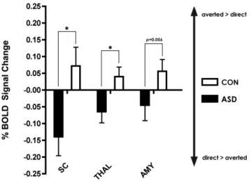
Regions of interest (ROIs) were selected to analyze activation of the subcortical route, known to be involved in the detection of biologically relevant stimuli, and consisting of the thalamus, the amygdala and the SC. To avoid circularity, ROIs were defined by independent anatomical constraints. The thalamus and the amygdala were identified using the respective label within the 25% probability Harvard-Oxford subcortical atlas. The SC was selected following anatomical landmarks [42]. Standard space anatomical ROIs were mapped back to subject space. Subse-quently, for each ROI, mean percentage BOLD signal change within that ROI was extracted from the contrast of parameter estimate at the subject-level using FSL’s Featquery. For each ROI Mann-Whitney U-test were conducted to assess differences between groups.
Eye-tracking
To control for potential between-group differences in looking behavior, we conducted an eye-tracking study on a separate day after the fMRI experiment in a subset of the participants. Nineteen ASD and 14 CON participated in this experiment, but 3 ASD had
to be excluded due to insufficient data (unsuccessful calibration or poor tracking quality). Data analysis was therefore conducted on 16 ASD and 14 CON.
Data collection and analysis
Eye-tracking data was collected using a T120 eye-tracking system running Tobii Studio (TOBII Technology, Sweden). Participants sat comfortably 60–65 cm away from a 17-inch flat screen in a dimly lit room. Corneal reflection was measured for both eyes with infrared light sources and cameras, integrated in the monitor. A 9-point calibration was run prior to the experiment and data were recorded at 60 Hz. The same stimuli as those used in the fMRI were presented for the same amount of time as in the fMRI experiment (300 ms), preceded by a fixation cross (1200 ms). Areas of interest (AOI) were drawn for the eye region, the face and the computer screen. The eye region consisted of one rectangle covering both eyes and the bridge of the nose between the eyes. One large oval was used as AOI for the face. The total time spent looking at those areas was measured using Tobii Studio v.3.0.2. Eye fixations were determined using the criterion of eye position remaining within a 35-pixel area for a time greater than 80 ms. Analysis was conducted on absolute time spent looking at the eye region and at the face as well as on the ratio of time spent on eye region to time spent on the computer screen and time spent on face to time spent on computer screen. For each AOI total fixation duration differences in averted vs. direct gaze conditions were investigated within group using non-parametric Wilcoxon signed-rank tests. Between-group differences (CON vs. ASD) were assessed using two-tailed non-parametric Mann-Whitney U-tests.
Results
Eye-tracking results
No differences were found for the time spent on theeyesbetween gaze conditions or groups (CON: fear direct: 211 ms623 (mean6SEM) and fear averted: 213 ms624, ASD: fear direct: 190 ms620 and fear averted: 195 ms620, allp.0.05,ns.) and for theratio of time spent on the eyesto time spent on the computer screen (CON: fear direct: 77.4%60.8 (mean6SEM) and fear averted: 78.0%60.9, ASD: fear direct: 69.0%60.7 and fear averted: 70.7%60.7, all p.0.05, ns.). There were also no significant differences for theface region (CON: fear direct: 272 ms62 and fear averted: 274 ms62, ASD: fear direct: 261 ms66 and fear averted: 270 ms64, allp.0.05,ns.) and for theratio of time spent on the faceto time spent on the computer screen (CON: fear direct: 100.0%60 and fear averted: 100.0%60, ASD: fear direct: 95.6%62.4 and fear averted: 98.9%60.8, all p.0.05, ns.). This was expected given the chosen paradigm, designed to have participants look in the eye region (fixation cross presented where eye region of face would later appear), and the very short presentation time (300 ms).
fMRI results
Within-group whole brain analysis -AVERTED.FIXATION and DIRECT.FIXATION. As ex-pected based on the eye-tracking data, both ASD and CON showed increased activation in striate and extrastriate areas for direct and for averted gaze when compared to fixation. In particular, ASD and CON exhibited fusiform face area (FFA) activation in both conditions, indicating that participants in both groups were looking at the faces.
in several brain regions including the frontal eye fields, the intraparietal sulcus, the superior temporal gyrus, the FFA, the insula and the supramarginal gyrus (see Table 1) whereas ASD failed to demonstrate increased activation in any area for this contrast, even at a very liberal threshold (p,0.05, uncorrected).
Within-group whole brain analysis -DIRECT.AVERTED. For DIRECT.AVERTED gaze, CON did not show increased activation even at a very liberal threshold (p,0.05, uncorrected). ASD participants did not show increased activation for direct gaze atpFWE,0.05. However, at a more liberal threshold (p,0.01, uncorrected), ASD showed increased activation for direct fearful gaze compared to avert in areas of the subcortical route, including SC and thalamus (but not the amygdala), and in fronto-insular cortex, anterior cingulate, posterior cingulate/precuneus, and cerebellum. See Table 2.
Between-group whole brain analysis -AVERTED.DIRECT. For the contrast AVERTED. DIR-ECT, CON showed increased activation compared to ASD in areas associated with gaze processing and attention including the
intraparietal sulcus, superior parietal lobule, frontal eye fields, STS, superior temporal gyrus, temporo-parietal junction and supramarginal gyrus. CON also exhibited increased emotion processing in brain areas involved in emotion processing, including the anterior insula, anterior cingulate and posterior cingulate/ precuneus cortex. In addition, increased activation was found for CON compared to ASD in the striate and extrastriate cortex, FFA, inferior occipital gyrus, inferior frontal gyrus, thalamus, hippocampus and cerebellum (pFWE,0.05,t.3.2, 20 contiguous voxels). See Figure 2, Table 3.
A prioriROI analysis
For all subcortical ROIs, values were numerically greater for CON for the contrast AVERTED.DIRECT, indicating activa-tion in controls for averted gaze. In contrast, ROI values for ASD for the contrast AVERTED.DIRECT were always negative, indicating that ASD showed more activation for the direct gaze condition. Significant between-group differences were observed for the SC (p= 0.01) and the right thalamus (p= 0.04), and showed a
Table 1.Within-group contrasts in CON.
Region Hemi MNI tvalue
x y z
AVERTED.DIRECT Inferior temporal cortex RH 58 264 216 5.96
LH 254 262 212 4.65
Intraparietal sulcus RH 14 254 52 5.84
LH 218 272 54 5.34
Frontal Eye Field RH 24 210 48 5.55
LH 226 210 50 5.1
Precuneus RH 9 252 55 5.39
LH 210 252 54 5.46
Lateral Occipital Cortex LH 240 278 12 5.22
RH 42 276 18 4.64
Superior temporal gyrus anterior RH 66 210 0 5.16
LH 258 210 22 4.68
Superior parietal cortex LH 226 254 62 5.04
RH 30 246 64 4.35
FFA RH 42 260 216 4.97
LH 242 60 220 3.42
Superior temporal gyrus posterior LH 262 242 14 4.82
RH 66 232 18 4.18
Parieto-occipital sulcus RH 24 261 22 4.82
LH 218 274 24 4.1
Supramarginal cortex RH 60 232 44 4.72
LH 256 232 44 4.11
Mid Cingulate cortex LH 28 0 42 4.44
RH 8 18 38 4.16
Insula RH 38 212 26 4.24
LH 242 22 2 3.64
Hippocampus RH 20 232 26 4.07
Inferior frontal gyrus, pars opercularis LH 256 2 6 3.65
DIRECT.AVERTED none
Brain regions for which CON showed increased activation for the contrast [AVERTED.DIRECT] gaze atpFWE,0.05,t.3.2. CON did not show increased activation for [DIRECT.AVERTED] gaze.
strong trend towards significance in the left amygdala (p= 0.056). See Figure 3.
Discussion
Previous studies in autism have mostly investigated gaze and facial expression separately, leaving aside their interactive effects. Here, by combining fearful expression with different gaze directions, we demonstrate that the observation of social cues implicitly indicating the presence of a danger does not result in activation of brain areas involved in gaze perception, attention, emotion processing and mental state attribution in adults with ASD.
In our study, ASD participants failed to show typical activation in the dorsal and ventral fronto-parietal attention networks for averted vs. direct gaze. The absence of activation of these
top-down and bottom-up attention networks suggests the lack of intuitive grasping of the biological relevance of the gaze cue and the absence of spontaneous reorientation. While studies using emoticons and studies using neutral faces have previously shown reflexive orienting in response to eye gaze cues in ASD [43,44], the present study is to our knowledge the first to address the perception of the meaning of an emotional and social cue using real faces with emotional expressions in adults with ASD.
In ASD, gaze following behavior is developmentally delayed, and joint attention deficits belong to the earliest markers of this disorder [45–48]. Deficits in joint attention, i.e. deficits in the ability to non-verbally coordinate attention between individuals in order to share information regarding the environment, remain present in adults with ASD. Individuals with ASD do not spontaneously react to joint attention cues in videos with avatars, still emoticons with a neutral expression, or during live interactive
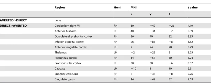
Table 2.Within-group contrasts in ASD.
Region Hemi MNI tvalue
x y z
AVERTED.DIRECT none
DIRECT.AVERTED Cerebellum right VI RH 30 242 226 4.19
Anterior fusiform RH 40 234 220 3.89
Dorsolateral prefrontal cortex RH 36 40 32 3.83
Inferior occipital cortex RH 26 286 28 3.82
Anterior cingulate cortex RH 2 24 28 3.29
Thalamus LH 22 222 2 3.25
Precuneus cortex RH 14 258 30 3.24
Fronto-insular cortex RH 30 30 26 3.07
Caudate LH 210 8 10 2.9
Superior colliculus RH 6 236 28 2.76
Cingulate gyrus RH 14 242 32 2.63
Areas, which showed increased activation in CON participants for the contrast [DIRECT.AVERTED] atp,0.01. Individuals with ASD did not show increased activation for [AVERTED.DIRECT].
doi:10.1371/journal.pone.0081206.t002
Figure 2. Cortical activation for averted gaze.Statistical maps of differences in fMRI activation for CON.ASD for the contrast averted.direct gaze (depicted in red to yellow). Group differences reflect increased activation for averted gaze in CON and lack of activation in ASD. Statistical maps are displayed on the lateral, medial and ventral views of both hemispheres, atpFWE,0.05. The light grey mask covers subcortical regions in which
video [12–14,49]. The capacity to attribute mental states to others, also known as theory of mind (ToM), has been suggested to arise from joint attention [50] and individuals with ASD show deficits in ToM, as demonstrated by their decreased performance in the ‘‘reading the mind in the eyes’’ task [51], as well as by their lack of spontaneous mental state attribution to others [52] or to animated shapes [53]. In this study, the ASD group showed significantly less activation in areas associated with the attribution of thoughts, actions and intentions to others. Notably, we observed absence of modulation in posterior STS in response to gaze cues, a finding previously reported in ASD [14,49]. The STS is involved in
biological motion and gaze perception [54] and abnormal STS activation has been repeatedly described in autism (for review see [55]).
Unlike typical individuals, ASD failed to show increased activation in the anterior insula for averted fearful gaze. The anterior insula, structurally connected with the posterior STS through the superior longitudinal fasciculus, is sensitive to the social significance of eye gaze [56]. The insular cortex has been associated with multiple functions, ranging from performance monitoring [57] and attention to sensory and sensorimotor processing [58], and the activation in the ventral part of the
Table 3.Between-group contrasts: CON.ASD for [AVERTED.DIRECT].
Region Hemi MNI k tvalue
x y z
Gaze & Attention Anterior STS RH 48 26 216 2127 3.93
LH 258 28 22 147 3.67
Intraparietal sulcus RH 18 266 64 1190 4.31
LH 216 270 54 130 3.64
Frontal eye fields RH 24 24 66 1340 4.47
LH 226 26 64 131 3.54
Superior parietal lobule RH 20 246 68 1190 3.31
LH 224 252 66 530 4.63
Supramarginal gyrus RH 60 232 46 309 4.02
LH 256 226 28 300 3.75
Inferior frontal gyrus, pars opercularis RH 54 20 14 31 3.38
Dorsolateral prefrontal cortex RH 40 48 30 183 4.39
MT/V5 LH 262 260 10 28 3.73
Emotion Anterior Insula RH 42 16 24 25 3.46
LH 242 10 26 219 3.46
Anterior cingulate RH 6 18 38 1340 3.77
LH 26 2 38 1340 4.75
Postcentral gyrus RH 66 216 28 65 3.69
LH 212 240 50 54 3.93
Hippocampus RH 24 222 212 2127 4.18
LH 234 218 214 101 3.29
Theory of Mind Posterior STS RH 70 236 4 24 3.73
Superior temporal gyrus ant RH 68 28 0 34 3.71
Temporo-parietal junction RH 66 230 28 58 3.38
Temporal pole RH 38 8 224 2127 4.07
LH 252 14 218 219 4.89
Posterior cingulate cortex/Precuneus RH 8 238 46 1190 4.57
Subcortical route Thalamus RH 10 218 8 58 3.77
Superior colliculus RH 8 230 28 2127 3.4
Face processing Fusiform, FFA RH 42 260 216 2127 4.3
Anterior fusiform gyrus RH 38 236 220 2127 4.22
LH 242 232 222 101 3.9
Inferior occipital gyrus RH 30 288 28 33 3.41
Lingual gyrus RH 8 254 22 91 4.06
Visual processing Parieto-occipital sulcus RH 16 282 36 1371 5.11
Inferior lateral occipital cortex LH 240 282 10 64 3.66
Other Cerebellum left crus I LH 236 258 240 115 3.79
anterior insula observed in controls is likely related to socio-emotional processing [58,59], that is absent in ASD. The anterior cingulate cortex (ACC), involved in appraisal and regulation of negative emotion [60] was also significantly less activated in ASD. Increased activation of the FFA for averted vs. direct gaze was observed in controls but not in ASD participants (although both groups showed FFA activation in both avert and direct conditions compared with fixation). The importance of the eye region in driving FFA activation has been shown in previous studies [61,62]. In the eye-tracking experiment, both groups spent the same amount of time looking at the eye region in both conditions, strongly suggesting that different fixation times on the eye region are not the cause of the observed difference between groups in the FFA for averted vs. direct gaze. Instead, a more likely explanation is that participants with ASD, not grasping the increased emotional meaning of the averted gaze stimulus in the fearful face (a phenomenon previously reported in typical individuals [30] and linked with both attentional and emotional processes [63]), fail to modulate FFA activation in response to this biologically-relevant cue.
The detection of threat-related facial expressions and the ability to quickly read gaze direction play a central role for adaptive responses. Based on the literature, we propose a conceptual scheme emphasizing that the combination of facial expression and gaze direction are directly linked with biological self-relevance (See Figure 1). In neutral expressions (1), direct gaze leads to more activation than averted gaze, as direct gaze represents a desire to engage in a social interaction. Direct gaze associated with a fearful emotion (2), leads to more activation than neutral direct gaze (reviewed in [64]. Even more activation is observed for briefly presented fearful faces with averted gaze (3). Averted gaze in a fearful face is biologically self-relevant, and leads to shorter reaction times and increased amygdala activation in typical individuals [7,28,30,31,65].
Figure 4 summarizes the findings for the processing of gaze in neutral and fearful faces in ASD. For neutral facial expression, individuals with ASD as well as controls show increased activation in response to direct neutral gaze as opposed to averted neutral gaze [66,67]. Recent data show that this process is supported by the subcortical route, as amygdala activation for neutral direct
gaze has been documented in a cortically blind patient [68]. The influence of direct gaze on behavior is referred to as ‘‘eye contact effect’’ reflecting the fact that perceived eye contact in others modulates cognitive processes (reviewed in [67]) and drives activation of areas associated with social processing, including the FFA, STS, amygdala and medial prefrontal cortex. In a study investigating the perception of socially relevant facial expressions either self- or other-directed (as indicated by gaze direction), ventromedial prefrontal cortex and medial temporal lobe/ amygdala were shown to play an important role [69]. In ASD atypical eye contact effect has been observed, reflecting altered processing of direct gaze [70]. Increased subcortical activation in response to direct gaze in fearful faces in ASD is in line with reports of atypical modulation of arousal in response to direct gaze in children with ASD reported by Kylliainen et al, who measured greater skin conductance in response to direct than averted gaze [66], increasing as a function of the degree of eye openness [71]. Studies investigating modulation byemotionin direct gaze have shown diminished modulation of the face-processing network in ASD [72,73], and a study conducted in adolescents with ASD reported that brain activations do not differ between averted and direct gaze in negative (anger and fear combined) facial emotions [74]. However, the results of this latter study do not allow to specifically draw conclusions about the interaction of gaze direction with a fearful facial expression as these two emotions were not analyzed separately. In addition, individuals with Asperger Syndrome do not have faster reaction times for fearful averted gaze, while controls show enhancement of joint attention by emotion [75].
To our knowledge, even though numerous studies have investigated the effect of fear vs. scrambled stimuli or the effect of various intensities of fear, no study has specifically compared fearful with neutral facial expressions (with direct gaze). Finally, in the current study, we show that individuals with ASD do not show increased activation for averted gaze in a fearful face. Instead, whole brain within-group and ROI analysis show increased activation of the subcortical face detection route in ASD for direct fearful gaze. This route, consisting of the SC, the thalamus and amygdala, [64,76–79], is activated in typical individuals by direct eye contact in neutral faces [67], to a greater extent by direct gaze in a fearful face [64,80] and to an even greater extent by an averted gaze in a briefly presented fearful face [29–31]. As shown by Senju and Johnson, the subcortical route may not appropriately modulate cortical and subcortical social brain networks in individuals with ASD [70], and the lack of top-down modulation together with decreased processing of mental and emotional states may therefore have lead to increased eye contact effect [81,82].
Figure 3. Region of interest analysis.Percent BOLD signal change (6SEM), for averted vs. direct gaze in selected subcortical ROIs. The thalamus (THAL) (p= 0.01), and superior colliculus (SC) (p= 0.04) were significantly different between ASD and CON while a strong trend was found for the amygdala (AMY) (p= 0.056).
doi:10.1371/journal.pone.0081206.g003
Figure 4. Atypical reactivity to social stimuli in ASD.Individuals with ASD show increased response to direct as opposed to averted gaze ((1) - Kylliainen 2006) but show atypical eye contact. While deficits in fearful face processing have been described in ASD, no study to our knowledge has specifically investigated fearful vs. neutral faces and it is unclear if individuals with ASD would show more activation in response to direct fearful gaze as opposed to direct neutral gaze. Finally, unlike controls, individuals with ASD do not show more activation for fearful averted gaze.
Future studies should investigate the role of gaze direction in other emotional expressions and neutral faces.
Conclusions
Using short stimulus presentation times, reflecting quick joint attention bids akin to how they occur in real life, we observed significant deficits in the activation of the distributed network of social attention in high-functioning individuals with ASD. Although both ASD and control participants looked similarly at the eye-region of the stimuli, networks involved in attention, gaze perception, emotion attribution and understanding of intentions were not engaged in individuals with ASD when processing social cues of danger. Instead participants with ASD showed hyper-activation of the subcortical route for direct gaze. This suggests that for individuals with ASD, eye contact with a fearful expression is more arousing than a fearful averted gaze signaling the potential presence of an environmental danger. These findings suggest that
in early behavioral therapies, emphasis should be placed on association between eye-gaze cues and emotions, in order to specifically train the integration of these cues, thereby allowing young children with ASD to gain access to their social meaning.
Acknowledgments
We warmly thank all participants and their families for their time and effort. We thank K. Me´trailler for her support in recruitment, J. Iranpour for help with eye-tracking data collection, A. Lissot for his technical support and C. Burget for her administrative support.
Author Contributions
Conceived and designed the experiments: NH NZ. Performed the experiments: NZ OR JB LH BR NG AH EL NH. Analyzed the data: NZ OR JB LH BR TR EL NH. Contributed reagents/materials/analysis tools: EL CG. Wrote the paper: NZ CG NH.
References
1. Blumberg SJ, Bramlett MD, Kogan MD, Schieve LA, Jones JR, et al. (2013) Changes in Prevalence of Parent-reported Autism Spectrum Disorder in School-aged U.S. Children: 2007 to 2011–2012. National Health Statistics Reports: CDC. 1–7 p.
2. CDC (2012) Prevalence of autism spectrum disorders-Autism and Developmen-tal Disabilities Monitoring Network, 14 sites, United States, 2008. MMWR Surveill Summ 61: 1–19.
3. American Psychiatric Association (2000) Diagnostic and Statistical Manual of Mental Disorders, DSM-IV-TR. Washington, DC: American Psychiatric Association.
4. Wing L, Gould J, Gillberg C (2011) Autism spectrum disorders in the DSM-V: better or worse than the DSM-IV? Research in developmental disabilities 32: 768–773.
5. Olsson A, Nearing KI, Phelps EA (2007) Learning fears by observing others: the neural systems of social fear transmission. Social cognitive and affective neuroscience 2: 3–11.
6. Mineka S, Davidson M, Cook M, Keir R (1984) Observational conditioning of snake fear in rhesus monkeys. J Abnorm Psychol 93: 355–372.
7. Adams RB, Jr., Kleck RE (2003) Perceived gaze direction and the processing of facial displays of emotion. Psychol Sci 14: 644–647.
8. Sander D, Grandjean D, Kaiser S, Wehrle T, Scherer KR (2007) Interaction effects of perceived gaze direction and dynamic facial expression: Evidence for appraisal theories of emotion. European Journal of Cognitive Psychology 19: 470–480.
9. Stevens SA, West GL, Al-Aidroos N, Weger UW, Pratt J (2008) Testing whether gaze cues and arrow cues produce reflexive or volitional shifts of attention. Psychon Bull Rev 15: 1148–1153.
10. Ashwin C, Baron-Cohen S, Wheelwright S, O9Riordan M, Bullmore ET (2007) Differential activation of the amygdala and the ‘social brain’ during fearful face-processing in Asperger Syndrome. Neuropsychologia 45: 2–14.
11. Humphreys K, Minshew N, Leonard GL, Behrmann M (2007) A fine-grained analysis of facial expression processing in high-functioning adults with autism. Neuropsychologia 45: 685–695.
12. Pitskel NB, Bolling DZ, Hudac CM, Lantz SD, Minshew NJ, et al. (2011) Brain mechanisms for processing direct and averted gaze in individuals with autism. J Autism Dev Disord 41: 1686–1693.
13. Greene DJ, Colich N, Iacoboni M, Zaidel E, Bookheimer SY, et al. (2011) Atypical neural networks for social orienting in autism spectrum disorders. Neuroimage 56: 354–362.
14. Redcay E, Dodell-Feder D, Mavros PL, Kleiner M, Pearrow MJ, et al. (2012) Atypical brain activation patterns during a face-to-face joint attention game in adults with autism spectrum disorder. Hum Brain Mapp.
15. Haxby JV, Hoffman EA, Gobbini MI (2000) The distributed human neural system for face perception. Trends Cogn Sci 4: 223–233.
16. Nummenmaa L, Passamonti L, Rowe J, Engell AD, Calder AJ (2010) Connectivity analysis reveals a cortical network for eye gaze perception. Cereb Cortex 20: 1780–1787.
17. Corbetta M, Shulman GL (2002) Control of goal-directed and stimulus-driven attention in the brain. Nat Rev Neurosci 3: 201–215.
18. Corbetta M, Patel G, Shulman GL (2008) The reorienting system of the human brain: from environment to theory of mind. Neuron 58: 306–324.
19. Ohman A (2005) The role of the amygdala in human fear: automatic detection of threat. Psychoneuroendocrinology 30: 953–958.
20. Vuilleumier P (2009) The role of the human amygdala in perception and attention. In: Whalen PJ, Phelps EA, editors. The human amygdala. New York: The Guilford Press. pp. 220–249.
21. Grieve KL, Acuna C, Cudeiro J (2000) The primate pulvinar nuclei: vision and action. Trends Neurosci 23: 35–39.
22. Baldwin MK, Balaram P, Kaas JH (2012) Projectionsof the superior colliculus to the pulvinar in prosimian galagos(Otolemur garnettii) and VGLUT2 staining of the visual pulvinar. The Journal of comparative neurology.
23. Tamietto M, Pullens P, de Gelder B, Weiskrantz L, Goebel R (2012) Subcortical connections to human amygdala and changes following destruction of the visual cortex. Current biology : CB 22: 1449–1455.
24. Petersen SE, Robinson DL, Morris JD (1987) Contributions of the pulvinar to visual spatial attention. Neuropsychologia 25: 97–105.
25. Saalmann YB, Pinsk MA, Wang L, Li X, Kastner S (2012) The pulvinar regulates information transmission between cortical areas based on attention demands. Science 337: 753–756.
26. Muller JR, Philiastides MG, Newsome WT (2005) Microstimulation of the superior colliculus focuses attention without moving the eyes. Proc Natl Acad Sci U S A 102: 524–529.
27. Nummenmaa L, Calder AJ (2009) Neural mechanisms of social attention. Trends Cogn Sci 13: 135–143.
28. Adams RB, Jr., Franklin RG, Jr., Kveraga K, Ambady N, Kleck RE, et al. (2012) Amygdala responses to averted vs direct gaze fear vary as a function of presentation speed. Soc Cogn Affect Neurosci 7: 568–577.
29. Adams RB, Jr., Franklin RG, Jr., Rule NO, Freeman JB, Kveraga K, et al. (2010) Culture, gaze and the neural processing of fear expressions. Soc Cogn Affect Neurosci 5: 340–348.
30. Hadjikhani N, Hoge R, Snyder J, de Gelder B (2008) Pointing with the eyes: the role of gaze in communicating danger. Brain Cogn 68: 1–8.
31. van der Zwaag W, Da Costa SE, Zurcher NR, Adams RB Jr, Hadjikhani N (2012) A 7 tesla FMRI study of amygdala responses to fearful faces. Brain Topogr 25: 125–128.
32. Lord C, Risi S, Lambrecht L, Cook EH Jr, Leventhal BL, et al. (2000) The autism diagnostic observation schedule-generic: a standard measure of social and communication deficits associated with the spectrum of autism. J Autism Dev Disord 30: 205–223.
33. Lord C, Rutter M, Le Couteur A (1994) Autism Diagnostic Interview-Revised: a revised version of a diagnostic interview for caregivers of individuals with possible pervasive developmental disorders. J Autism Dev Disord 24: 659–685. 34. Nygren G, Hagberg B, Billstedt E, Skoglund A, Gillberg C, et al. (2009) The Swedish version of the Diagnostic Interview for Social and Communication Disorders (DISCO-10). Psychometric properties. Journal of autism and developmental disorders 39: 730–741.
35. American Psychiatric Association (2013) Diagnostic and statistical manual of mental disorders (5th ed). Arlington, VA: American Psychiatric Publishing. 36. Baron-Cohen S, Wheelwright S, Skinner R, Martin J, Clubley E (2001) The
autism-spectrum quotient (AQ): evidence from Asperger syndrome/high-functioning autism, males and females, scientists and mathematicians. J Autism Dev Disord 31: 5–17.
37. Wechsler D (1999) Wechsler Abbreviated Scale of Intelligence (WASI): San Antonio, TX: Harcourt Assessment.
38. Wechsler D, Naglieri J (2006) Wechsler Nonverbal Scale of Ability. San Antonio, TX: PsychCorp Edition, A Brand of Harcourt Assessment.
39. Tottenham N, Tanaka JW, Leon AC, McCarry T, Nurse M, et al. (2009) The NimStim set of facial expressions: judgments from untrained research participants. Psychiatry Res 168: 242–249.
41. Ashburner J, Friston KJ (2000) Voxel-based morphometry-the methods. Neuroimage 11: 805–821.
42. Duvernoy HM (1999) The Human Brain: surface, three-dimensional sectional anatomy with MRI, and blood supply. Wien New York: Springer Verlag. 43. Kylliainen A, Hietanen JK (2004) Attention orienting by another’s gaze
direction in children with autism. J Child Psychol Psychiatry 45: 435–444. 44. Senju A, Tojo Y, Dairoku H, Hasegawa T (2004) Reflexive orienting in response
to eye gaze and an arrow in children with and without autism. J Child Psychol Psychiatry 45: 445–458.
45. Nation K, Penny S (2008) Sensitivity to eye gaze in autism: is it normal? Is it automatic? Is it social? Dev Psychopathol 20: 79–97.
46. Osterling JA, Dawson G, Munson JA (2002) Early recognition of 1-year-old infants with autism spectrum disorder versus mental retardation. Dev Psychopathol 14: 239–251.
47. Stone WL, Ousley OY, Yoder PJ, Hogan KL, Hepburn SL (1997) Nonverbal communication in two- and three-year-old children with autism. J Autism Dev Disord 27: 677–696.
48. Charman T (2003) Why is joint attention a pivotal skill in autism? Philos Trans R Soc Lond B Biol Sci 358: 315–324.
49. Pelphrey KA, Morris JP, McCarthy G (2005) Neural basis of eye gaze processing deficits in autism. Brain 128: 1038–1048.
50. Korkmaz B (2011) Theory of mind and neurodevelopmental disorders of childhood. Pediatr Res 69: 101R–108R.
51. Baron-Cohen S, Jolliffe T, Mortimore C, Robertson M (1997) Another advanced test of theory of mind: evidence from very high functioning adults with autism or asperger syndrome. Journal of child psychology and psychiatry, and allied disciplines 38: 813–822.
52. Senju A, Southgate V, White S, Frith U (2009) Mindblind eyes: an absence of spontaneous theory of mind in Asperger syndrome. Science 325: 883–885. 53. Castelli F, Frith C, Happe F, Frith U (2002) Autism, Asperger syndrome and
brain mechanisms for the attribution of mental states to animated shapes. Brain 125: 1839–1849.
54. Allison T, Puce A, McCarthy G (2000) Social perception from visual cues: role of the STS region. Trends Cogn Sci 4: 267–278.
55. Ristic J, Mottron L, Friesen CK, Iarocci G, Burack JA, et al. (2005) Eyes are special but not for everyone: the case of autism. Brain research Cognitive brain research 24: 715–718.
56. Ethofer T, Gschwind M, Vuilleumier P (2011) Processing social aspects of human gaze: a combined fMRI-DTI study. Neuroimage 55: 411–419. 57. Taylor SF, Stern ER, Gehring WJ (2007) Neural systems for error monitoring:
recent findings and theoretical perspectives. Neuroscientist 13: 160–172. 58. Kurth F, Zilles K, Fox PT, Laird AR, Eickhoff SB (2010) A link between the
systems: functional differentiation and integration within the human insula revealed by meta-analysis. Brain Struct Funct 214: 519–534.
59. Jezzini A, Caruana F, Stoianov I, Gallese V, Rizzolatti G (2012) Functional organization of the insula and inner perisylvian regions. Proc Natl Acad Sci U S A 109: 10077–10082.
60. Etkin A, Egner T, Kalisch R (2011) Emotional processing in anterior cingulate and medial prefrontal cortex. Trends Cogn Sci 15: 85–93.
61. Dalton KM, Nacewicz BM, Johnstone T, Schaefer HS, Gernsbacher MA, et al. (2005) Gaze fixation and the neural circuitry of face processing in autism. Nat Neurosci 8: 519–526.
62. Morris JP, Pelphrey KA, McCarthy G (2007) Controlled scanpath variation alters fusiform face activation. Soc Cogn Affect Neurosci 2: 31–38.
63. Vuilleumier P, Armony JL, Driver J, Dolan RJ (2001) Effects of attention and emotion on face processing in the human brain: an event-related fMRI study. Neuron 30: 829–841.
64. Adolphs R (2002) Neural systems for recognizing emotion. Curr Opin Neurobiol 12: 169–177.
65. N9Diaye K, Sander D, Vuilleumier P (2009) Self-relevance processing in the human amygdala: gaze direction, facial expression, and emotion intensity. Emotion 9: 798–806.
66. Kylliainen A, Hietanen JK (2006) Skin conductance responses to another person’s gaze in children with autism. J Autism Dev Disord 36: 517–525. 67. Senju A, Johnson MH (2009) The eye contact effect: mechanisms and
development. Trends Cogn Sci 13: 127–134.
68. Burra N, Hervais-Adelman A, Kerzel D, Tamietto M, de Gelder B, et al. (2013) Amygdala activation for eye contact despite complete cortical blindness. The Journal of neuroscience : the official journal of the Society for Neuroscience 33: 10483–10489.
69. Schilbach L, Wohlschlaeger AM, Kraemer NC, Newen A, Shah NJ, et al. (2006) Being with virtual others: Neural correlates of social interaction. Neuropsycho-logia 44: 718–730.
70. Senju A, Johnson MH (2009) Atypical eye contact in autism: models, mechanisms and development. Neurosci Biobehav Rev 33: 1204–1214. 71. Kylliainen A, Wallace S, Coutanche MN, Leppanen JM, Cusack J, et al. (2012)
Affective-motivational brain responses to direct gaze in children with autism spectrum disorder. J Child Psychol Psychiatry 53: 790–797.
72. Dawson G, Webb SJ, Carver L, Panagiotides H, McPartland J (2004) Young children with autism show atypical brain responses to fearful versus neutral facial expressions of emotion. Dev Sci 7: 340–359.
73. Pelphrey KA, Morris JP, McCarthy G, Labar KS (2007) Perception of dynamic changes in facial affect and identity in autism. Soc Cogn Affect Neurosci 2: 140– 149.
74. Davies MS, Dapretto M, Sigman M, Sepeta L, Bookheimer SY (2011) Neural bases of gaze and emotion processing in children with autism spectrum disorders. Brain Behav 1: 1–11.
75. Uono S, Sato W, Toichi M (2009) Dynamic fearful gaze does not enhance attention orienting in individuals with Asperger’s disorder. Brain Cogn 71: 229– 233.
76. LeDoux JE (1996) The Emotional Brain: Simon & Schuster, New York. 77. de Gelder B, Vroomen J, Pourtois G, Weiskrantz L (1999) Non-conscious
recognition of affect in the absence of striate cortex. Neuroreport 10: 3759–3763. 78. Morris JS, Ohman A, Dolan RJ (1999) A subcortical pathway to the right amygdala mediating "unseen" fear. Proc Natl Acad Sci U S A 96: 1680–1685. 79. Garrido MI, Barnes GR, Sahani M, Dolan RJ (2012) Functional evidence for a
dual route to amygdala. Curr Biol 22: 129–134.
80. Winston JS, Vuilleumier P, Dolan RJ (2003) Effects of low-spatial frequency components of fearful faces on fusiform cortex activity. Curr Biol 13: 1824– 1829.
81. Akechi H, Senju A, Kikuchi Y, Tojo Y, Osanai H, et al. (2010) The effect of gaze direction on the processing of facial expressions in children with autism spectrum disorder: an ERP study. Neuropsychologia 48: 2841–2851.