Gephyrin-Independent GABA
A
R Mobility and Clustering
during Plasticity
Fumihiro Niwa1,2, Hiroko Bannai1*, Misa Arizono1,3, Kazumi Fukatsu1, Antoine Triller4, Katsuhiko Mikoshiba1
1Laboratory for Developmental Neurobiology, Brain Science Institute (BSI), RIKEN, Saitama, Japan,2Laboratory of Functional Genomics, Department of Medical Genome Science, Graduate School of Frontier Science, The Institute of Medical Science, The University of Tokyo, Minato-ku, Tokyo, Japan,3Division of Neuronal Network, The Institute of Medical Science, The University of Tokyo, Tokyo, Japan,4Institut de Biologie de l’E´cole Normale Supe´rieure (IBENS), Institut National de la Sante´ et de la Recherche Me´dicale U1024, Centre National de la Recherche Scientifique UMR8197, Paris, France
Abstract
The activity-dependent modulation of GABA-A receptor (GABAAR) clustering at synapses controls inhibitory synaptic transmission. Several lines of evidence suggest that gephyrin, an inhibitory synaptic scaffold protein, is a critical factor in the regulation of GABAAR clustering during inhibitory synaptic plasticity induced by neuronal excitation. In this study, we tested this hypothesis by studying relative gephyrin dynamics and GABAAR declustering during excitatory activity. Surprisingly, we found that gephyrin dispersal is not essential for GABAAR declustering during excitatory activity. In cultured hippocampal neurons, quantitative immunocytochemistry showed that the dispersal of synaptic GABAARs accompanied with neuronal excitation evoked by 4-aminopyridine (4AP) orN-methyl-D-aspartic acid (NMDA) precedes that of gephyrin. Single-particle tracking of quantum dot labeled-GABAARs revealed that excitation-induced enhancement of GABAAR lateral mobility also occurred before the shrinkage of gephyrin clusters. Physical inhibition of GABAAR lateral diffusion on the cell surface and inhibition of a Ca2+ dependent phosphatase, calcineurin, completely eliminated the 4AP-induced decrease in gephyrin cluster size, but not the NMDA-induced decrease in cluster size, suggesting the existence of two different mechanisms of gephyrin declustering during activity-dependent plasticity, a GABAAR-dependent regulatory mechanism and a GABAA R-independent one. Our results also indicate that GABAAR mobility and clustering after sustained excitatory activity is independent of gephyrin.
Citation:Niwa F, Bannai H, Arizono M, Fukatsu K, Triller A, et al. (2012) Gephyrin-Independent GABAAR Mobility and Clustering during Plasticity. PLoS ONE 7(4): e36148. doi:10.1371/journal.pone.0036148
Editor:Laurent Groc, Institute for Interdisciplinary Neuroscience, France
ReceivedDecember 16, 2011;AcceptedMarch 27, 2012;PublishedApril 26, 2012
Copyright:ß2012 Niwa et al. This is an open-access article distributed under the terms of the Creative Commons Attribution License, which permits unrestricted use, distribution, and reproduction in any medium, provided the original author and source are credited.
Funding:This work is supported by Grant-in-Aid for Scientific Research (20220007 to KM, 20700300 to HB), grants from the Kato Memorial Bioscience Foundation, and Toray Science Foundation (to HB). FN and MA were supported by the Junior Research Associate program of RIKEN. The funders had no role in study design, data collection and analysis, decision to publish, or preparation of the manuscript.
Competing Interests:The authors have declared that no competing interests exist.
* E-mail: [email protected]
Introduction
Inhibitory neurotransmission plays a critical role in the regulation of neuronal excitability and information processing in the brain. GABA-A receptors (GABAARs) are neurotransmitter
receptors that mediate fast inhibitory neurotransmission in the central nervous system [1]. The number of GABAARs at the
synapse is a factor that controls the efficacy of GABAergic transmission [2,3]. The number of synaptic GABAARs can be
altered within a few minutes depending on neuronal inputs in the hippocampus. A brief application of N-methyl-D-aspartic acid (NMDA), which induces a chemical form of long-term depression at excitatory synapses, results in elevated inhibitory synaptic transmission through the increase of surface GABAAR expression
and synaptic accumulation of GABAARs [4,5]. By contrast, the
decrease in the number of functional postsynaptic GABAARs and
GABAergic synaptic currents is induced by brief high-frequency stimulation of Schaffer collateral fibers that produce long-term potentiation of excitatory synaptic transmission or induction of status epilepticus [6,7,8,9,10]. The latter process, i.e., activity-dependent reduction in the number of synaptic GABAARs, is
mediated by the increase in intracellular Ca2+ concentration
followed by the activation of a Ca2+/calmodulin-activated phosphatase, calcineurin [7,10]. Several lines of evidence have indicated that calcineurin modulates the number of synaptic GABAARs by regulating their lateral mobility through the
dephosphorylation of Ser327 in the GABAAR c2 subunit
[11,12,13]. However, the detailed molecular mechanism underly-ing the activity-dependent change in postsynaptic GABAAR
number remains unclear.
The interaction between neurotransmitter receptors and postsynaptic density proteins is an important factor that deter-mines synaptic receptor number and density [14,15]. Gephyrin is a scaffold protein that directly binds to the a1–a3 subunit of GABAARs [16,17,18] and multiple proteins including tubulin,
forming clusters at the GABAergic synapse [19]. Gephyrin plays a critical role in the regulation of synaptic GABAAR stability
because gene knockout, RNAi knockdown, and prevention of GABAAR–gephyrin interaction result in a decrease in the number
and density of synaptic GABAARs and an increase in GABAAR
A previous study revealed that the amount of postsynaptic gephyrin decreases when the number of synaptic GABAARs
decreases as a result of excitatory activity [11]. In the present study, we tested the hypothesis that gephyrin declustering could be the starting point of this activity-induced regulation of GABAAR
lateral mobility and the number of postsynaptic GABAARs.
Contrary to this hypothesis, we found evidence suggesting that excitatory activity impacts clustering of GABAARs first and
gephyrin later.
Results
Activity-dependent decrease in synaptic GABAARs precedes that in gephyrin
We have previously shown the decrease in synaptic GABAARs
and gephyrin when excitatory activity is increased [11]. To examine the timing of this process, we tracked changes in the immunofluorescence of synaptic GABAAR, gephyrin, and the
presynaptic marker protein synapsin after pharmacological neuronal stimulation every 2.5 min in cultured rat hippocampal neurons. For GABAAR labeling, we developed a custom-made
antibody that recognizes the extracellular domain of rat GABAAR
(amino acids 39–67). We confirmed that this antibody specifically recognized mouse GABAARc2 subunits expressed in HeLa cells
(Fig. S1A–C). The antibody labeled clusters on the dendrites and cell bodies of cultured hippocampal neurons (Fig. S1D), as visualized by immunocytochemical staining with the antibody against the GABAAR c2 subunit (amino acids 39–53) used in a
previous study [11] (Fig. S1E). We therefore concluded that the anti-GABAAR c2 antibody selectively recognizes the rodent
GABAARc2 subunit.
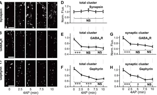
Excitatory neuronal activity was induced by incubating cells with the potassium channel blocker 4-aminopyridine (4AP; 50mM) for 2.5, 5, 7.5, and 10 min before fixation. Treatment
with 4AP did not affect the immunofluorescent intensity of synapsin (Fig. 1A and D), suggesting that the increase in neuronal activity has only a minor effect on the size of presynaptic terminals. By contrast, the immunoreactivity associated with total (synaptic and extrasynaptic) GABAARs significantly decreased to
75.3%62.3% of non-treated control cells within 2.5 min of incubation with 4AP (0 vs. 2.5 min,p,0.005, ANOVA;p,0.005, Tukey’s range test in ANOVA), and no further decrease was induced by longer incubation (2.5–10 min,p.0.05, Tukey’s range test in ANOVA; Fig. 1B and E). Total gephyrin immunoreactivity also decreased to 78.5%62.7% of control cells (0 vs. 2.5 min,
p,0.005, Tukey’s range test in ANOVA) within 2.5 min. However, we observed a further decrease of gephyrin immuno-reactivity to 63.9%61.6% after incubation with 4AP for 7.5 min (2.5 vs. 7.5 min,p,0.005, Tukey’s range test in ANOVA; Fig. 1C and F). Synaptic GABAAR and gephyrin clusters exhibited a time
course similar to that of total GABAAR and gephyrin clusters
(Fig. 1G and H). These results indicated that the activity-dependent decrease in the number of synaptic GABAAR clusters
reached a steady state more quickly than that of synaptic gephyrin clusters.
Furthermore, we investigated the time courses of GABAA
R-and gephyrin-associated immunofluorescence recovery after washout of 4AP (Fig. S2A–C). Both synaptic GABAAR and
gephyrin immunoreactivity gradually recovered to almost the same level as that of non-treated cells within 10 min with a similar time course (Fig. S2E and F). No significant change in the size of the presynaptic terminals was detected by synapsin-associated immunofluorescence during the washout (Fig. S2D).
The comparison of the time courses of GABAAR and gephyrin
clusters raised the possibility that the excitatory activity-induced reduction in GABAAR immunofluorescence precedes that in
gephyrin immunofluorescence. Therefore, we further examined the 4AP-induced changes in GABAAR- and gephyrin-associated
immunoreactivity within 2.5 min (150 s). Stimulation by 4AP for 60 s induced the reduction in synaptic GABAAR
immunoreactiv-ity to 73.0%62.5% of control cells (p,0.005, Welch’s t-test; Fig. 2A). However, synaptic gephyrin-associated immunofluores-cence in the cells stimulated by 4AP for 60 s maintained the same intensity as observed in 4AP non-treated cells (106.6%63.8% of control cells,p.0.05, Welch’st-test; Fig. 2B). We then examined the timing of NMDA-induced changes in GABAAR- and
gephyrin-associated immunoreactivities, as the activation of the NMDA receptor and subsequent Ca2+influx is also involved in the neuronal excitatory activity-dependent decrease in GABAergic synaptic transmission and declustering of GABAARs at inhibitory
synapses [7,12,28]. When neurons were stimulated by 50mM
NMDA with its co-agonist, glycine, and TTX for 60 s, synaptic GABAAR immunoreactivity declined to 76.1%62.3% of control
cells (p,0.005, Welch’s t-test; Fig. 2C). By contrast, synaptic gephyrin-associated immunofluorescence was unaffected by NMDA stimulation for 60 s (96.8%64.7% of control cells,
p.0.05, Welch’s t-test; Fig. 2D). Longer NMDA treatment (150 s) resulted in the reduction of synaptic gephyrin immunore-activity, as similarly observed with 4AP treatment; synaptic gephyrin immunoreactivity was reduced to 77.9%62.2% of control cells (p,0.005, Welch’s t-test; Fig. 2E). These results, together with the results of the time-course analysis of 4AP treatment, indicate that the excitatory activity-induced decrease in the number of synaptic GABAARs at postsynapses takes place
before the shrinkage of synaptic gephyrin clusters.
Modulation of GABAAR diffusion is complete before that of gephyrin clustering
The increase in GABAAR lateral diffusion dynamics plays a key
role in neuronal activity-dependent decrease in GABAAR
clustering at inhibitory synapses [11,12]. Therefore, we conducted a time-course analysis of GABAAR lateral diffusion dynamics after
4AP stimulation using single-particle tracking with quantum dots (QD-SPT) [29]. Endogenous GABAARs were targeted with an
antibody against the extracellular domain of thec2 subunit (Fig. S1D) and subsequently labeled with an intermediate biotinylated Fab fragment and streptavidin-coated QDs. The lateral diffusion parameters after 4AP stimulation were calculated from the trajectories of GABAARs labeled with QDs (GABAAR-QDs)
(Fig. 3A). The location of the active synapse was visualized by labeling with the amphiphilic dye FM4–64, induced after a burst of activity with 40 mM KCl. We confirmed that this FM4–64 labeling did not affect the GABAAR diffusion coefficient both in
the absence and presence of 4AP treatment (Fig. S3A). The diffusion coefficient of GABAAR-QD at the synapse obtained by
synaptic trajectories (red in Fig. 3A) was significantly increased within 2.5 min after the onset of 4AP stimulation (0–10 min,
p,0.005, Kruskal–Wallis test; 0 vs. 2.5 min, p,0.005, Mann– WhitneyUtest; Fig. 3B and C). An additional increase in diffusion coefficient was not induced by longer incubation (2.5–10 min,
p.0.05, Kruskal–Wallis test). In the absence of FM4–64 labeling, a 4AP-induced increase in diffusion coefficient was observed within 4 min (Fig. S3B), suggesting that the KCl-induced burst during FM4–64 labeling does not significantly impact the time course of 4AP-induced changes in the GABAAR diffusion
coefficient. Forty to fifty percent of synaptic GABAAR-QD
Figure 1. Time-course analysis of 4AP-induced decrease in GABAAR- and gephyrin-associated immunofluorescence. A–C: Representative examples of immunoreactivity associated with synapsin (A), GABAAR (B), and gephyrin (C) on the dendrites of hippocampal
neurons (21–27 DIV) treated with 50mM 4AP for 0–10 min. Scale bars: 5mm.D–F: Time-course plots of changes in normalized fluorescence intensities (averages6SEM) of total clusters of synapsin (D), GABAAR (E), and gephyrin (F) following 4AP treatment.G,H: Time-course plots of 4AP-induced
reduction in the normalized fluorescence intensities of synaptic GABAAR (G) and gephyrin (H) clusters. NS:p.0.05, *:p,0.05, ***:p,0.005, Tukey’s
range test in ANOVA, n = 40 cells/condition (4 cultures). doi:10.1371/journal.pone.0036148.g001
Figure 2. Activity-dependent decrease in synaptic clusters of GABAAR preceding that of gephyrin.Left: Representative examples of immunoreactivity associated with GABAAR (A,C) and gephyrin (B,D,E) in the presence (A,B: 4AP,C–E: NMDA) or absence (Con) of stimulation for
the indicated times. Right: Normalized fluorescence intensities (averages6SEM) of synaptic GABAAR (A,C) and gephyrin (B,D,E) clusters following
stimulation. Note that fluorescence intensity of gephyrin was unchanged 60 s after the onset of stimulation (4AP:B, NMDA:D), while that of GABAAR
significantly decreased at 60 s (4AP:A, NMDA:C). Scale bars: 5mm. NS:p.0.05, ***:p,0.005, Welch’st-test, n = 30 cells/condition (3 cultures). doi:10.1371/journal.pone.0036148.g002
small surface area [30], as reported previously [11]. The size of confinement was calculated for this population (see Materials and Methods). The average confinement size was significantly increased to 131.4%66.9% of control cells by 4AP treatment for 2.5 min (0 vs. 2.5 min, p,0.005, Tukey’s range test in ANOVA) and then maintained during further incubation (2.5– 10 min, p.0.05, Tukey’s range test in ANOVA; Fig. 3D). Furthermore, the synaptic dwell time of GABAAR-QD decreased
to 73.8%63.8% of control cells at 2.5 min (0 vs. 2.5 min,
p,0.005, Tukey’s range test in ANOVA); however, no further decrease was observed after 2.5 min (2.5–10 min, p.0.05, Tukey’s range test in ANOVA) (Fig. 3E). These results indicate that 4AP-dependent modification of GABAAR lateral diffusion
reaches a steady state within 2.5 min, which probably leads to the decrease in the number of synaptic GABAARs (Fig. 1G). The time
taken by gephyrin-associated immunofluorescence to reach a steady state was 7.5 min (Fig. 1H). This is 5 min longer than the time taken for GABAAR diffusion dynamics to reach a steady
state. Therefore, our results indicate that the activity-dependent change in the lateral diffusion of GABAARs is completed before
the dispersion of gephyrin clusters.
4AP-dependent modulation of gephyrin clusters depends on GABAAR lateral mobility
It is well established that synaptic gephyrin clustering also requires synaptic localization of GABAARs [13,22,23,24,25,
26,27]. Based on the finding that the excitatory activity-induced modulation of GABAAR lateral diffusion was accomplished before
gephyrin declustering, we hypothesized that gephyrin clustering could be sensitive to GABAAR diffusion dynamics, in addition to
its existence and localization. To confirm this hypothesis, we manipulated GABAAR diffusion dynamics by artificially
cross-linking (XL) the GABAAR c2 subunits using antibodies, as
performed previously for AMPA receptors and metabotropic glutamate receptors [31,32]. Successful XL of GABAARs was
confirmed by the appearance of fluorescent clusters labeled with the Alexa FluorH-conjugated antibody used for XL of primary antibodies targeted to GABAARs (Fig. 4A). The fluorescence
intensities of these cross-linked GABAAR clusters were not affected
by 4AP treatment (Fig. 4B). Next GABAAR mobility was
examined by QD-SPT. Trajectories revealed that the area explored by GABAAR-QDs were greatly reduced when surface
GABAARs were cross-linked, both inside (red, Fig. 4C) and outside
(blue, Fig. 4C) the synapses. In the absence of 4AP, XL induced an approximately 100-fold reduction in GABAAR-QD diffusion
coefficients (Fig. 4D), an approximately 3.7-fold increase in the percentage of immobilized GABAAR-QD (Fig. 4E), a 13.6%
decrease in the confinement size (Fig. 4F), and an approximately 3.4-fold increase in the synaptic dwell time (Fig. 4G), indicating that GABAAR-QD lateral diffusion is greatly inhibited by XL.
Moreover, XL blocked the 4AP-induced significant increase in the diffusion coefficient, enlargement of confinement size, and decrease in the synaptic dwell time of GABAAR-QDs (Fig. 4D–G).
We also confirmed that 4AP-induced increase in intracellular Ca2+ remained unaffected under XL conditions, which is
responsible for the increase in GABAAR lateral diffusion. Ca2+
imaging with fluo-4 at proximal dendrites revealed that increase in intracellular Ca2+was successfully induced by 4AP treatment even under XL conditions (Fig. 5B) as observed in the absence of XL (Fig. 5A), and that there was no significant difference in the peak amplitudes (Fig. 5C) and levels of increase in intracellular Ca2+as represented by the area under the curve (Fig. 5D) between control and XL cells. Taken together, these experiments indicate that XL could inhibit GABAAR lateral diffusion without affecting
intra-cellular Ca2+elevation. Next we examined 4AP-induced declus-tering of gephyrin under XL conditions (Fig. 5E). Although a previous study showed that a 12-h XL of GABAAR resulted in the
formation of extrasynaptic gephyrin clusters [33], the total number of gephyrin clusters in GABAAR XL conditions was not different
from that without XL (Fig. 5F), suggesting that extrasynaptic artificial gephyrin clusters are not formed under our XL conditions. In the cells without GABAAR XL, 4AP incubation
for 15 min significantly decreased gephyrin-associated immuno-reactivity [Fig. 5G (2XL)]. Conversely, the same 4AP stimulation failed to induce reduction in gephyrin immunofluorescence in the cells with GABAAR XL [Fig. 5G (+XL)).
XL of surface GABAARs is an extreme condition in which a
large proportion of surface GABAARs are immobilized.
There-fore, we also examined the effect of a calcineurin inhibitor, cyclosporin A (CysA), which does not immobilize surface GABAARs but suppresses the NMDA-induced increase in
GABAAR mobility [11,12], on gephyrin clustering. We confirmed
that the 4AP-driven increase in the synaptic diffusion coefficient (Fig. 6A) and reduction in the synaptic dwell time (Fig. 6B) were completely inhibited in the presence of 1mM CysA (Fig. 6C and D), which is in agreement with previous studies of NMDA stimulation [11,12]. Ca2+ imaging with fluo-4 revealed that increase in intracellular Ca2+, sustained for at least 15 min, was normally induced by 4AP even in the presence of CysA (Fig. 6E). The peak amplitude (Fig. 6F) and Ca2+influx level represented by the area under the curve (Fig. 6G) was not significantly affected by CysA (p.0.05, Welch’s t-test). Under this condition, the size of synaptic clusters of GABAAR and gephyrin was quantified by
immunocytochemistry. The shrinkage of synaptic GABAAR
clusters induced by 4AP stimulation for 30 min (Fig. 6H) was blocked completely in the presence of CysA (Fig. 6I). Furthermore, 4AP-driven gephyrin declustering at the synapse (Fig. 6J) was also prevented by CysA treatment (Fig. 6K), despite the increase in cytosolic Ca2+.
In summary, the above results indicate that 4AP-driven gephyrin declustering is inhibited when there is no increase in GABAAR lateral diffusion in response to neuronal excitation. Our
results also imply that synaptic gephyrin clustering is dependent on the mobility of GABAARs during sustained activity induced by
4AP.
NMDA-driven gephyrin declustering is independent of GABAAR mobility
The result of GABAAR XL and CysA experiments with 4AP
stimulation suggested the existence of a mechanism, dependent on GABAAR surface mobility, which regulates gephyrin clustering.
Finally, we examined whether gephyrin clustering is constantly subjected to this GABAAR-dependent regulation during sustained
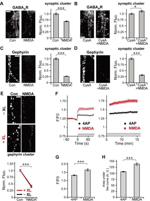
neuronal excitation. NMDA stimulation was applied to increase neuronal activity, and effects of CysA treatment on synaptic GABAAR and gephyrin clusters were examined. In agreement
with previous reports that CysA inhibits NMDA-induced increase in GABAAR lateral diffusion [11,12] and declustering of
GABAARs [12], the dispersal of synaptic GABAAR observed after
30 min of NMDA treatment (Fig. 7A) was completely blocked by the presence of CysA (Fig. 7B). NMDA stimulation significantly diminished the size of gephyrin clusters to 26.7%60.9% of control cells (Fig. 7C). Unlike the GABAAR clusters, synaptic gephyrin
clusters were reduced (31.0%62.1% of control cells, Fig. 7D) even in the presence of CysA. XL of surface GABAARs also failed to
(Fig. 7F). The average peak amplitude of Ca2+elevation evoked by
NMDA was 1.2 times larger than that induced by 4AP (p,0.005, Welch’st-test; Fig. 7G) and the level of increase in Ca2+during
NMDA stimulation was 1.3 times higher than that during 4AP stimulation (p,0.005, Welch’s t-test; Fig. 7H). Taken together, these results suggest that gephyrin clustering is not dependent on GABAAR mobility during sustained activity induced by NMDA,
possibly at high levels of increase in Ca2+. More importantly, despite the loss of synaptic gephyrin clustering by NMDA stimulation (Fig. 7D), Cys A blocked NMDA-induced declustering of GABAARs (Fig. 7B) and the increase in lateral diffusion [11,12].
These results clearly indicate that lateral diffusion of GABAARs at
the synapse and synaptic GABAAR clustering during inhibitory
synaptic plasticity are independent of the amount of synaptic gephyrin present.
Discussion
The main finding of this study is that changes in lateral diffusion dynamics and number of synaptic GABAARs preceded gephyrin
declustering during excitatory activity. In addition, our results indicate that synaptic GABAAR diffusion and clustering are Figure 3. Modulation of GABAAR lateral diffusion by 4AP completed within 2.5 min. A: Examples of trajectories of GABAAR-QDs recorded
for 38.9 s in neurons treated for the indicated times with 4AP, inside (red) and outside (blue) the synapse. Scale bar: 1mm.B,C: The diffusion coefficients (D) of GABAAR-QDs inside the synapses in neurons incubated with 4AP for 0–2.5 min (2.5 min), 2.5–5 min (5 min), 5–7.5 min (7.5 min),
and 7.5–10 min (10 min) and that in control cells (0 min). Median diffusion coefficients (triangle) and plus (hatched) and minus (solid) interquartile ranges (IQR) (B) and cumulative probability (C) of the synaptic diffusion coefficients of GABAAR-QDs. 0 min: n = 974 QDs; 2.5 min: n = 290; 5 min:
n = 295; 7.5 min: n = 278; 10 min: n = 258. ***:p,0.005, NS:p.0.05, Kruskal–Wallis test.D: The average (6SEM) confinement sizes of GABAAR-QDs in
synapses. 0 min: n = 500 QDs; 2.5 min: n = 122; 5 min: n = 106; 7.5 min: n = 127; 10 min: n = 111.E: The average synaptic dwell times (6SEM) of GABAAR-QD. 0 min: n = 3466 events; 2.5 min: n = 1147; 5 min: n = 1320; 7.5 min: n = 1131; 10 min: n = 1098. NS:p.0.05, ***:p,0.005, Tukey’s range
test in ANOVA (D,E). Data were obtained from 3 or 4 cultures. doi:10.1371/journal.pone.0036148.g003
independent of the status of gephyrin clusters during sustained excitatory activity.
Gephyrin is considered a key protein that controls GABAAR
stability at the postsynapse [13,16,20,21]. In this study, we tested the hypothesis that the excitatory activity-dependent reduction in postsynaptic GABAARs [11,12], which could be involved in
GABAergic synaptic plasticity, is initiated by the dispersion of gephyrin from clusters. If this hypothesis were correct, excitatory activity should have affected gephyrin first or at least at the same time when affecting GABAARs. Contrary to this expectation, a
detailed time-course analysis indicated that the dispersal of GABAAR clusters induced by the enhancement of GABAAR Figure 4. Inhibition of GABAAR lateral diffusion by cross-linking (XL). A: Examples of cross-linked GABAAR clusters on the dendrites of
hippocampal neurons with (XL+4AP) or without (XL) 4AP stimulation. Scale bar: 5mm.B: Normalized average fluorescent intensities of cross-linked GABAAR clusters with (XL+4AP) or without (XL) 4AP stimulation (averages6 SEM). XL: n = 39 cells; XL+4AP: n = 40 cells, from 4 cultures. C:
Representative trajectories of GABAAR-QDs recorded for 38.9 s with (+XL) or without (2XL) XL in the presence or absence of 4AP, inside (red) and
outside (blue) the synapse. Scale bar: 1mm.D–G: Effects of XL and 4AP treatment on diffusion coefficients (D), percentage of immobile receptors (E), synaptic confinement sizes (F), and synaptic dwell times (G) of GABAAR-QDs.D: The diffusion coefficients of GABAAR-QDs (median6IQR). The
number of GABAAR-QDs analyzed: Con: n = 556; 4AP: n = 418; XL: n = 321; XL+4AP: n = 321.E: The percentage of immobile GABAAR-QDs (average6
SEM). Con: n = 15 cells; 4AP: n = 14; XL: n = 14; XL+4AP: n = 19.F: The confinement size (average6SEM) of synaptic GABAAR-QDs. Con: n = 366 QDs;
4AP: n = 203; XL: n = 200; XL+4AP: n = 196.G: The average dwell times (6SEM) of GABAAR-QDs in synapses. Con: n = 2039 events; 4AP: n = 1576; XL:
n = 473; XL+4AP: n = 493. Data inD–Gwere obtained from 3 cultures. NS:p.0.05, **:p,0.01, ***:p,0.005, Welch’st-test forB,E–G, and Mann– WhitneyUtest forD.
lateral mobility preceded the dispersal of gephyrin. Our results suggest that neuronal activity-induced rapid decrease in GABAAR
numbers at mature inhibitory synapses is not mediated by gephyrin declustering. This notion was further supported by the observation that synaptic GABAAR mobility and clustering were
not affected by NMDA in the presence of CysA, while gephyrin cluster largely decreased under the same conditions. Our findings suggest that excitatory activity-induced plasticity in GABAergic synapses is induced independent of the status of gephyrin clusters. There was no remarkable difference in the recovery time course of GABAAR and gephyrin cluster size after 4AP removal, similar
to the process of synaptogenesis in hippocampal neurons [34,35]. This suggests that the reaccumulation of GABAAR and gephyrin
to the inhibitory synapse occurs simultaneously. It remains unclear whether gephyrin is critical for the recovery of GABAAR clusters.
Furthermore, our results suggested that there are existence two regulatory mechanisms of gephyrin clustering during sustained activity: GABAAR-dependent and GABAAR-independent
mech-anisms. The amount of gephyrin in clusters was maintained even in the presence of 4AP, when surface GABAARs were immobilized
by XL and when 4AP-induced increase in GABAAR diffusion was
prevented by CysA-treatment. This finding indicates that GABAAR lateral diffusion dynamics can affect clustering of the
scaffold protein gephyrin. Recent theoretical modeling of postsynaptic structures based on chemical potential proposed another concept which states that the stabilization of the postsynaptic structure is reciprocal. In other words, scaffold proteins stabilize receptors and receptors stabilize scaffold proteins [36]. Together with the fact that gephyrin is crucial for the stabilization of postsynaptic GABAARs [16,20,21], our data
provide direct evidence of a reciprocal mechanism that stabilizes
Figure 5. Suppression of 4AP-induced reduction in gephyrin immunofluorescence by GABAAR immobilization under increased levels of cytosolic Ca2+. A,B: Top: Representative pseudocolor images of hippocampal neurons loaded with fluo-4 AM without (A) or with (B) surface GABAAR XL, before (Con) and 10 s after 4AP application (4AP). Scale bars: 20mm. Bottom: Time-course plots of F/F0 ratio changes (averages6
SEM) measured on proximal dendrites with the addition of 4AP, in the absence (A) or presence (B) of GABAAR XL. 4AP was applied at time = 0 as
indicated by the gray horizontal bar in the traces.C,D: Peak amplitudes (C) and areas under the curve (D) for the F/F0-time plot during 90 s after addition of 4AP. Values indicate averages6SEM. NS:p.0.05, Welch’st-test.2XL: n = 30 cells,+XL: n = 28, from 3 cultures. Note that normal increase in Ca2+was induced by 4AP even under XL conditions.E: Examples of gephyrin-immunoreactive clusters in dendrites with (+XL) or without (2XL) surface GABAAR XL in the presence (4AP) and absence (Con) of 4AP treatment for 10 min. Scale bar, 5mm.F,G: Effects of GABAAR XL and 4AP
treatment on the normalized number of clusters (F) and normalized fluorescence intensities (G) of gephyrin clusters (averages6SEM). NS:p.0.05; ***:p,0.005, Welch’st-test, n = 30 cells/condition (3 cultures). 4AP-induced reduction in gephyrin cluster size was completely suppressed by GABAAR
XL.
doi:10.1371/journal.pone.0036148.g005
the structure of GABAergic synapses. Regulation of postsynaptic scaffolds by neurotransmitter receptors is involved in synaptogen-esis and the maintenance of GABAergic synapses, as evidenced by the fact that the absence of some GABAAR subunits results in the
disappearance of gephyrin clusters [22,23,24,25,26,27]. Our
present results, which imply that activity-induced mobilization of surface GABAARs destabilizes gephyrin clusters, also raise the
possibility that GABAAR lateral mobility, in addition to its
existence and localization, could be a primary determinant of stability of mature GABAergic synaptic structures during synaptic
Figure 6. Prevention of 4AP-induced gephyrin declustering by calcineurin inhibitor CysA. A–D: Inhibition of 4AP-driven mobilization of GABAAR-QDs by CysA treatment. Diffusion coefficients in the synapse (AandC, median6IQR) and synaptic dwell times (BandD, averages6SEM)
in the absence (A,B) or presence (C,D) of 1mM CysA. NS:p.0.05, ***:p,0.005, Mann–WhitneyUtest forA,C(Con: n = 535 QDs, 4AP: n = 537, CysA: n = 478, CysA+4AP: n = 506.), Welch’st-test forB,D(Con: n = 2107 events, 4AP: n = 2505, CysA: n = 1930, CysA+4AP: n = 2094). Data were obtained from 3 cultures.E–G: Intact 4AP-induced increase in cytosolic Ca2+
concentration under CysA treatment. Changes in intracellular Ca2+
levels indicated as F/F0 of fluo-4 after the addition of 4AP (black) or CysA+4AP (red) (E). Drugs, i.e. 4AP or CysA+4AP, were applied at time = 0 as indicated by the gray horizontal bar in the traces. Peak amplitudes of F/F0 (F) and areas under the F/F0-time curve (G) during 90 s after the onset of stimulation. Values indicate averages6SEM. NS:p.0.05, Welch’st-test. 4AP: n = 26 cells, CysA+4AP: n = 28 cells (3 cultures).H–K: The effect of CysA treatment on 4AP-driven declustering of GABAAR and gephyrin. Left: Example of immunoreactivity associated with GABAAR (H,I) and gephyrin (J,K) on the dendrites
treated with 4AP for 30 min, in the absence (H,J) and presence (I,K) of CysA. Scale bars: 5mm. Right: Normalized fluorescence intensities (averages 6SEM) of synaptic GABAAR (H, I) and gephyrin (J,K) clusters. NS:p.0.05, ***:p,0.005, Welch’st-test. Con, CysA: n = 30 cells/condition, 4AP,
Figure 7. GABAAR-independent gephyrin declustering during sustained activity induced by NMDA stimulation. A–D: Effect of CysA treatment on NMDA-driven dispersal of GABAAR and gephyrin clusters. Left: Examples of GABAAR (A,B) and gephyrin (C,D) immunoreactivity in
neurons incubated with NMDA for 30 min, with (B,D) and without CysA (A,C). Scale bars, 5mm. Right: Normalized fluorescence intensities (averages 6SEM) of synaptic GABAAR (A,B) and gephyrin (C,D) clusters. *:p,0.05, ***:p,0.005, Welch’st-test. Con, CysA, CysA+NMDA: n = 30 cells/condition,
NMDA: n = 25 cells/condition (3 cultures). CysA suppressed NMDA-induced dispersal of GABAAR clusters, but not that of gephyrin clusters.E:
NMDA-induced gephyrin dispersal under GABAAR XL. Top: Gephyrin immunoreactive clusters in neurons with (+XL) and without (2XL) surface GABAAR XL
after NMDA stimulation. Bottom: Effects of GABAAR XL and NMDA treatment on the normalized fluorescence intensity of gephyrin clusters (average
6SEM). ***:p,0.005, Welch’st-test, n = 30 cells/condition (3 cultures).F–H: Comparison of the Ca2+
influx level induced by 4AP and NMDA. Increase in Ca2+
after the addition of 4AP (black) or NMDA (red) (F). Gray horizontal bars in the traces indicate the presence of 4AP or NMDA. Peak amplitudes (G) and areas under the curve (H) for F/F0-time plots during 90 s after the onset of stimulation. Values indicate averages6SEM. ***:p,0.005, Welch’st-test. 4AP: n = 21 cells, NMDA: n = 23 cells (3 cultures).
doi:10.1371/journal.pone.0036148.g007
plasticity. Changes in the chemical potential associated with GABAARs and gephyrin, which are induced by the enhancement
of lateral diffusion and subsequent decrease in synaptic GABAAR
density, could lead to a new steady state of postsynaptic molecular assembly [36].
The observation that gephyrin dispersed after NMDA stimu-lation regardless of GABAAR mobility suggested that another
GABAAR-independent regulatory mechanism may control
ge-phyrin clustering. Considering that NMDA induced a 1.3 times larger Ca2+elevation than 4AP, the Ca2+influx level could be one of the factors determining whether gephyrin is subjected to GABAAR-dependent regulation or independently destabilized in
response to Ca2+elevation. Gephyrin is a substrate of the Ca2+ -dependent non-lysosomal cysteine protease calpain-1, which is activated when NMDA receptors are stimulated [37], and turnover of gephyrin is regulated by calpain-1 activity [38]. Therefore, it is possible that gephyrin stability is also controlled by the activation of calpain-1 during NMDA stimulation [39]. However, it must be noted that the same NMDA stimulation (50mM, with glycine and TTX) did not induce gephyrin declustering in cultured spinal cord neurons [40], in which calpain-1 is also activated by NMDA stimulation [41]. Thus, the molecular mechanism for this GABAAR-independent gephyrin
regulation remains to be elucidated by future studies.
Activity-dependent regulation of GABAAR lateral diffusion and
clustering at inhibitory synapses is mediated by Ca2+influx and subsequent activation of calcineurin [11,12,13]. Our present findings provide several insights into the molecular mechanism of how Ca2+signaling enhances GABAAR lateral diffusion. In the
present study, we found that GABAAR diffusion and clustering
were independent of gephyrin clustering during NMDA stimula-tion in the presence of CysA. This finding strongly suggests that calcineurin-dependent regulation of GABAAR mobility does not
require gephyrin. Because alterations in receptor–scaffold inter-actions can modulate the lateral diffusion of receptors [15], we propose the existence of other GABAAR-interacting protein(s) that
contribute to GABAAR stabilization in a gephyrin-independent
manner. GABAAR accumulation at the inhibitory synapse occurs
before gephyrin accumulation during synaptogenesis in spinal cord neurons [42], suggesting the existence of a gephyrin-independent stabilization mechanism of GABAARs. This gephyrin-independent
pathway may enhance GABAAR lateral diffusion via the
calcineurin-dependent dephosphorylation of Ser327 in the GA-BAARc2 subunit [12]. We speculate that the dephosphorylation
of Ser327 upon neuronal excitation induces the dissociation of unidentified GABAAR-associating protein(s) from GABAARs,
which leads to the observed increase in GABAAR lateral mobility.
The Ca2+-dependent increase in GABAAR lateral mobility is
involved in synaptic plasticity at inhibitory synapses that may underlie neuronal disorders resulting from pathological disinhibi-tion [11,12]. Therefore, elucidating the detailed molecular mechanism of the gephyrin-independent regulation of GABAAR
lateral mobility might contribute not only to understanding the basis of learning and memory but also to discovering therapeutic targets for neuropathies such as epilepsy.
Materials and Methods
Ethics statement
All animal procedures in this study were performed in accordance with the guidelines issued by the Japanese Ministry of Education, Culture, Sports, Science and Technology. All animal procedures in this study were approved by the Animal Experiment Committee of the RIKEN (H23-2-204). All efforts
were made to minimize animal suffering and reduce the number of animals used.
Anti-GABAARc2 subunit antibody production
The rabbit anti-GABAAR c2 subunit antibody
(anti-GA-BAARc2) was raised against the peptide
‘‘QKSDDDYE-DYASNKTWVLTPKVPEGDVTV(C)’’ corresponding to amino acid residues 39–67 of the rat GABAAR c2 subunit, as shown
previously [43]. The peptide was synthesized by the Support Unit for Bio-material Analysis at the RIKEN BSI Research Resources Center (RRC) and was subsequently injected into rabbits to obtain the antibody by the Support Unit for Animal Resources Development at the RIKEN BSI RRC.
The specificity of the antibody was confirmed using HeLa cells (RIKEN BioResource Center, Ibaraki, Japan) expressinga1,b3, and c2 subunits of GABAAR (Fig. 1A and C). HeLa cells were
plated onto 18-mm diameter glass coverslips and cultured in DMEM (Nacalai Tesque, Kyoto, Japan) supplemented with 10% fetal bovine serum and antibiotics. For transfection, a coverslip in 1 ml culture medium was incubated with the transfection mixture containing 100ml OPTI-MEM (Invitrogen, Tokyo, Japan), mixture of DNA (a1,b3,c2; 0.7mg each), and 4.2ml TransIT-LT1 (Mirus, WI, USA) for 24 h before observation. Plasmids encodinga1,b3, andc2 subunits of GABAAR were generated by
subcloning the coding region into the mammalian expression vector [pcDNA3.1/Zeo(+/2); Invitrogen] using FANTOM3 clones as PCR templates (a1: C630037M06; b3: C630014N19;
c2: B930018F17 and C230063G02) [44].
Primary cultures
Primary cultures of hippocampal neurons co-cultured with astrocytes were prepared from E18–21 Wistar rat embryos as previously described [45] with some modifications. Hippocampal cells were dissociated in plating medium comprising minimum essential medium (MEM; Invitrogen) supplemented with B27 (Invitrogen), 2 mM L-glutamine, 1 mM sodium pyruvate (Invitro-gen), and antibiotics, and were plated at a density of 1.46105
cells/ml onto 18-mm diameter glass coverslips precoated with 0.04% polyethyleneimine (Sigma, Tokyo, Japan). Three days after plating, the culture medium was replaced with maintenance medium comprising Neurobasal-A medium (Invitrogen) supple-mented with B27, 2 mM L-glutamine, and antibiotics. Cells were cultured for 21–27 daysin vitro before the experiments. At least three independent cultures were used for each experiment.
Drug treatment
To increase excitatory activity, cultured hippocampal neurons were incubated with 50mM 4AP (Nacalai Tesque) or 50mM NMDA (Tocris, MO, USA), glycine (5mM), and TTX (1mM; Tocris) at 37uC in the imaging medium comprising MEM without phenol red (Invitrogen), 20 mM HEPES, 33 mM glucose, 2 mM glutamine, 1 mM sodium pyruvate, and B27. For time-course analysis of cluster recovery, neurons were treated with 50mM 4AP
for 10 min and subsequently incubated with the imaging medium for 0–15 min before fixation. For QD-SPT experiments, 4AP (final concentration, 50mM) was added to the imaging medium immediately before recording. For Ca2+imaging, recording were done for 1 min in the absence of drugs, then drugs were bath applied to the cells during the recording.
Immunocytochemistry and quantitative analysis
For GABAAR immunostaining of cultured neurons with drug
neurons were labeled with ourc2 antibodies by incubating live cells with 2.0mg/ml antibody diluted in imaging medium for 30 min at 37uC. Subsequently, cells were stimulated by 4AP or NMDA and fixed with 4% (w/v) paraformaldehyde (PFA) in PBS-0.02% NaN3at room temperature (24–26uC) for 15 min. After
permeabilization with 0.1% triton X-100 for 3 min and incubation with 5% (w/v) bovine serum albumin (BSA; Sigma) for 30 min to block nonspecific staining, cells were labeled with the mouse anti-synapsin I antibody (1:3000; Synaptic Systems, Goettingen, Germany) in 2.5% BSA for 60 min. After washes, the cells were incubated in Alexa FluorH-conjugated secondary antibodies (5–10mg/ml, Alexa Fluor 488 or Alexa Fluor 594;
Invitrogen) for 30 min, washed, and mounted on slides with Vectashield (Vector Laboratories, CA, USA). In the experiments using the calcineurin inhibitor CysA (1mM; Santa Cruz
Biotechnology, CA, USA), cells were incubated with our c2 antibodies (2.0mg/ml) for 30 min in the presence of drug (i.e., 4AP, NMDA+TTX+Gly, CysA) and subsequently fixed by 4% PFA. After fixation, the procedures were the same as those of experiments without CysA treatment. In some experiments (Fig. S1E), GABAAR was labeled with commercially available rabbit
anti-c2 subunit antibodies (6.0mg/ml; Alomone Labs, Jerusalem,
Israel), which were used in a previous study [11]. GABAARs on
the GABAAR-expressing HeLa cells were labeled with our
custom-made anti-GABAARc2 antibody (0.8mg/ml) as described above,
and nuclei of HeLa cells were stained with DAPI.
For labeling of gephyrin, cells were fixed with 4% PFA after drug stimulation and permeabilized with 0.1% Triton X-100. After blocking with 5% BSA, cells were incubated with anti-gephyrin antibody (0.33mg/ml, clone mAb7a; Synaptic Systems) and the rabbit polyclonal anti-synapsin I antibody (1:400; Millipore, MA, USA) in the presence of 2.5% BSA for 90 min, and subsequently labeled with Alexa Fluor 488 or Alexa Fluor 594 (5–10mg/ml; Invitrogen).
Immunofluorescence from isolated neurons was acquired on an inverted microscope (IX-70; Olympus, Tokyo, Japan) equipped with a Plan Apo 606oil immersion objective with a numerical
aperture (NA) of 1.42 (Olympus), cooled CCD camera (Orca-II-ER; Hamamatsu Photonics, Shizuoka, Japan), and appropriate filter sets for Alexa Fluor 488 (ex: 480610 nm, em: 530620 nm) and Alexa Fluor 594 (ex: 535615 nm, em: 580 nm long pass). All images from a given culture were acquired with the same subsaturation exposure time.
Quantification of GABAAR-, gephyrin-, and
synapsin-associat-ed immunofluorescence was performsynapsin-associat-ed using ‘‘Integratsynapsin-associat-ed Mor-phometry Analysis’’ function of the MetaMorph software (Molecular Device Japan, Tokyo, Japan). GABAAR- and
gephyrin-immunoreactive clusters and synapsin-positive presy-napses were defined by processing images with multidimensional image analysis (MIA) interface, i.e., a 2D object segmentation by wavelet transform [46] and ‘‘auto threshold for light object (isodata method)’’ function of MetaMorph. Synaptic GABAAR or
gephyrin clusters were defined as clusters that overlapped at least 1 pixel with presynaptic terminals. For each culture, all cluster fluorescence intensity was normalized to the average value in control cells.
QD-SPT experiments
Neurons were incubated with the custom-made anti-GA-BAARc2 antibody (2.0mg/ml) for 5 min, washed, and incubated
with the biotinylated anti-rabbit Fab antibody (2.2mg/ml; Jackson ImmunoResearch, PA, USA) for 5 min. Following washes, the coverslips were incubated with 1.0 nM streptavidin-coated QDs emitting at 605 nm or 625 nm (Invitrogen) in borate buffer for
1 min [29]. After washes, functional presynaptic boutons were labeled with 2mM FM4–64 (Invitrogen) in imaging medium containing 40 mM KCl for 15 s. Incubation with antibodies and washes were performed at 37uC in the imaging medium.
The diffusive behavior of GABAAR-QD and FM4–64 signals
was recorded at 37uC in the imaging medium using an inverted microscope (IX-71, Olympus) equipped with an oil immersion objective (NA 1.45, 606; Olympus) and an EM-CCD camera (C9100; Hamamatsu Photonics) or an inverted microscope (IX-70; Olympus) equipped with an oil immersion objective (NA 1.42, 606; Olympus) and cooled CCD camera (Orca-II-ER;
Hama-matsu Photonics). Fluorescent signals were detected using appropriate filter sets for QD (ex: 455670 nm, em: 605620 nm) and FM4–64 (ex: 535615 nm, em: 580 nm long pass). GABAAR-QD lateral diffusion was recorded with an
integration time of 76 ms with 512 consecutive frames (38.9 s). All recordings were taken within 30 min.
Data analysis for QD-SPT experiments
The trajectory of GABAAR-QD was obtained by
cross-correlating images with a Gaussian model of the point spread function [47], and diffusion coefficients and confinements were calculated using TI workbench software written by Dr. T. Inoue (Waseda University), as described previously [11]. Only single QDs identified by intermittent fluorescence (i.e., blinking) were analyzed. The synaptic area was defined by processing FM4–64 images with wavelet decomposition [46]. GABAAR-QDs were
classified as ‘‘synaptic’’ when overlapping with synaptic area+2 pixels (284 nm). For the calculation of diffusion parameters in the synapse except for synaptic dwell time, the longest sub-trajectories of single GABAAR-QDs with greater than or equal to 30 points in
each compartment were taken into account.
To obtain the diffusion parameters, such as the diffusion coefficient and confinement size, values of the mean square displacement (MSD) plot versus time were calculated for each trajectory by applying the following equation:
MSD(nt)~ 1 N{n
X
N{n
i~1
x iððznÞtÞ{x ið Þt
ð Þ2zðy iððznÞtÞ{y ið ÞtÞ2
h i ð1Þ
([48]), wheretis the acquisition time, Nis the total number of
frames, andnandi are positive integers withnrepresenting the time increment. Diffusion coefficients (D) were calculated by fitting first four points of the MSD versus time curves with the following equation:
MSD(nt)~4Dntzb, ð2Þ
wherebis a constant reflecting the spot localization accuracy. In this system, GABAAR-QDs with a diffusion coefficient (D) less
than 0.0002mm2/s were defined as immobile.
The confinement domain size, in which the diffusion of GABAAR-QD was restricted, was obtained by fitting the
MSD-ntplot to the following equation:
MSD(nt)~L
2
3 1{exp {
12Dnt
L2
z4Dmacnt ð3Þ
[30], whereL2is the confined area in which diffusion is restricted, and Dmac is the diffusion coefficient on a long time scale. The
diffusion of GABAAR-QD with MSD-ntplot that does not apply
|D-Dmac|,0.16D or L,0.001 was defined as restricted motion,
and only GABAAR-QDs meeting this criteria were considered for
calculations of confinement domain sizes [49].
The GABAAR-QD dwell time inside the synapse was defined as
the duration of synaptic sub-trajectories.
GABAAR XL experiments
GABAARs on the cell surface were cross-linked by incubating
neurons with the anti-c2 subunit antibody (8.0mg/ml; Alomone
Labs) for 10 min, washing, and incubating with Alexa FluorH -conjugated anti-rabbit antibodies (20mg/ml; Invitrogen) for 5 min in the imaging medium. Cells were further incubated with the biotinylated anti-rabbit Fab antibody and streptavidin-coated QDs for QD-SPT, or fixed and subsequently immunolabeled with the gephyrin antibody for quantitative immunocytochemistry, as mentioned previously. In all experiments, it was confirmed that surface GABAARs were successfully cross-linked by fluorescence
from GABAAR-associated clusters (Fig. 4A).
Ca2+imaging
Neurons were loaded with 0.5mM fluo-4 AM (Invitrogen) for 5 min at 37uC. Fluo-4 fluorescence was acquired at 0.2 Hz with a 200-ms exposure at room temperature (24–26uC), with an inverted microscope (IX-70; Olympus) equipped with a 406objective (NA 0.85, UPlanApo; Olympus), a cooled CCD camera (Orca-II-ER; Hamamatsu Photonics), and appropriate filters (ex, 480610 nm; em, 530620 nm). For longer recording (Figs. 6E and 7E), images were further acquired at 0.1 Hz from 6 min to 15 min after drug application. Data were analyzed using a TI Workbench. The ratio of the fluorescence intensities F/F0, where F is a fluorescence intensity and F0 is the intensity at t = 0, was calculated after subtraction of the background fluorescence. To estimate the level of Ca2+elevation, the area under the curve was calculated using Igor Pro software (WaveMetrics, OR, USA).
Statistical analysis and image preparation
Statistical differences of data in the time course were determined using the Kruskal–Wallis (for the diffusion coefficient)
and one-way ANOVA (p= 0.05) tests, followed by Tukey’s post-hoc tests (for others). For comparisons between two groups, the Mann–Whitney U test or Welch’s t-test were performed as indicated. All statistical analysis was performed using Kaleida-Graph (Synergy Software, PA, USA). Images were prepared for printing using MetaMorph, Adobe Photoshop, and Adobe Illustrator.
Supporting Information
Figure S1 Specificity of the anti-GABAAR c2 subunit
antibody.
(PDF)
Figure S2 Recovery of GABAAR and gephyrin immuno-fluorescence after 4AP washout.
(PDF)
Figure S3 Lateral diffusion of GABAAR with or without FM4–64 labeling.
(PDF)
Acknowledgments
We thank the members of the Research Resource Center for invaluable technical support in the development of the GABAARc2 antibody and for
providing FANTOM clones. We specially thank Takafumi Inoue, Maxim Dahan, Victor Racine, and Jean-Baptiste Sibarita for data analysis programs, and Charles Yokoyama, Chihiro Hisatsune, Yoshiyuki Yamada, and Masahiro Enomoto for their valuable comments on the manuscript. We thank two anonymous reviewers for their insightful and constructive comments on the manuscript.
Author Contributions
Conceived and designed the experiments: HB AT KM. Performed the experiments: FN HB MA. Analyzed the data: FN HB. Contributed reagents/materials/analysis tools: FN HB KF. Wrote the paper: FN HB MA AT KM.
References
1. Moss SJ, Smart TG (2001) Constructing inhibitory synapses. Nat Rev Neurosci 2: 240–250.
2. Kilman V, van Rossum MC, Turrigiano GG (2002) Activity deprivation reduces miniature IPSC amplitude by decreasing the number of postsynaptic GABA(A) receptors clustered at neocortical synapses. J Neurosci 22: 1328–1337. 3. Nusser Z, Cull-Candy S, Farrant M (1997) Differences in synaptic GABA(A)
receptor number underlie variation in GABA mini amplitude. Neuron 19: 697–709.
4. Marsden KC, Beattie JB, Friedenthal J, Carroll RC (2007) NMDA receptor activation potentiates inhibitory transmission through GABA receptor-associat-ed protein-dependent exocytosis of GABA(A) receptors. J Neurosci 27: 14326–14337.
5. Marsden KC, Shemesh A, Bayer KU, Carroll RC (2010) Selective translocation of Ca2+
/calmodulin protein kinase IIalpha (CaMKIIalpha) to inhibitory synapses. Proc Natl Acad Sci U S A 107: 20559–20564.
6. Goodkin HP, Joshi S, Mtchedlishvili Z, Brar J, Kapur J (2008) Subunit-specific trafficking of GABA(A) receptors during status epilepticus. J Neurosci 28: 2527–2538.
7. Lu YM, Mansuy IM, Kandel ER, Roder J (2000) Calcineurin-mediated LTD of GABAergic inhibition underlies the increased excitability of CA1 neurons associated with LTP. Neuron 26: 197–205.
8. Naylor DE, Liu H, Wasterlain CG (2005) Trafficking of GABA(A) receptors, loss of inhibition, and a mechanism for pharmacoresistance in status epilepticus. J Neurosci 25: 7724–7733.
9. Terunuma M, Xu J, Vithlani M, Sieghart W, Kittler J, et al. (2008) Deficits in phosphorylation of GABA(A) receptors by intimately associated protein kinase C activity underlie compromised synaptic inhibition during status epilepticus. J Neurosci 28: 376–384.
10. Wang J, Liu S, Haditsch U, Tu W, Cochrane K, et al. (2003) Interaction of calcineurin and type-A GABA receptor gamma 2 subunits produces long-term depression at CA1 inhibitory synapses. J Neurosci 23: 826–836.
11. Bannai H, Le´vi S, Schweizer C, Inoue T, Launey T, et al. (2009) Activity-dependent tuning of inhibitory neurotransmission based on GABAAR diffusion
dynamics. Neuron 62: 670–682.
12. Muir J, Arancibia-Carcamo IL, MacAskill AF, Smith KR, Griffin LD, et al. (2010) NMDA receptors regulate GABAA receptor lateral mobility and
clustering at inhibitory synapses through serine 327 on the gamma2 subunit. Proc Natl Acad Sci U S A 107: 16679–16684.
13. Luscher B, Fuchs T, Kilpatrick CL (2011) GABAAreceptor trafficking-mediated
plasticity of inhibitory synapses. Neuron 70: 385–409.
14. Bruneau EG, Esteban JA, Akaaboune M (2009) Receptor-associated proteins and synaptic plasticity. FASEB J 23: 679–688.
15. Gerrow K, Triller A (2010) Synaptic stability and plasticity in a floating world. Curr Opin Neurobiol 20: 631–639.
16. Mukherjee J, Kretschmannova K, Gouzer G, Maric HM, Ramsden S, et al. (2011) The Residence Time of GABAARs at Inhibitory Synapses Is Determined
by Direct Binding of the Receptora1 Subunit to Gephyrin. J Neurosci 31: 14677–14687.
17. Tretter V, Jacob TC, Mukherjee J, Fritschy JM, Pangalos MN, et al. (2008) The clustering of GABA(A) receptor subtypes at inhibitory synapses is facilitated via the direct binding of receptor alpha 2 subunits to gephyrin. J Neurosci 28: 1356–1365.
18. Tretter V, Kerschner B, Milenkovic I, Ramsden SL, Ramerstorfer J, et al. (2011) Molecular basis of the GABAAreceptora3 subunit interaction with gephyrin.
J Biol Chem.
20. Jacob TC, Bogdanov YD, Magnus C, Saliba RS, Kittler JT, et al. (2005) Gephyrin regulates the cell surface dynamics of synaptic GABAA receptors.
J Neurosci 25: 10469–10478.
21. Kneussel M, Brandstatter JH, Laube B, Stahl S, Muller U, et al. (1999) Loss of postsynaptic GABA(A) receptor clustering in gephyrin-deficient mice. J Neurosci 19: 9289–9297.
22. Essrich C, Lorez M, Benson JA, Fritschy JM, Luscher B (1998) Postsynaptic clustering of major GABAAreceptor subtypes requires the gamma 2 subunit and
gephyrin. Nat Neurosci 1: 563–571.
23. Kralic JE, Sidler C, Parpan F, Homanics GE, Morrow AL, et al. (2006) Compensatory alteration of inhibitory synaptic circuits in cerebellum and thalamus of gamma-aminobutyric acid type A receptor alpha1 subunit knockout mice. J Comp Neurol 495: 408–421.
24. Li RW, Yu W, Christie S, Miralles CP, Bai J, et al. (2005) Disruption of postsynaptic GABA receptor clusters leads to decreased GABAergic innervation of pyramidal neurons. J Neurochem 95: 756–770.
25. Panzanelli P, Gunn BG, Schlatter MC, Benke D, Tyagarajan SK, et al. (2011) Distinct mechanisms regulate GABAA receptor and gephyrin clustering at
perisomatic and axo-axonic synapses on CA1 pyramidal cells. J Physiol. 26. Schweizer C, Balsiger S, Bluethmann H, Mansuy IM, Fritschy JM, et al. (2003)
The gamma 2 subunit of GABA(A) receptors is required for maintenance of receptors at mature synapses. Mol Cell Neurosci 24: 442–450.
27. Studer R, von Boehmer L, Haenggi T, Schweizer C, Benke D, et al. (2006) Alteration of GABAergic synapses and gephyrin clusters in the thalamic reticular nucleus of GABAA receptor alpha3 subunit-null mice. Eur J Neurosci 24:
1307–1315.
28. Stelzer A, Slater NT, ten Bruggencate G (1987) Activation of NMDA receptors blocks GABAergic inhibition in an in vitro model of epilepsy. Nature 326: 698–701.
29. Bannai H, Le´vi S, Schweizer C, Dahan M, Triller A (2006) Imaging the lateral diffusion of membrane molecules with quantum dots. Nat Protoc 1: 2628–2634. 30. Kusumi A, Sako Y, Yamamoto M (1993) Confined lateral diffusion of membrane receptors as studied by single particle tracking (nanovid microscopy). Effects of calcium-induced differentiation in cultured epithelial cells. Biophys J 65: 2021–2040.
31. Heine M, Groc L, Frischknecht R, Beique JC, Lounis B, et al. (2008) Surface mobility of postsynaptic AMPARs tunes synaptic transmission. Science 320: 201–205.
32. Renner M, Lacor PN, Velasco PT, Xu J, Contractor A, et al. (2010) Deleterious effects of amyloid beta oligomers acting as an extracellular scaffold for mGluR5. Neuron 66: 739–754.
33. Le´vi S, Logan SM, Tovar KR, Craig AM (2004) Gephyrin is critical for glycine receptor clustering but not for the formation of functional GABAergic synapses in hippocampal neurons. J Neurosci 24: 207–217.
34. Dobie FA, Craig AM (2011) Inhibitory synapse dynamics: coordinated presynaptic and postsynaptic mobility and the major contribution of recycled vesicles to new synapse formation. J Neurosci 31: 10481–10493.
35. Le´vi S, Grady RM, Henry MD, Campbell KP, Sanes JR, et al. (2002) Dystroglycan is selectively associated with inhibitory GABAergic synapses but is dispensable for their differentiation. J Neurosci 22: 4274–4285.
36. Sekimoto K, Triller A (2009) Compatibility between itinerant synaptic receptors and stable postsynaptic structure. Phys Rev E Stat Nonlin Soft Matter Phys 79: 031905.
37. Kawasaki BT, Hoffman KB, Yamamoto RS, Bahr BA (1997) Variants of the receptor/channel clustering molecule gephyrin in brain: distinct distribution patterns, developmental profiles, and proteolytic cleavage by calpain. J Neurosci Res 49: 381–388.
38. Tyagarajan SK, Ghosh H, Yevenes GE, Nikonenko I, Ebeling C, et al. (2011) Regulation of GABAergic synapse formation and plasticity by GSK3beta-dependent phosphorylation of gephyrin. Proc Natl Acad Sci U S A 108: 379–384.
39. Tyagarajan SK, Fritschy JM (2010) GABA(A) receptors, gephyrin and homeostatic synaptic plasticity. J Physiol 588: 101–106.
40. Le´vi S, Schweizer C, Bannai H, Pascual O, Charrier C, et al. (2008) Homeostatic regulation of synaptic GlyR numbers driven by lateral diffusion. Neuron 59: 261–273.
41. Das A, Sribnick EA, Wingrave JM, Del Re AM, Woodward JJ, et al. (2005) Calpain activation in apoptosis of ventral spinal cord 4.1 (VSC4.1) motoneurons exposed to glutamate: calpain inhibition provides functional neuroprotection. J Neurosci Res 81: 551–562.
42. Dumoulin A, Le´vi S, Riveau B, Gasnier B, Triller A (2000) Formation of mixed glycine and GABAergic synapses in cultured spinal cord neurons. Eur J Neurosci 12: 3883–3892.
43. Benke D, Mertens S, Trzeciak A, Gillessen D, Mohler H (1991) GABAA
receptors display association of gamma 2-subunit with alpha 1- and beta 2/3-subunits. J Biol Chem 266: 4478–4483.
44. Carninci P, Kasukawa T, Katayama S, Gough J, Frith MC, et al. (2005) The transcriptional landscape of the mammalian genome. Science 309: 1559–1563. 45. Goslin K, Asmussen H, Banker G (1998) in Culturing nerve cells; Banker G,
Goslin K, eds. MIT press, Cambridge.
46. Racine V, Sachse M, Salamero J, Fraisier V, Trubuil A, et al. (2007) Visualization and quantification of vesicle trafficking on a three-dimensional cytoskeleton network in living cells. J Microsc 225: 214–228.
47. Bonneau S, Cohen L, Dahan M (2004) A multiple target approach for single quantum dot tracking. Proceedings of the IEEE International Symposium on Biological Imaging. 664 p.
48. Saxton MJ, Jacobson K (1997) Single-particle tracking: applications to membrane dynamics. Annu Rev Biophys Biomol Struct 26: 373–399. 49. Ehrensperger MV, Hanus C, Vannier C, Triller A, Dahan M (2007) Multiple
association states between glycine receptors and gephyrin identified by SPT analysis. Biophys J 92: 3706–3718.