Mn
2
+
Repression Enhances Anthropogenic and Natural
Substrate Degradation
Tomer M. Salame1, Doriv Knop1, Dana Levinson1, Sameer J. Mabjeesh2, Oded Yarden1, Yitzhak Hadar1*
1Department of Plant Pathology and Microbiology, The Robert H. Smith Faculty of Agriculture, Food and Environment, The Hebrew University of Jerusalem, Jerusalem, Israel,2Department of Animal Sciences, The Robert H. Smith Faculty of Agriculture, Food and Environment, The Hebrew University of Jerusalem, Jerusalem, Israel
Abstract
The versatile-peroxidase (VP) encoded bymnp4 is one of the nine members of the manganese-peroxidase (MnP) gene family that constitutes part of the ligninolytic system of the white-rot basidiomycetePleurotus ostreatus(oyster mushroom). VP enzymes exhibit dual activity on a wide range of substrates. As Mn2+supplement to P. ostreatuscultures results in
enhanced degradation of recalcitrant compounds and lignin, we examined the effect of Mn2+on the expression profile of
the MnP gene family. InP. ostreatus(monokaryon PC9),mnp4was found to be the predominantly expressedmnpin Mn2+
-deficient media, whereas strongly repressed (to approximately 1%) in Mn2+-supplemented media. Accordingly, in-vitro
Mn2+-independent activity was found to be negligible. We tested whether release ofmnp4from Mn2+repression alters the
activity of the ligninolytic system. A transformant over-expressingmnp4(designated OEmnp4) under the control of theb -tubulinpromoter was produced. Now, despite the presence of Mn2+in the medium, OEmnp4producedmnp4transcript as
well as VP activity as early as 4 days after inoculation. The level of expression was constant throughout 10 days of incubation (about 0.4-fold relative tob-tubulin) and the activity was comparable to the typical activity of PC9 in Mn2+-deficient media.
In-vivodecolorization of the azo dyes Orange II, Reactive Black 5, and Amaranth by OEmnp4preceded that of PC9. OEmnp4 and PC9 were grown for 2 weeks under solid-state fermentation conditions on cotton stalks as a lignocellulosic substrate. [14C]-lignin mineralization, in-vitro dry matter digestibility, and neutral detergent fiber digestibility were found to be
significantly higher (about 25%) in OEmnp4-fermented substrate, relative to PC9. We conclude that releasing Mn2+
suppression of VP4 by over-expression of themnp4gene inP. ostreatusimproved its ligninolytic functionality.
Citation:Salame TM, Knop D, Levinson D, Mabjeesh SJ, Yarden O, et al. (2012) Release ofPleurotus ostreatusVersatile-Peroxidase from Mn2+
Repression Enhances Anthropogenic and Natural Substrate Degradation. PLoS ONE 7(12): e52446. doi:10.1371/journal.pone.0052446
Editor:Fanis Missirlis, Queen Mary University of London, United Kingdom
ReceivedSeptember 12, 2012;AcceptedNovember 13, 2012;PublishedDecember 21, 2012
Copyright:ß2012 Salame et al. This is an open-access article distributed under the terms of the Creative Commons Attribution License, which permits unrestricted use, distribution, and reproduction in any medium, provided the original author and source are credited.
Funding:The funders had no role in study design, data collection and analysis, decision to publish, or preparation of the manuscript. This research was partially supported by the Ministry of Science and Technology, Israel, and by grant No. 2011505 from the U.S.-Israel Binational Science Foundation (BSF). No additional external funding received for this study.
Competing Interests:The authors have declared that no competing interests exist.
* E-mail: [email protected]
Introduction
Pleurotus ostreatus, the oyster mushroom, is a commercially important edible ligninolytic white-rot filamentous basidiomycete and a good model for the study of mechanisms involved in lignin biodegradation [1–3]. Mn2+ supplements to P. ostreatus cultures
have been shown to enhance degradation of anthropogenic aromatic compounds [4–5] and lignin [6–10]. Extracellular manganese peroxidases (MnPs) are considered to be key players in the ligninolytic system [4–18].
The MnP gene family (mnps) ofP. ostreatusis comprised of five Mn2+
-dependent peroxidases (mnp3, 6, 7, 8 and 9) and four versatile-peroxidases (mnp1, 2, 4and 5; VPs), all having related gene and protein structure. Mn2+
-dependent peroxidases (EC 1.11.1.13) exclusively oxidize Mn2+
to Mn3+
[4,15]. Mn2+
is an obligatory co-substrate for these enzymes, as it is required to complete the catalytic cycle. VPs (EC 1.11.1.16) possess two catalytic sites, one for the direct oxidation of low- and high-redox-potential compounds, and the second for oxidation of Mn2+
in a preferred manner [4,14,15,19,20]. This dual activity mode of action enables VPs to modify a wide range of substrates, and
makes them attractive potential catalysts for a variety of bio-technological applications [15,20]. VPs have been isolated and thoroughly characterized inPleurotusandBjerkandera. The existence of VPs was also reported in Panus, Trametes, Dichomitus and
Spongipellis[15,17].
Gene-expression analyses of P. ostreatuscultures have revealed that its ninemnps are transcribed in glucose-peptone medium (GP) and their expression is differentially affected by Mn2+supplements.
This resulted in drastic up-regulation (200-fold increase) of the predominantly expressed Mn2+
-dependent peroxidase-encoding genesmnp3andmnp9, obtaining 0.2 level of expression relative to b-tubulin. In contrast, Mn2+
supplement resulted in drastic down-regulation (0.03-fold level of expression relative tob-tubulin) of the VP4-encoding gene mnp4, which is by far the predominantly expressed gene in Mn2+
-deficient medium, exhibiting 2.5-fold level of expression relative to b-tubulin. These findings provided an explanation for the lower activity levels detected in Mn2+
-containing cultures [4,8–10]. Conclusive proof for the predomi-nance of VP4 under Mn2+deficiency was provided by inactivation
[21] and Collins et al. [22] portrayed these phenomena as a ‘‘biological contradiction’’: the preferred substrate for VPs activity, Mn2+
, also reduces their expression.
In this report we investigated the ligninolytic functionality of a genetically engineeredP. ostreatusstrain in whichmnp4, the gene encoding the predominant VP, was released from repression by using a constitutive promoter, resulting in over-expression ofmnp4
despite the presence of Mn2+
in the culture. Hence, in this strain VP4 is active under the conditions favoring aromatic compounds and lignin degradation. This trait may be harnessed for its use in various biotechnological applications, such as for bioremediation and pretreatment of lignocellulosic substrates to provide feedstocks for ruminants feed and the biofuels industry.
Materials and Methods
Fungal and Bacterial Strains and Growth Conditions Pleurotus ostreatusmonokaryon strain PC9 (Spanish Type Culture Collection accession number CECT20311), which is a protoclone derived by dedikaryotization of the commercial dikaryon strain N001 (Spanish Type Culture Collection accession number CECT20600), was used throughout this study [23]. Fungal strains were grown and maintained in YMG medium [1% w/v glucose, 1% w/v malt extract (Difco), 0.4% w/v yeast extract (Difco)] [24] or GP medium [2% w/v glucose, 0.5% w/v peptone (Difco), 0.2% yeast extract (Difco), 0.1% w/v K2HPO4, 0.05% w/v
MgSO4?7H2O] containing 27mM Mn2+[4,25]. When required,
1.5% (w/v) agar was added to the appropriate medium. Liquid cultures were maintained in stationary 100-ml Erlenmeyer flasks containing 10 ml media. Solid-state fermentation was conducted using cotton stalks as a substrate (obtained from a cotton field after defoliation and harvest, dried at 60uC and ground to pass a 2-mm-pore-size screen in a Wiley mill), moistened with 8 ml of deionized water in either the presence or absence of 73mM Mn2+
, in glass jars (125-ml, 60 mm diameter668 mm height, Wheaton). Cul-tures were incubated at 28uC in the dark. The inoculum for all growth conditions was one disk (5 mm diameter) of mycelium obtained from the edge of a young colony grown on solid medium and positioned at the center of the Petri dish, flask or jar. The azo dyes Orange II, Reactive Black 5, Amaranth and fungicide carboxin (Sigma-Aldrich) were added to a final concentration of 100 mg/l and 2 mg/l (LD50= 0.16 mg/l), respectively, as
speci-fied.Escherichia coliJM109 cells (Promega) were used for standard cloning procedures according to the manufacturer’s protocol. Culture biomass production was measured as dry weight (oven-dried to a constant weight at 65uC) in liquid GP culture containing Orange II. Mycelial linear growth rate was determined by measuring the position of the advancing mycelial front (leading hyphae) in solid GP culture containing Orange II.
Nucleic Acid Manipulation and Analyses
Molecular manipulations were carried out on the basis of standard protocols as described by Sambrook et al. [26]. Genomic DNA was extracted with the DNeasy Plant Mini Kit (Qiagen) from culture biomass first ground under liquid nitrogen with mortar and pestle. Nucleic acid concentration and purity measurements were performed using a NanoDrop-2000 apparatus (Thermo Scientific). PCR was performed in an Eppendorf Mastercycler Gradient Thermocycler using Phusion High-Fidelity PCR Master Mix (Finnzymes), with the primers listed in Table 1. Isolation and purification of DNA fragments from agarose gel or PCR amplification were performed using the Wizard SV Gel and PCR Clean-Up System (Promega). Cloning into plasmids was performed using the pGEM-T Vector System II (Promega).
Plasmid DNA was purified using the QIAprep Spin Miniprep Kit (Qiagen). DNA endonuclease restriction and ligation were performed using restriction enzymes and T4 DNA Ligase from Fermentas. Total RNA was extracted from culture biomass first ground under liquid nitrogen with mortar and pestle, then homogenized with QIA shredder spin columns (Qiagen) and purified from the lysate using the RNeasy Plus Mini Kit (Qiagen). cDNA was synthesized using the qScript cDNA Synthesis Kit (Quanta Biosciences) (Invitrogen). Gene-expression analyses were performed on an ABI StepOnePlus Real-Time PCR Sequence Detection System and software (Applied Biosystems), using Power SYBR Green PCR Master Mix (Applied Biosystems), with the primers listed in Table 1 and an annealing temperature of 63uC, according to the manufacturer’s default operating procedures. cDNA synthesis and real-time PCR can be inhibited by contaminants (e.g. plant polyphenols and polysaccharides) co-extracted with the RNA isolated from the cotton stalks after solid-state fermentation [10]. To eliminate bias resulting from the presence of possible inhibitors, serial dilutions of the isolated RNA were prepared and, using real-time PCR, confirmed to produce amplification plots accurately corresponding to the dilution factor, all within the recommended working limits of the system (i.e. an RNA concentration of approximately 0.1mg for cDNA synthesis, and the targeted gene’s amplification threshold cycle (Ct) ranging between 24 and 27 during real-time PCR). DNA fragments, plasmid inserts and RT-PCR amplicons were fully sequenced at the Center for Genomic Technologies of The Hebrew University of Jerusalem.
Construction of themnp4Over-expression Cassette
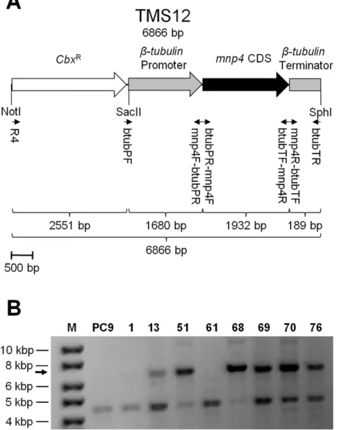
b-tubulinandmnp4were identified in the JGI genome database of PC9 v1.0 (http://genome.jgi-psf.org/PleosPC9_1/PleosPC9_1. home.html) as protein IDs 117235 and 137757, respectively, corresponding to previously identified protein IDs 16119 and 186006, respectively, in PC15 v1.0 [4,15,18]. The promoter and terminator regions of b-tubulin [8,18] and the coding sequence (CDS) ofmnp4were amplified from genomic DNA, using primers btubPF and mnp4F-btubPR, mnp4R-btubTF and btubTR, btubPR-mnp4F and btubTF-mnp4R, respectively (Figure 1A, Table 1). The resulting amplicons were fused together using the double-joint (fusion) PCR technique [27,28] and then ligated into plasmid pTM1 [29] using the SphI and SacII restriction sites to produce a conjunct mnp4 expression and carboxin-resistance (CbxR
) cassette designated TMS12 (Figure 1A), which was cloned to produce plasmid pTMS12.
Fungal Transformation
Transformation was performed based on the PEG-CaCl2
protocol previously adapted forP. ostreatus. Carboxin was used as a selection marker, and resistance was conferred via introduction of the carboxin-resistance cassette (CbxR) (Figure 1A). Either plasmid pTMS12 or the linear cassette TMS12, extracted from pTMS12 by restriction with NotI and SphI, were used as the transforming DNA (Figure 1A). Competent protoplasts were produced by digestion of vegetative mycelium ofP. ostreatusfrom YMG liquid culture with lytic enzymes. The lytic enzyme solution consisted of 2% (w/v) Lysing enzymes fromTrichoderma harzianum
(Sigma-Aldrich, product number L1412) and 0.05% (w/v) Chitinase fromT. viride(Sigma-Aldrich, product number C8241) in 0.5 M sucrose as an osmotic stabilizer. The protoplasts were washed (by centrifugation at 450g, 8 min, 4uC) in STC solution (18.2% w/v sorbitol, 50 mM Tris-HCl pH 8.0, 50 mM CaCl2,
Table 1.Oligonucleotides used in this study.
Primer designation Sequence (59R39)
btubPF CCGCGGCCGCGGATGCTGTTGGGAGGAAACTAAAT
mnp4F-btubPR GGAGAGCGTCTTGAAAGACATTCTGCATGGAAAAGAAGTTAGTCG
btubPR-mnp4F CGACTAACTTCTTTTCCATGCAGAATGTCTTTCAAGACGCTCTCC
btubTF-mnp4R GGTGATATGGACAATCCAACGATTACGATCCAGGGCTGTAGGA
mnp4R-btubTF TCCTACAGCCCTGGATCGTAATCGTTGGATTGTCCATATCACC
btubTR GCATGCGCATGCAAGGGCACAAAATGACATGAA
R4 GCTGGTAGCGGTGGTTTTT
Gene-expression see reference [4]
doi:10.1371/journal.pone.0052446.t001
Figure 1. Strategy for producing amnp4over-expressingP. ostreatusstrain.(A) Map of the VP4 (encoded bymnp4) over-expression and carboxin-resistance-conferring (CbxR) cassette, TMS12. Small arrows indicate the location of primers (Table 1) used for construction and detection of
the construct. (B) PCR screening ofP. ostreatusgenomic DNA targeting TMS12, using primers R4 and btubTR (Figure 1A, Table 1). The arrow indicates the expected 6866 bp amplicon. M – DNA size marker (GeneRuler DNA Ladder Mix, Fermentas); PC9– wild-type; 1, 13, 51, 61, 68, 69, 70, 76– carboxin-resistant transformant strains.
transforming DNA (300 ng/ml), 150ml heparin solution (Sigma-Aldrich, product number H4784) (5 mg dissolved in 1 ml STC solution), and 300ml single-strand l phage carrier DNA (Fermentas, product number SD0011) (500mg/ml, after de-naturation at 95uC for 5 min and immediate transfer to ice). After 40 min of incubation on ice, 10 ml PTC solution (40% w/v PEG#4000, 50 mM Tris-HCl pH 8.0, 50 mM CaCl2, 0.5 M
sucrose) was added, and the mixture was incubated for 20 min at room temperature. The mixture was then plated on selective solid YMG regeneration medium, containing 0.5 M sucrose and carboxin at a final concentration of 2 mg/l. Transformants were isolated after 10 days of incubation at 28uC. Transformant stability was verified by three successive transfers (inoculated from the edge of a 10-day-old colony) to solid medium without the selection drug, and then returning the transformant to solid culture conditions in which the selective drug was present.
Dye Decolorization Analyses
Decolorization capacity was estimated in GP cultures supple-mented with one of the tested compounds: Orange II (lmax
483 nm), Reactive Black 5 (lmax 597 nm) or Amaranth (lmax
521 nm). In solid culture, decolorization capacity was estimated according to the visually decolourized area, as measured from the center of the inoculation point. In liquid culture, 100ml of media were mixed with 900ml phosphate buffered saline (0.1 M, pH 7.4), and the dye concentration in the media was quantified according to the absorption reading of the solution at its lmax, using a BioMate 3 spectrophotometer (Thermo Scientific), according to a standard curve. Non-inoculated media supplemen-ted with the corresponding dye were used as a control. No decolorization was observed in non-inoculated media for any of the dyes [4].
Elemental Analyses
Samples were either solid-state fermentation products after lyophilization and grinding to pass a 1-mm-pore-size screen in a Wiley mill or sections of fresh solid culture medium, excised using a scalpel. Metal element concentrations were determined by plasma emission spectrometry after acid digestion, via End-On-Plasma Inductively Coupled End-On-Plasma Atomic Emission Spectros-copy (ICP-AES) model ARCOS (Spectro GMBH), at The Z.B.M. Analytical Laboratory of The Hebrew University.
Enzymatic Activity Assays
Samples of culture fluids were collected, centrifuged (4720 g, 10 min, 4uC) and maintained at 4uC. Enzymatic activity assays were conducted in a volume of 200ml in microtiter plates at 32uC, using the Synergy 2 Multi-Mode Microplate Reader (BioTek). Mn2+
-dependent and Mn2+
-independent activities were deter-mined using phenol red (Sigma-Aldrich) as the substrate. Oxidation of phenol red was measured by monitoring the A610
(e= 22.0 mM21
cm21). The reaction mixture contained 0.1 mM MnSO4, 0.1 mM H2O2, 0.01% (w/v) phenol red, 25 mM lactate,
0.1% (w/v) bovine serum albumin and 20 mM sodium succinate buffer (pH 4.5). After incubation, the reaction was terminated by addition of NaOH to a final concentration of 80 mM. Activity in the absence of either MnSO4or H2O2, was measured to establish
specific (either Mn2+
-dependent or Mn2+
-independent) peroxidase activity. Accordingly, Mn2+
-independent activity was deduced from the reaction in the absence of Mn2+after subtraction of its
corresponding reaction in the absence of H2O2, and Mn2+
-dependent activity was deduced from the reaction containing Mn2+ by subtraction of the corresponding Mn2+-independent
activity. Corresponding boiled samples served as blanks. One unit
(U) of enzymatic activity was defined as the amount of enzyme that catalyzes the formation of 1.0mmol of product per minute per milliliter of culture filtrate.
Protein Expression Profile
Culture fluids were filtered through Whatman No. 1 filter paper followed by 0.45-mm mixed cellulose ester filter papers (Whatman). The sample was then concentrated 60-fold using a 10-kDa cutoff PM-10 membrane (Amicon Division) and treated with cOmplete, EDTA-free Protease Inhibitor Cocktail Tablets. The concentrated eluate was separated on a NuPAGE 4–12% bis-Tris gel in MES-SDS running buffer (Invitrogen), and the 40–45 kDa gel fraction was excised and maintained at 4uC. The sample was subsequently analyzed by HPLC/mass spectrometry/mass spectrometry (LC-MS/MS) in an Orbitrap (Thermo Scientific) mass spectrometer and identified by Sequest 3.31 software against the JGI genome database of P. ostreatus PC9 v1.0 (http://genome.jgi-psf.org/ PleosPC9_1/PleosPC9_1.home.html), at The Smoler Proteomics Center of The Israel Institute of Technology (Technion).
[14C]-lignin Mineralization Analysis
[14C]-lignin preparation and mineralization during solid-state fermentation were performed according to the methods described by Kerem and Hadar [6–8]. 14C-radiolabelled substrate (556104dpm [14C]-lignin, 2560.1 mg) was added to each of three replicates of 2 g of lignocellulosic substrate (cotton stalks) in 20-ml polyethylene cups (27 mm diameter661 mm height), then sterilized, inoculated and sealed in a 300 ml biometer flask with two gas-tight caps.14CO2evolved in each flask was trapped every
3–4 days for 6 h on a cellulose filter paper (2610 cm) presoaked with 0.25 ml of 6 N NaOH. Prior to removal of the filters, the flasks were flushed for 1 min with moistened, sterile, atmospheric air. Subsequently, the amount of trapped radioactive14CO2was
measured in Hionic-Fluor (PerkinElmer) scintillation liquid by a TriCarb 1900 TR Liquid Scintillation Analyzer (Packard Instrument).
Lignocellulose Digestibility Analyses
Non-inoculated substrate and solid-state fermentation products were lyophilized and ground to pass a 1-mm-pore-size screen in a Wiley mill.In-vitrodry matter digestibility (IVDMD) values were determined according to Tilley and Terry et al. [30], using rumen fluid obtained from two wether sheep (Ovis aries). Neutral detergent fiber digestibility (NDFD) values were determined by quantifying the NDF soluble content of samples pre-in-vitro-digestion and
post-in-vitro-digestion according to the Van Soest methodology [31], adapted for the ANKOM-200 fiber analyzer (Ankom Technolo-gy). Acid detergent soluble compound, cellulose, acid-insoluble lignin and ash contents were determined according to the Van Soest methodology [31], adapted for the ANKOM-200 fiber analyzer (Ankom Technology).
Statistical Analyses
Analysis of variance with Tukey-Kramer HSD test (significance accepted at P,0.05) was used to analyze differences among treatments, using JMP 7.0 Statistical Analysis Software (SAS Institute).
Results
Engineering a Strain Over-expressingmnp4(OEmnp4)
P. ostreatusVP4 is negatively regulated by Mn2+
in the culture medium [4,8–10]. However, due to its potential importance and high ability to oxidize Mn2+
ostreatusstrain over-expressingmnp4despite the presence of Mn2+
in the culture.
Introduced expression of mnp2 [32] and mnp3 [25], and knockdown (silencing) of mnp3 by RNAi [4,33] have been previously reported. In those studies, the transforming constructs were driven by theP. ostreatus iron-sulfur protein subunit of succinate dehydrogenase(sdi1) promoter. Recently, we have shown that theP. ostreatusb-tubulinpromoter yields expression levels superior to those achieved with thesdi1promoter [18].
Therefore, we used theb-tubulinpromoter to drivemnp4CDS in the TMS12 cassette (Figure 1A) transformed into the wild-type strain (PC9). About 100 carboxin-resistant colonies were isolated and, after confirming their stability, grown without antibiotic selection.
Screening for candidate strains over-expressing mnp4 was performed by evaluating Orange II decolorization in solid GP cultures [4]. More than 50% of the transformants exhibited markedly enhanced decolorization compared to PC9, while showing a similar linear growth rate. Similar results were observed using Reactive Black 5 and Amaranth (Figure 2) [4], Orange G, Bromocresol green, Poly R-478, Poly B-411 and Remazol Brilliant Blue R (data not shown).
An additional screen consisted of monitoring the appearance of dark brown to black precipitates typically visualized in solid GP cultures containing more than 54mM Mn2+
(Figure 2). Elemental analyses confirmed that the precipitates formed in a culture containing 108mM Mn2+were composed of oxidized Mn2+, i.e.
MnO2, as the dark areas contained 22.362.1 mg/kg Mn 2+ in
comparison to 5.061.4 mg/kg Mn2+
in the non-darkened areas. No differences were found in Mg2+
and K+
concentrations (internal controls) in these areas. An indication of enhanced Mn2+
oxidation by the transformants was the formation of larger and darker areas of MnO2precipitates when grown in solid GP culture
containing 108mM Mn2+
(Figure 2).
Genomic integration of TMS12 was confirmed by PCR, using primers R4 and btubTR, on DNA extracted from eight transformants (designated 1, 13, 51, 61, 68, 69, 70 and 76) and PC9 (Figure 1, Table 1). Integration of the full-length TMS12 cassette was verified in strains 13, 51, 68, 69, 70 and 76 (Figure 1B). It is possible that although the TMS12 amplicon was not detected in strains 1 and 61, they did integrate sufficient essential segments from TMS12 to produce the phenotypes described above. A non-specific amplicon observed in all of the tested strains provided a convenient internal control, verifying the integrity of the DNA used (Figure 1B). Strain 51 was selected for further study, referred to herein as OEmnp4. Both PC9 and OEmnp4 showed similar linear growth rates (7.360.2 and 6.860.2 mm/24 h, respectively) and biomass production (125.766.9 and 114.8611.7 mg/flask, after 10 days of growth, respectively).
Constitutive Over-expression ofmnp4Results in Higher
Peroxidase Activities and Earlierin-vivoAzo Dye
Decolorization
To determine whether OEmnp4over-expressesmnp4in culture despite the presence of Mn2+, the MnP gene family expression
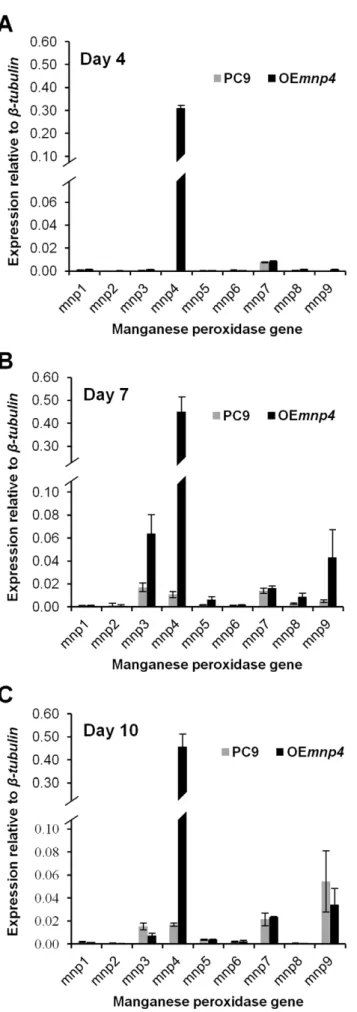
profiles of OEmnp4and PC9 were quantitatively evaluated in total RNA extracted from liquid GP cultures, at 4, 7 and 10 days of incubation (Figure 3).
Expression of the introducedmnp4was apparent at high levels as early as day 4, while the expression of the endogenousmnps (in particularmnp3,mnp4andmnp9) was evident from day 7 onwards.
mnp4 expression increased throughout the incubation period (Figure 3).
To support these results, the 40–45 kDa fraction of the fungal secretome, which contains the peroxidases, also based onin-silico
genomic analyses [14,15], was analyzed for MnP isoenzymes in extracellular fluids collected from liquid GP culture at 7 days. 8, 6 and 4 unique peptides corresponding to VP4, MnP3 and MnP9, respectively, were detected, corresponding to the presence of significant amounts ofmnp4,mnp3, andmnp9transcript.
Taken together, the genetic manipulation resulted in modifica-tion of three major traits with respect to mnp4 expression: (a) release from Mn2+
repression, (b) markedly higher expression
Figure 2.In-vivodecolorization of Orange II, Reactive Black 5 and Amaranth, and MnO2 precipitation. P. ostreatus wild-type
strain (PC9) andmnp4over-expressing strain (OEmnp4) were grown for 10 days on solid GP medium containing 27mM Mn2+ supplemented with 100 mg/l of the corresponding dye. For Mn2+oxidation assay, the
level, and (c) earlier expression. Specifically, OEmnp4expression of
mnp4was at least 27-fold higher than that of PC9 throughout the incubation period.
To directly study the effect of mnp4 over-expression on extracellular oxidation activity in the presence of Mn2+
in the culture, a 10-day time-course assay was conducted. Both of the VP activities, Mn2+-dependent and Mn2+-independent
peroxidase-catalyzed reactions, were measured in liquid GP cultures of OEmnp4and PC9 (Figure 4). Phenol red was used as a substrate, and its oxidation was monitored in the presence and absence of Mn2+in the reaction mixture.
In PC9, Mn2+
-dependent activity was negligible until 8 days, reaching 0.016 U/ml, and gradually increased to a maximum 0.036 U/ml at 10 days. In OEmnp4it was detected 4 days earlier and was higher throughout the incubation period, reaching 0.032 U/ml at 4 days, and constantly increasing to 0.153 U/ml at 10 days, while still maintaining a rising trend (Figure 4). Thus, the level of Mn2+
-dependent activity in OEmnp4was at least 4-fold higher than in PC9 throughout the incubation period.
As expected, owing to negative regulation of mnp4 by Mn2+
[4,8–10], Mn2+
-independent activity in PC9 was negligible throughout the incubation period. In OEmnp4, the activity profile correlated with mnp4 transcript levels, as higher activity was observed throughout the incubation period, reaching 0.004 U/ml at 4 days and constantly increasing to 0.062 U/ml at 10 days (Figure 4). This level of activity in OEmnp4was comparable to that observed in a Mn2+
-deficient culture of PC9 [9,18].
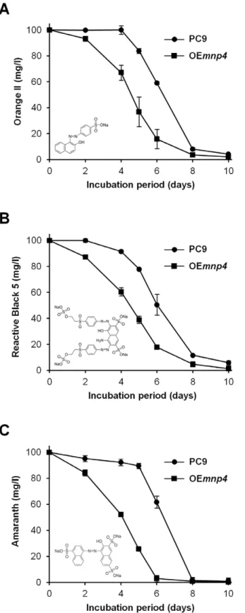
To evaluate the effect ofmnp4over-expression on the function of the ligninolytic system, thein-vivodecolorization capacities of the azo dyes Orange II, Reactive Black 5 and Amaranth by OEmnp4 Figure 3. Expression of theP. ostreatus manganese peroxidase genes in PC9 and OEmnp4strains.Total RNA was extracted fromP. ostreatus wild-type strain (PC9) and mnp4 over-expressing strain (OEmnp4) cultures at (A) 4 days, (B) 7 days and (C) 10 days of incubation. The fungi were grown in GP medium containing 27mM Mn2+ supplemented with 100 mg/l Orange II. Data represent the
average of three biological replicates. Bars denote SD. doi:10.1371/journal.pone.0052446.g003
Figure 4. Time-course assay of peroxidase activity. Mn2+ -dependent (+Mn2+) and Mn2+-independent (2Mn2+) peroxidase
and PC9 were determined in liquid GP cultures during 10 days of incubation (Figure 5).
The results clearly indicate earlierin-vivoazo dye decolorization capacity by OEmnp4 (Figure 5). Furthermore, the earlier de-colorization by OEmnp4correlated with both the high and early expression level of mnp4 and the presence of Mn2+
-mediated peroxidase activity at 4 days of incubation in OEmnp4 culture, while at this time point the expression of the othermnps and all the
mnps in PC9 were tenuous, as was the activity in PC9 culture (Figure 3, Figure 4).
In spite of the constant increased accumulation of VP activity in OEmnp4(Figure 4), the rate of decolorization of each of the tested dyes was comparable between OEmnp4and PC9 (Figure 5). This may suggest the existence of an as yet unknown rate-limiting factor restricting the expected accelerated decolorization by OEmnp4due to over-expression of VP4 (Figures 3 and 4).
Enhanced [14C]-lignin Mineralization and Lignocellulose
Digestibility by OEmnp4
To assess the effect of mnp4 over-expression on lignin degradation and the digestibility of lignocellulose, cotton stalks (non-supplemented or supplemented with Mn2+
) were inoculated with OEmnp4or PC9 and incubated for 14 days under solid-state fermentation conditions. The cultures were then analyzed to quantify mnp4expression level in total RNA (Figure 6A), [14 C]-lignin mineralization (Figure 6B), and the fermentation product was analyzed for in-vitro dry matter digestibility (IVDMD) (Figure 6C) [30] and neutral detergent fiber digestibility (NDFD) (Figure 6D) [31]. Non-inoculated substrate was used as a control.
Mn2+
concentrations in the cotton stalks were 8.061.4 mg/kg dry matter (basal Mn2+
concentration) and 23.362.1 mg/kg dry matter (after Mn2+
addition), which is consistent with 0.5 kg stalks treated with 2 liters of a 73mM Mn2+
solution.
mnp4expression in PC9 grown on cotton stalks that were either non- supplemented or supplemented with Mn2+was 0.97 and 0.24
relative tob-tubulin, respectively, whilemnp4expression in OEmnp4
grown under the same conditions was 2.03 and 1.93 relative tob -tubulin, respectively (Figure 6A). This clearly shows that Mn2+
supplements down-regulatedmnp4expression in PC9 (being four-fold higher under non-supplemented conditions) whereas it did not have a significant effect in OEmnp4, demonstrating that the latter indeed expressesmnp4in a Mn2+
-independent manner. Moreover, marked over-expression of mnp4 was measured in OEmnp4
compared to PC9 (two- and eight-fold higher in cotton stalks non-supplemented and supplemented with Mn2+
, respectively). This is in agreement with previously reported down-regulation ofmnp4by Mn2+
in liquid GP cultures of PC9, albeit at a different level [4,8]. While the expression ofmnp4in GP not-supplemented with Mn2+(containing less than 0.1
mM Mn2+) was seventy-fold
higher than in the presence of 27mM Mn2+
[4,8], in cotton stalks not-supplemented with Mn2+
it was only four-fold higher than in cotton stalks in which Mn2+(Figure 6A) was added. The fact that
cotton stalks contain a significant basal level of Mn2+
, likely enforcing down-regulation of mnp4, may explain the smaller difference observed between the two conditions using cotton stalks as a substrate.
[14C]-lignin mineralization of cotton stalks not-supplemented with Mn2+
was found to be 1.00% and 1.28% for PC9 and OEmnp4, respectively, after 2 weeks of solid-state fermentation. When the stalks were supplemented with Mn2+, mineralization
increased in both strains, reaching 1.40% and 1.69% for PC9 and OEmnp4, respectively. The mineralization rate of OEmnp4
surpassed that of PC9 throughout the incubation period in each of the culture conditions (Figure 6B).
Figure 5. Time-course assay ofin-vivoazo dyes decolorization.
P. ostreatus wild-type strain (PC9) and mnp4 over-expressing strain (OEmnp4) were grown for 10 days in GP medium containing 27mM Mn2+supplemented with 100 mg/l of (A) Orange II, (B) Reactive Black 5
Kerem et al. (6,7) and Cohen et al. (8) previously reported that Mn2+
supplements increase lignin degradation. As this phenom-enon was shown, here, to also occur by Mn2+
-independent over-expression ofmnp4, direct evidence for the correlation between the level of mnp4 expression and lignin degradation is now sub-stantiated. Moreover, the fact the over-expression of mnp4
enhanced lignin mineralization indicates that the basal level of
mnp4expression limits the natural lignin degradation capacity by
P. ostreatus.
IVDMD of non-inoculated cotton stalks (regardless of Mn2+
supplements) were 12%. This value was not altered in cotton stalks in which PC9 was cultured in the absence of Mn2+
supplements. However, when OEmnp4was cultured under the same conditions it increased to 16%. These values increased to 16% and more than 20% when PC9 or OEmnp4, respectively, were cultured in the presence of the Mn2+
supplements (Figure 6C). The NDFD values supported the IVDMD results, and in addition revealed a signif-icant increase, from 6% to 10%, in the case of PC9 cultured on stalks not-supplemented with Mn2+, compared to the
non-inoculated controls (Figure 6D).
From these results, we deduced that solid-state fermentation of cotton stalks with PC9 increases lignocellulose digestibility, and that Mn2+
supplements improve this process by more than 33%.
Evidently, this process can be greatly enhanced by application of OEmnp4, producing digestibility values higher than that of PC9 by more than 33% (non-supplemented) and 25% (supplemented), respectively.
Moreover, these marked improvements in digestibility resulted from a relatively short-duration of solid-state fermentation (14 days), thus limiting consumption of the cellulose constituent (48.3– 49.5% of the produce dry matter) by the fungus in all treatments. Comparison of the gene-expression data (Figure 6A) with the mineralization (Figure 6B) and digestibility data (Figure 6C, D) of PC9 and OEmnp4leads to several fundamental conclusions: (a) the higher expression level ofmnp4in OEmnp4correlates with higher mineralization and digestibility; (b) since OEmnp4shows similar
mnp4expression in both Mn2+
-supplemented and non-supplemen-ted substrates, the higher mineralization and digestibility levels in the presence of Mn2+
supplement is most probably due to the production of greater amounts of reactive Mn3+
by VP4, as more of its substrate, Mn2+
, becomes available for oxidation, and (c) in PC9, Mn2+
supplements down-regulate mnp4expression while at the same time increase mineralization and digestibility. These results strongly support the hypothesis that the amount of mnp4
produced by PC9 is a limiting factor for its lignin-degrading
Figure 6. Gene-expression, lignin degradation and lignocellulose digestibility under solid-state fermentation.(A)mnp4expression level, (B) [14C]-lignin mineralization (percentage of14CO2 emitted from the total initial radiolabelled [14C]-lignin is presented), (C)in-vitrodry matter digestibility (IVDMD) and (D) neutral detergent fiber digestibility (NDFD) byP. ostreatus. The wild-type strain (PC9) andmnp4over-expressing strain (OEmnp4) were incubated for 14 days under solid-state fermentation conditions, using a lignocellulosic substrate of cotton stalks containing either 8.0 (basal Mn2+concentration) or 23.3 (concentration in Mn2+supplemented stalks) mg/kg dry matter (DM) of Mn2+. Non-inoculated substrate was
used as a control. Data represent the average of three biological replicates. Bars denote SD. Statistical analysis was performed by analysis of variance with Tukey-Kramer HSD test (significance accepted atP,0.05).
capacity, as the highermnp4expression by OEmnp4was proven to enhance lignocellulose digestibility.
Discussion
Gene families of multiple ligninolytic isoenzymes encoded by various, and apparently redundant structurally-related genes, are common among ligninolytic fungi [17]. For example,Phanerochaete chrysosporium,Ceriporiopsis subvermisporaandPhlebia chrysocreashave at least 5, 4 and 7 different genes encoding MnPs, respectively [12,34,35], and P. ostreatus, Coprinopsis cinerea, and Laccaria bicolor
have at least 7, 17 and 11 different genes encoding laccases, respectively [36–38].
Differential gene-expression among the members of a gene family is also characteristic. Studies of the catalytic properties of various ligninolytic isoenzymes reflect distinct differences in both the culture conditions and substrate specificities associated with their transcription and kinetic constants [4,39,40]. For example, 7 isoforms of MnPs were isolated from C. subvermisporacultured in salt medium, whereas 4 isoenzymes were fractioned in extracts derived from wood chips. The requirement for Mn2+by each of
these MnPs varied on the basis of the nature of the aromatic substrate (vanillylacetone, o-dianisidine, p-dianisidine, guaiacol) added to the reaction mixture [41]. Expression of the P. chrysosporium MnP gene family is regulated by nitrogen levels, Mn2+
, heat shock, agitation and other factors. Whilemnps 1–3are expressed under various culture conditions,mnp4andmnp5seem to be actively transcribed only when the fungus is grown on wood-containing soil samples, and wood pulp, respectively [34,42]. Recently, a comprehensive systematic transcriptomic analysis showed that Postia placenta and P. chrysosporium gene-expression patterns, e.g. cytochrome P450s and extracellular glycoside hydrolases, are substantially influenced by the wood species used as a substrate [i.e. when grown on aspen (Populus grandidentata) or pine (Pinus strobus)] [39].
The fact theP. ostreatus has a large number of distinct genes encoding transcripts of various isoenzymes belonging to the MnP gene family points to the potential redundancy and/or diversity of these enzymes. It also may imply that complex and versatile strategies are employed by this fungus for the degradation of aromatic and recalcitrant compounds such as amorphic lignin and azo dyes. As the enzymatic systems for lignin degradation operate in concert for the successful degradation of lignin or recalcitrant anthropogenic aromatic compounds, regulation of the MnP gene family expression and corresponding enzymatic activity is also dependent on environmental conditions and nutrient availability [1,4,15].
Mn2+
is both an active mediator of MnP, and a regulator of MnP, laccase, and lignin peroxidase (LiP) expression in various ligninolytic fungi, usually exhibiting significantly higher MnP expression in Mn2+
-supplemented compared to Mn2+
-deficient media [6,21,34,35,43]. Accordingly, in the model white-rot fungus
P. chrysosporium, significantly higher expression of MnP was obtained in Mn2+
-supplemented cultures, whereas significantly higher expression of LiP was obtained in Mn2+
-deficient cultures [34]. These phenomena can be explained by the fact that MnP catalyzes Mn2+oxidation, whereas LiP oxidizes phenols and
non-phenolic aromatics, hence the production of these enzymes alternates depending on the substrates present in the culture, to best fit the required mode of action.
While both of the predominantP. ostreatus MnPs, MnP3 and MnP9, comply with this dogma, the predominant VP4 does not [4,9]. As mentioned above, an apparent biological contradiction
exists: although VP efficiently oxidizes Mn2+, its expression is
repressed by Mn2+. One proposed explanation is that VP is
expressed during the later stages of lignin degradation, when much of the Mn2+present in wood has been exhausted, and functions in
the further oxidation of lignin subunits. Another possible explanation is that the Mn2+-independent activity of VP functions
similarly to that ofP. chrysosporiumLiP, but this activity only occurs at relatively higher concentrations of phenolic substrates due to lower catalytic efficiency (kcat/Km) compared to Mn2+as a substrate
[21,22]. Furthermore, sinceP. ostreatusdoes not possess LiP, but does possess LiP-like activity as part of VP (which may be regarded as a MnP-LiP hybrid enzyme), it might also retain an expression pattern similar to that of LiP. In short, VP repression by Mn2+
does not yet have a functional explanation and remains an enigma. This study focused on investigating this phenomenon by implementing a genetic manipulation that facilitates constitutive expression ofmnp4and characterizing the corresponding pheno-types associated with ligninolytic functionality.
Introduced expression ofmnp4CDS under the control of theb -tubulin promoter enabled circumventing the Mn2+
expression-repressive effect targeting the endogenousmnp4, thereby achieving an expression level that was one order of magnitude higher than naturally present in Mn2+-containing culture. Furthermore, a high
level of mnp4 expression was detectable days before significant levels of any othermnps were detected. This expression level was on the same scale as that of endogenousmnp4in Mn2+-deficient
culture, makingmnp4(encoding VP4) the predominantly expressed member of the MnP gene family in Mn2+-containing culture, while
under natural conditions this predominance is overtaken bymnp3
andmnp9(both encoding MnPs) [4].
As a consequence of mnp4 over-expression, the naturally negligible VP activity levels were markedly and constantly increased as the incubation period progressed, to levels compa-rable to those naturally present in Mn2+
-deficient cultures [9,18]. Moreover, these VP activities were detected 4 days earlier than the first indication of MnP activity. This enzymatic activity profile correlated with the aforementioned gene-expression profile.
mnp4over-expression in an environment where Mn2+
is present resulted in enhanced decolorization capacity of azo dyes, lignin mineralization, and conversion of lignocellulosic substrate into product with greater digestibility. Taken together, these data prove that the VP encoded by mnp4 makes a key contribution to ligninolytic functionality inP. ostreatus.
The improved degradation of anthropogenic and natural substrates shown here by genetic modification ofP. ostreatusmay provide a basis for harnessing its qualities for various bio-technological processes, such as improving lignocellulose digest-ibility for use as animal feed or biofuel and for bioremediation.
Acknowledgments
We are deeply grateful to Prof. Takashi Watanabe and Prof. Yoichi Honda, Research Institute for Sustainable Humanosphere, Kyoto University, Japan, for generously providing plasmid pTM1. We thank the Joint Genome Institute (US Department of Energy) and thePleurotus Genome Consortium for access to theP. ostreatusgenome database.
Author Contributions
References
1. Cohen R, Persky L, Hadar Y (2002) Biotechnological applications and potential of wood-degrading mushrooms of the genusPleurotus. Appl Microbiol Biotechnol 58: 582–594.
2. Stajic´ M, Vukojevic´ J, Duletic´-Lausˇevic´ S (2009) Biology ofPleurotus eryngiiand role in biotechnological processes: a review. Crit Rev Biotechnol 29: 55–66. 3. Sa´nchez C (2010) Cultivation ofPleurotus ostreatusand other edible mushrooms.
Appl Microbiol Biotechnol 85: 1321–1337.
4. Salame TM, Yarden O, Hadar Y (2010)Pleurotus ostreatusmanganese-dependent peroxidase silencing impairs decolourization of Orange II. Microb Biotechnol 3: 93–106.
5. Golan-Rozen N, Chefetz B, Ben-Ari J, Geva J, Hadar Y (2011) Transformation of the recalcitrant pharmaceutical compound carbamazepine by Pleurotus ostreatus: role of cytochrome P450 monooxygenase and manganese peroxidase. Environ Sci Technol 45: 6800–6805.
6. Kerem Z, Hadar Y (1993) Effect of manganese on lignin degradation byPleurotus ostreatusduring solid-state fermentation. Appl Environ Microbiol 59: 4115–4120. 7. Kerem Z, Hadar Y (1995) Effect of manganese on preferential lignin degradation byPleurotus ostreatusduring solid-state fermentation. Appl Environ Microbiol 61: 3057–3062.
8. Cohen R, Hadar Y, Yarden O (2001) Transcript and activity levels of different
Pleurotus ostreatus peroxidases are differentially affected by Mn2+
. Environ Microbiol 3: 312–322.
9. Cohen R, Persky L, Hazan-Eitan Z, Yarden O, Hadar Y (2002) Mn2+
alters peroxidase profiles and lignin degradation by the white-rot fungus Pleurotus ostreatus under different nutritional and growth conditions. Appl Biochem Biotechnol 102: 415–429.
10. Cohen R, Yarden O, Hadar Y (2002) Lignocellulose affects Mn2+
regulation of peroxidase transcript levels in solid-state cultures ofPleurotus ostreatus. Appl Environ Microbiol 68: 3156–3158.
11. Martı´nez AT, Speranza M, Ruiz-Duen˜as FJ, Ferreira P, Camarero S, et al. (2005) Biodegradation of lignocellulosics: microbial chemical, and enzymatic aspects of the fungal attack of lignin. Int Microbiol 8: 195–204.
12. Morgenstern I, Klopman S, Hibbett DS (2008) Molecular evolution and diversity of lignin degrading heme peroxidases in the Agaricomycetes. J Mol Evol 66: 243–257.
13. Hammel KE, Cullen D (2008) Role of fungal peroxidases in biological ligninolysis. Curr Opin Plant Biol 11: 349–355.
14. Hofrichter M, Ullrich R, Pecyna MJ, Liers C, Lundell T (2010) New and classic families of secreted fungal heme peroxidases. Appl Microbiol Biotechnol 87: 871–897.
15. Ruiz-Duen˜as FJ, Ferna´ndez E, Martı´nez MJ, Martı´nez AT (2011) Pleurotus ostreatusheme peroxidases: anin-silicoanalysis from the genome sequence to the enzyme molecular structure. Crit Rev Biol 334: 795–805.
16. Chen M, Zeng GM, Tan ZY, Jiang M, Li H, et al. (2011) Understanding lignin-degrading reactions of ligninolytic enzymes: binding affinity and interactional profile. PLoS ONE 6: e25647.
17. Floudas D, Binder M, Riley R, Kerrie B, Blanchette RA, et al. (2012) The Paleozoic origin of enzymatic lignin decomposition reconstructed from 31 fungal genomes. Science 336: 1715–1719.
18. Salame TM, Knop D, Tal D, Levinson D, Yarden O, et al. (2012) Predominance of a versatile-peroxidase-encoding gene,mnp4, as demonstrated by gene replacement via a gene targeting system forPleurotus ostreatus. Appl Environ Microbiol 78: 5341–5352.
19. Kamitsuji H, Honda Y, Watanabe T, Kuwahara M (2004) Production and induction of manganese peroxidase isozymes in a white-rot fungusPleurotus ostreatus. Appl Microbiol Biotechnol 65: 287–294.
20. Garcı´a-Ruiz E, Gonza´lez-Pe´rez D, Ruiz-Duen˜as FJ, Martı´nez AT, Alcalde M (2012) Directed evolution of a temperature-, peroxide- and alkaline pH-tolerant versatile peroxidase. Biochem J 441: 487–498.
21. Martı´nez MJ, Ruiz-Duen˜as FJ, Guille´n F, Martı´nez AT (1996) Purification and catalytic properties of two manganese-peroxidase isoenzymes from Pleurotus eryngii. Eur J Biochem 237: 424–432.
22. Collins PJ, O’Brien MM, Dobson ADW (1999) Cloning and characterization of a cDNA encoding a novel extracellular peroxidase fromTrametes versicolor. Appl Environ Microbiol 65: 1343–1347.
23. Larraya LM, Pe´rez G, Pen˜as MM, Baars JJP, Mikosch TSP, et al. (1999) Molecular karyotype of the white rot fungusPleurotus ostreatus. Appl Environ Microbiol 65: 3413–3417.
24. Irie T, Honda Y, Watanabe T, Kuwahara M (2001) Efficient transformation of filamentous fungus Pleurotus ostreatususing single-strand carrier DNA. Appl Microbiol Biotechnol 55: 563–565.
25. Irie T, Honda Y, Watanabe T, Kuwahara M (2001) Homologous expression of recombinant manganese peroxidase genes in ligninolytic fungusPleurotus ostreatus. Appl Microbiol Biotechnol 55: 566–570.
26. Sambrook J, Fritsch EF, Maniatis T (1989) Molecular cloning: a laboratory manual, 2nd edition. Cold Spring Harbor, NY: Cold Spring Harbor Laboratory Press.
27. Ninomiya Y, Suzuki K, Ishii C, Inoue H (2004) Highly efficient gene replacements in Neurospora strains deficient for nonhomologous end-joining. Proc Natl Acad Sci USA 101: 12248–12253.
28. Yu JH, Hamari Z, Han KH, Seo JA, Reyes-Domı´nguez Y, et al. (2004) Double-joint PCR: a PCR-based molecular tool for gene manipulations in filamentous fungi. Fungal Genet Biol 41: 973–981.
29. Honda Y, Matsuyama T, Irie T, Watanabe T, Kuwahara M (2000) Carboxin resistance transformation of the homobasidiomycete fungusPleurotus ostreatus. Curr Genet 37: 209–212.
30. Tilley JMA, Terry RA (1963) A two-stage technique for thein-vitrodigestion of forage crops. J Brit Grass Soc 18: 104–111.
31. Van Soest PJ, Robertson JB, Lewis BA (1991) Methods for dietary fiber, neutral detergent fiber, and nonstarch polysaccharides in relation to animal nutrition. J Dairy Sci 74: 3583–3597.
32. Tsukihara T, Honda Y, Watanabe T (2006) Molecular breeding of white rot fungusPleurotus ostreatusby homologous expression of its versatile peroxidase MnP2. Appl Microbiol Biotechnol 71: 114–120.
33. Salame TM, Ziv C, Hadar Y, Yarden O (2011) RNAi as a potential tool for biotechnological applications in fungi. Appl Microbiol Biotechnol 89: 501–512. 34. Kersten P, Cullen D (2007) Extracellular oxidative systems of the lignin-degrading basidiomycetePhanerochaete chrysosporium. Fungal Genet Biol 44: 77–87. 35. Gutie´rrez M, Rojas LA, Mancilla-Villalobos R, Seelenfreund D, Vicun˜a R, et al. (2008) Analysis of manganese-regulated gene-expression in the ligninolytic basidiomyceteCeriporiopsis subvermispora. Curr Genet 54: 163–173.
36. Kilaru S, Hoegger PJ, Ku¨es U (2006) The laccase multi-gene family inCoprinopsis cinereahas seventeen different members that divide into two distinct subfamilies. Curr Genet 50: 45–60.
37. Pezzella C, Autore F, Giardina P, Piscitelli A, Sannia G, et al. (2009) The
Pleurotus ostreatuslaccase multi-gene family: isolation and heterologous expression of new family members. Curr Genet 55: 45–57.
38. Courty PE, Hoegger PJ, Kilaru S, Kohler A, Bue´e M, et al. (2009) Phylogenetic analysis, genomic organization, and expression analysis of multi-copper oxidases in the ectomycorrhizal basidiomyceteLaccaria bicolor. New Phytol 182: 736–750. 39. Wymelenberg AV, Gaskell J, Mozuch M, BonDurant SS, Sabat G, et al. (2011) Significant alteration of gene expression in wood decay fungiPostia placentaand
Phanerochaete chrysosporiumby plant species. Appl Environ Microbiol 77: 4499– 4507.
40. MacDonald J, Suzuki H, Master ER (2012) Expression and regulation of genes encoding lignocellulose-degrading activity in the genus Phanerochaete. Appl Microbiol Biotechnol 94: 339–351.
41. Urzu´a U, Larrondo LF, Lobos S, Larraı´n J, Vicun˜a R (1995) Oxidation reactions catalyzed by manganese peroxidase isoenzymes from Ceriporiopsis subvermispora. FEBS Lett 371: 132–136.
42. Gettemy JM, Ma B, Alic M, Gold MH (1998) Reverse transcription-PCR analysis of the regulation of the manganese peroxidase gene family. Appl Environ Microbiol 64: 569–574.