Re vie w o f the Y chro m o so m e
and hype rte nsio n
Department of Biology, University of Akron, Akron, O H, USA D. Ely, M. Turner
and A. Milsted
Abstract
The Y chromosome from spontaneously hypertensive rats (SHR) has a locus that raises blood pressure 20-25 mmHg. Associated with the SHR Y chromosome effect is a 4-week earlier pubertal rise of testos-terone and dependence upon the androgen receptor for the full blood pressure effect. Several indices of enhanced sympathetic nervous system (SNS) activity are also associated with the SHR Y chromo-some. Blockade of SNS outflow reduced the blood pressure effect. Salt sensitivity was increased by the Y chromosome as was salt appetite which was SNS dependent. A strong correlation (r = 0.57, P<0.001) was demonstrable between plasma testosterone and angio-tensin II. Coronary collagen increased with blood pressure and the presence of the SHR Y chromosome. A promising candidate gene for the Y effect is the Sry locus (testis determining factor), a transcription factor which may also have other functions.
Co rre spo nde nce
D. Ely
Department of Biology University of Akron Akron, O H 44325-3908 USA
Fax: + 1-330-972-8445 E-mail: Ely1@ Uakron.edu
Presented at the III International Symposium on Vasoactive Peptides, Belo Horizonte, MG, Brasil, O ctober 8-10, 1999.
Research partially supported by NIH (No. Ro1 HL-48072-7), the American Heart Association (No. 95010670) and the State of O hio Board of Regents (No. 5-34540).
Received November 26, 1999 Accepted February 9, 2000
Ke y wo rds
·Sympathetic nervous system
·Testosterone
·Androgen receptor
·Collagen
Intro ductio n
The mammalian Y chromosome has evolved as an efficient mechanism of sex determination. There are two major compo-nents of the mammalian Y chromosome that are necessary for this process: a pseudoauto-somal region (PAR) and a Y unique region. The PAR has loci that are homologous to a region on the X chromosome. These regions pair during meiosis, resulting in the homolo-gous pairing and separation during meiosis I of the X and Y chromosomes. The unique region, which does not recombine with the X chromosome, contains the dominant testes
determining locus (Sry). This locus is
prima-rily responsible for male sexual phenotype. Other than the small PAR, the Y chromo-some is not involved in recombination (cross-ing over) dur(cross-ing meiosis. This results in the majority of the Y chromosome being
inher-ited without variation from father to son. The classic genetic tools of linkage analysis and crossing over to locate a gene of interest are of no use for a Y chromosome locus. From a genomic view the human Y chromosome is the most thoroughly studied mammalian chro-mosome, with a complete physical map and overlapping YAC contig (1,2). A Y chromo-some contains fewer loci than predicted by its size when compared to autosomal chro-mosomes, since most of its length is com-posed of species specific repeats. There are about 20 identified loci on the human Y chromosome but not all these are conserved on the rat Y chromosome (3). The Y chromo-somes of different mammalian species con-tain a few conserved Y chromosome loci,
such as Sry, but other loci are species
a gene locus (or loci) that is instrumental in increasing blood pressure in this strain. Re-sults from both reciprocal crosses and consomic lines indicate that the Y chromo-some is responsible for about 20-25% of the total blood pressure increase in the SHR strain. The following review will cover the fol-lowing topics: 1) the Y chromosome blood pressure effect in humans and animals re-ported by other laboratories, 2) the develop-ment of consomic strains, 3) the sympathetic nervous system and salt sensitivity, which is modulated by the Y chromosome, 4) the Y chromosome effect on testosterone, the renin-angiotensin system, and renal norepinephrine (NE) release which is modulated by the andro-gen receptor, 5) a testosterone-coronary artery collagen deposition link, and 6) new molecu-lar Y chromosome candidate genes.
Y Chro mo so me blo o d pre ssure e ffe ct in o the r labo rato rie s - animal and human studie s
It is always important to have new find-ings validated by similar results in independ-ent laboratories. In 1991 Hilbert et al. (4) and Jacob et al. (5) reported results consistent with a Y chromosome effect in stroke-prone SHR (SHRSP) of about 17 mmHg in systolic pressure. Dominiczaks group (6) (Glasgow) reported a similar Y chromosome effect in SHRSP males crossed with WKY of 20 mmHg. Also male SHRSP have larger in-farcts and more spontaneous strokes than females (7). The only negative report which did not show a Y chromosome effect was from Samanis laboratory (8) using the Charles River SHR (SHR/crl). It appears that the SHR/crl has a different Y chromo-some. We crossed SHR/crl with our WKY rats and failed to find an SHR Y chromo-some blood pressure effect (9). Recent hu-man studies further support our hypothesis and show that a hypertensive father but not a hypertensive mother affects blood pressure in normotensive male offspring through body
mass index (10). Also, Lemne (11) showed that there was increased blood pressure reac-tivity in children of borderline hypertensive fathers. Recently, the use of a mathematical model to analyze data from cattle that devel-oped pulmonary hypertension at high alti-tude showed that those data were consistent with a Y chromosome effect (12).
Co nso mic line de ve lo pme nt and substrain analysis
After our original observation of the hy-pertensive effect of the SHR Y chromosome, we began a breeding protocol to develop consomic strains for the SHR and WKY Y chromosomes. From an original WKY x
SHR cross, F1 sons were selected and
backcrossed to a WKY . In each subsequent generation sons were selected and back-crossed to a WKY . This strain has been designated SHR/y as it has the SHR Y chro-mosome in a normotensive WKY back-ground. A second consomic was developed concurrently from an SHR x WKY cross, and sons were backcrossed to an SHR . This strain is designated SHR/a, as it has the SHR autosomal, X chromosomal and pseudoau-tosomal regions of SHR with a WKY Y chromosome. These strains have been back-crossed for 19 generations. Blood pressures of the two consomic strains have been pub-lished (13), and in each the addition (or deletion) of the SHR Y chromosome adds (or subtracts) 15-20 mmHg from the paren-tal strain systolic blood pressure. The consomic strains are maintained by back-crossing to the original female strain rather than by interbreeding of the consomic strain. This prevents random differentiation between the congenic strains and the parental strains. This also insures that the SHR and WKY strains are genetic control strains for the consomics in each generation.
SHR Y chromosome into the normotensive King Holztman (KH) genetic background. In these hybrid animals those with the SHR Y chromosome had higher blood pressure than those with a KH Y chromosome (14). Also animals with a deficient androgen receptor (feminized male) had lower blood pressure than the siblings with normal receptors. The SHR strains from different vendors have different Y chromosomes. Our original ex-periments were conducted on animals from our breeding colony which had originally been purchased from Harlan Sprague Dawley (SHR/hsd and WKY/hsd). Our data are con-sistent with the SHR/hsd and SHR/crl strains having different Y chromosomes, most likely the result of mutation and genetic drift since the two vendor strains have been separated. Y chromosomes are more prone to these types of change than the other chromosomes because of reduced number of copies in each breeding pair. For example, for each breed-ing pair there are four copies of each autoso-mal locus (two in each parent) and three copies of each X chromosome locus (two in the female and one in the male) but only one copy of the Y chromosome.
Sympathe tic ne rvo us syste m
The Y blood pressure effect is associated with indices of increased sympathetic ner-vous system (SNS) activity including in-creased adrenal gland NE and chromogranin A content, increased heart and renal NE turnover, increased plasma NE response to acute stress, and reduction in blood pressure after chemical sympathectomy or clonidine treatment (13,15,16). There is a large body of evidence that suggests that the SNS is involved in both human and animal neuro-genic models of hypertension, by many dif-ferent mechanisms. Lee et al. (17) showed that one of the primary roles of the overac-tive SNS in SHR was through the trophic effects on the arteries, especially through hyperplasia of smooth muscle cells. The
de-velopment of the SNS in SHR appears to be tissue specific.
A substantial portion of patients with borderline hypertension show signs of in-creased sympathetic and dein-creased parasym-pathetic tone. The research of Esler et al. (18) and Wallin et al. (19) has shown strong evidence for increased regional sympathetic nervous activity in human hypertension. The source of the increased SNS is unknown, but evidence is mounting that it may have a central nervous system origin. Wysss (20) and Oparils (21) laboratories have devel-oped the hypothesis that inappropriate CNS regulation of SNS activity is a major con-tributor to NaCl-sensitive hypertension in the salt-sensitive SHR. Excessive dietary NaCl leads to a significant decrease in NE release in the anterior hypothalamic area (20), which results in decreased inhibition of SNS activity and a resultant rise in arterial pressure (21). Also direct SNS nerve record-ing has shown enhanced activity in SHR renal and splanchnic beds (22).
Our laboratory has also shown increased SNS in SHR on a high salt diet that under chronic stress causes increased blood pres-sure (23); SNS blockers prevented this salt-induced blood pressure rise (16,24). Since the NE turnover rate is an indicator of sym-pathetic tone, we hypothesized that the kid-ney and heart NE content and turnover rate would be increased in animals with the SHR Y chromosome (SHR and SHR/y) as com-pared to the WKY. In this study, adult male SHR/a, WKY and SHR/y (N = 8/group) kidneys and heart were removed at time 0
after saline injection and 3 h after a
-methyl-DL-p-tyrosine injection (200 mg/kg, ip) and
stored until they were homogenized and an-alyzed for NE by HPLC with electrochemi-cal detection. The SHR and SHR/y kidneys showed a significant (P = 0.0125) elevation in NE content as compared to WKY rats. A significant (P<0.0001)) increase was also found in the NE turnover rate between both
the SHR (27.5 ng g-1
h-1
g-1
h-1
) strains when compared to the WKY
strain (12.0 ng g-1 h-1). Also there was
in-creased heart NE turnover in SHR/y (25
ng g-1
h-1
) and SHR (8 ng g-1
h-1
) as
com-pared to WKY (4.6 ng g-1 h-1) (15).
These data suggest that the Y chromo-some from SHR increased the NE turnover rate in the heart, since removal of the SHR Y chromosome (SHR/a strain) reduced the turn-over rate and addition of the SHR Y chromo-some (SHR/y strain) increased the turnover rate. The potential mechanisms for increased turnover rate are increased synthesis of the rate-limiting enzyme tyrosine hydroxylase, or increased degradation by monoamine oxi-dase and catechol-O methyltransferase. In conclusion, the SHR Y chromosome in-creased kidney and heart NE turnover rate.
To examine the effect of the Y chromo-some on blood pressure, circadian rhythm, and the role of the SNS, colonies of socially interacting WKY and SHR/y and individu-ally caged control animals were maintained on normal rat chow (25). Both caged and colony groups were also treated with cloni-dine to reduce SNS outflow. Blood pressure, heart rate and activity were continuously monitored using radiotelemetry. The hyper-tensive Y chromosome increased blood pres-sure in both the colony and caged groups as compared to WKY (P<0.0001, respectively). The WKY colony did not show a blood pressure circadian rhythm. In comparison, there was a significant nocturnal increase in blood pressure in the SHR/y colony (P<0.0001; light: 132 mmHg, dark: 143 mmHg). The blood pressure of the SHR/y colony on clonidine was reduced to baseline levels similar to those of the WKY colony. Although both strains showed a heart rate circadian rhythm in the colony, there was no significant difference between strains on a normal diet or receiving clonidine. Heart rate was reduced (P<0.05) in both strains by clonidine during the dark, but not under light conditions. The observation that clonidine reduced the blood pressure in the SHR/y
strain supports the hypothesis that SNS ac-tivity is involved in the Y chromosome ef-fect The activity profile increased in the SHR/y animals, a fact possibly due to in-creased SNS activity. Also the long-term indicators of SNS activity were increased, as measured by chromogranin A levels in the adrenal gland (13).
Salt se nsitivity
The SHR exposed to a high sodium (3% Na) diet has salt sensitivity and increased blood pressure partly due to the SNS, and when placed in a territorial colony, its blood pressure rise is exacerbated. The objective of this experiment was to test the hypotheses that a 3% Na diet increases blood pressure in male WKY rats with the SHR Y chromo-some (SHR/y) and that the presence of tes-tosterone potentiates the SNS effect (26). A 2 x 4 factoral design was used with strain (WKY and SHR/y) and treatment [0.3% Na (NNa), 3% Na (HNa), 3% Na-clonidine (C), and 3% Na-flutamide (F)]. Blood pressure (24 h) was significantly (P<0.0001) higher in the SHR/y as compared to the WKY on the NNa diet. However, blood pressure rose in both strains when they were exposed to HNa (P<0.001). The F diet reduced blood pressure in both strains as compared to HNa (P<0.0001). Also, the F treatment signifi-cantly reduced the SHR/y blood pressure below the levels for the SHR/y NNa group. There was a rise in NE levels with the HNa diet as compared to NNa (P<0.02) which was reduced by the addition of C to the diet. In conclusion, sodium sensitivity is not mediated through the Y chromosome alone. However, our findings support the hypothesis that testos-terone potentiates Na sensitivity in both strains and the hypertensive effect of the Y chromo-some.
Salt appe tite
experi-ments were to determine 1) if female rats have higher Na intake than males and if social stress increases Na intake, 2) if the SNS mediates the stress effect, and the gen-der effect, and 3) if the Y chromosome from a hypertensive father increases Na intake (27). Four rat strains (N = 10/group) of both sexes were used: 1) normotensive WKY, 2)
an F16 backcross with a Y chromosome from
a hypertensive father (SHR/y), 3) SHR, and
4) an F16 backcross with a Y chromosome
from a normotensive father (SHR/a). Fe-males showed greater baseline Na intake than males (hypertensive strains), intruder stress increased Na intake, and clonidine decreased Na intake, but not in WKY or SHR females. SHR/y males had higher base-line Na intake compared to WKY males. In conclusion, the higher Na intake in females during baseline and stress was partially me-diated through the SNS in hypertensive strains and the SHR Y chromosome was partially responsible for the increased Na intake in SHR/y and SHR males compared to WKY.
Te sto ste ro ne link
Plasma testosterone and LH levels are higher in SHR than WKY (28). Also the SHR Y chromosome when placed in a WKY background causes an earlier testosterone rise and elevated blood pressure (29). Block-ing the androgen receptor or castration pre-vents the blood pressure rise (14,30). Tes-tosterone influences many target organs both during development and in the adult that could have a significant impact upon blood pressure. The exact mechanisms are not known but several possibilities exist.
One potential mechanism that our labo-ratory has studied is that greater amounts of collagen deposition in the aorta and resis-tance vessels are laid down in response to testosterone. Indeed, testosterone exerts an anabolic effect on arterial connective tissue by increasing the synthesis of collagen and elastin (31,32). Testosterone may also
in-crease collagen by causing a dein-crease in degradation. We have shown an increase in coronary collagen associated with testoster-one and the SHR Y chromosome (33).
Te sto ste ro ne -co llage n link
To examine further the role of testoster-one (T) upon coronary and myocardial col-lagen deposition the following study was conducted. The objective was to test the hypothesis that T raises blood pressure, which is associated with increased coronary adven-titial collagen, whereas, the hemodynamic force of blood pressure increases the coro-nary media/lumen ratio. Five groups of SHR were used (N = 8-10/group): controls, hy-dralazine (HYZ), castration, castration + HYZ, and castration + HYZ + T + captopril. At 12 weeks of age, the castration + HYZ group was divided so that the mean blood pressure was the same in both groups (162 mmHg). Both groups continued HYZ treat-ment, but one group received T implants. Also, at 12 weeks of age the castration + HYZ + T + captopril group received T im-plants. Blood pressure in the HYZ group was reduced (192 mmHg) compared to controls (218 mmHg, P<0.01). Castration lowered blood pressure to 170 mmHg (P<0.01) com-pared to controls. However, T implants in-creased blood pressure 15 mmHg (P<0.02) in the castration + HYZ group, and 44 mmHg in the castration + HYZ + captopril group (P<0.01). Captopril in combination with HYZ significantly reduced blood pressure com-pared to controls, but T replacement increased blood pressure and coronary collagen depo-sition in spite of hydralazine and captopril treatment.
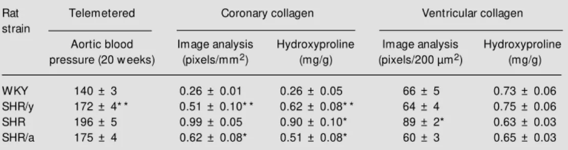
Y chromosome (SHR/y) increased collagen and removal of the SHR Y chromosome (SHR/a) decreased coronary collagen. The correlation between image analysis and bio-chemical analysis showed that both tech-niques were measuring the same thing (r = 0.88, P<0.001; Figure 2). Table 1 shows the results of telemetered blood pressure and coronary and ventricular collagen in four strains of rats with different Y chromosomes. The SHR Y chromosome placed in the WKY (SHR/y) doubled the coronary collagen meas-ured by either of the two techniques (image analysis and hydroxyproline content) com-pared to WKY. Removal of the SHR Y chromosome (SHR/a) reduced coronary col-lagen compared to SHR. Addition or re-moval of the Y chromosome did not change ventricular collagen, although SHR had more collagen than WKY (Seachrist D, Dunphy G, Daneshvar H, Caplea A, Milsted A and Ely D, unpublished observations).
Te sto ste ro ne and the re ninangio -te nsin sys-te m
The renin-angiotensin system (RAS) is an important participant in the development and maintenance of hypertension and is acti-vated in hypertensive rats. Sexual dimor-phism in the RAS is well known (34). An-drogens are known to potentiate the devel-opment of hypertension. Tissue specific regu-lation of renin mRNA by androgens has been shown in both adrenals and brain of
Im a g e a n a ly s is ( p ix e ls /2 0 0 µ m 2) 110 100 90 80 70 60 50 40
0.5 0.6 0.7 0.8 0.9 1.0
Hydroxyproline (mg/g) 0.4
Figure 2 - Pearson’s correlation of coronary artery collagen im-age analysis (pixels/200 µm2)
versus coronary artery hydroxy-proline content (mg/g) (r = 0.88, P<0.001).
Figure 1 - A, Coronary adventitial collagen measurement by com-puter histological image analysis (pixels/mm2)for four rat strains
(means ± SEM , * P<0.05 com-pared to WKY group). B, Bio-chemical analysis of coronary ar-tery hydroxyproline (mg/g coro-nary) by rat strain (m eans ± SEM , * P< 0.05 com pared t o
WKY strain). His
to lo g ic a l im a g e a n a ly s is (S ir iu s r e d ) (p ix e ls /m m 2) 1.25 1234 1234 1234 1234 1234 1234 1234 1234 1234 1234 1234 1234 1234 1234 1234 1234 1234 1234 1234 1.00 0.75 0.50 0.25 0.00
WKY SHR/y SHR/a SHR
Rat strain H y d ro x y p ro lin e (m g /g c o ro n a ry ) 1.25 1.00 0.75 0.50 0.25 0.00
WKY SHR/y SHR/a SHR
Rat strain
*
*
A
B
Table 1 - Blood pressure and coronary and ventricular collagen by rat strain (males, 20 w eeks).
* P<0.05, * * P<0.01 SHR/y compared to WKY and SHR/a compared to SHR.
Rat Telemetered Coronary collagen Ventricular collagen
strain
Aortic blood Image analysis Hydroxyproline Image analysis Hydroxyproline
pressure (20 w eeks) (pixels/mm2) (mg/g) (pixels/200 µm2) (mg/g)
WKY 140 ± 3 0.26 ± 0.01 0.26 ± 0.05 66 ± 5 0.73 ± 0.06
SHR/y 172 ± 4* * 0.51 ± 0.10* * 0.62 ± 0.08* * 64 ± 4 0.75 ± 0.06
SHR 196 ± 5 0.99 ± 0.05 0.90 ± 0.10* 89 ± 2* 0.63 ± 0.03
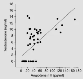
mice (35). Chen et al. (36) showed that renal and hepatic angiotensinogen mRNA levels in SHR are dependent on androgen in both sexes. Plasma renin activity was significant-ly higher in gonadalsignificant-ly intact male SHR than in females, and testosterone treatment in-creased plasma renin activity. Also, in some studies, genotypes of angiotensinogen codon 179 were significantly associated with varia-tion in systolic blood pressure in men (37). We have measured plasma testosterone and angiotensin II (Ang II) simultaneously in male SHR and found a significant correla-tion at 15 weeks of age (r = 0.57, P<0.001; Figure 3). It appears that testosterone en-hances circulating Ang II. Brilla et al. (38) showed that Ang II can increase fibroblast collagen turnover.
Both the renin and angiotensinogen genes are responsive to steroid hormones, includ-ing androgens, and glucocorticoids. Tissue-and age-related differences in angiotensino-gen mRNA levels have been reported in SHR and WKY (39). Both genes can be regulated at transcriptional and post-tran-scriptional (mRNA stability) levels. The re-nin gene is on chromosome 13 and the an-giotensinogen gene is on chromosome 19 in the rat. Yet these genes on different chromo-somes, both originating from WKY rats, are not expressed at the same level in our WKY and SHR/y females. Differences in renin gene structure in various rat strains have been described. In the human angiotensino-gen angiotensino-gene, several molecular variant alleles are known; in some populations the M235T polymorphism is associated with hyperten-sion (40). We have not yet analyzed renin or angiotensinogen gene structure in our rat strains. Our hypothesis was that changes in steroid hormones occurring during the first 15 weeks of life would produce changes in renal renin and angiotensinogen mRNA that would reflect differences due to the genetic origin of each rat strain. Blood pressure in WKY was lower than in strains with SHR
autosomes (P<0.003 vs SHR/a), SHR Y
chro-mosome (P<0.03 vs SHR/y), or both SHR
autosomes and SHR Y chromosome (P<0.03
vs SHR). Angiotensinogen mRNA was not
detected until 5 weeks of age, and no strain differences were seen at that age. Angioten-sinogen mRNA levels increased by 2-4-fold between 5 and 15 weeks of age. Renin mRNA levels were highest in all strains at 1 week and decreased by as much as 20-fold with age. At one week of age the strains contain-ing SHR autosomes (SHR and SHR/a) had higher renin mRNA levels than WKY and SHR/y, and overall, levels of renin mRNA appeared to decrease at slightly earlier ages in SHR and SHR/a than in WKY and SHR/y (41).
In summary, our results fail to reveal consistent patterns of RAS differences that reflect the presence or the absence of the SHR Y chromosome. In male rats we find no evidence of either coordinate regulation of renin and angiotensinogen gene expression
or of imprinting. In both WKY and SHR/y
male rats, the X chromosome is always con-tributed by their WKY mothers; in SHR and SHR/a males, the X chromosome always comes from their SHR mothers.
We have also investigated the effects of estrogen removal (ovariectomy, OVX) and androgen addition (testosterone implants) in 3 groups of female SHR/y rats and the paren-tal rat strain, WKY: group 1) intact (control), 2) ovariectomy at 3 weeks old, and 3)
ova-T
e
s
to
s
te
ro
n
e
(
n
g
/m
l)
12
10
8
6
4
2
0
80 100 120 140 160 180 Angiotensin II (pg/ml)
60 18
16
14
40 20 0
riectomy with testosterone implant at 3 weeks old. SHR/y females have the parental WKY autosomes and X chromosomes and have no chromosomes of SHR origin; thus they are genetically equivalent to female WKY rats. One-way ANOVA showed significant blood pressure differences between WKY and SHR/ y at 10 weeks and at 16 weeks (P<0.0001). Treatment had a significant effect on blood pressure (P<0.0001). Blood pressure was highest in the 16-week SHR/y OVX + T
group, reaching 204 ± 9 mmHg (P<0.05 vs
all other groups). Plasma renin activity was significantly higher in WKY than in SHR/y (P<0.01, two-way ANOVA).
Levels of renin mRNA and angiotensino-gen mRNA in the kidney followed similar patterns in each strain. There were signifi-cant strain differences in renin mRNA levels (P<0.02) and age differences in angiotensin-ogen mRNA levels (P<0.003). Correlation analysis indicated that renal angiotensino-gen mRNA was positively correlated with renal renin mRNA in all groups of both strains (P<0.001, r = 0.7515). The combina-tion of removing estrogen early in develop-ment and suppledevelop-menting the ovariectomized females with testosterone revealed strain dif-ferences in blood pressure response.
Renin and angiotensinogen mRNA lev-els appear to be regulated coordinately within each strain, although actual mRNA levels differ between strains. Strain differences in regulation of RAS genes may result from epigenetic mechanisms such as genome im-printing of these genes or of another gene that functions as a common regulator of renin and angiotensinogen (42).
Strain differences to OVX and response to OVX + T were seen. Age-dependent ef-fects on blood pressure were also found. OVX increased blood pressure in both strains in 10-week-old females but not in old animals. The addition of T to 16-week-old SHR/y females resulted in a very large increase in blood pressure, to 204 ± 9 mmHg. In 13-week-old SHR females, Reckelhoff et
al. (43) have reported no differences in blood pressure between OVX and intact females, untreated or treated with enalapril. The in-teractions of steroid hormones with the RAS are complex and appear to be influenced by age and genetic background.
It might be that the rat liver, like the mouse liver, is androgenized at an early stage and the hepatic angiotensinogen system might play a key role in the cascade of events involving testosterone and its effects on hy-pertension. Sex differences in the develop-mental pattern of blood pressure may also be related to organizational effects of perinatal sex steroids on the immature CNS. We have not explored this possibility. However, since female SHR also develop hypertension, it is possible that the rise in testosterone level may be responsible only for an earlier onset of hypertension in males. Several actions of testosterone have not been explained by known metabolites or receptors. Therefore, the idea has emerged that for some testoster-one actions the androgen receptor may not be necessary. Also we have reported that in males lacking a functional androgen recep-tor there appear to be direct cell membrane testosterone-mediated effects which influ-ence calcium flux (14). Thus, it is evident that a cell can recognize and respond to testosterone by a variety of independent mechanisms some of which remain to be defined. The androgen receptor is gaining a wider role and there is evidence that it is an important mediator of gene expression and signal transduction pathways (44).
Te sto ste ro ne -re nal link
chromosome backgrounds. The study in-volved male SHR, WKY and two consomic strains with different Y chromosomes (N = 5-8/group). Adult animals were castrated and implanted at the base of the neck with Silastic tubing (Dow Corning, Midland, MI, USA) containing testosterone propionate (14). Blood T levels were measured by RIA two weeks after castration. The left kidney was isolated and perfused with oxygenated Krebs solution at a constant flow and temperature with electrical stimulation of the renal nerves. Perfusate was collected and analyzed for NE by HPLC. Renal perfusate and renal tissue NE levels were significantly elevated by T. The average T value with a single T implant was 13 ng/ml, and for a double T implant 30 ng/ml. The Y chromosome from the SHR produced a significant increase in renal NE release compared to the WKY Y chromo-some. Significance was shown between all
groups: 1 vs 2 implants (P = 0.0067), 1 vs
sham implants (P = 0.015), and 2 vs sham
implants (P<0.001). In conclusion, T caused an enhanced renal NE release that was strain specific, with the Y chromosome raising renal NE content and release.
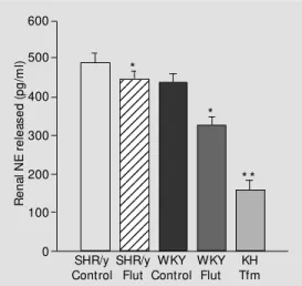
Follow-up research using the isolated kid-ney with an androgen receptor (AR)-defi-cient model (testicular feminized male, Tfm) or flutamide treatment has shown that the lack of a functional AR resulted in a signifi-cant reduction in renal NE release (Figure 4). Flutamide reduced renal NE release in both SHR/y and WKY. Also in the Tfm there was a large renal NE reduction. Three hybrid strains of adult male rats were used and within each strain normal AR kidneys were compared to deficient AR kidneys: SHR x KH (N = 21), KH x KH (N = 11), SHR/y x KH (N = 15). Kidneys were isolated and perfused with oxygenated Krebs-Henseleit solution and pressure and flow measured as well as NE in the effluent by HPLC under nonstimulated and electrically stimulated conditions. In each strain the lack of a func-tional AR resulted in a significant reduction
in the release of renal NE: SHR = 23% (440 to 340 pg/ml), KH = 38% (265 to 165 pg/ml), and SHR/y = 42% (180 to 105 pg/ml), P = 0.0017. LDH values were low, suggesting minimal cell damage after perfusion. In con-clusion, the AR increased renal NE release and AR blockade or mutation reduced renal NE release in the isolated kidney in 3 differ-ent strains of rats (47).
Inte ractio n o f te sto ste ro ne with the sympathe tic ne rvo us syste m
Testosterone interacts with several hor-monal systems and requires interaction with them for a full androgen response. For in-stance, there is evidence that testosterone influences NE metabolism, storage and re-lease (48). Testosterone has been reported to
increase a-adrenergic receptors in rat tail
arteries, whereas gonadectomy attenuated the total apparent number of binding sites in SHR (49). Philippe et al. (50) have shown a
more than two-fold increase in the a1
-adre-nergic receptors in response to both testos-terone and dihydrotestostestos-terone. The early rise in testosterone in SHR could induce greater sensitivity to NE, which in turn could produce higher blood pressure at an early age. There may also be an early developmen-tal testosterone interaction with tissue NE that cannot be detected by plasma
measure-R
e
n
a
l
N
E
r
e
le
a
s
e
d
(
p
g
/m
l)
600
12345 12345 12345 12345 12345 12345 12345 12345 12345 12345 12345 12345 12345 12345 12345 12345 12345 12345 12345 12345
Figure 4 - Y Chromosome and androgen receptor influence on renal norepinephrine (NE) re-lease (pg/ml) from isolated kid-ney by treatment group. Data are reported as means ± SEM . * P<0.05 and * * P<0.001 com-pared to SHR/y control. Flut, Flutamine; Tfm, testicular femi-nized male.
500
400
300
200
100
0
SHR/y Control
SHR/y Flut
WKY Control
WKY Flut
KH Tfm
*
*
ments. Indeed, Mayerhofer and Hodges (51) showed that NE regulates the number of luteinizing hormone receptors in the adult hamster testis and affects Leydig cell func-tion, especially during acute stress. The same group further showed (52) that in immature hamster testes catecholamines can act through
both aand ß adrenergic receptors and may
be potent stimulators of testosterone produc-tion. Isoproterenol treatment resulted in marked Leydig cell hypertrophy in young Sprague Dawley rats (53).
Androgens also have an effect on uptake and release of NE from sympathetic fibers (49). Testosterone also increases s-adenosyl-methionine, a major cellular methylator, and may influence the catecholamine pathway (54). The rate-limiting enzyme for the cate-cholamine pathway, tyrosine hydroxylase, and NE concentration in the abdominal aorta and mesenteric artery were decreased by castration and restored by testosterone in SHR but not in WKY (50). The mechanism for this effect could either be directly on the neurons or through tissue trophic factors.
Also in hypothalamic regions steroids modu-late tyrosine hydroxylase mRNA in dopami-nergic neurons (55) (Table 2).
Mo le cular pro bing fo r he at sho ck pro te in (Hsp) po lymo rphism
Preliminary data had shown a male spe-cific band in WKY from Southern blots
probed with rat Hsp70. This is consistent
with the rat Y chromosome containing an Hsp70-like locus and/or homologous se-quences. We made five sets of PCR primers that covered different conserved regions from rat Hsp70 DNA sequences in GenBank. These primers were used to amplify DNA from SHR and WKY males and females. Reactions were run at two annealing
tem-peratures (55 and 60o
C) and a variety of
MgCl2 concentrations. All primers had single
or multiple bands at 55o
C and three of the
five had bands at 60o
C. None of the bands observed were male specific and no amplifi-cations showed obvious amplification dif-ferences between males and females. With these five primer sets we were not able to
identify or isolate a Y chromosome Hsp70
-related sequence. These data are consistent with the hypothesis that the Southern results
were from Hsp70-related partial sequences,
rather than a full length potentially active Hsp70 homolog.
We contracted with Stratagene to con-struct a male genomic library in lambda DASHII (average insert 9-22 kb) from our SHR strain. From this library we have iso-lated clones for at least three rat Y
chromo-some loci (Ube 1y, Smcy, and Sry). We have
begun subcloning and sequencing the Sry
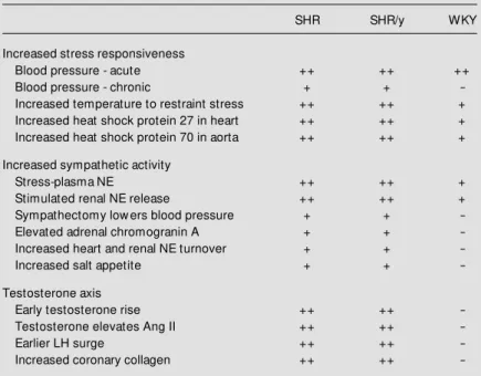
clones. Clone 96s15 contains regions of at least 75 bp homologous to brain and testis transcripts that are identical to regions in rat Hsp70-1 and Hsp70-3, as well as many other rat loci. It is possible that this type of DNA sequence homology could result in male spe-cific bands from Southern blotting to restric-tion enzyme-digested genomic DNA, with-Table 2 - Summary of evidence of Y-SNS-testosterone interaction.
The table summarizes the data show ing the involvement of the SHR Y chromosome w ith the SNS, renin-angiotensin and testosterone axes.
SHR SHR/y WKY
Increased stress responsiveness
Blood pressure - acute ++ ++ ++
Blood pressure - chronic + +
-Increased temperature to restraint stress ++ ++ +
Increased heat shock protein 27 in heart ++ ++ +
Increased heat shock protein 70 in aorta ++ ++ +
Increased sympathetic activity
Stress-plasma NE ++ ++ +
Stimulated renal NE release ++ ++ +
Sympathectomy low ers blood pressure + +
-Elevated adrenal chromogranin A + +
-Increased heart and renal NE turnover + +
-Increased salt appetite + +
-Testosterone axis
Early testosterone rise ++ ++
-Testosterone elevates Ang II ++ ++
-Earlier LH surge ++ ++
-out a complete Hsp70 sequence on the rat Y chromosome.
In conclusion, the data are consistent with the Y chromosome containing DNA sequences identical with portions of rat Hsp70, but we do not have evidence of a full
length potentially active copy of Hsp70 on
the rat Y chromosome.
The rat Y chro mo so me and Sry
The publication of the rat map has per-mitted a rapid exploration of candidate genes for hypertension on all chromosomes except the Y. The human and mouse Y chromo-somes are the best characterized mammalian Y chromosomes. Although there are some similarities, the genetic organization of these two chromosomes is very different. The knowledge we do have about the rat Y chro-mosome shows similarities to the human and mouse chromosome but there are some very basic differences. The mouse, human and rat
Y chromosomes all contain Sry and Zfy loci,
but the mouse has 2 copies of Zfy, the rat and
human one. The rat has multiple copies of
Sry and humans and mice only one. A third
locus identified in mouse, human and rat is
the steroid sulfatase locus (Sts). The Sts
struc-tural locus is Y linked and pseudoautosomal in the mouse, Y linked but not pseudoauto-somal in humans and X linked in the rat. We have identified a heat shock-related sequence on the rat Y chromosome and another labo-ratory has identified a mitochondrial D-loop-related sequence on the rat Y and neither of these sequences has been identified on either the human or mouse Y chromosome. Be-cause of the observed differences between these three Y chromosomes, each may serve as a model for the others but differences may
be more common than similarities.
We compared an Sry sequence from our
genomic library to other published rat Sry, to
study the relationships among multiple cop-ies of this gene. As many as four copcop-ies of
the Sry gene have been reported in rodents.
We obtained the 5-flanking sequence (5-FLK), complete coding sequence and 3-untranslated region (3-UTR), including a stop codon and a polyadenylation signal, from the Y chromosome clone, 96s15. The
three available rat Sry partial sequences in
GenBank were compared to 5-FLK and
cod-ing regions of the SHR gene, designated
Sry-1. Within the coding region, minimal
differ-ences (0.032) were found across strains and
species (sequences are from Rattus
norvegi-cus, R. exulans and Brown Norway rats). Greater sequence divergence (0.105) was present in the 5-FLK region sequences. No comparisons of 3-UTR sequences are
pos-sible at this time, since the other rat Sry
genes are partial sequences, not including
that region. With the SHR full length rat Sry
gene, we can now begin to evaluate the
functional significance of the other Sry gene
copies (56).
In conclusion, the SHR Y chromosome has a locus that raises blood pressure. Other autosomal loci, like genes controlling testos-terone biosynthetic enzyme production and the biosynthetic catecholamine enzymes, may interact with the Y loci for maximum blood pressure expression.
Ackno wle dgm e nts
Re fe re nce s
1. Foote S, Vollrath D, Hilton A & Page DC (1992). The human Y chromosome: over-lapping DNA clones spanning t he euchromic region. Science, 258: 60-66. 2. Vollrath D, Foote S, Hilton A, Brow n LG,
Beer-Romero P, Bogan JS & Page DC (1992). The human Y chromosome: a 43-interval map based on naturally occurring deletions. Science, 258: 52-59.
3. Turner M E, Ely D & M ilsted A (1998). The rat Y chromosome. Rat Genome, 4: 78-83.
4. Hilbert P, Lindpainter K, Beckmann JS, Serikaw a T, Soubrier F, Dubay C, Cartw right P, DeGouyon B, Julien C, Takahasi S, Vincent M , Ganten D, Georges M & Lathrop GM (1991). Chromosomal mapping of tw o genetic loci associated w ith blood pressure regulation in heredi-tary hypertensive rats. Nature, 353: 521-529.
5. Jacob HJ, Lindpainter K, Lincoln SE, Kasumi K, Bunker RK, M ao Y-P, Ganten D, Dzau V & Lander E (1991). Genetic mapping of a gene causing hypertension in the stroke-prone SHR. Cell, 67: 213-224.
6. Davidson AO, Schork N, Jaques BC, Kelman AW, Sutcliffe RG, Reid JL & Dominiczak AF (1995). Blood pressure in genetically hypertensive rats. Influence of the Y chromosome. Hypertension, 26: 452-459.
7. Carsw ell HVO, Anderson NH, Clark JS, Graham D, Jeffs B, Dom iniczak A & M acrae IM (1999). Genetic and gender influences on sensitivity to focal cerebral ischemia in the stroke-prone spontane-ous hypertensive rat. Hypertension, 33: 681-685.
8. Vincent M , Kaiser M A, Orea V, Lodw ick D & Samani N (1994). Hypertension in the SHR and the sex chromosomes. Hyper-tension,23: 161-166.
9. Turner M & Ely D (1995). Tw o Y chromo-somes in the SHR strain: polymorphism or interaction? FASEB Journal, 9: A50 (Ab-stract).
10. Uehara Y, Shin Ws, Watanabe T, Osanai T, M iyazaki M , Kanase H, Taguchi R, Sugano K & Toyo-Oka T (1998). A hyper-t ensive f ahyper-t her, buhyper-t nohyper-t hyperhyper-t ensive mother, determines blood pressure in nor-motensive male offspring through body mass index. Journal of Human Hyperten-sion, 12: 441-445.
11. Lemne C (1998). Increased blood pres-sure reactivity in children of borderline
hypertensive fathers. Journal of Hyper-tension,16: 1243-1248.
12. Darling RWR & Holt T (1999). Genetic models w ith reduced penetrance related to the Y chromosome. Biometrics,55: 55-64.
13. Ely D, Caplea A, Dunphy G, Daneshvar H, Turner M , M ilsted A & Takiyyuddin M (1997). Spontaneously hypertensive rat Y chromosome increases indices of sympa-thetic nervous system activity. Hyperten-sion, 29: 613-618.
14. Ely DL, Salisbury R, Hadi D, Turner M & Johnson M L (1991). Androgen receptor and the testes influence hypertension in a hybrid rat model. Hypertension, 17: 1104-1110.
15. Caplea A, Dunphy G, Seachrist D & Ely D (1999). The SHR Y chrom osom e in-creases the norepinephrine content and turnover rate in the kidney. FASEB Jour-nal,13: A724 (Abstract).
16. Ely D, Caplea A, Dunphy G, Turner M , Takiyyuddin M , Tremblay J & Hamet P (1996). Interaction of the Y chromosome, heat shock protein and sympathetic ner-vous system. In: M cCarty R, Aguilera G, Subban E & Kvet nansky R (Edit ors),
Stress: M olecular Genetic and Neurobio-logical Advances. Gordon & Breach Sci-ence Pub., New York, 281-297.
17. Lee RM KW, Triggle CR, Cheng DWT & Coughlin M D (1987). Structural and func-tional consequences of neonatal sympa-thectomy on the blood vessels of SHR.
Hypertension,10: 328-338.
18. Esler M , Lambert G & Jennings G (1990). Increased regional sympathetic nervous activity in human hypertension: causes and consequences. Journal of Hyperten-sion, 8 (Suppl VII): VII-553-VII-557. 19. W allin B, Kunem oto M & Sellgren J
(1993). Possible genetic influence on the strength of human muscle nerve sympa-thetic activity at rest. Hypertension, 22: 282-284.
20. Wyss JM (1993). The contribution of the sympathetic nervous system to hyperten-sion. Current Opinion in Nephrology and Hypertension, 2: 265-273.
21. Chen CW, Chen YF, M eng QC, Wyss JM & Oparil S (1991). Decreased norepineph-rine released in anterior hypothalamus of NaCl-sensitive SHR during high NaCl in-take. Brain Research,565: 135-141. 22. Lundin S & Thoren P (1982). Renal
func-tion and sympathetic activity during men-tal stress in spontaneously hypertensive
rats. Clinical Science, 63: 327-330. 23. Ely D & Weigand J (1983). Stress and high
sodium effects on blood pressure and brain catecholamines in spontaneously hypertensive rats. Clinical and Experimen-tal Hypertension,A5: 1559-1567. 24. Ely DL (1995). Organization of
cardiovas-cular and neurohumoral responses to stress implications for health and disease. In: Chrousos G, M cCarty R, Pacak K, Cizza G, Sternberg E, Gold P & Kvetnansky R (Editors), Stress. Annals of the New York Academy of Sciences, 771: 594-608. 25. Caplea A, Dunphy G, Seachrist D & Ely D
(1999). The SHR Y chrom osom e en-hances the nocturnal blood pressure rise in socially interacting rats. FASEB Jour-nal,13: A739 (Abstract).
26. Seachrist D, Caplea A, Dunphy G & Ely D (1999). Testosterone potentiates sodium sensitivity and the SHR Y chromosome hypertensive effect. FASEB Journal,13: A509 (Abstract).
27. Ely D, Herman M , Ely L, Barrett L & M ilsted A (1999). Sodium intake is in-creased by social stress and the Y chro-mosome and reduced by clonidine. Ameri-can Journal of Physiology.Regulatory, In-tegrative and Comparative Physiology (in press).
28. Rodriguez-Padilla M , Bellido C, Pinilla L & Aguilar E (1987). Secretion of LH in spon-taneously hypertensive rats. Journal of Endocrinology, 113: 255-260.
29. Ely D, Falvo J, Dunphy G, Caplea A, Salisbury R & Turner M (1994). The spon-taneously hypertensive rat Y chromo-some produces an early testosterone rise in normotensive rats. Journal of Hyper-tension,12: 769-774.
30. Ely D, Daneshvar H, Turner M , Johnson M L & Salisbury RL (1993). The hyperten-sive Y chromosome elevates blood pres-sure in F11 normotensive rats.
Hyperten-sion,21: 1071-1075.
31. Wolinsky H (1973). Comparative effects of castration and antiandrogen treatment on the aortas of hypertensive and normo-tensive male rats. Circulation Research, 33: 183-189.
32. Fischer GM & Sw ain M L (1977). Effect of sex hormones on blood pressure and vas-cular connective tissue in castrated and noncastrated male rats. American Journal of Physiology,232: H617-H621. 33. Ely D, Chonko D, Rittgers D, Smith D &
coro-nary arteries of normotensive rats w ith a hypert ensive Y chrom osom e. FASEB Journal, 8: A9 (Abstract).
34. Bachmann J, Feldmer M , Ganten U, Stock G & Ganten D (1991). Sexual dimorphism of blood pressure: possible role of the renin-angiotensin system. Journal of Ste-roid Biochemistry andM olecular Biology, 40: 511-515.
35. Wagner D, M etzger R, Paul M , Ludw ig G, Suzuki F, Takahashi S, M urakami K & Ganten D (1990). Androgen dependence and tissue specificity of renin messenger RNA expression in mice. Journal of Hy-pertension,8: 45-52.
36. Chen Y, Naftilan AJ & Oparil S (1992). Androgen-dependent angiotensinogen and renin messenger RNA expression in hypertensive rats. Hypertension, 19: 456-463.
37. Hegele RA, Brent JH & Connelly PW (1994). A polymorphism of the angioten-sinogen gene associated w ith variation in blood pressure in a genetic isolate. Circu-lation,90: 2207-2212.
38. Brilla CG, Zhou G, Rupp H, M aisch B & Weber KT (1995). Role of angiotensin II and prostaglandin E2 in regulating cardiac fibroblast collagen turnover. American Journal of Cardiology, 76: 8D-13D. 39. Lodw ick D, Kaiser M A, Harris J, Cumin F,
Vincent M & Samani NJ (1995). Analysis of the role of angiotensinogen in sponta-neous hypertension. Hypertension, 25: 1245-1251.
40. Jeunemaitre X, Soubrier F, Kotelevtsev YV, Lifton RP, Williams CS & Charru A (1992). M olecular basis of human hyper-tension: role of angiotensinogen. Cell, 71: 169-180.
41. Lee TK, Neves LAA, Turner M E, Ely DL & M ilsted A (1996). Developmental
differ-ences in renal renin gene expression in hypertensive and nonhypertensive rats.
Ohio Journal of Science, 96: A-19-A-20 (Abstract).
42. M ilsted A, M arcelo M C, Turner M E & Ely DL (1998). Female WKY and SHR/y rats have the same genotype but different pat-terns of expression of renin and angioten-sinogen. Journal of Hypertension, 16: 823-828.
43. Reckelhoff JF, Zhang H, Srivasta K & Hooker KD (1999). Gender differences in the development of hypertension in SHR: Role of the renin-angiotensin system. Hy-pertension, 34: 337 (Abstract).
44. Cato ACB & Peterziel H (1998). The an-drogen receptor as mediator of gene ex-pression and signal transduction path-w ays. Trends in Endocrinology and M e-tabolism, 9: 150-154.
45. Gong G, Dobin A, M cArdle S, Sun L, Johnson M L & Pettinger WA (1994). Sex influence on renal 2-adrenergic receptor density in the spontaneously hyperten-sive rat. Hypertension, 23: 607-612. 46. Jones TJ, Dunphy G, M ilsted A & Ely D
(1998). Testosterone effects on renal nor-epinephrine content and release in rats w ith different Y chromosomes. Hyperten-sion, 32: 880-885.
47. Ely DL, Ebbs A, Dunphy G & M ilsted A (1999). Renal norepinephrine release is enhanced by t he androgen recept or.
FASEB Journal, 13: A509 (Abstract). 48. Lara H, Galleguillos X, Arrau J & Belman J
(1985). Effect of castration and testoster-one norepinephrine storage on the re-lease of [3H]-norepinephrine from rat vas deferens. Neurochemical International, 7: 667-674.
49. Salt PJ (1972). Inhibition of noradrenaline uptake-2. In the isolated rat heart by
ste-roids, clonidine and methoxylated phenyl-ethylamines. European Journal of Phar-macology,20: 329-340.
50. Philippe M , Saunders T & Bangalore S (1991). A mechanism for testosterone modulation of alpha-1 adrenergic receptor expression in the DDT1, M F-2 smooth muscle myocyte. M olecular and Cellular Biochemistry, 100: 79-90.
51. M ayerhofer A & Hodges S (1988). Cat-echolam ines augm ent t est icular re-sponses to gonadotropin stimulation by direct action at the gonadal level. Biologi-cal Reproduction, 38 (Suppl I): I.85 (Ab-stract).
52. M ayerhofer A, Steger RW, Gow G & Bartke A (1992). Catecholamines stimu-late testicular testosterone release of the immature golden hamster via interaction w ith alpha- and beta-adrenergic receptors.
Acta Endocrinologica, 127: 526-530. 53. Bergh A, Blom H, Damber JE &
Henriks-son R (1987). The effect of long-term variation in sympathetic activity on tes-t icular m orphology in im m ates-t ure rates-t s.
Andrologia, 19: 448-451.
54. M ant euf f el-Cym borow ska M , Chm ur-zynska W & Grzelakow ska-Sztabert B (1992). Tissue specific effects of testos-terone on s-adenosylmethionine forma-tion and utilizaforma-tion in the mouse. Biochimi-ca et BiophysiBiochimi-ca Acta, 1116: 166-172. 55. Kohama SG & Bethea CL (1995). Steroid
regulation of tyrosine hydroxylase mes-senger ribonucleic acid in dopaminergic subpopulations of monkey hypothalamus.
Endocrinology, 136: 1790-1800. 56. M ilsted A, Codispoti CD, Ely DL, Turner
M E & M artins AS (1999). Comparison of