Vol-7, Special Issue3-April, 2016, pp1960-1967 http://www.bipublication.com Research Article
Mammogram contrast enhancement in wavelet
domain using fuzzy denoising
Hadi Amirpour1,*, Bita Tarfiee2
And Seyyed Mohammad Shams3
1Department of Electrical Engineering, K. N. Toosi University of Technology, Tehran, Iran.
2 Department of Biomedical Engineering, K. N. Toosi University of Technology, Tehran, Iran. 3Department of Biomedical Engineering, Faculty of Engineering,
Islamic Azad University, South Thran Branch, Tehran, Iran. Correspondence author: Email:[email protected]
ABSTRACT
Breast cancer is one of the leading death causes among many women around the world. Diagnosing breast cancer in early stage before it gets the chance to spread helps patients treatment.Mammography is an effective screening test that helps to early detection and diagnosis of breast cancer in women. In many cases, due to the low-energy X-ray beams usedin mammography and microcalcifications position, mammograms are low contrast and noisy images and it is difficult for radiologiststo distinguish between normal and neoplastic breast tissues.Therefore, image enhancement algorithms have been proposed to improve mammograms quality for better detection. The main focus of this paper is to propose a procedure for enhancingthe contrast of mammogram images by using a fuzzy based wavelet transform. At the first step of the procedure, a multiscale wavelet transform is obtained in four levels. In the second step,the fuzzy logic approach is applied to the fourth level for denoising and improving image quality. In the final step, wavelet coefficients in all scales are manipulated by an enhancing factor, to improve the image contrast. The performance of the proposed procedure was investigated according to various factors by simulation data.
Keywords—Mammograms; Wavelet transform; fuzzy logic; Contrast enhancement 1. INTRODUCTION
Breast cancer is the most commonly diagnosed and second leading cause of cancer death among women known as the first cancer’s type ranked in the list of National Cancer Institute in 2016 in the United State with more than 249,000 new case expected [1]. Advances in diagnostic procedures which leads to early detection of cancer, contribute to the ongoing decline in breast cancer mortality among women. Annual Report to the Nation on the Status of Cancer shows that among women between 10 years (2003-2012) breast cancer death rate decreased 1.9% per year. More
trials have demonstrated a mortality benefit for women from 40 to 74 years old. Some studies have shown that mammography may be particularly
beneficial for
women who are 80 years of age and older. Age distribution of breast cancer is shown in Fig1 [2].
Fig 1: Age distribution of breast cancer
In mammogram, radiologist can find Malignant tumour or breast tissue abnormalities before symptoms appeared so Screening mammography accounts for the greatest contribution to early detection and decrease in breast cancer mortality, although its use has resulted in a minor increase in the number of in situ cancers detected.Breast cancer death rate evaluation which is done by American Cancer society in the United States shows that from 1990, the year of widespread screening begin, increasing breath cancer
deathwas stop and also decreased in next 20 years by the rate of 34% [3].In screening mammography applying low dose of x-ray to prevent possible breast tissue damage is really serious point. Thus low dose of X-ray resulted low contrast image that is difficult to diagnose abnormalities for radiologist. Accordingly, nowadays, Computer aided program to enhance mammogram’s contrast becomes One big issue. An example of a
mammography image is shown in Fig2.
Fig 2: An example of mammography image
Some works have been done for enhancing the contrast of mammogram images. Generally, contrast enhancement methods classified in two main groups: indirect contrast enhancement and direct contrast enhancement.Indirect contrast enhancement algorithms work on the modification of image histogram. The intensities or color
(CLAHE) [5], Brightness Preserving Bi-Histogram Equalization (BBHE) [6], Minimum Mean Brightness Error Bi-Histogram Equalization (MMBEBHE) [7] and Recursive Mean Separate Histogram Equalization (RMSHE) [8].
In direct algorithm, contrast of the image enhanced directly such as (DE) [9] and HVS [10] methods.
In this paper, mammogram imagesare
decomposed into multiscale representation by wavelet transform and wavelet coefficients are denoised unshapely by applying fuzzy.
Section II introduces multiscale representation of images in wavelet domain and explains direct
enhancement (DE) method of contrast
enhancement. Section III explains how fuzzy helps to improve performance of DE by denoising.
Section IV shows experimental results and Section V concludes paper.
2. MULTISCALE REPRESENTATION OF IMAGES
Mammograms have distinguishing types of features in different scales [11]. For instance, calcifications that have small scale have subtle features while coarse scales such as masses are larger objects with smooth borders [12]. In order to consider different scales of an mammogram, using multiscale representation of mammograms will be helpful. Each scale contains different features and to improve feautures in all scales a mammogram is divided into different scales. For the original image A, k-Level wavelet transform is :
1 ,
( , ) ( ) ( ) (2 ,2 )
N N
m n Z
A i j h m h n A i m j N
1S1
1 ,
( , ) ( ) ( ) (2 ,2 )
N N
m n Z
D i j h m g n A i m j N
2S2
1 ,
( , ) ( ) ( ) (2 ,2 )
N N
m n Z
D i j g m h n A i m j N
3S3
1 ,
( , ) ( ) ( ) (2 ,2 )
N N
m n Z
D i j g m g n A i m j N
4SThe original image A decomposed into four subband images. Therefore, one approximation subband and three detail subbands are obtained as a result of wavelet transform. In this
transformation,
hn ,
gn are analysis filters,N
A that is very close to the input image is
low-frequency subband of AN1. The subband that contains vertical high-frequency and horizontal
low-frequency of AN1 is called D1N. Vertical
low-frequency and horizontal high-frequency of
1
N
A and also vertical high-frequency and
horizontal high-frequency of AN1 are
decomposed in 2 N
D and 3 N
D , respectively [9].
When the desired reforms are applied to the subbands enhanced image can be reconstructed by
using following formula considering
hn and
gn
as synthesis filters:
1 , 1 , 2 , 3 ,
( , ) 4 ( ) ( ) ( , )
2 2 ( ) ( ) ( , ) 2 2 ( ) ( ) ( , ) 2 2 ( ) ( ) ( , ) 2 2
[
]
N N NN m n Z N
m n Z
m n Z
m n Z
i m j n
A i j h m h n A
i m j n h m g n D
i m j n g m h n D
i m j n g m g n D
So, original image can be represented by:
0( , ) ( , ), ( , )
k
N l
A k j A i j D i j 6S
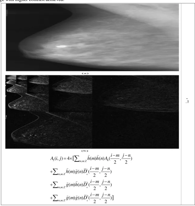
Fig 3 represents an example of original image and its multiscale decomposition.
In order to enhance contrast of an image, all coefficients of detail subbands in fourth level
1 2 3
4( , ), 4( , ), 4( , )
D i j D i j D i j are manipulated in a constant factor λ:
1 1
4( , ) 4( , )
D i j D i j 7S
2 2
4( , ) 4( , )
D i j D i j 8S
3 3
4( , ) 4( , )
D i j D i j 9S
By applying recostructed formula (5) to these new subband detail and repeated for other level the new image with higher contrast achieved.
Fig 3: An example of multiscale decomposition. a) Original image b) multiscale decomposition [9]. 4
4
4
3 , 4
1 ,
2 ,
3 ,
( , ) 4 ( ) ( ) ( , )
2 2
( ) ( ) ( , )
2 2
( ) ( ) ( , )
2 2
( ) ( ) ( , )
2 2
[
]
m n Z
m n Z
m n Z
m n Z
i m j n
A i j h m h n A
i m j n h m g n D
i m j n g m h n D
i m j n g m g n D
3. FUZZY DENOISING
Manipulating detail sub bands
D i jl1( , )
,
2
( , ) l
D i j ,
D i jl3( , )
, in a factor (λ) canenhances contrast of an image and helps radiologist to distinguish normal and abnormal breasts as a result have a accurate decision on a mammograms. On the other hand, in addition to
image information, noises are manipulated in λ,
too and denoising can help to improve quality of an image. First strategy is defining a threshold for coefficients of detail subbands and determining higher coefficients as a noise and eliminating
them before manipulating in λ. However, in order
to have an efficient denoising a fuzzy based denoising is proposed in this paper.Instead of determining high confidents as noise and setting them to zero, two if-then rules in fuzzy domain have been used in this paper. These if-then rules are as follow:
if input is small, then output is large. if input is large, then output is small.
A ‘zmf’ membership function (MF) is used for small MF in input and large output and a ‘smf’ MF is used for large input and small output. MF of input and output are shown in Fig 4.
After implementing fuzzy system, detail subband coefficients are considered as input and proportion to applied coefficient an output is
obtained. Then, λ is manipulated in coefficients
and contrast enhanced image is reconstructed. Due to large number of coefficients especially in lower subbands, in order to reduce computational load, fuzzy denoising is applied only to fourth level detail subband coefficients. Also, parameters of MFs will be determined dynamically on based on maximum and minimum of each detail subband.
a
b
Fig 4: MF of a) input b) output.
4. EXPERIMENTAL RESULTS
dense-glandular group, fatty group, and fatty-dense-glandular group which consist of three types of mammograms as normal, benign, and malignant. In order to measure performance of proposed algorithm, two parameters are considered. First, the region contrast of an image I, Cwthat is defined as:
1
( ) ( , ) log ( , ) w
w
C I c x y c x y
m
100For local contrast at pixel (x,y), C(x,y) defined as:
( , ) 4 ( , ) ( 1, ) ( , 1) ( 1, ) ( , 1)
C x y I x y I x y
I x y I x y I x y
11
where I(x,y) is an image pixel intensity value for the pixel location (x,y), w is a region of image I(x,y), and the parameter m is the number of the pixels in the region over which the contrast is evaluated. In this paper, entire image is considered as the selected region.On the other, although increasing λ enhance contrast of an image, but visual quality of image is become worse because of enhancing noise and background variations. Applying fuzzy will help to enhance visual quality that can be measured by PSNR:
255 10 log( ) PSNR
MSE
122
1 1
2
0 0
1
( , ) ( , ) *
m n
o r
i j
MSE f i j f i j
m n
133Where fo is original image,fr is reconstructed image and m, n are size of image.
Table 1 shows contrast measurement for original image, DE algorithm [9] and proposed algorithm. It’s clear that introduced contrast enhancement method has been increased contrast significantly. Table (2) shows PSNR values and Fig 5 shows an example of proposed algorithm.
Table1:Region contrast Cw
Lambda=2 Lambda=3 No. Original DE Proposed DE Proposed mdb001 0.0068 0.0137 0.0137 0.0206 0.0206 mdb002 0.0100 0.0200 0.0201 0.0301 0.0302 mdb003 0.0090 0.0181 0.0182 0.0272 0.0273 mdb004 0.0091 0.0183 0.0185 0.0275 0.0277 mdb005 0.0110 0.0221 0.0222 0.0332 0.0333 mdb006 0.0113 0.0226 0.0227 0.0340 0.0341 mdb007 0.0093 0.0188 0.0189 0.0282 0.0283 mdb008 0.0031 0.0063 0.0063 0.0094 0.0095 mdb009 0.0119 0.0240 0.0242 0.0361 0.0364 mdb010 0.0075 0.0151 0.0151 0.0226 0.0227
Table 2: PSNR
mdb007 28.8321 31.1851 22.8115 24.8718 mdb008 30.5408 32.8157 24.5202 26.5653 mdb009 24.8720 27.5407 18.8514 21.2904 mdb010 31.7742 34.3142 25.7536 27.9883
Fig 5: Original image and enhanced mammograms with lambda 2 and 3.
5. CONCLUSION
Image enhancement can help radiologists to diagnose the normal and abnormal mammograms better. In this paper, a multiscale contrast enhancement algorithm in wavelet has been proposed. An image is decomposed to 4-level wavelet subbands and in order to enhance contrast, coefficients of detail subbands are manipulated in enhance factor and then image is reconstructed. With increasing enhance factor, contrast enhances but visual quality decreases. Due to increase visual quality of image that reduces in contrast enhancement process, a fuzzy denoising method is proposed that eliminate noises in detail subbands and help to increase PSNR considerably. Experimental results show that proposed algorithm not only has good contrast, but also has acceptable PSNR.
6. REFERENCES
1. American Cancer Society: Cancer Facts and Figures 2016. Atlanta, Ga: American Cancer Society, 2016. Last accessed February 1, 2016.
2. Brenda K., Holly L. Howe, Lynn AG Ries, Michael J. Thun, Harry M. Rosenberg, Rosemary Yancik, Phyllis A. Wingo, Ahmedin Jemal, and Ellen G. Feigal. Edwards, (2002), "Annual report to the nation on the status of cancer, 1973–1999, featuring implications of age and aging on US cancer burden.," Cancer 94, no. 10.
3. Breast Cancer Facts & Figures 2014-2015," American Cancer Society.
4. M. H. Kabir, M. A. Akber Dewan and O. Chae, M. Abdullah-Al-Wadud, (May 2007), "A Dynamic Histogram Equalization for Image Contrast Enhancement," IEEE Transactions on Consumer Electronics, vol. 53, no. 2, pp. 593-600.
5. Shelda, and M. Ravishankar Mohan,(2013), "Modified contrast limited adaptive histogram equalization based on local contrast enhancement for mammogram images,"
Mobile Communication and Power
6. Sheng Hoong, Nor Ashidi Mat Isa, Chen Hee Ooi, and Kenny KalVinToh Lim, (2015), "A new histogram equalization method for digital
image enhancement and brightness
preservation," Signal, Image and Video Processing, pp. 675-689.
7. Soong-Der, and AbdRahmanRamli Chen,
(2003), "Minimum mean brightness error
bi-histogram equalization in contrast
enhancement," Consumer Electronics, IEEE Transactions on, vol. 49, no. 4, pp. 1310-1319.
8. Soong-Der, and AbdRahmanRamli
Chen, (2003), "Chen, S. D., & Ramli, A. R. (2003). Contrast enhancement using recursive mean-separate histogram equalization for scalable brightness preservation," Consumer Electronics, IEEE Transactions, vol. 49(4), pp. 1301-1309.
9. Jinshan, Xiaoming Liu, and Qingling Sun Tang, (2009), "A direct image contrast enhancement algorithm in the wavelet domain for screening mammograms," Selected Topics in Signal Processing, IEEE Journal of, vol. 3, no. 1, pp. 74-80.
10.Yicong, Karen Panetta, and SosAgaian Zhou, (2010), "Human visual system based mammogram enhancement and analysis," in 2nd International Conference on Image Processing Theory Tools and Applications (IPTA), pp. 229-234.
11.Etta D., R. Edward Hendrick, Martin Yaffe, Emily F. Conant, and Constantine Gatsonis Pisano, (2007), "Should Breast Imaging Practices Convert to Digital Mammography? A Response from Members of the DMIST Executive Committee 1," Radiology, vol. 245, no. 1, pp. 12-13.
12.P., L. Costaridou, and G. Panayiotakis Sakellaropoulos, (2003), "A wavelet-based spatially adaptive method for mammographic contrast enhancement," Physics in Medicine and Biology, vol. 48, no. 6, p. 787.
13.The mini-Mammographic Image Analysis Society database ofmammograms (MIAS) webpage.[Online].

![Table 1 shows contrast measurement for original image, DE algorithm [9] and proposed algorithm](https://thumb-eu.123doks.com/thumbv2/123dok_br/18153589.327873/6.892.265.616.458.578/table-shows-contrast-measurement-original-algorithm-proposed-algorithm.webp)
