ÓRgÃO OfICIAL DA SOCIEDADE PORTUgUESA DE REUMATOLOgIA
273 CARTA AO EDITOR
To the Editor,
Livedoid Vasculopathy (LV) is a chronic recurrent thrombo-occlusive vasculopathy of unknown patho-genesis that has been associated with hypercoagulable states and several connective tissue diseases1. It parti -cularly affects middle-aged women and presents as painful purpuric papules, mainly distributed over the lower legs that subsequently ulcerate and slowly heal with residual white atrophic stellate scars2. Neurologi-cal involvement is rare and probably secondary to the ischemia of the vasa nervorum3. Treatment is often chal-lenging and usually based on case series. We present a refractory LV case associated with peripheral neuropa-thy, successfully treated with rituximab.
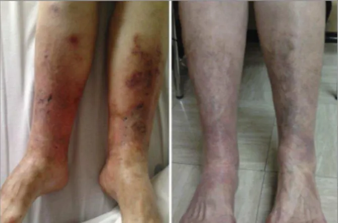
A 58-year-old woman presented with a 3-year his-tory of pain, paresthesiae and multiple leg ulcers. She had an irrelevant medical history and her physical exa -mination was normal except for the presence of multi-ple reticulate and ulcerated lesions in both the ante rior surface of her legs and around the ankles, with areas of intense red/violaceous macules, very painful to touch. Neurological examination revealed hypoesthe-sia in the dorsum of both feet.
Blood counts, hepatic and renal function, erythro-cyte sedimentation rate and C reactive protein were normal; homocysteine, protein C and S activity, acti-vated protein C resistance and antithrombin III showed normal levels; cryoglobulins, cryoagglutinins, lupus anticoagulant and anti-phospholipids antibodies were absent; G2021A prothrombin and C677T methylenete-trahydrofolate reductase mutations were negative. Antinuclear antibodies, anti-dsDNA, antineutrophil cy-toplasmatic antibodies, rheumatoid factor, circula ting immune complexes and anti-ENA (jo-1, RNP, SCL-70, Sm, SSa and SSb) were also negative. Immuno glo -bulins, C3c and C4c were within normal ranges and serum proteins electrophoresis had no alterations. HIV
and HCV serologies were negative. Ag HBs was nega-tive but AcHBc and AcHBs were posinega-tive (5.08 and 714 respectively UI/L), although HBV DNA was unde-tectable. Electromyography showed lower limbs sensi-tive polyneuropathy but skin and sural nerve biopsies were inconclusive.
For suspected vasculitis, a 3-month course of 1mg/ /Kg/day prednisolone was tried, unsuccessfully. A symptom control strategy based on gabapentin was im-plemented but 2 years later, recrudescent pain justified a new skin biopsy that revealed the presence of eosi no -philic, PAS positive material within dermal vessels with minimal inflammatory infiltrate and, on immunofluo-rescence, perivascular immunorreactivity to fibrinogen and dermal vessels deposits of IgM and C3c, consistent with LV. The patient was medicated with warfarin and immunoglobulins (IvIg) (2 gr/Kg over five days, every 4 weeks). With treatment maintenance, the patient ex-perienced improvement of pain and dysesthesiae, and cicatrization of leg ulcers. However, 8 years later, aggra -vation of pain and cutaneous ulcers was again noticed. Warfarin and IvIg were stopped and the patient was started on rituximab 1.0 g, 2 infusions, 14 days apart, associated with entecavir for HBV reactivation
pro-1. Rheumatology, Hospital Dr. Nélio Mendonça; Centro Hospitalar São João
2. Rheumatology, Centro Hospitalar São João
Livedoid vasculopathy – a challenging disease
ACTA REUMATOL PORT. 2016;41:273-274 Vieira R1, Bernardes JM2, Pinto JA2, Costa L2
ÓRgÃO OfICIAL DA SOCIEDADE PORTUgUESA DE REUMATOLOgIA
274
Livedoid vascuLopathy – a chaLLenging disease
phylaxis. Since then, the patient presented gradual improvement and currently, 1 year later, she has full cica -tri zation of her leg ulcers (Figure 1) and no need of analgesia, referring only mild dorsal feet hypoesthe-sia.
A previous case report4describes improvement of both pain and ulcers related to cutaneous LV after treat-ment with rituximab.
The case presented emphasizes that even after pro-longed involvement, peripheral neuropathy may im-prove with rituximab treatment. Despite the typical absence of neutrophil infiltration on the blood vessels, accumulating clinical experience suggests that B cells may be key players in LV pathogenesis.
coRREpondEncE to
Romana Vieira
Rua Padre José de Pinho, nº 91 4465-184, São Mamede de Infesta Portugal
E-mail: romi_rv@hotmail.com
REFEREncEs
1. Alavi A, Hafner J, Dutz JP, Mayer D, Sibbald RG, Criado PR, et al. Livedoid vasculopathy: an in-depth analysis using a modi-fied Delphi approach. J Am Acad Dermatol 2013; 69: 1033-1042.e1.
2. Criado PR, Rivitti EA, Sotto MN, de Carvalho JF. Livedoid vas-culopathy as a coagulation disorder. Autoimmun Rev 2011;10: 353-360.
3. Tubone MQ, Escobar GF, Peruzzo J, Schestatsky P, Maldonado G. Livedoid vasculopathy associated with peripheral neuro-pathy: a report of two cases. An Bras Dermatol 2013; 88: 227--229.