Lateral decubitus position vs. lithotomy position: which is
the best way to minimize patient’s pain perception during
transrectal prostate biopsy?
_______________________________________________
Phil Hyun Song
1, Young Hwii Ko
11Department of Urology, College of Medicine, Yeungnam University, Daegu, Korea
ABSTRACT
ARTICLE
INFO
______________________________________________________________ ______________________
Introduction: Considering the distinctive nature in terms of psychological stress and anal tone of position which is generally selected between lithotomy and left lateral decubitus (LLD), we postulated its effect on pain perception during biopsy, and inves-tigated their association.
Materials and Methods: A prospective study for comparison of two biopsy positions
which were perform in a different working day was conducted for 208 men (lithotomy position=86, LLD=122). The decision on the position was made solely based on the patient’s preference for the biopsy day, and all procedures were performed according to the identical protocol (12-core biopsy with intrarectal lidocaine gel), probe, and ne-edle. The maximal degree of pain during the entire process was assessed using a visual analogue scale (VAS), immediately after biopsy. After propensity matching, a total of 152 patients were finally selected (lithotomy group=76, LLD=76), then peri-biopsy parameters were compared.
Results: Between groups, no differences were observed across all variables including age, obesity, prostate volume, serum PSA, international prostate symptom score, and cancer detection rate, except mean (±standard deviation) VAS score (3.89±2.01 vs. 4.58±2.22, p=0.049). VAS score showed significant association solely with patient’s position (Pearson’s coefficient=-0.165, p=0.042). In multiple linear regression models regarding the effect of clinical variables on VAS score, patient position was a single independent predictor favoring lithotomy position to decrease perceived pain (B=-0.928, p=0.024).
Conclusions: These data suggest lithotomy position as a proper way to perform trans-rectal prostate biopsy with routine use of topical lidocaine gel in comparison with conventional LLD position.
Keywords:
Pain Measurement; Patient Positioning; Biopsy
Int Braz J Urol. 2017; 43: 462-9
_____________________
Submitted for publication: October 01, 2015
_____________________
Accepted after revision: February 29, 2016
_____________________
Published as Ahead of Print: January 27, 2017
INTRODUCTION
In identification of prostate cancer, the most common malignant disease in Caucasians also with persistently increasing incidence in Asian populations, the use of extended number of biopsy cores became a contemporary standard
in management of low risk prostate cancer (3). In addition, along with increasing incidence of biop-sy led by increased public awareness of the disease as well as escalation of biopsy cores obtained, the number of subjects who consider the procedure uncomfortable and painful is also increasing (4). Thus, efforts to minimize the discomfort associa-ted with the procedure are crucial not only in de-tection, but also in a proper control of the disease through the patient’s life expectancy.
Although techniques including intrarectal lidocaine gel, periprostatic nerve block, and in-travenous sedation with opioid drug, have been suggested to decrease the discomfort (5-7), the majority of patients still experience a considera-ble degree of pain (8). In an attempt to obtain a further relief, the effect of position which is gene-rally selected between lithotomy and conventional left lateral decubitus (LLD) position had been re-searched (9-11). However, outcomes from several western studies reached contrasting conclusions, and there are no data for Asian population, who have a relatively smaller prostate. Considering the distinctive nature in terms of psychological stress, physician’s movement, and anal tone of each po-sition, we postulated its effect on pain perception during the procedure, and investigated their asso-ciation in Korean men in a prospective manner.
MATERIAL AND METHODS
The patients enrolled
Our indications for prostate biopsy were identical, including elevated serum prostate spe-cific antigen (PSA) level over 3.5ng/mL and/or an abnormal digital rectal examination (DRE). In our institution, TRBx was performed in lithotomy position or LLD position and the decision on the position was made solely based on the patient’s preference for the biopsy date (lithotomy position on Monday and Friday; LLD position on Tuesday and Thursday). Biopsy in a lithotomy position was performed by a single urologist in the operative room and in a LLD position by a single radiologist in the radiologic department. At the initiation of this study, both physicians had over 10 years’ ex-perience (minimal 500 cases each) in TRBx using identical position. Patients with anal and/or rectal
pathologies, chronic pelvic pain syndrome, pre-sence of urinary tract infection, or contraindica-tion for the lithotomy posicontraindica-tion were excluded in this study. After approval of the local review bo-ard, 208 patients (122 subjects in LLD and 86 sub-jects in lithotomy position) were enrolled in this prospective study conducted from September 2013 to February 2014. Propensity score matching was then performed to control the imbalances between the groups, and 152 patients were finally selected for each group (76 subjects in LLD and 76 subjects in lithotomy position).
Institutional transrectal prostate biopsy proce-dure
using an 18-gauge 20-cm disposable needle (Bax-ter, USA), under the guidance of the same model of ultrasonography device (Hitachi HIGH VISION 5500; Hitachi Aloka Medical, Ltd, Tokyo, Japan) using the UST-675P prostate probe. When typical hypoechoic lesions suspicious of tumor were iden-tified during procedure, additional biopsies were performed.
The highest degree of pain across the entire procedure from insertion of probe to completion of biopsy was assessed by a third person (non--physician coordinator) who was not participating in the procedure at the time of questioning, using a visual analogue scale (VAS) graded from 0 to 10 (0=painless, 10=intolerable pain), immediately after biopsy.
Matching the patients and statistical analysis
Comparison of variables between litho-tomy and LLD position was performed using chi--square test and Student’s T-test. Based on this comparison, the parameters that showed a statis-tically significant difference between groups were selected, and then used for propensity score ma-tching. Propensity scores were calculated for each patient using multivariable logistic regression.
The relationship between clinical variables and VAS was analyzed using simple correlation (Pearson’s correlation) and multivariable analy-sis using linear regression models. All statistical analyses were performed using SPSS, version 21.0 (SPSS Inc., Chicago, IL, USA) using two-sided tests with a significance level of 5%.
RESULTS
Basic demographics and matching the patient
The characteristics of the patients are sum-marized in Table-1. Before matching, the patients in lithotomy position had significantly severe lo-wer urinary tract symptoms, which was assessed using international prostate symptom score (IPSS; 15.49±6.19 vs. 13.24±9.11, p=0.042) and margi-nally higher pre-biopsy PSA (22.76±27.50ng/dL vs. 16.01±18.81ng/dL, p=0.005). Propensity sco-re matching was then performed for 4 psco-re-biopsy variables, including IPSS, PSA, prostate volume, and age, considering previously reported links
be-tween the last two variables for the first two va-riables. For finally selected 152 patients, statistical similarity was obtained for all pre-biopsy varia-bles (Table-1).
After biopsy, the overall cancer detection rate was 36.8%, which was similar across each group (p=0.867) despite of significantly more biopsy cores were obtained in lithotomy position (12.30±0.673 vs. 12.08±0.271, p=0.009). During the procedure, while no differences were obser-ved in overall distribution of VAS and equinoctial distribution using cutoff score of 5 (p=0.157 and 0.099), the mean value of VAS was significantly lower in the lithotomy position group (3.89±2.01 vs. 4.58±2.22, p=0.049), when it was treated as continuous variables.
Clinical variables associated with VAS
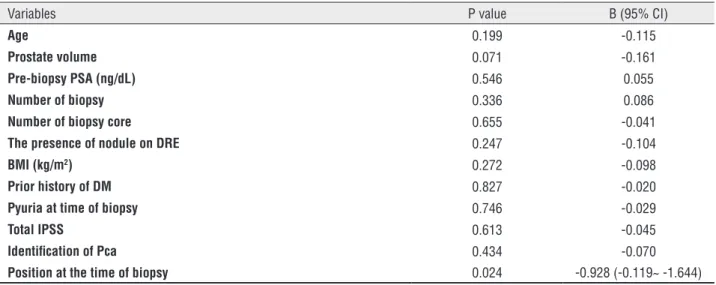
In simple correlation analysis, VAS sco-re showed significant association solely with patient’s position preferring lithotomy position to decrease perceived pain (Pearson’s coeffi-cient=-0.165, p=0.042, Table-2). In multiple line-ar regression models (stepwise method, R2=0.042, p=0.024) regarding the effect of clinical variables on VAS score, patient position was a single in-dependent predictor (B=-0.928, p=0.024, Table-3) favoring lithotomy position.
DISCUSSION
Table 1 - Characteristics of the enrolled patients.
Before matching (n=208) After matching (n=152)
Total LLD position (n=122)
lithotomy position
(n=86) p-value Total LLD position (n=76)
lithotomy position
(n=76) p-value
Pre-biopsy
variables
Age (years) 67.81±8.33 67.73±8.881 67.93±7.539 0.861 67.16±8.45 66.72±9.19 67.59±7.67 0.528
Prostate volume (g) 42.17±25.86 40.33±20.25 44.78±32.14 0.258 42.84±27.86 40.56±21.14 45.12±33.24 0.315
Pre-biopsy PSA
(ng/dL) 22.99±18.8 16.01±18.81 22.76±27.50 0.050 18.77±22.87 16.11±17.88 21.44±26.82 0.151
Number of biopsy
(%)
first 180 (86.5) 103 (84.4) 77 (89.5)
0.456
135 (88.8) 67 (88.2) 68 (89.5)
0.763 second 21 (10.1) 15 (12.3) 6 (7.0) 12 (7.9) 7 (9.2) 5 (6.6)
third 7 (3.4) 4 (3.3) 3 (3.5) 5 (3.3) 2 (2.6) 3 (3.9)
Nodule on DRE (%)
with palpable nodule 9 (5.4) 5 (5.3) 4 (5.4)
0.620
6 (4.9) 2 (3.4) 4 (6.3)
0.682 without palpable
nodule
159 (94.6) 89 (94.7) 70 (94.6) 116 (95.1) 56 (96.6) 60 (93.8)
BMI (kg/m2) 23.82±2.67 23.71±2.57 23.97±2.77 0.485 23.86±2.60 23.92±2.45 23.79±2.76 0.754
Prior history of
DM (%)
with DM 36 (17.3) 20 (16.4) 16 (18.6)
0.712
31 (20.4) 16 (21.1) 15 (19.7)
1.000 without DM 172 (82.7) 102 (83.6) 70 (81.4) 121 (79.6) 60 (78.9) 61 (80.3)
Pyuria at time of
biopsy (%)
0.601
with pyuria 10 (4.8) 6 (4.9) 4 (4.7) 6 (3.9) 4 (5.3) 2 (2.6)
0.681 without pyuria 198 (95.2) 116 (95.1) 82 (95.3) 146 (96.1) 72 (94.7) 74 (97.4)
Total IPSS 14.12±8.15 13,24±9.11 15.49±6.19 0.042 15.28±7.94 15.07±9.40 15.49±6.19 0.745
Intra &
post -
biopsy
variables
VAS score 4.29±2.20 4.54±2.28 3.89±2.02 0.041 4.24±2.14 4.58±2.22 3.89±2.01 0.049
0 4 (2.1) 4 (3.4) - 0.105 3 (2.0) 3 (3.9) - 0.157
1 11(5.6) 4 (3.4) 7 (9.2) 10 (6.6) 3 (3.9) 7 (9.2)
2 37 (19.0) 19 (16.0) 18 (23.7) 27 (17.8) 9 (11.8) 18 (23.7)
3 22 (11.3) 11 (9.2) 11 (14.5) 19 (12.5) 8 (10.5) 11 (14.5)
4 38 (19.5) 24 (20.2) 14 (18.4) 30 (19.7) 16 (21.1) 14 (18.4)
5 19 (9.7) 15 (12.6) 4 (5.3) 11 (7.2) 7 (9.2) 4 (5.3)
6 37 (19.0) 23 (19.3) 14 (18.4) 32 (21.1) 18 (23.7) 14 (18.4)
7 8 (4.1) 4 (3.4) 4 (5.3) 6 (3.9) 2 (2.6) 4 (5.3)
8 15 (7.7) 11 (9.2) 4 (5.3) 13 (8.6) 9 (11.8) 4 (5.3)
9 - - -
-10 4 (2.1) 4 (3.4) - 1 (0.7) 1 (1.3)
-Number of biopsy
core
12.13±0.627 12.02±0.589 12.29±0.648
0.006 12.19±0.524 12.08±0.271 12.30±0.673 0.009
12 core (%) 179 (86.1) 112 (91.8) 67 (77.9) 129 (84.9) 70 (92.1) 59 (77.6) 0.022
over 12 core
(13-16%)
29 (13.9) 10 (8.2) 19 (22.1)
0.007 23 (15.2) 6 (7.9) 17 (22.4)
Pca detected (%) 85 (40.9) 54 (44.3) 31 (36.0) 0.254 56 (36.8) 29 (38.2) 27 (35.5) 0.867
Table 2 - Outcome of simple correlation among clinical variables associated with VAS.
Variables Pearson’s coefficeint p-value
Age -0.068 0.402
Prostate volume 0.005 0.954
Pre-biopsy PSA (ng/dL) -0.003 0.974
Number of biopsy 0.067 0.410
Number of biopsy core -0.036 0.661
The presence of nodule on DRE -0.176 0.053
BMI (kg/m2) -0.060 0.463
Prior history of DM 0.041 0.620
Pyuria at time of biopsy -0.015 0.856
Total IPSS -0.117 0.152
Identification of Pca -0.015 0.852
Position at the time of biopsy -0.165 0.042
PSA = prostate-specific antigen; DRE = Digital rectal examination; BMI = body mass index; DM = Diabetes mellitus; Pca = Prostate cancer
Table 3 - Outcome of multiple linear regression model among clinical variables associated with VAS.
Variables P value B (95% CI)
Age 0.199 -0.115
Prostate volume 0.071 -0.161
Pre-biopsy PSA (ng/dL) 0.546 0.055
Number of biopsy 0.336 0.086
Number of biopsy core 0.655 -0.041
The presence of nodule on DRE 0.247 -0.104
BMI (kg/m2) 0.272 -0.098
Prior history of DM 0.827 -0.020
Pyuria at time of biopsy 0.746 -0.029
Total IPSS 0.613 -0.045
Identification of Pca 0.434 -0.070
Position at the time of biopsy 0.024 -0.928 (-0.119~ -1.644)
PSA = prostate-specific antigen; DRE = Digital rectal examination; BMI = body mass index; DM = Diabetes mellitus; Pca = Prostate cancer
worse as the test proceeded in the remaining patients (17). Familiarity with the procedure at the repeat biopsy did not decrease pain or an-xiety at al. (18).
In contrast, only 4% and 11% of patients reported no pain or discomfort, respectively, and 3% had no complaint during TRBx (18). Of men who were interviewed, 19% would not wish to un-dergo the procedure again without aid of analge-sia, and 6% would like biopsies to be done under
general anesthesia (19). Because the biopsy itself is still invasive in nature, a high degree of dis-comfort associated with it may result in failure of the patient to return for the future biopsy even though it will be necessary.
was reported in 64% of biopsy events (18), pain on insertion of the transrectal ultrasonic probe (15) or during DRE (20), and age of the patients (19). However, all of these previously reported characteristics were not chosen or adjustable by the physician, without providing a substantial clue to minimize the discomfort at the time of the procedure despite usefulness in identification of the risk group. Conversely, the outcomes from this series suggest lithotomy position as a simple method for the majority of subjects who have no limitation in range of motion in the hip joint. A significant decrease of mean VAS score was observed in lithotomy position in comparison with LLP, and a multivariable model showed li-thotomy position as a single significant predictor to minimize VAS score.
Then, what is the potential explanation for our findings? While the mechanism of pain associated with TRBx is complex, recent studies have gradually enlightened this area. Between li-thotomy position and LLP, there exist two funda-mental differences; the visibility of the procedu-re by the subject or eye contact by the physician which thereby influences the embarrassment of the subject, and the convenience in relaxation of pelvic floor muscle which affects the anal sphinc-ter contraction thereby enabling easier probe in-sertion and lesser pain perception. Because a sense of vulnerability or defenselessness associated with patient positioning may interfere with physical and psychological distress (14), lithotomy position which allows the patient to identify visual infor-mation on the progress of the procedure improves the tolerance of the patient while the position of the legs in this position obviously creates additio-nal discomfort. A more direct relationship betwe-en patibetwe-ent’s positioning and the degrees of pelvic floor muscle relaxation was recently identified. Using electromyographic evaluation with eight--channels for 29 women, Resende et al. demons-trated that the lateral positon presented a signifi-cantly greater myoelectrical signal of pelvic floor resting tone among lithotomy, supine, and lateral positions (21). In the same context, several rando-mized controlled trails which assessed the effect of topical muscle relaxant during TRBx consistently confirmed the effectiveness and safety in
dimi-nishing the patient’s discomfort, particularly du-ring the insertion of an ultrasound probe (22, 23). Our hypothesis regarding the positive in-fluence of lithotomy position on TRBx was su-pported by other research, which demonstrated that use of a larger probe (74mm) results in much higher VAS pain perception than same size and smaller (58mm) probe in the absence of injectable local anesthesia (24). In addition, probe insertion was reported to produce a significantly higher pain scale than biopsy using a 12 core prostate biopsy scheme (25). Due to the similarities of the procedure, discomfort during DRE can reflect that of the patient during TRBx, and several studies have reported an association between the patient’s position and pain during DRE (20, 26). Among four positions including LLD and supine position, more than half of their patients chose the supine position for DRE (27).
with us, and we believe these distinctive natures as a main reason for inconsistent conclusions on the advantage of lithotomy position during TRBx in prior series which used different biopsy setting in terms of the use of analgesics, the number of biopsy cores, and the number and experiences of physicians (9-11).
It is also obvious that not all men expe-rience severe endurable pain during the procedu-re. However, in performing prostate biopsy, one of the goals should be to minimize the patient’s discomfort associated with the procedure in era of active surveillance strategy in which the accep-tance of repeat biopsy is crucial. In acquisition of this, our data indicating an obvious influence of position on pain may contribute to establishment of the best clinical setting for TRBx.
CONCLUSIONS
The position of the patient was a single factor associated with pain perception during transrectal prostate biopsy with an extended biop-sy scheme. With routine use of topical lidocaine gel, lithotomy position significantly decreased the patient’s pain without compromising detectabili-ty of prostate cancer. Based on these findings, we suggest lithotomy position as a proper way to per-form TRUS guided prostate biopsy in comparison with conventional lateral decubitus position.
ACKNOWLEDGMENT
This work was supported by the 2015 Yeungnam University Research Grant.
CONFLICT OF INTEREST
None declared.
REFERENCES
1. Presti JC Jr, O’Dowd GJ, Miller MC, Mattu R, Veltri RW. Extended peripheral zone biopsy schemes increase cancer detection rates and minimize variance in prostate specific antigen and age related cancer rates: results of a community multi-practice study. J Urol. 2003;169:125-9.
2. Ukimura O, Coleman JA, de la Taille A, Emberton M, Epstein JI, Freedland SJ, et al. Contemporary role of systematic prostate biopsies: indications, techniques, and implications for patient care. Eur Urol. 2013;63:214-30.
3. Heidenreich A, Bastian PJ, Bellmunt J, Bolla M, Joniau S, van der Kwast T, et al. European Association of Urology. EAU guidelines on prostate cancer. part 1: screening, diagnosis, and local treatment with curative intent-update 2013. Eur Urol. 2014;65:124-37.
4. Lee C, Woo HH. Current methods of analgesia for transrectal ultrasonography (TRUS)-guided prostate biopsy -- a systematic review. BJU Int. 2014;113:48-56.
5. Inal G, Yazici S, Adsan O, Ozturk B, Kosan M, Cetinkaya M. Effect of periprostatic nerve blockade before transrectal ultrasound-guided prostate biopsy on patient comfort: a randomized placebo controlled study. Int J Urol. 2004;11:148-51.
6. Ozden E, Yaman O, Göğüs C, Ozgencil E, Soygür T. The optimum doses of and injection locations for periprostatic nerve blockade for transrectal ultrasound guided biopsy of the prostate: a prospective, randomized, placebo controlled study. J Urol. 2003;170:2319-22.
7. Berger AP, Frauscher F, Halpern EJ, Spranger R, Steiner H, Bartsch G, et al. Periprostatic administration of local anesthesia during transrectal ultrasound-guided biopsy of the prostate: a randomized, double-blind, placebo-controlled study. Urology. 2003;61:585-8.
8. Collins GN, Lloyd SN, Hehir M, McKelvie GB. Multiple transrectal ultrasound-guided prostatic biopsies--true morbidity and patient acceptance. Br J Urol. 1993;71:460-3. 9. Bruyère F, Faivre d’Arcier B, Haringanji DC, Boutin JM, Haillot
O, Lanson Y. Effect of patient position on pain experienced during prostate biopsy. Urol Int. 2007;78:351-5.
10. Kilciler M, Demir E, Bedir S, Erten K, Kilic C, Peker AF. Pain scores and early complications of transrectal ultrasonography-guided prostate biopsy: effect of patient position. Urol Int. 2007;79:361-3.
11. Lodeta B, Lodeta M. Prostate Biopsy in the Left Lateral Decubitus Position is Less Painful than Prostate Biopsy in the Lithotomy Position: A Randomized Controlled Trial. Korean J Urol. 2012;53:87-91.
12. Bastide C, Lechevallier E, Eghazarian C, Ortega JC, Coulange C. Tolerance of pain during transrectal ultrasound-guided biopsy of the prostate: risk factors. Prostate Cancer Prostatic Dis. 2003;6:239-41.
13. Zisman A, Leibovici D, Kleinmann J, Siegel YI, Lindner A. The impact of prostate biopsy on patient well-being: a prospective study of pain, anxiety and erectile dysfunction. J Urol. 2001;165:445-54.
15. Romero FR, Romero AW, Brenny Filho T, Bark NM, Yamazaki DS, de Oliveira FC. Patients’ perceptions of pain and discomfort during digital rectal exam for prostate cancer screening. Arch Esp Urol. 2008;61:850-4.
16. Chopra S, Rowe EW, Laniado M, Patel A. A prospective study analysing the effect of pain on probe insertion, and the biopsy strategy, on the patients’ perception of pain during TRUS-guided biopsy of the prostate. N Z Med J. 2008;121:39-43. 17. Horninger W, Reissigl A, Fink K Results of a prospective
randomised study comparing the prostate cancer detection rates in PSA screening volunteers undergoing 10 vs 14 transrectal ultrasound guided biopsies: J Urol 1998;159 suppl:180.
18. Rodríguez-Patrón Rodríguez R, Mayayo Dehesa T, Lennie Zucharino A, González Galán A, Peral Amorós M. Complications of prostatic echo-guided transrectal biopsy and tolerance depending on the patient and the operator. Study of 205 patients. Arch Esp Urol. 2002;55:509-21. 19. Zisman A, Leibovici D, Kleinmann J, Siegel YI, Lindner A.
The impact of prostate biopsy on patient well-being: a prospective study of pain, anxiety and erectile dysfunction. J Urol. 2001;165:445-54.
20. Irani J, Fournier F, Bon D, Gremmo E, Doré B, Aubert J. Patient tolerance of transrectal ultrasound-guided biopsy of the prostate. Br J Urol. 1997;79:608-10.
21. Djavan B, Zlotta A, Remzi M, Ghawidel K, Basharkhah A, Schulman CC, et al. Optimal predictors of prostate cancer on repeat prostate biopsy: a prospective study of 1,051 men. J Urol. 2000;163:1144-8.
22. Resende AP, Petricelli C, Zanetti MRD. Which of the recumbent positions promoves better pelvic floor muscle relaxation? ICS 2010 abstract number 1015
23. Rochester MA, LE Monnier K, Brewster SF. A double-blind, randomized, controlled trial of topical glyceryl trinitrate for transrectal ultrasound guided prostate biopsy. J Urol. 2005;173:418-20.
24. McCabe JE, Hanchanale VS, Philip J, Javle PM. A randomized controlled trial of topical glyceryl trinitrate before transrectal ultrasonography-guided biopsy of the prostate. BJU Int. 2007;100:536-8.
25. Koprulu S, Cevik I, Unlu N, Dillioglugil O. Size of the transrectal ultrasound probe makes no difference in pain perception during TRUS-Bx under adequate local anesthesia. Int Urol Nephrol. 2012;44:29-33.
26. Philip J, McCabe JE, Roy SD, Samsudin A, Campbell IM, Javlé P. Site of local anaesthesia in transrectal ultrasonography-guided 12-core prostate biopsy: does it make a difference? BJU Int. 2006;97:263-5.
27. Furlan AB, Kato R, Vicentini F, Cury J, Antunes AA, Srougi M. Patient’s reactions to digital rectal examination of the prostate. Int Braz J Urol. 2008;34:572-5.
_______________________ Correspondence address: