Article
Functional Role of Parkin against Oxidative Stress in
Neural Cells
Minyoung Hwang1, Ja-Myong Lee1, Younghwa Kim2, Dongho Geum1
1
Department of Biomedical Sciences, Korea University College of Medicine, Seoul; 2
Department of Emergency Medical Technology, Kyungil University College of Nursing and Public Health, Gyeongsan, Korea
Background: Parkinson disease (PD) is caused by selective cell death of dopaminergic neurons in the substantia nigra. An early onset form of PD, autosomal recessive juvenile parkinsonism has been associated with a mutation in the parkin gene. The func-tion of parkin is known to remove misfolding proteins and protect cell death. We aimed to investigate the role of parkin against oxidative stress in neuronal cells.
Methods: Parkin knockout embryonic stem cells (PKO ES cells) were differentiated into neurons by adherent monolayer culture method. Oxidative stress was induced by the treatment of 1-methyl-4-phenylpyridinium (MPP+
) in neurons derived from wild type and PKO ES cells, and cell viability was examined by MTT assay. After exposure to MPP+
, Tuj1-positive cell population was compared between PKO and wild type cells by fluorescence activated cell sorter (FACS) analysis. The activated caspase3 protein level was also measured by Western blot analysis, FACS and immunocytochemistry.
Results: There was no difference in the efficiency of neuronal differentiation between wild type and PKO ES cells. After exposure to MPP+
, no significant differences were found in cell viability and Tuj1-positive cell population between the two groups deter-mined by MTT assay and FACS analysis, respectively. The activated caspase3 protein levels exadeter-mined by Western blot analysis, FACS and immunocytochemistry were not changed in PKO cells compared with those of wild type cells after MPP+
treatment. Conclusion: These results suggest that PKO neuronal cells including dopaminergic neurons are not sensitive to caspase3-depen-dent cell death pathway during the response against MPP+
-induced oxidative stress.
Keywords: Parkin; Dopaminergic neurons; Oxidative stress; Cell death; Embryonic stem cells
INTRODUCTION
Neurodegenerative diseases, such as Alzheimer disease,
Par-kinson disease (PD), Huntington disease, and amyotrophic lat-eral sclerosis are characterized by the progressive loss of spe-cific neural cells, and are associated with protein aggregation
Received: 27 July 2013, Accepted: 11 October 2013
Corresponding authors:
Younghwa Kim
Department of Emergency Medical Technology, Kyungil University College of Nursing and Public Health, 50 Gamasil-gil, Hayang-eup, Gyeongsan 712-701, Korea
Tel: +82-53-600-5681, Fax: +82-53-600-5681, E-mail: [email protected]
Dongho Geum
Department of Biomedical Sciences, Korea University College of Medicine, 73 Inchon-ro, Seongbuk-gu, Seoul 136-705, Korea
Copyright © 2014 Korean Endocrine Society
and accumulation [1,2]. Recently, it has been reported that ox-idative stress might be involved in the dysfunction or death of neural cells that contributes to the pathogenesis of neurode-generative diseases [3-6]. Oxidative stress is the result of un-regulated production of reactive oxygen species, which lead to impaired cellular function through oxidative damage of lipids, proteins, and DNA [7].
PD is one of the most common neurodegenerative diseases, which is caused by selective cell death of dopaminergic neu-rons in the substantia nigra. Parkin is a 465 amino acid protein that contains two really interesting new gene (RING)finger domains and one in between RING domain that serves as an E3 ubiquitin ligase. Parkin functions to remove misfolded pro-teins and protect against cell death [8]. Mitochondrial dysfunc-tion and oxidative damage also have been reported in parkin null mice [9], which suggests a protective role of parkin against oxidative stress. Recently, parkin and its interaction with PTEN induced putative kinase 1 have been shown to participate in the maintenance of mitochondrial function [10-12].
To determine whether parkin is associated with neuronal cell death caused by oxidative stress, parkin knockout embryonic stem cells (PKO ES cells) were used. PKO ES cells have the potential to differentiate into neuronal cells including dopami-nergic neurons. In vitro, this system more closely reflects
neu-ronal cells than established cell lines, such as SH-SY5Y, PC12, and NM9D. 1-Methyl-4-phenylpyridinium (MPP+
) was used as an oxidative stressor, which is well known to induce cellular oxidative stress and PD-like symptoms.
METHODS
Maintenance and differentiation of ES cells
PKO ES cells were generated by deleting exon 2 of the parkin gene (unpublished data), and was provided by Dr. Richard Pal-miter at University of Washington. Wild-type (WT) and PKO ES cells were maintained on mouse embryonic fibroblast feed-er cells with leukemia inhibitory factor (1,000 U/mL, Chemi-con, Temecula, CA, USA) in ES cell medium consisting of Dulbecco’s modified Eagle’s medium supplemented with 15% fetal bovine serum, 100 mM nonessential amino acid, 0.5% antibiotics, and 0.55 mM 2-mercaptoethanol, at 37°
C in a 5% CO2. The adherent monolayer culture method was used for the
differentiation of ES cells into neural cells as previously de-scribed without any modifications [13].
Reverse-transcriptional polymerase chain reaction
Total RNA was isolated using a ToTALLY RNA kit (Ambion, Austin, TX, USA). One microgram of the RNA template was reverse-transcribed using a Transcript First Strand cDNA Syn-thesis kit (Roche Diagnostics, Basel, Switzerland) according
to the manufacturer’s guidance. Subsequently, a 2 μL aliquot
of each sample was subjected to real-time polymerase chain
reaction (PCR) in a 20 μL reaction mixture containing 4 mM
MgCl2, 10 pmol of upstream and downstream primers, and 2
μL of 10X Light Cycler Fast Start DNA Master SYBR Green
1 (Roche Diagnostics). Data were analyzed with Light Cycler software version 3.5 (Roche Diagnostics). The following prim-er sets wprim-ere chosen for real-time PCR analyses.
Glyceraldehyde 3-phosphate dehydrogenase: 5′-GTGTTC-CTACCCCCAATGTG-3′; 5′-TGTGAGGGAGATG- CTCA-GTG-3′ (400 bp), Nurr1: 5′-CGGTTTCAGAAGTGCCTA-GC-3′; 5′-CTGGGTTGGACCTGTATGCT-3′ (420 bp), Pitx3:
5′-ACAAAGTGGAAC- CCCTATGAG-3′; 5′-TTCTTGGC
-CAATCTGTAGGA-3′ (255 bp), tyrosine hydroxylase (TH): 5′-TTGGCTGACCGCACATTTG-3′; 5′-ACGAGAGGC-
ATAGTTCCTGAGC-3′ (336 bp), aromatic L-amino acid de
-carboxylase (AADC): 5′-AGAAGAGGCAAGGAGATG
-GTGG-3′; 5′-AAGCGAAGAAAT- AGGGAC-TCTGC-3′
(214 bp), dopamine receptor 2 (D2R): 5′-CCTT- CATCGT
-CACCCTGCTGG-3′; 5′-CTCCATTTCCAGCTCCTGAG-3′
(245 bp/158 bp).
Immunocytochemistry
Cells were fixed with 4% paraformaldehyde in phosphate buffered saline (PBS) for 20 minutes at room temperature, and treated with 0.3% Triton X-100 in 10% normal goat serum for 45 minutes. Cells were incubated at 4°
PKO WT
WT PKO
14 13 12 11 10 9 8 7 6 5 4 3 2 1 0
TH
+/total cell (%)
B
WT
Day 0 Day 5 Day 10 Day 15
PKO
A
MAP2/DAPI
Fig. 1. Induction of dopaminergic neurons from wild-type (WT) and parkin knockout embryonic stem (PKO ES) cells by the adherent monolayer culture method. (A) Induction of neural cells from WT and PKO ES cells by the adherent monolayer method. Representative images were taken 0, 5, 10, and 15 days after differentiation. Immunocytochemistry shows MAP2-positive mature neural cells, and 6-di-amidino-2-phenylindole (DAPI) was used for nuclear staining. (B) Expression of tyrosine hydroxylase (TH) 15 days after differentia-tion. TH+ neurons were analyzed by immunocytochemistry with an anti-TH antibody 15 days after differentiation. The 10% to 12% of
cells were identified as TH-positive cells. (C) Reverse-transcriptional polymerase chain reaction analysis of dopaminergic neuron mark-ers, such as Nurr1, TH, Pitx3, aromatic L-amino acid decarboxylase (AADC), and dopamine receptor 2 (D2R) at 15 days after differenti-ation. Glyceraldehyde 3-phosphate dehydrogenase (GAPDH) was used as an internal control. Data are expressed as mean±SEM (n=3
TH
Pitx 3 AADC D2R Nurr 1
14 13 12 11 10 9 8 7 6 5 4 3 2 1 0
% of control
WT PKO
C
Nurr 1
AADC
D2R
GAPDH Pitx3
TH
Fluorescence-activated cell sorting
WT and PKO ES cells were differentiated into neural cells for 12 days. Subsequently, cells were treated with or without MPP+
(1 mM, Sigma, St. Louis, MO, USA) for 24 hours. Cells were harvested using 0.25% trypsin/ethylenediaminetetraace-tic acid (Invitrogen, Carlsbad, CA, USA) containing DNase I (4 U/mL, Roche Diagnostics), and gently dissociated into a single-cell suspension. Then, cells were fixed and treated us-ing a Cytofix/Cytoperm kit (BD Pharmus-ingen, San Diego, CA, USA) according to the manufacturer’s guidelines. Primary an-tibodies were added and incubated for 1 hour, and cells were then washed, incubated with Alexa Fluor 488 and/or 594 fluo-rescent secondary antibodies for 30 minutes, and subsequently washed. Fluorescence-activated cell sorting (FACS) was per-formed using a FACSAria cell sorter and FACSDiva software (BD Biosciences, San Jose, CA, USA). All FACS experiments were repeated at least three times.
MTT assay
ES cells (6×10²
cells/well) were seeded onto 12-well plates, differentiated into neural cells for 12 days, and then treated with MPP+ (0, 100, 500, 750, 100, 1,250, and 1,500 μM, Sig
-ma) for 24 hours. Next, 3-(4,5-Dimethylthiazol-2-yl)-2,5-di-phenyltetra- zolium bromide solution (0.5 g/L) was added into each well and incubated at 37°
C for 2 hours. Supernatants were removed and formazan crystals were dissolved in dimethyl-sulfoxide. Finally, optical density was determined at 570 nm by a SPECTRA max Plus (Molecular Devices, Sunnyvale, CA, USA).
Western blot analysis
To analyze the cell signaling pathways, Western blot analysis was performed. ES cells were differentiated into neural cells on 60 mm dishes and were treated with or without 1 mM MPP+
for 24 hours. Following treatment, the media were aspirated and the cells were washed twice in ice-cold PBS and
suspend-ed in 100 μL of lysis buffer. The samples were then heatsuspend-ed for
5 minutes at 95°
C, and centrifuged for 5 minutes. The quantita-tive analysis of protein was carried out via bicinchoninic acid assay. The samples were separated on sodium dodecyl sulfate polyacrylamide gel electrophoresis (8%) gels, and transferred to nitro cellulose membranes. The blots were incubated with 5% skim milk at room temperature for 1 hour. Next, the blots were incubated overnight at 4°
C with primary antibodies, and then washed three times in Tris-buffered saline/0.1% Tween 20 prior to 1 hour of probing with horseradish
peroxidase-conju-gated secondary antibodies at room temperature. The blots were then visualized via enhanced chemiluminescence (Amer-sham Biosciences, Buckinghamshire, UK). In some cases, the blots were stripped and reprobed using other antibodies.
Data analysis
Data are expressed as means±SEM. Statistic analyses were
conducted using SigmaStat (SPSS Inc., Chicago, IL, USA). P
values of <0.05 were considered statistically significant.
RESULTS
Differentiation of PKO and WT ES cells into neural cells by adherent monolayer culture method
To examine differences in efficiency of neural differentiation, WT and PKO ES cells were differentiated into neurons by the adherent monolayer culture method. Morphological changes were observed during a differentiation period, and immunocy-tochemistry was performed with MAP2, a mature neuron marker. There were no differences in the morphology or differ-entiation of MAP2-positive cells between WT and PKO cells (Fig. 1A). Specifically, the efficiency of neural differentiation into dopaminergic neurons showed no difference between WT and PKO ES cells, as determined by immunocytochemistry with TH, a dopaminergic neuron marker (Fig. 1B). Real-time RT-PCR analysis with dopaminergic neuron markers such as Nurr1, Pitx3, AADC, TH, and D2R also showed no difference
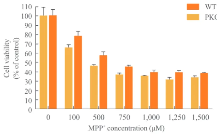
0 100 500 750 1,000 1,250 1,500 110
100 90 80 70 60 50 40 30 20 10 0 Cell viability (% of control)
MPP+ concentration (μM)
WT PKO
Fig. 2. Cell viability analysis after treatment with 1-methyl-4-phenylpyridinium (MPP+) by MTT assay. MPP+ was treated at
various concentrations (0, 100, 500, 750, 1,000, 1,250, and 1,500
μM) for 24 hours, at 15 days after differentiation. MTT assay was
performed for viability of differentiated cells. Data are expressed as mean±SEM (n=6 per group). Statistical analysis was
per-formed by two-way analysis of variance. No significant differenc-es in the MPP+ susceptibility were observed between wild-type
100 101 102 103 104 Tuj1
Activated caspase 3
10
0
10
1
10
2
10
3
10
4
100 101 102 103 104 Tuj1
Activated caspase 3
10
0
10
1
10
2
10
3
10
4
100 101 102 103 104 Tuj1
Activated caspase 3
10
0
10
1
10
2
10
3
10
4
100 101 102 103 104 Tuj1
Activated caspase 3
10
0
10
1
10
2
10
3
10
4
CTL MPP+
4
3
2
1
0
D
ouble
+ cells
in
T
uj1
+ cells
(%)
CTL MPP+
A
B
WT
CTL MPP+ CTL MPP+ PKO
Activated caspase 3
β-Actin
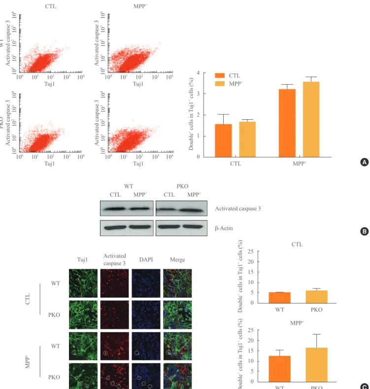
Fig. 3. Activity of caspase 3 in 1-methyl-4-phenylpyridinium (MPP+) treated wild-type (WT) and parkin knockout (PKO) neuronal cells.
(A) Fluorescence-activated cell sorting analysis of WT and PKO cells treated with 1 mM MPP+ for 24 hours with anti-Tuj1 and
antiacti-vated caspase 3 antibodies. Double+ cells represent colabeling with Tuj1 and activated caspase 3. The right panel shows the percentage of
double+ cells per Tuj1 positive cells. (B) Western blot analysis of caspase 3 activity in WT and PKO cells. Cells were harvested 24 hours
after 1 mM MPP+ treatment and Western blot analysis was performed with an activated caspase 3 antibody. β-Actin was used as an inter
-nal control. (C) Immunocytochemistry of WT and PKO cells treated with 1 mM MPP+ for 24 hours with anti-Tuj1 (green) and activated
caspase 3 (red) antibody (scale bar, 20 μm). While MPP+ treatment causes a significant increase in caspase 3 activity, no significant
dif-C
Tuj1 Activated
caspase 3 DAPI Merge
WT
PKO
WT
PKO
CTL
MPP
+
WT PKO
25
20
15
10
5
0
Double
+ cells in
T
uj1
+ cells (%)
MPP+
WT PKO
25
20
15
10
5
0
Doubl
e
+ c
el
ls i
n
T
uj
1
+ c
el
ls (%)
CTL CTL
WT
PKO
between WT and PKO cells (Fig. 1C).
Cell death of PKO and WT neural cells by oxidative stress
To investigate the response of PKO neural cells against oxida-tive stress, MPP+
, a metabolite of 1-methyl-4-phenyl-1, 2, 3, 6-tetrahydropyridine (MPTP), was treated with serial
concen-trations (0, 100, 500, 750, 1,000, 1,250, and 1,500 μM) after
15 days of differentiation for 24 hours. MTT assay showed
that almost 60% of cells had died from 750 μM to 1.5 mM of
MPP+
treatment, and there were no significant differences in the profile of cell death between WT and PKO neural cells (Fig. 2).
Activation of caspase 3 in WT and PKO neural cells by oxidative stress
To test whether MPP+
-induced neuronal cell death is associat-ed with apoptotic cell death, caspase 3 activity was determinassociat-ed with FACS analysis, Western blot analysis, and immunocyto-chemistry. Differentiated cells were treated with 1 mM MPP+
, at which dose almost half of the cells died (Fig. 2); Tuj1 (a neural cell marker) and activated caspase 3 double positive cells were examined by FACS analysis. The Tuj1 and activat-ed caspase 3 double positive cells were increasactivat-ed in MPP+
-treated groups by 3-fold. However, there were no significant differences in MPP+
-induced caspase 3-dependent apoptotic cell death between WT and PKO neural cells (Fig. 3A). West-ern blot analysis (Fig. 3B) and immunocytochemistry (Fig.
WT PKO
CTL 45
40 35 30 25 20 15 10 5 0
Double
+ cells in
TH
+ cells (%)
WT PKO
MPP+ 45
40 35 30 25 20 15 10 5 0
Double
+ cells in
TH
+ cells (%)
A
WT
PKO
WT
PKO
CTL
MPP
+
TH Activated
caspase 3 DAPI Merge
WT PKO
CTL 30
25 20 15 10 5 0
Double
+ cells TH + cells (%)
WT PKO
MPP+ 30
25
20
15
10
5 0
Double
+ cells TH + cells (%)
Fig. 4. Activity of caspase 3 in 1-methyl-4-phenylpyridinium (MPP+) treated wild-type (WT) and parkin knockout (PKO) dopaminergic
neurons. (A) Fluorescence-activated cell sorting analysis of WT and PKO cells treated with 1 mM MPP+ for 24 hours with tyrosine
hy-droxylase (TH) and activated caspase 3 antibodies. Double+ cells represent colabeling with TH and activated caspase 3 antibodies.
Graphs show the percentage of double positive cells per TH-positive cells. (B) Immunocytochemistry of WT and PKO cells treated with 1 mM MPP+ for 24 hours with TH (red) and activated caspase 3 (green) antibodies (scale bar, 20 μm). No significant differences in cas
-pase 3 activity were observed between WT and PKO dopaminergic neurons. Data are expressed as mean±SEM (n=3 per group).
Statis-tical analysis was performed by Student t test. CTL, control without MPP+ treatment; DAPI, 4, 6-diamidino-2-phenylindole.
3C) also showed no differences in MPP+
-induced caspase 3-dependent apoptotic cell death between WT and PKO neural cells.
Activation of caspase 3 in WT and PKO dopaminergic neurons by oxidative stress
The sensitivity of PKO dopaminergic neurons against MPP+
-induced oxidative stress was examined by FACS analysis. Ini-tially, the Tuj1 and TH double positive cell population was ex-amined. The double positive cell population represented about 20% of the Tuj1-positive cells (data not shown). After expo-sure to 1 mM MPP+
, FACS analysis was performed with TH and activated caspase 3 antibodies. Double positive cells per TH-positive cells (26.14%±15.62% in WT cells and 19.81%±
12.75% in PKO cells) were similar in the two groups (Fig. 4A). Immunocytochemistry with TH and activated caspase 3 antibodies also showed a parallel result with the FACS analy-sis of the two groups (Fig. 4B).
DISCUSSION
It has been reported that Parkin, which is located in the cyto-plasm or mitochondrial outer membrane, inhibits cell death induced by various stressors [14]. Most studies concerning the function of parkin against oxidative stress were performed in established cell lines, such as SH-SY5Y, PC12, and NM9D, which may not faithfully represent real conditions. To over-come these issues, PKO neural cells differentiated from PKO ES cells were used to examine the function of the parkin gene in cell death caused by oxidative stress. Consistent with the goals of our study, there were no differences in the neural dif-ferentiation efficiency between PKO and WT cells. This result was in accord with an in vivo study, which showed the number
of TH-positive neurons were not different between WT and PKO mice [15].
As an oxidative stressor, MPP+
, which is an active metabo-lite of MPTP, was used. MPTP is known to inhibit the mito-chondrial complex 1, and induce cellular oxidative stress [16, 17]. Previous studies suggest that MPP+
is able to induce apoptotic cell death in vitro in various cell lines [18-21]. In
this study, PKO neural cells showed no significant differences compared with WT cells against MPP+
-induced oxidative stress as examined with MTT assay (Fig. 2) and caspase 3 ac-tivity-dependent apoptotic cell death (Figs. 3, 4). These results match with in vivo studies using PKO mice [22], and suggest
caused by MPP+
-induced oxidative stress in neuronal cells dif-ferentiated from ES cells in vitro. However, these results differ
from previous studies, which showed a protective role of par-kin through ectopic over expression [23-26]. From these dif-ferences, it is apparent from this study that the protective func-tion against oxidative stress is not a major component in the anticell death machinery.
This study analyzed the induced neural cells from ES cells. To exclude the possible contamination of other cell types dif-ferentiated from ES cells, we performed FACS analysis and immunocytochemistry to detect contamination at the single cell level. FACS analysis is based on cell population, hence a relatively small difference between groups is not easily distin-guishable. Therefore, immunohistochemical marker analysis was carried out together. Immunocytochemistry data showed that more cells were activated caspase 3-positive in PKO cells after MPP+
treatment (Figs. 3, 4).
In summary, PKO neuronal cells showed no significant dif-ference in caspase 3-dependent cell death against MPP+
-in-duced oxidative stress.
CONFLICTS OF INTEREST
No potential conflict of interest relevant to this article was re-ported.
ACKNOWLEDGMENTS
This research was supported by the Korea Research Founda-tion, funded by the Korean Government (Ministry of Educa-tion, Science and Technology) (Grant KRF-20120007428).
REFERENCES
1. Bossy-Wetzel E, Schwarzenbacher R, Lipton SA.
Molecu-lar pathways to neurodegeneration. Nat Med 2004;10 Suppl:S2-9.
2. Taylor JP, Hardy J, Fischbeck KH. Toxic proteins in
neuro-degenerative disease. Science 2002;296:1991-5.
3. Alam ZI, Jenner A, Daniel SE, Lees AJ, Cairns N, Marsden
CD, Jenner P, Halliwell B. Oxidative DNA damage in the parkinsonian brain: an apparent selective increase in 8-hy-droxyguanine levels in substantia nigra. J Neurochem 1997; 69:1196-203.
neu-Curr Opin Lipidol 2002;13:289-94.
5. Gabbita SP, Lovell MA, Markesbery WR. Increased
nucle-ar DNA oxidation in the brain in Alzheimer’s disease. J Neurochem 1998;71:2034-40.
6. Pedersen WA, Fu W, Keller JN, Markesbery WR, Appel S,
Smith RG, Kasarskis E, Mattson MP. Protein modification by the lipid peroxidation product 4-hydroxynonenal in the spinal cords of amyotrophic lateral sclerosis patients. Ann Neurol 1998;44:819-24.
7. Finkel T, Holbrook NJ. Oxidants, oxidative stress and the
biology of ageing. Nature 2000;408:239-47.
8. Shimura H, Hattori N, Kubo S, Mizuno Y, Asakawa S,
Minoshima S, Shimizu N, Iwai K, Chiba T, Tanaka K, Su-zuki T. Familial Parkinson disease gene product, parkin, is a ubiquitin-protein ligase. Nat Genet 2000;25:302-5. 9. Palacino JJ, Sagi D, Goldberg MS, Krauss S, Motz C,
Wacker M, Klose J, Shen J. Mitochondrial dysfunction and oxidative damage in parkin-deficient mice. J Biol Chem 2004;279:18614-22.
10. Clark IE, Dodson MW, Jiang C, Cao JH, Huh JR, Seol JH,
Yoo SJ, Hay BA, Guo M. Drosophila pink1 is required for mitochondrial function and interacts genetically with par-kin. Nature 2006;441:1162-6.
11. Park J, Lee SB, Lee S, Kim Y, Song S, Kim S, Bae E, Kim
J, Shong M, Kim JM, Chung J. Mitochondrial dysfunction in Drosophila PINK1 mutants is complemented by parkin. Nature 2006;441:1157-61.
12. Dodson MW, Guo M. Pink1, Parkin, DJ-1 and
mitochon-drial dysfunction in Parkinson’s disease. Curr Opin Neuro-biol 2007;17:331-7.
13. Ying QL, Stavridis M, Griffiths D, Li M, Smith A.
Con-version of embryonic stem cells into neuroectodermal pre-cursors in adherent monoculture. Nat Biotechnol 2003;21: 183-6.
14. Darios F, Corti O, Lucking CB, Hampe C, Muriel MP,
Ab-bas N, Gu WJ, Hirsch EC, Rooney T, Ruberg M, Brice A. Parkin prevents mitochondrial swelling and cytochrome c release in mitochondria-dependent cell death. Hum Mol Genet 2003;12:517-26.
15. Goldberg MS, Fleming SM, Palacino JJ, Cepeda C, Lam
HA, Bhatnagar A, Meloni EG, Wu N, Ackerson LC, Klap-stein GJ, Gajendiran M, Roth BL, Chesselet MF, Maid-ment NT, Levine MS, Shen J. Parkin-deficient mice exhib-it nigrostriatal deficexhib-its but not loss of dopaminergic neu-rons. J Biol Chem 2003;278:43628-35.
16. Langston JW, Ballard P, Tetrud JW, Irwin I. Chronic
Par-kinsonism in humans due to a product of meperidine-ana-log synthesis. Science 1983;219:979-80.
17. Schapira AH, Cooper JM, Dexter D, Jenner P, Clark JB,
Marsden CD. Mitochondrial complex I deficiency in Par-kinson’s disease. Lancet 1989;1:1269.
18. Dipasquale B, Marini AM, Youle RJ. Apoptosis and DNA
degradation induced by 1-methyl-4-phenylpyridinium in neurons. Biochem Biophys Res Commun 1991;181:1442-8. 19. Du Y, Dodel RC, Bales KR, Jemmerson R, Hamilton-Byrd
E, Paul SM. Involvement of a caspase-3-like cysteine pro-tease in 1-methyl-4-phenylpyridinium-mediated apoptosis of cultured cerebellar granule neurons. J Neurochem 1997; 69:1382-8.
20. Hartley A, Stone JM, Heron C, Cooper JM, Schapira AH.
Complex I inhibitors induce dose-dependent apoptosis in PC12 cells: relevance to Parkinson’s disease. J Neurochem 1994;63:1987-90.
21. Itano Y, Nomura Y. 1-methyl-4-phenyl-pyridinium ion
(MPP+
) causes DNA fragmentation and increases the Bcl-2 expression in human neuroblastoma, SH-SY5Y cells, through different mechanisms. Brain Res 1995;704:240-45. 22. Thomas B, von Coelln R, Mandir AS, Trinkaus DB, Farah
MH, Leong Lim K, Calingasan NY, Flint Beal M, Dawson VL, Dawson TM. MPTP and DSP-4 susceptibility of sub-stantia nigra and locus coeruleus catecholaminergic neu-rons in mice is independent of parkin activity. Neurobiol Dis 2007;26:312-22.
23. Fallon L, Belanger CM, Corera AT, Kontogiannea M,
Re-gan-Klapisz E, Moreau F, Voortman J, Haber M, Rouleau G, Thorarinsdottir T, Brice A, van Bergen En Henegouwen PM, Fon EA. A regulated interaction with the UIM protein Eps15 implicates parkin in EGF receptor trafficking and PI(3)K-Akt signalling. Nat Cell Biol 2006;8:834-42. 24. Henn IH, Bouman L, Schlehe JS, Schlierf A, Schramm JE,
Wegener E, Nakaso K, Culmsee C, Berninger B, Krappma-nn D, Tatzelt J, Winklhofer KF. Parkin mediates neuropro-tection through activation of IkappaB kinase/nuclear factor-kappaB signaling. J Neurosci 2007;27:1868-78.
25. Jiang H, Jiang Q, Feng J. Parkin increases dopamine
up-take by enhancing the cell surface expression of dopamine transporter. J Biol Chem 2004;279:54380-6.
26. Jiang H, Ren Y, Zhao J, Feng J. Parkin protects human