Disease and Stress Biology Laboratory
Instituto de Tecnologia Química e Biológica António Xavier Universidade NOVA de Lisboa
Av. da República
2781-901 Oeiras – Portugal
Cell and Molecular Neuroscience Unit Instituto de Medicina Molecular
Faculdade de Medicina da Universidade de Lisboa Avenida Professor Egas Moniz
1649-028 Lisboa – Portugal
Supervised by:
Doctor Maria Cláudia Nunes dos Santos Doctor Sandra Nogueira Tenreiro Prof. Doctor Tiago Fleming Outeiro
Acknowledgements ... i
Resumo ... ix
Thesis outline ... xiii
Abbreviations list ... xvii
Introduction ... 1
Chapter 1: Blocking alpha-synuclein phosphorylation impairs its
clearance through autophagy ... 58
Chapter 2: (Poly)phenols protect from alpha-synuclein toxicity by
promoting its clearance through autophagy ... 92
Chapter3: (Poly)phenol-digested metabolites protect against
alpha-synuclein toxicity ... 150
iii I was born in a small and humble island where I thought, for a long time, that
scientists were mere characters in children’s books. If I have reached this far, I must acknowledge those who walked alongside with me throughout these years of scientific
discovery.
First of all, to my supervisors Cláudia N. Santos, Sandra Tenreiro and Tiago F.
Outeiro for receiving me so well on their laboratories, for the technical support and
guidance over the development of the research. Thank you for the critical manuscript
revisions, for the opportunities to participate in challenging projects and for the
motivation and words of encouragement.
I am proud to have been part of the DSB and UNCM labs, which become my
second home. Past and present members contributed to my scientific path through
exchange of knowledge, technical and soft skills, thank you also for the brainstorming
meetings and kindness. Furthermore, I would like to acknowledge my colleagues from
the ITQB PhD program for the companionship.
The support of my family and friends was crucial for the success of this thesis,
I must acknowledge João for understanding the demands of a science career and for
being supportive through every challenge. Bina, Vitor, Avó Laura e Avô Zeca for the
day-to-day small treats, lovely Sunday lunches and kind-heartedness. Glória and
Francisco, thank you for the easy-going friendship and the encouragement.
Thank you to my brother and best friend Rui, who always makes me think
outside-of-the-box and inspires me to follow my dreams. Finally, I would like to express
my gratitude to my parents Zita and Manuel for their unconditional support, the
perseverance, resilience and optimism I see in you were essential for this
accomplishment.
vi
Parkinson’s disease (PD) is the second most common neurodegenerative disease, and affects 1-2% of the population worldwide after the age of 65 years. Currently, there are
only symptomatic treatment for PD and these do not mitigate the underlying causes of the
disease.
There is strong hope that the deeper we scrutinize the pathobiology of PD, the closer
we will become to identifying useful drug targets and design an effective pleiotropic drug.
This drug should be able to act in the multitude of events leading to disease, ultimately
fighting neurodegeneration.
PD is intimately associated with the misfolding and aggregation of alpha-synuclein
(aSyn), a protein primarily found in pre-synaptic neurons. aSyn is found aggregated in
cytosolic inclusions known as Lewy bodies (LBs), the main pathological hallmark of PD and
other synucleinopathies.
Strikingly, 90% of aSyn appears to be phosphorylated on serine 129 (pS129) in LBs
of patients with synucleinopathies. Nevertheless, the normal and pathological function of this
post-translational modification is still unresolved. The present study analysed the role of
pS129 in a simple but versatile yeast model of PD, by using a phospho-resistant aSyn form,
S129A. Blocking aSyn phosphorylation on S129 increased aSyn toxicity and promoted the
formation of cytosolic inclusions. Furthermore, unphosphorylated aSyn exacerbated
vesicular trafficking defects and failed to induce autophagy, leading to the accumulation of
aSyn.
Autophagy is the main pathway by which large protein aggregates are degraded and
cleared from the cell. Modulations of phosphorylation on S129 and of autophagy are
considered potential drug targets in PD. However, in addition to the identification of drug
targets, it is urgent to develop novel therapeutic strategies, and to identify molecules able to
modify the cellular mechanisms involved in neurodegeneration. Thus, we further explored
PD pathways using the yeast model of PD to study the potential of (poly)phenols as
neuroprotectants.
The ubiquity of (poly)phenols in fruits and vegetables, allied with
epidemiological/research studies reporting their ability to reverse and prevent age-related
vii (Poly)phenols occur naturally in plants, which constitute a valuable source of these
compounds to be explored in pharmacological or nutritional approaches. In this thesis, we
explored Portuguese plant species to study the potential of (poly)phenols in the pathobiology
of PD.
In an initial pharmacological/nutraceutical perspective, (poly)phenols were extracted
and identified from leaves or fruits from the species C. album, A. unedo, G. biloba and R. idaeus. Interestingly (poly)phenols from the leaves of C. album presented the strongest
properties in phenotypic assays of cell viability, metabolism, and aSyn aggregation.
The mechanism of action underlying the protection provided by these (poly)phenols
was found to be related with the inhibition of aSyn fibrillization and stabilization of oligomeric
species in vitro and, remarkably, these species were not toxic in a human cell model of PD.
Furthermore, (poly)phenols reduced oxidative stress, the formation of aSyn inclusions in
yeast, and likely promoted the clearance of aSyn through the promotion of autophagy, both
in yeast and in human H4 cell models of PD.
To address the nutritional role of (poly)phenols, an in vitro digestion procedure that
mimics the upper gastro-intestinal digestion was used. In a previous study, we reported the
effect of digestion in the chemical profile of fruits and leaves from A. unedo and their ability
to protect yeast cells from an oxidative insult. Thus, metabolites from A. unedo were
analysed in the yeast model of PD. The digested metabolites from the leaves of A. unedo
were protective against aSyn toxicity, and reduced the formation and size of aSyn inclusions.
Importantly, it regulated pathways intimately associated with protein misfolding, including
oxidative stress, mitochondrial dysfunction, ER-stress and autophagy and proteasomal
function.
In conclusion, a better understanding of the molecular mechanism underlying PD, in
particular the role of S129 aSyn phosphorylation, opens novel avenues for the identification
of new drug targets and the development of more effective therapies. (Poly)phenols proved
to be a promising source of therapeutic agents for neurodegenerative diseases, oxidative
stress or unfolded protein disorders, as a pharmacological/nutraceutical or nutritional
approach.
x
A doença de Parkinson (DP) é a segunda doença neurodegenerativa mais comum
e afecta 1-2% da população mundial com idade superior a 65 anos. Atualmente apenas
existem tratamentos sintomáticos para a DP e estes não mitigam as causas subjacentes da
doença.
Existe grande espectativa de que quanto mais profundamente estudarmos a
patobiologia da DP, mais nos aproximemos da identificação de alvos terapêuticos úteis e
de desenhar um fármaco pleiotrópico eficaz. Este fármaco deve ser capaz de agir na
multiplicidade de eventos que levam à doença, combatendo a neurodegeneração.
A DP está intimamente associada com a conformação incorreta e a agregação de
alfa-sinucleína (aSyn), uma proteína principalmente presente em neurónios pré-sinápticos.
aSyn é encontrada agregada em inclusões citosólicas conhecidas por corpos de Lewy, a
principal característica patológica de DP e outras sinucleinopatias. Apesar da intensa
pesquisa e do vasto conhecimento criados nos últimos anos, um claro exemplo de que a
patobiologia da DP ainda representa um desafio vem da falta de compreensão sobre o
papel da fosforilação na agregação da aSyn.
Surpreendentemente, 90% da aSyn parece ser fosforilada na serina 129 (fS129)
nos corpos de Lewy em pacientes com sinucleinopatias. No entanto, a função normal e
patológica desta modificação pós-transcripcional está por resolver. No presente estudo
analisamos o papel da fS129 num modelo simples mas versátil de DP em levedura, usando
uma aSyn fosfo-resistente, S129A. O bloqueio da fosforilação na S129 da aSyn aumentou
a toxicidade da aSyn e promoveu a formação de inclusões citosólicas. Além disso, aSyn
não fosforilada exacerbou o deficiente tráfico vesicular e não conseguiu induzir autofagia,
levando à acumulação de aSyn.
A autofagia é a principal via pela qual grandes agregados proteicos são
degradados e eliminados da célula. A modulação da fosforilação na S129 e da autofagia
são considerados como potenciais alvos terapêuticos em DP. No entanto, além da
identificação de alvos terapêuticos é urgente o desenvolvimento de novas estratégias
terapêuticas, e a identificação de moléculas capazes de modificar os mecanismos celulares
xi DP, usando a levedura como modelo, para estudar o potencial neuroprotector de
(poli)fenóis.
A ubiquidade de (poli)fenóis em frutas e legumes, aliado aos estudos
epidemiológicos/científicos relatando a sua capacidade de reverter e prevenir o declínio
cognitivo relacionado com a idade, tornou os (poli)fenóis moléculas atrativas no estudo na
DP.
Os (poli)fenóis ocorrem naturalmente em plantas, que constituem uma valiosa
fonte destes compostos a ser analisada em abordagens farmacológicas ou nutricionais.
Nesta tese, exploramos espécies de plantas portuguesas e estudamos o potencial dos seus
(poli)fenóis na DP.
Numa inicial perspetiva farmacológica/nutracêutica, (poli)fenóis foram extraídos e
identificados a partir de folhas ou frutos das espécies C. album, A. unedo, G. biloba e R. idaeus. Curiosamente, os (poli)fenóis das folhas de C. album apresentaram propriedades
promissores em ensaios fenotípicos de viabilidade, metabolismo ou agregação de aSyn.
O mecanismo de ação subjacente à proteção oferecida por estes (poli)fenóis
estava relacionado com a inibição da fibrilização da aSyn e estabilização de espécies
oligoméricas in vitro. Notavelmente, estas espécies não exibiram toxicidade num modelo
celular humano de DP. Adicionalmente, os (poli)fenóis reduziram o stresse oxidativo, as
inclusões de aSyn em levedura, e estimularam a degradação de aSyn através da promoção
de uma autofagia funcional, em modelos de PD em levedura e células humanas H4.
Para contemplar o papel nutricional dos (poli)fenóis estes foram submetidos a um
procedimento de digestão in vitro que mimetiza a digestão gastrointestinal superior. Num
estudo anterior, reportamos o efeito de digestão no perfil químico de frutos e folhas de A. unedo e a sua capacidade de proteger as células de levedura de um insulto oxidativo.
Consequentemente, os metabolitos de A. unedo foram analisados no modelo de PD em
levedura. Os metabolitos digeridos das folhas de A. unedo exibiram proteção contra a
toxicidade da aSyn, reduziram a formação e o tamanho dos agregados de aSyn. É
importante realçar que os (poli)fenóis regularam vias intimamente associadas com a
mal-conformação proteica, incluindo stresse oxidativo, disfunção mitocondrial, stresse do
xii
Em conclusão, a melhor compreensão dos mecanismos moleculares da DP, em
particular, o papel da fosforilação no resíduo S129 da aSyn, abre caminho para a
identificação de novos alvos terapêuticos e para o desenvolvimento de terapias mais
eficazes. Os (poli)fenóis revelaram-se uma promissora fonte de agentes terapêuticos em
doenças neurodegenerativas, relacionadas com o stresse oxidativo ou agregados
xv This thesis is organized in five parts: introduction, three chapters of experimental
work and a general discussion. In the general introduction the topics necessary for the broad
comprehension of this thesis are described. It is briefly defined neurodegenerative diseases,
PD pathology and its key players and pathways. Followed by information concerning yeast
as a model for PD and how it can be used to screen for protective molecules. (Poly)phenols
are also described as well as its pharmacological and nutritional importance, and the main
findings regarding its neuroprotective properties. Finally, a short introduction to the plant
species used in this study is made. Part of this section was included in a review published
in Microbial cell.
The experimental work is divided in 3 chapters. The first chapter exploits a yeast
model of PD to investigate the molecular mechanisms underlying the pathobiology of the
disease. More specifically, we explored the role of aSyn phosphorylation in PD, and its effect
on protein clearance. The work presented in this chapter was integrated in a publication in
PLoS Genetics.
In the second chapter, a (poly)phenol enriched-fraction from Corema album leaves,
who exhibited neuroprotective potential, was tested for protection against aSyn toxicity in yeast and H4 cell models of Parkinson’s disease. Furthermore, its mechanisms of protection where explained, relying on a pharmacological/nutraceutical approach to (poly)phenols. This
chapter was published in Human Molecular Genetics.
The third chapter is the continuation of a manuscript in preparation for publication
(Jardim et al.), and comprises a nutritional approach to (poly)phenols. The (poly)phenols
from Arbutus unedo leaves where submitted to an in vitro gastrointestinal process,
subsequently its neuroprotective potential was determined and its protection mechanisms
were analysed. From this work resulted a manuscript awaiting for peer review (Macedo et
al.).
Finally an integrated discussion of the findings obtained is presented, as well as
xvii
Abbreviations list
AAPH: 2, 2’ azobis (2-amidopropane) dihydrochloride Aβ: β-amyloid
AD: Alzheimer’s disease APP: amyloid precursor protein
aSyn: alpha-synuclein
Atg8: autophagy-related protein 8
CSM: complete supplement mixture DCF: 2’,7’-dichlorfluorescein
DCFHDA: 2’,7’-dichlorfluorescein-diacetate DT: doubling time
ER: endoplasmatic reticulum
ERAD: endoplasmatic reticulum associated degradation
ERAA: endoplasmatic reticulum associated autophagy
GAE: gallic acid equivalents
iNOS: inducible nitric oxide synthase
MDR: multi-drug resistance
MFI: median fluorescence intensity
OD: optical density
ORAC: oxygen radical absorbance capacity PD: Parkinson’s disease
PDA: photo diode array
PDM: (poly)phenol-digested metabolites
PEFs: (poly)phenol-enriched fractions
xviii
PI: propidium iodide
ROS: reactive oxygen species
RNOS: reactive oxygen and nitrogen species
SC: synthetic complete
SD: standard deviation
Sirt1: sirtuin 1
SOD: superoxide dismutase
SPE: solid phase extraction
TCA: trichloroacetic acid
TE: trolox equivalents
ThT: thioflavin T
UPS: ubiquitin-proteasome system
YAP1: yes-associated protein
2
Table of contents
1. Neurodegenerative diseases ... 3
1.1. Synucleinopathies ... 3
1.2. Parkinson’s disease ... 4
1.2.1. Parkinson’s disease pathobiology ... 5
1.2.1.1. Alpha-synuclein misfolding and aggregation ... 8
1.2.1.2. Alpha-synuclein phosphorylation ... 11
1.2.1.3. Oxidative stress and mitochondrial dysfunction ... 12
1.2.1.4. Quality control systems ... 16
1.3. Yeast Parkinson’s disease model... 19
1.3.1. Testing small molecules and natural compound in yeast ... 23
2. (Poly)phenols ... 25
2.1. (Poly)phenols in neurodegenerative diseases ... 26
2.2. Neuroprotective mechanisms of (poly)phenols ... 29
3. Plants ... 34
3.1. Ericaceae family ... 34
3.1.1. Corema album ... 35
3.1.2. Arbutus unedo ... 35
4. Aim ... 36
3
Introduction
1.
Neurodegenerative diseases
Neurodegenerative diseases are a group of incurable and debilitating conditions that
emerge from a progressive nervous system dysfunction, as a consequence of the loss of
neuronal cells 1. These diseases can arise from hereditary and sporadic conditions, they
have a complex multifactorial pathogenesis and are often associated with atrophy of the
central or peripheral structures of the nervous system 2. Neurodegenerative diseases
comprise disorders such as Alzheimer's disease (AD), Parkinson's disease (PD), multiple
sclerosis (MS), amyotrophic lateral sclerosis (ALS), Huntington's disease (HD) and prion
diseases.
Since neurodegenerative diseases strike primarily in mid- to late-life, their incidence
is expected to rise due to the increase in life expectancy observed in the last century. In
2004, the WHO estimated that neurodegenerative diseases reached 29.4 million people
worldwide, posing tremendous social and economic challenges to modern societies.
1.1.
Synucleinopathies
Neurodegenerative diseases are highly heterogeneous pathologies, affecting
different parts of the brain and having different etiologies. The diseases that rise from the
misfolding or aggregation of proteins are collectively described protein conformational
disorders. Proteins, as the main effectors in the cell, play underpinning roles in all biological
processes. Therefore, as new proteins are identified and their functions understood, the list
of protein conformational disorders expands incessantly.
The aggregation and toxicity of amyloidogenic proteins have been implicated in the
pathobiology of disorders such as AD, HD, PD, and prion encephalopathies 2, 3. The
neurodegenerative diseases that share the aggregation of the protein α-synuclein (aSyn) as a primary feature are collectively known as synucleinopathies. These disorders include PD, dementia with Lewy bodies, pure autonomic failure, and multiple
systems atrophy. Clinically, they are characterized by a chronic and progressive decline in
motor, cognitive, behavioural, and autonomic functions, depending on the distribution of the
4
aSyn is a ubiquitous and abundant brain protein that belongs to a family of three
distinct proteins, aSyn, -synuclein and -synuclein, that have only been found in vertebrates 4. It is known that aSyn is a small protein comprising 140 amino acid residues,
and is encoded by the SNCA gene. It is expressed at high levels in the brain and in neural
synaptic terminals. However, although its function is still partially unclear, it is thought to be
involved in the regulation of dopamine neurotransmission, vesicular trafficking and in
synaptic function and plasticity 5, 64, 7, 8.
The synucleins were associated with neurodegenerative diseases after the initial
report of an AD non-amyloid- (A) component, consisting of a 35 amino acid residue polypeptide generated by cleavage of aSyn9. A few years after, the PARK1 locus, which
encodes for aSyn, was associated to PD 10. This discovery was then followed by a report
identifying aSyn in Lewy bodies (LBs), described as concentric hyaline cytoplasmic inclusion
bodies11, 12. All LBs were shown to contain the protein aSyn13, 14. After this initial discovery,
this protein was detected in cellular inclusions in several other neurodegenerative diseases
including dementia with LBs and multiple systems atrophy4.
1.2.
Parkinson’s disease
PD was first characterized by James Parkinson in 1817, who described this
synucleinopathy as a loss of dopaminergic neurons in the substantia nigra pars compacta,
accompanied by muscle rigidity, bradykinesia, resting tremor and postural instability15.
LBs, the pathological hallmark of this disease, can be detected in the surviving
dopaminergic neurons via histological analysis of the brains of patients. These cytoplasmic
inclusions enclose the protein aSyn, as well as molecular chaperones, proteasomal and
lysosomal subunits12.
PD is one of the most common neurodegenerative diseases, with a prevalence of
1-2% in people over 65 years old and 4-5% in people over 85 16. European studies on the
prevalence and incidence of PD showed that the ageing of the human population will lead
to tremendous increase in the number of affected individuals17. In Portugal, a study on the
prevalence of PD showed that age standardized rates are 1.4/1000 and 1.3/1000 for male
5
Introduction
with yearly increments ranging from 0.6% in 65 year old patients to 3.5% in 85 or older. In
Portugal, PD reaches 9/1000 in individuals with "75 or more" years18.
Almost two centuries past after the first description of PD, we still lack an effective
treatment for this illness. The drug discovery process for synucleinopathies has been
delayed, in part, by its complex and multifactorial etiology, which poses a challenge to the
understanding of the mechanisms leading to neurodegeneration. Thus, it is imperative to
target research to investigate the genes and proteins involved in PD, both in terms of their
normal physiological function and their role in this synucleinopathy 19.
1.2.1. Parkinson’s disease pathobiology
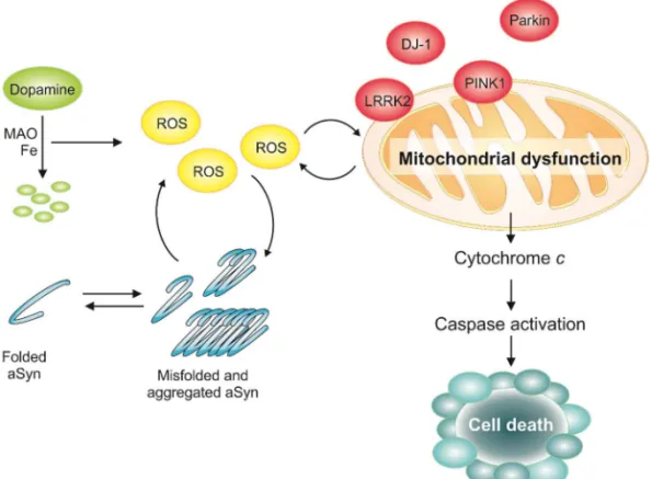
PD symptoms arise from the loss of dopaminergic neurons and reduced levels of
dopamine 20 (Fig. 1). Dopamine functions as a neurotransmitter in the brain. Dopamine plays
a major role in the brain reward system and is involved in motor control. Its chemical
structure comprises an amine that is formed by removing a carboxyl group from a molecule
of L-DOPA 20.
The presence of unfolded, aggregated and ubiquitylated proteins in the LBs of
dopaminergic neurons indicates that proteostasis dysfunction is a common theme
underlying the different etiologies of PD 12, 21. These alterations play a significant role in the
initiation, development and/or progression of the neurodegenerative process in PD.
Concomitantly, one of the pathways from which cell toxicity arises in PD is through aSyn
misfolding and aggregation 22, 23. The SNCA gene, encoding for aSyn, was the first loci
associated with familiar and sporadic cases of PD 10. Genetic alteration in this gene,
including triplication24 and duplication25, as well as missense mutation A30P26, E46K27,
H50Q28, G15D29, A53T10 and A53E30 are described as causing autosomal dominant forms
of this disease. These mutations affect aSyn interaction with membranes and propensity to
6
7
Introduction
Despites the clear relevance of aSyn in PD pathogenesis, several other genes
linked to heritable, monogenic PD have also been described. Including the leucine-rich
repeat kinase 2 LRRK2; the E3 ubiquitin-ligase Parkin; the mitochondrial PTEN-induced
putative kinase 1 PINK1; the oxidation-sensitive chaperone DJ-1; and the lysosomal ATPase
ATP13A2 35. Additionally, mutations in several genes are known to increase the risk of
developing PD, standing out the vacuolar protein sorting 35 homolog VPS35; the ubiquitin
carboxyl-terminal esterase L1 UCH-L1; the translation initiation factor 4-gamma 1 EIF4G1;
and the beta-glucocerebrosidase (GBA)35. The public online database ‘PDGene’ (http://
www.pdgene.org) provides a comprehensive list of PD genetic risk factors, recently
identified in large-scale meta-analysis of genome-wide association studies 36, 37. The proteins
encoded by these genes are involved in processes such as synaptic vesicles 38, 39,
autophagy 40, 41, neurite outgrowth 42, endocytosis 43, mitochondrial morphology 43 and
function 44, mitophagy 45-50 and oxidative stress responses 51. Overall, the genetics of PD
highlights the multifactorial aspect of the disease, underscoring the importance of
mitochondrial function, membrane trafficking and protein quality control systems in the
pathophysiology of PD (Fig. 1).
aSyn aggregation is implicated in the loss of dopaminergic neurons and defects in
dopamine transport. Soluble aSyn is a competitive inhibitor of tyrosine hydroxylase, the
rate-limiting step in tyrosine to L-DOPA biosynthesis 52. aSyn in its functional form is bonded to
membranes. Although, pathological conditions results in aSyn impaired vesicular binding,
which abrogates lipid-mediated signalling cascades and vesicle trafficking 19, 52. Therefore,
the equilibrium between lipid-associated (folded) and cytoplasmic (aggregated) aSyn seems
to provide a mechanistic link between dopamine production, packaging and vesicle
dynamics. Moreover, impaired neurotransmitter release might lead to the formation of
reactive oxygen species (ROS) 53 (Fig. 1).
Oxidative stress and dysfunctional mitochondrial metabolism is also responsible for
cell toxicity in PD 54. There is evidence that a reduction in oxidative phosphorylation and a
decrease in complex 1 activity, leading to ROS formation and oxidative stress, are related
8
release of cytochrome C from the intermembrane space to the cytosol, and activation of the
intrinsic apoptotic pathway, resulting in caspase activation and cell death 56.
To overcome aSyn-induced toxicity cells have protein quality control systems to deal
with misfolded and aggregated proteins. aSyn can be degraded by the ubiquitin-proteasome
pathway (UPS), chaperone mediated autophagy (CMA) and macroautophagy 57. However,
failure of protein clearance pathways and accumulation of misfolded proteins with age leads
to the formation of protein aggregates and cell toxicity 22 (Fig. 1).
The dysfunction in either of the pathways, protein clearance or mitochondrial
pathway, leads to oxidative stress which causes malfunction of these very same pathways
by feedback and feed-forward mechanisms, resulting in irreversible cell damage and death
19 (Fig. 1).
Despite the significant progress in understanding the molecular basis of
neurodegeneration, the lack of known useful molecular targets for therapeutic intervention
has slowed down the scientific discovery processes. Thus, combating PD by preventing or
reversing the build-up of toxic protein aggregates, mainly composed of aSyn, may be an
interesting therapeutic approach.
1.2.1.1. Alpha-synuclein misfolding and aggregation
Intense efforts have been made to characterize the sequence and structure of aSyn.
This 14.5 kDa protein has an amphipathic lysine-rich terminus, an aggregation prone central
region and a disordered acidic carboxy-terminal tail58, 59 (Fig. 2).
The N-terminal region of aSyn contains four 11 residue imperfect repeats with a
highly conserved hexameric motif (KTKEGV)10, 60. This region is responsible for aSyn
interaction with membranes and the PD aSyn mutations were found in this terminal region10,
60 (Fig. 2).
The central region of aSyn was first identified in AD amyloid plaques and was named
non-amyloid component region (NAC), it is composed predominantly by hydrophobic
residues9. The NAC region is necessary for aSyn aggregation, as shown by the deletion of
9
Introduction
Figure 2: Biochemical structure and schematic representation of aSyn. A) Biochemical structure of micelle-bound aSyn (Protein Data Bank ID:1XQ8). The N-terminal region, the non-amyloid-β component of Alzheimer’s disease amyloid plaques (NAC) region and the C-terminal part are coloured orange, green and blue, respectively. Numbers refer to amino acid residues flanking the different regions. B) Schematic representation of aSyn showing the aSyn residues that can be phosphorylated in blue and the mutations associated with familial PD are shown in purple. The N-terminal amphipathic region of the protein is represented in orange, the hydrophobic central region containing the NAC peptide is represented in green and the highly acidic C-terminal is represented in blue. The imperfect KTKEGV repeats are represented in purple.
The C-terminal region is highly enriched in acidic (glutamate and aspartate) and
proline residues and is highly disordered 63, 64. It has been suggested that this region plays
a critical role in modulating the stability, structure, aggregation and function of aSyn in vivo
65. Additionally, it has been implicated in the regulation of aSyn interactions with proteins 66,
6768, 69, metals 70, 71 and small molecules (e.g. dopamine and polyamines) 72. The C-terminal
region also contains the majority of the known aSyn PTMs sites 60, including the
phosphorylation sites (Fig. 2).
In solution, aSyn (WT, A30P, E46K and A53T) exists as an ensemble of disordered
conformations 73. The study of the familiar forms of PD contributed significantly to the
understanding of PD intimate relation with aSyn misfolding and aggregation. The missense
10
or mature fibrils (A53T and E46K), and as well as genomic multiplications of the aSyn
encoding gene, led to an increase in the cytoplasmic accumulation of the aSyn monomer
and potentiated its aggregation and toxicity 74-76.
Several other factor also contribute to triggering the aggregation of aSyn, including
post-translational modifications (PTMs) and interactions with specific metals 70, 71 and small
molecules 72. Fibril formation by aSyn proceeds through a series of discrete oligomeric
intermediates, known as protofibrils of different sizes and morphologies, including spherical,
annular and chain-like structures 13, 20 (Fig. 3).
Figure 3: Putative pathway of aSyn aggregation. In the cytosol, aSyn can unfold due to SNCA mutations, PTMs, oxidized dopamine, reactive oxygen species (ROS) or toxins. This process can be reverted by chaperones. Unfolded aSyn monomers interact to form initially unstable dimers, which grow slowly to generate oligomers of varying morphologies, including transient spherical and ring-like oligomers that eventually originate fibrils. The aSyn oligomers are in equilibrium with monomers and convert to fibrils by monomer addition via a nucleated polymerization mechanism. The accumulation of these amyloid fibrils leads to the formation of intracellular inclusions called Lewy bodies.
The ‘amyloid hypothesis’ states that the aggregation of proteins into an ordered
fibrillar structure is causally related to aberrant protein interactions that culminate in neuronal
dysfunction and ultimately neurodegeneration77.
Despite the well-known connection between protein misfolding, aggregation and
disease, the way by which misfolding results in disease is not clearly understood. In the “gain
of toxic function” theory proteins acquire toxic conformations, which may disrupt the
homeostasis of cells, and the respective tissues and organs. In “the loss of function” theory
the lack of functional protein, due to its recruitment into the aggregates, results in the failure
11
Introduction
Recent experimental evidence suggests the formation of neurotoxic oligomers as a
key pathological event not only in PD but also in other synucleinopathies81, 82. Although the
molecular basis for oligomer neurotoxicity is still elusive, oligomeric species likely have a
negative impact on the UPS machinery and on mitochondrial function83. These species also
seem to trigger cellular defence pathways, such as the stress-response and caspase
activation84. The aggregation of aSyn and the putative gain of toxic function may contribute
to haploinsufficiency by entrapping the wild type aSyn protein, thus reducing the amount of
available functional aSyn 80.
Therefore, in neurodegenerative diseases, it appears that the symptoms arise from
the destruction of cells by a “gain of toxic function” that results from the aggregation process
itself or by a combination of the “gain of toxic function” and a “loss of normal function” of the
proteins in the cell 78-80.
1.2.1.2. Alpha-synuclein phosphorylation
Several PTMs have been described as modulators of aSyn aggregation, including
phosphorylation85, ubiquitination86, sumoylation87, nitration88, enzymatic cross-linking, and
C-terminal truncation60, 88. Phosphorylation at the aSyn serine 129 (pS129) is the dominant
pathological modification of aSyn, and has emerged as a defining hallmark of PD and related
synucleinopathies 85, 89-91. Although aSyn is phosphorylated at low levels under physiological
conditions, a striking 90% of aSyn appears to be phosphorylated on S129 in LBs and
inclusions isolated from brains of patients who died with PD, multiple system atrophy (MSA),
dementia with Lewy bodies (DLB), or other synucleinopathies 85, 89-92.
In addition to S129, serine 87 (S87), tyrosine 39, 125, 133 and 135 (Y39, Y125, Y133
and Y135) are also phosphorylated 93. pS87 is increased in brains of transgenic models of
synucleinopathies and human brains from AD, DLB and MSA patients94.
The phosphorylation sites in aSyn are highly conserved and, with the exception of
S87 and Y39, are located in the C-terminal region, suggesting important roles for these
modifications in regulating aSyn interaction with other proteins, ligands, metals and small
molecules60 (Fig. 2). Accordingly, increasing evidence from pathological, genetic, animal
12
or multiple sites may play an important role in regulating its structure, membrane binding,
oligomerization, fibril formation, LB formation and neurotoxicity in vivo 85, 89. However, the
significance of pS129 in the pathogenesis of synucleinopathies is still unresolved.
In vitro studies demonstrated that pS129 and pS87 significantly inhibits aSyn
fibrillization 94, 95 96. In rodent and Drosophila models of PD, pS129 promotes aSyn
oligomerization but does not influence inclusion formation 97, 98. Accordingly, abolishing
pS129, using the phospho-resistant aSyn form S129A, promotes aSyn fibrillization and
inclusion formation, favouring the hypothesis that pS129 results in kinetic stabilization or
accumulation of toxic aSyn oligomers, in fly and rodent models97, 98.
Concerning aSyn toxicity, pS129 accelerated neuronal loss in Drosophila, suggesting
a toxic effect for aSyn pS129 97. Contrarily, PLK2, a major contributor of aSyn S129
phosphorylation, was shown to be protective in yeast 99, 100, Caenorhabditis elegans and rat
neurons 101. Consistently, over-expression of phospho-resistant aSyn form S129A in the
brain of rat led to increased aSyn toxicity 102, 103, supporting the hypothesis that aSyn pS129
is protective. Nevertheless, whether pS129 promotes or prevents aSyn aggregation and
toxicity, and the pathways affected by pS129, remains largely controversial96, 104, 105.
1.2.1.3. Oxidative stress and mitochondrial dysfunction
Redox reactions are the basis for numerous biochemical pathways and cellular
chemistry, biosynthesis and regulation. They are also important for understanding biological
oxidation and radical/antioxidant effects 106.
A crucial advance of scientific interest in the field of oxygen toxicity and free radicals
occurred when Fridovich described in 1969 107 the existence of superoxide dismutase (SOD)
in almost all aerobic cells, leading to the description of the superoxide theory of oxygen
toxicity, which became the focus of much research and debate associated with ageing,
development, diseases and cell signalling 106.
Oxygen-derived pro-oxidants can be classified as radical and non-radical oxygen
metabolites, commonly named reactive oxygen species (ROS), which can cause damage to
biological targets such as lipids, DNA, proteins and on the defending systems of the cell
13
Introduction
The organism must continuously confront and control the presence of both oxidants
and antioxidants. This balance, often referred to as the redox potential, is specific for each
organelle and biological site, and any interference of the balance in any direction might be
deleterious for the cell and organism.
An increased generation of ROS or reactive nitrogen species (RNS), collectively
known as RNOS, can occur via several cellular insults, including ultraviolet irradiation,
redox-cycling of quinones, the metabolism of xenobiotics, ageing, environmental mitochondrial
toxins, and mutant toxic proteins: e.g. Aβ in AD, mutant huntingtin in HD, aSyn in PD, SOD1
loss of function mutant in ALS 106.
Oxidative stress plays a central role in the pathology of PD as it arises during
dopamine metabolism and during oxidative phosphorylation 108, 109. Moreover, reduced
complex I activity, observed in PD, increases ROS production 110-112 (Fig. 4).
Metabolism of dopamine leads to the formation of several cytotoxic molecules,
including superoxide anions, dopamine–quinone species, and hydroxyl radicals 113, 114.
Dopamine breakdown may occur spontaneously in the presence of iron, or can be catalysed
by monoamine oxidase (MAO) in a reaction that generates hydrogen peroxide 20.
Normally, cells scavenge these deleterious molecules using several antioxidant systems.
For instance, reduced glutathione (GSH) peroxidase, oxidized glutathione,
GSH-S-transferase, SOD and catalase20. In PD, however, an abnormal increase in the production
of ROS might tilt the balance between production and elimination, leading to enhanced
oxidative stress 20 which may also contribute to aSyn pathology in PD. In fact, oxidative
modified aSyn is more prone to aggregation than the native protein115. Additionally, aSyn
protofibrils may cause toxicity directly, inducing an oxidative stress that subsequently
disables UPS by several mechanisms: reduction of ATP levels, inhibition of the proteasome,
and oxidative modification of Parkin, which results in an increasing accumulation of
aggregates, creating a vicious cycle of ROS and aggregates production. Furthermore,
elevated aSyn expression can itself cause oxidative stress116. There is accumulating
evidence for impaired oxidative phosphorylation and decreased complex I activity in PD,
which leads to ROS formation and oxidative stress117. In parallel, aSyn expression induces
14
the mitochondrial permeability transition pore (mPTP), release of cytochrome C from the
intermembrane space to the cytosol, and activation of mitochondrial-dependent apoptosis,
resulting in caspase activation and cell death118, in yeast cells 119, 120 and human fetal
dopaminergic neurons 121. Suggesting that aSyn triggers the apoptotic cell death program.
15
Introduction
Normal mitochondrial function is notably compromised by loss of function of LRRK2,
PINK1, Parkin and DJ-1, resulting in early-onset Parkinsonism118. Concomitantly, there is
evidence that these proteins might have neuroprotective effects against mitochondrial
dysfunction, although their exact site of action remains unknown 55, 118 (Fig. 4).
PINK1 is a serine/threonine kinase located in mitochondria, it appears to
phosphorylate specific mitochondrial proteins exerting a protective cellular function. It has
been shown to inhibit the release of cytochrome C following ceramide-induced stress 56.
Although the exact pathophysiological role of PINK1 is not clear, disruption of this kinase in
Drosophila increased the vulnerability to oxidative stress and mitochondrial morphological
defects, ultimately resulting in a gradual loss of dopaminergic neurons 46.
Parkin is an E3 ubiquitin-ligase, besides labelling specific substrates for degradation,
it can control sorting and trafficking of proteins. Furthermore, observations made in cellular
and transgenic models suggest a role for Parkin in mitochondria 46, 122, a hypothesis
strengthened by its physical and genetic interaction with PINK1 46, 122.
It was shown that Parkin acts as an effector of PINK1 affecting mitochondrial function
46, 123, since the phenotypes induced by loss of PINK1 were rescued by overexpression of
Parkin, but the inverse was not observed 46, 123. In vitro studies further demonstrated Parkin
interaction with LRKK2 124. LRRK2 is a kinase that localizes at membranous and vesicular
structures, including mitochondria 125. Recent studies reported its function in the regulation
of membrane dynamics relevant to endocytosis, mitochondrial morphology and function 43,
44. Continuing to explore the role of PD associated genes with mitochondria, studies reported
a physical interaction of Parkin and PINK1 with DJ-1126, 127. DJ-1 was shown to confer
protection against ROS128 and very diverse functions has been assigned. Although, the
presumed anti-oxidative properties of DJ-1 are probably the most relevant for PD, chaperone
activity and autophagy regulation have also been ascribed to this protein 41, 126.
The discovery of new therapies, preferably natural blood-brain barrier (BBB)
permeable products with protective capacity, including food supplements which could help
16
addressed. The beneficial effects of certain (poly)phenols (e.g. catechins and resveratrol)
on PD and aSyn oligomerization are well documented 129-134.
1.2.1.4. Quality control systems
Aggregation of misfolded proteins appears to be a complex physical-chemical
process, being highly regulated by the cellular environment and modulated by various
cellular components, as molecular chaperones, proteolytic enzymes and other factors135.
Failure of such regulatory mechanisms is likely to be a major factor in the onset and
development of misfolding diseases. Thus, proteostasis is a central event in the
pathobiology of several disorders136. When the accumulation of unfolded proteins surpasses
the capacity of the endoplasmatic-reticulum (ER) to cope with the protein load, diverse
quality control mechanisms actively sequester and degrade these proteins 137-139. The quality
control mechanisms are conserved from yeast to mammalian cells 140 and the unfolded
protein response, triggered upon ER stress, is one of the most important regulators. It leads
to the transcription of genes related with restoration of protein-folding homeostasis, including
chaperones, protein degradation and secretion pathways 137, 140.
Strong evidence on the involvement of the quality control systems in
neurodegeneration came from genetic studies of familial PD cases141. PD causative
mutations were identified in the UPS related genes, Parkin 142 and UCH-L1 143, encoding an
ubiquitin E3 ligase and an ubiquitin hydrolase, respectively. Homozygous mutations in the
DJ-1 gene product, a redox-sensitive molecular chaperone144 involved in normal UPS
function126, have also been described in families with PD inheritance145.
The involvement of molecular chaperones on aSyn toxicity came from the
observation that enhanced aSyn inclusion formation was observed upon deletion of
individual chaperones in yeast 146. Concomitantly, a mild heat shock, treatment with
geldanamycin (a heat shock response activator), or overexpression of the chaperones
Hsp70 119, Jem1 or Hsp90 147, protected yeast cells against aSyn-induced ROS and
subsequent toxicity. The involvement of chaperones in PD was also supported by evidences
17
Introduction
Figure 5: Cellular quality control systems are involved in cytoprotection at different stages of aSyn aggregation. aSyn can misfold and form pathological oligomers that fibrillize and deposit into larger aggregates, ultimately forming LBs. In healthy cells, the cellular quality control systems are able to counteract this cascade of events. The first steps of aggregation can be prevented or reversed by promoting the degradation of misfolded proteins by the ubiquitin-proteasome system while the later ones are counteracted by degradation mediated by macroautophagy. Chaperones can aid the correct conformation of proteins in the several states of aggregation.
Finally, the heat shock-induced protective mechanism may involve Hsp104 and its
co-chaperones, which were described to relieve cells from ER-stress 152. It was shown that
Hsp104 degraded aSyn in a concentration-dependent manner and decreased aSyn
fibrillation in vitro 153-155. In agreement, Hsp104 antagonized aSyn aggregation and reduced
dopaminergic degeneration in a rat model of PD 156. It was shown that at endogenous levels
the presence of Hsp104 had a deleterious effect on aSyn aggregation, and deletion of
Hsp104 in yeast expressing WT or mutant aSyn resulted in lower oxidative stress,
cytotoxicity, increased cell viability and rescue of endocytotic defects 157. Nevertheless,
another study reported that it is possible to reprogram Hsp104 to rescue aSyn proteotoxicity
18
aggregate dissolution, restored protein localization, suppressed proteotoxicity 158, and in a
C. elegans PD model attenuate dopaminergic neurodegeneration158.
aSyn expression promotes the impairment of the proteasome, as described in the
pioneer study of the yeast PD model159. The reduced proteasome activity was found to be
the result of a deficient proteasome composition160. Furthermore, the failure of the UPS
enhanced aSyn toxicity161, 162 and led to the accumulation of inclusions163, either by impairing
the UPS using proteasomal mutants 161, 162 or the chemical inhibitor, lactacystin 163. Despite
the clear evidence of proteasome dysfunction in PD, a recent study reported that the
degradation of aSyn aggregates is more dependent on autophagy than on proteasome
function, in the yeast PD model164, suggesting that the proteasome is more likely to be
responsible for the degradation of soluble forms of aSyn, and that there is a complex
cross-talk between the different proteolytic pathways involved in aSyn degradation165 (Fig. 5).
Autophagy involves the formation of an autophagosomal vesicle that transports the
misfolded and aggregated proteins to degradation, in the lysosome in higher eukaryotes or
the vacuole in yeast. The term autophagy was coined approximately 50 years ago and the
molecular insights were significantly explored after the discovery of autophagy in yeast
subjected to starvation 166 167. Autophagy is regulated by the kinase “target of rapamycin”
(TOR) 168. Lst8, a TOR interacting protein was identified as an aSyn toxicity modulator in
yeast169. Moreover Ypk9 (the yeast orthologue of ATP13A2), a vacuolar P-type ATPase,
was identified as a suppressor of aSyn toxicity 169 and aggregation101. PD-associated
mutations in ATP13A2 appear to cause a loss-of-function as the protein no longer localizes
at the lysosome 170. The role of this ATPase in neuropathology is not completely understood,
but its putative lysosomal function may have important consequences for
autophagy-mediated clearance of protein aggregates and organelles. Concomitant with the beneficial
influence of autophagy in aSyn toxicity, rapamycin treatment, which induces autophagy by
inhibiting TOR, was reported to reduce aSyn inclusions 163. These observations are in line
with experiments indicating that under pathological conditions, autophagy is required for the
degradation of aSyn aggregates, rather than the proteasome164. However, the autophagic
19
Introduction
There are controversial findings regarding the role of autophagy on aSyn toxicity. A
study reported that rapamycin treatment lead to increased aSyn toxicity in yeast169. It was
also shown that WT or A53T mutant aSyn were not able to enter the vacuole and promoted
vacuolar fusion defects in yeast146. Additionally, aSyn-mediated mitophagy, specific
degradation of mitochondria through autophagy, was reported to be deleterious in aged
yeast cells 172. To intensify the discussion, aSyn LB-like aggregates resisted degradation
and impaired autophagy in cellular models 172.
It is clear that the interplay between autophagy and aSyn toxicity and aggregation is
still elusive and needs to be explored.
1.3.
Yeast as a model of PD
Saccharomyces cerevisiae, also known as baker’s yeast, is one of the most versatile
biological systems model. This unicellular eukaryotic organism is extremely useful for
molecular biologists and it has been extensively used in the study of numerous complex and
devastating disorders, such as HD and PD 173.
Multiple characteristics make this simple eukaryote a model system of choice.
Despite the obvious absence of a nervous system, basic mechanisms and pathways
underlying neurodegenerative diseases, such as mitochondrial dysfunction, transcriptional
deregulation, trafficking defects and proteasomal dysfunction, are highly conserved between
yeast and humans, enabling detailed studies of the molecular events involved in those
conditions. In fact, several remarkable insights into the understanding of brain diseases have
been recently achieved with yeast models84, 173, 174. To develop disease models, it is essential
that some relevant aspects of the disease phenotype are reiterated. It is possible to study
directly a gene function implicated in a disease, if provided a yeast homolog. On the other
hand, if the gene is absent in yeast, and causes disease by a toxic gain-of-function in
humans (eg. SNCA), it can still be modelled via the heterologous expression of the human84.
Notably, several PD associated genes, such as aSyn, LRRK2, Parkin, DJ-1, VSP35,
EIF4G1 and ATP13A3, haven been successfully expressed in yeast, by heterologous or
20
Nevertheless, the yeast PD model most used is based on the heterologous
expression of aSyn and within this models various phenotypes can be achieved, depending
on the expression system, given that this affects the level of aSyn expression 159, 175, 176. This
feature has been also explored according to the objectives of the studies.
The use of multicopy plasmids revealed that yeast cells reduce the average plasmid
copy number in order to reduce aSyn expression and toxicity 159. To avoid this, insertions of
the aSyn coding sequence in the yeast genome enabled more stable expression and the
levels of toxicity could be manipulated by varying the number of copies of the aSyn cDNA
inserted in the genome 176, 177. The use of a galactose-inducible promoter provided additional
control for the synchronous induction of expression of aSyn, avoiding the negative pressure
during routine cell manipulations.
Using these various expression systems, several genetic modifiers (enhancers and
suppressors) of aSyn toxicity were identified in genetic screenings in yeast 175, 176.
In other studies, yeast cells expressing different levels of aSyn, hence displaying
different levels of cytotoxicity, revealed the involvement of multiple cellular pathways in the
toxicity 159, 176-181.
The drug discovery process for synucleinopathies has been delayed by the elusive
nature of the mechanisms leading to neurodegeneration and lack of reliable model
organisms to screen for compounds. Moreover, aSyn is a ”natively unstructured protein”,
missing a defined secondary structure under physiological conditions, which constitutes an
additional challenge for drug discovery73. As a major effort to overcome these difficulties
several phenotypic high-throughput assays have been developed. These are cell-based
assays that recapitulate phenotypic and functional aspects of disease, focused on
viability/toxicity readouts, leading to the identification of small molecules and also potential
novel drug-targets. However, it is essential to scrutinize the mechanism of action of the small
molecules and candidate compounds. Here yeast cells offer a remarkable advantage, it
allows target and mechanisms identification thought diverse and complementary genetic
approaches, empowering the development of pre-clinical candidates. Furthermore, several
cell-based assays targeting key molecular aspects of PD, such as aSyn cytotoxicity,
21
Introduction
yeast159, 176, 182. All of these processes are well conserved between yeast and higher
eukaryotes, including mammals.
Figure 6: Yeast models of Parkinson’s disease. Proteins associated with PD are shown, as well as the type of expression (heterologous or homologous). The structure of the proteins are represented for aSyn, LRRK2, Parkin, EIF4G1, DJ-1, and VPS35 (Protein Data Bank ID: 1XQ8, 2ZEJ, 4I1H, 2VSX, 4OQ4 and 2R17 respectively. ATP13A2 structure is not determined).
The main drawback for phenotypic high-throughput assays using stable immortalized
cell lines is the resistance to proteotoxic insults, resulting in relatively low aSyn toxicity. This
resistance is in part caused by the reduced apoptotic mechanisms necessary to enable
proliferation in culture. However, assays that monitor cell viability through mitochondrial
potential or membrane leakage could be of use in low toxicity models, however extensive
optimization is required. Primary rat neurons infected with aSyn-expressing lentivirus, are
more stable and sensitive but they also present technical limitations related with virus and
cell quantities, besides being costly and labour demanding. Indeed, the lack of published
cell culture toxicity screens reflects the difficulty in their employment in high-throughput
22
Yeast has numerous advantages at the early stages of the drug development
process, in comparison to mammalian cells and animal models. Notwithstanding the obvious
lack of a nervous system the simplicity of yeast, missing the complex neuronal
communication with other cell types makes it ideal to study early-onset events in disease
pathology. Some useful properties of yeast include the ease of experimental and genetic
manipulation, low cost, functional similarity to higher eukaryotes and fast growth.
Additionally, several major drugs have hit the same targets and elicit the same responses in
yeast as they do in humans, including statins, methotrexate, omeprazole, tacrolimus
(FK506) and bortezomib (Velcade) 183, 185. The yeast platform enables rapid screen and
analysis of therapeutic targets, providing a direct and important linkage between pathways
and chemicals. Several examples of pharmacological discoveries using yeast-based assays
have been reported. These have been performed in 384- and 1536-plate format, illustrating
their high-throughput potential 186.
Nevertheless, it is important to acknowledge that yeast has its limitations, as with all
model organisms. It is a unicellular organism, some genes important in neurodegeneration
may be absent and it has a cell wall, which might constitute a barrier to some molecules.
Yet, this last undesirable attribute can be attenuated by genetic manipulation of the efflux
pump system or the ergosterol biosynthesis, reducing the capability of yeast cells to export
drugs or by increasing yeast cells permeability, respectively. Though, the unparalleled
discovery characteristics of yeast outweigh any potential drawbacks, particularly since many
genes tied to neurodegenerative disorders are ubiquitously expressed and highly conserved
187.
Yeast is considered a robust primary drug-screening platform to filter for compounds
with cytoprotective activity, for further complementation with assays in more physiologically
relevant models. Approaches involving the sequential use of different model systems,
starting with simpler cellular models and ending with more complex animal models, already
resulted in the discovery of promising small molecules with therapeutic potential (described
below). Recently, it was established a yeast-to-human discovery platform for
23
Introduction
PD patient derived neurons. Subsequently, yeast was used for clarification of the
mechanism of action, due to yeast unmatched genetic tools 188.
Ultimately, yeast-to-human discovery and validation platforms will provide more
significant findings into disease pathology. By iteratively moving between simple cellular
models and patient derived cells we will be able to elucidate mechanisms and evaluate
patient-specific drug targets, this will allow to pursue more significant animal and clinical
trials in order to overcome neurodegenerative diseases.
1.3.1. Testing small molecules and natural compound in
yeast
The reduced number of reported high-throughput drug screens highlights the
difficulty in establishing robust, meaningful PD models amenable to screening. Yeast,
despite its obvious differences from neuronal cells, has actually provided potentially
beneficial lead compounds against aSyn toxicity.
The yeast discovery platform has been used as an approach to understand basic
mechanisms of protein aggregation and toxicity. The events leading to protein
oligomerization are likely amenable to modulation by small molecules. Thus, notably, yeast
has been used to screen for small molecules that can inhibit aSyn aggregation and toxicity.
Screening of large libraries of compounds lead to the identification of aSyn toxicity
suppressors in yeast. In a large-scale genetic screen, ~115 000 compounds were tested for
the ability to rescue aSyn toxicity at micromolar concentration and a class of structurally
related 1,2,3,4-tetrahydroquinolinones were identified177. These compounds reduced the
formation of aSyn inclusions, re-established ER-to-Golgi trafficking, and ameliorated
mitochondria defects induced by aSyn. The targets were further confirmed in nematode
neurons and in primary rat neuronal midbrain cultures. Interestingly, these compounds also
rescued rotenone toxicity in neuronal cultures, a toxin used to study mitochondrial deficit in
PD177.
The ease of manipulation makes yeast a suitable tool to explore unconventional
compounds and its mechanisms. Mannosylglycerate, a compatible solute typical of marine
microorganisms thriving in hot environments, was found to reduce aSyn aggregation in a
24
against aSyn by inducing its degradation through autophagy, representing a novel scaffold
for discovery of robust pro-autophagic/anti-neurodegeneration compounds190.
A novel class of molecules, cyclic peptides (CPs), was also screened in yeast191. CPs
are natural-product-like chemicals with potent bioactivity. Yeast was exploited to express a
plasmid-derived self-splicing intein that liberates a CP. This approach enabled the scale-up
of high-throughput screens to 10–100 times the size of a typical small molecule screen. A
pool of 5 million yeast transformants were screened and two related CP constructs with the
ability to reduce aSyn toxicity were identified. These cyclic peptide constructs also prevented
dopaminergic neuron loss in a nematode model of PD191.
Small molecules have been the centre of drug high-throughput screenings, and
amongst them phytochemicals have emerged as attractive molecules in the context of
neurodegeneration. It is largely accepted that dietary products such as green tea, small fruits
and even olive oil have health benefits, however the major advances regarding its
mechanisms and targets were only achieved in the last decade. The yeast allied with animal
and chemical studies has significantly contributed for these discoveries.
The first small compound high-throughput screen in yeast tested ~10 000 compounds
and identified a group of protective flavonoids, quercetin and epigallocatechin gallate
(EGCG), which protected aSyn-expressing yeast in the presence of iron192. The protection
promoted by these compounds was further analysed. The positive effect was virtue of its
anti-oxidant and metal-chelating activities. Importantly, (poly)phenols, and particularly
quercetin and EGCG, have been proven beneficial in cellular and animal models of PD 193,
194.
The advances in biochemical tools and the multidisciplinary teams also gave a major
contribution to drug discovery. In fact, the green tea benefits have been deciphered by
combining HPLC fractionation in a microplate format with yeast screening and parallel
electrospray mass spectrometry195. This integrated process enabled the rapid assess of the
efficacy of fractions and systematically target their bioactive constituents. The green tea
metabolites were individually examined for their pharmacological effects and interestingly,
25
Introduction
metabolites196. This study emphasizes the prominence of yeast high-throughput screenings
to dissect natural extracts and explore the numerous synergistic effect of its metabolites.
Clues for candidate compounds for aSyn pathology can also be obtained from
screenings in other neurodegenerative disease models, as the case of ALS yeast model,
based on the expression of the protein TDP-43 197. The yeast powerful genetics was used
to identify multiple protective 8-hydroxyquinolines, which are natural plant alkaloids 197.
Some of these compounds were also protective in aSyn yeast and nematode models. Its
protective mechanisms were related with its ionophores and intracellular metal chelation
activity 197. From this screening N-aryl benzimidazole (NAβ) proved more potent and
effective against aSyn toxicity than TDP-43. Thus, the yeast aSyn platform was explored to
identify NAβ mechanisms. It reversed diverse phenotypes induced by aSyn, including the
accumulation of aSyn inclusions, the generation of ROS, the block of ER–Golgi trafficking
and the nitration of proteins 198. Moreover, this compound was used in an iterative
yeast-to-human neuron platform to understand how translational its targets were 181.
Taken together, identifying disease therapies is an incredible challenge.
Nevertheless, rapidly improving methodologies and iterative processes, allied with an
evolving mechanistic understanding of disease, is nurturing more interdisciplinary
approaches to research and fostering drug discovery, with the ultimate goal of discovering
novel therapeutics for humans.
2.
(Poly)phenols
Phenolic compounds, commonly referred to as polyphenols constitute one of the
most extensive and ubiquitous group of secondary metabolites in the plant kingdom. It is
estimated that more than 8000 compounds have been isolated and described 199. These
compounds are characterized structurally by the presence of, at least, one hydroxyl
functional group (-HO) linked to an aromatic ring. Polyphenols are usually conjugated to
sugars, carboxylic and organic acids, amines, lipids and other phenols. They are classified
according to the number of phenols rings and the structural elements that bind these rings199.