O
h
r
c
i
r
g
a
in
e
a
s
l
R
e
Şule Taş Gülen1, Onur Yazıcı1, İmran Kurt Ömürlü2 1Chest Disease Department, 2Biostatistics Department, Adnan Menderes University, Aydın, Turkey Role of Inlammation in Pulmonary Embolism
The Prognostic Value of Hematological
Parameters in Patients with Pulmonary Embolism
Pulmoner Emboli Hastalarında Hematolojik
Parametrelerin Prognostik Değeri
DOI: 10.4328/JCAM.4800 Received: 05.09.2016 Accepted: 12.10.2016 Printed: 01.05.2017 J Clin Anal Med 2017;8(3): 207-10 Corresponding Author: Şule Taş Gülen, Department of Chest Diseases, Adnan Menderes University School of Medicine, Aydin, Turkey.
GSM: +90 5056919099 E-Mail: [email protected] Özet
Amaç: PE’li hastaların tanı anındaki ve tedavi sonrasındaki hemogram değer-leri incelenerek, hematolojik parametredeğer-lerin tedavi yanıtı ve hastalığın prog-nozu ile ilişkisi araştırılmıştır. Gereç ve Yöntem: Çalışmaya Aralık 2014-Ara-lık 2015 tarihleri arasında hastanemiz Göğüs Hasta2014-Ara-lıkları Kliniği’nde PTE ta-nısı ile yatırılarak tedavi edilmiş toplam 48 hasta alındı. Olgu dosyaları diji-tal arşiv sisteminden retrospektif olarak incelendi ve demografik verileri, kli-nik değerlendirmeleri ile tanı anındaki ve tedavi sonrasındaki hemogramları retrospektif olarak değerlendirildi. Hemogram parametrelerinden WBC, NLR, MPV’nin ölçümleri incelendi. İstatistiksel analizler için SPSS 17,0 programı ile Kolmogorov-Smirnov, Mann-Whitney U ve bağımsız grup t testleri kullanıldı. Bulgular: Çalışmamıza alınan toplam 48 hastanın ortalama yaşı 62.68 olup, 27’si (%56.2) erkek idi. Ortalama yatış süresi 9,2 gün olarak bulundu. Tanı anında NLR, MPV ve WBC ortanca değerleri sırasıyla 4.82 (3.04-9.32), 10.2 (7.40-11.8), 9215 (6507.5-13255) olup tedavi sonunda ise; 2.60 (1.82-3.74), 9.3 (7-12.9), 7265 (6125-8872.5) olarak bulundu. NLR, MPV ve WBC değerle-rinin tedavi sonrası istatistiksel olarak anlamlı düştüğü saptandı. (p <0,001) Tartışma: Çalışmamızın sonuçları nötrofil lenfosit oranı, ortalama trombosit hacmi ve beyaz kürenin pulmoner embolinin tanısı ve tedavi yanıtını gösteren prognostik inlamatuar belirteçler olabileceğini düşündürmektedir.
Anahtar Kelimeler
Pulmoner Emboli; Beyaz Küre Sayısı; Nötrofil Lenfosit Oranı; Ortalama Trom-bosit Hacmi
Abstract
Aim: By analyzing the hemogram values of patients with PE during and fol-lowing the treatment, the relationships of hematological parameters with response to treatment and prognosis of the disease were investigated. Ma-terial and Method: Forty-eight patients, who were hospitalized and treated with the diagnosis of PTE in the Pulmonary Diseases Clinic of our hospital between December 2014 and December 2015, were included in the study. The patients’ charts, located in the digital archive system, were analyzed ret-rospectively and demographic characteristics, clinical evaluations, and their hemogram results during and following the treatment were retrospectively evaluated. Among the hemogram parameters, the values of WBC,NLR and MPV were statistically analyzed. For statistical analysis, SPSS 17.0 sotware and the Kolmogorov-Smirnov test, Mann-Whitney U test and independent sample t test were used. Results: The average age of the 48 patients included in our study was 62.68, and 27 (56.2%) of them were males. The average duration of hospitalization was 9.2 days. The median values of NLR, MPV, and WBC at the time of diagnosis were 4.82 (3.04-9.32), 10.2 (7.40-11.8), and 9215 (6507.5-13255), respectively. Following treatment, these values were 2.60 (1.82-3.74), 9.3 (7-12.9), and 7265 (6125-8872.5), respectively. It was determined that the NLR, MPV, and WBC values were statistically signiicant-ly reduced following treatment (p <0,001). Discussion: Our study suggested that NLR,MPV and WBC can be used as prognostic inlammatory indicators for diagnosing and treating pulmonary embolism.
Keywords
Pulmonary Embolism; White Blood Cell Count; Neutrophil Lymphocyte Ratio; Mean Platelet Volume
Bu araştırma, Türk Toraks Derneği 19. Yıllık Kongresinde (6-10 Nisan 2016, Antalya) Poster olarak sunulmuştur.
Role of Inlammation in Pulmonary Embolism
Introduction
Pulmonary embolism (PE) is a preventable, important clinical problem with high morbidity and mortality [1]. Simple tests can be used to determine prognosis and to evaluate patients’ responses to treatment. Hemogram parameters such as white blood cell count (WBC), platelet count, neutrophil lymphocyte ratio (NLR), and mean platelet volume (MPV) have been recently investigated as inlammatory indicators in numerous disorders [2,3]. In recently conducted studies on patients with PE, the elevation of NLR has been found to be correlated with early mortality [4]. Studies that examine MPV have determined that MPV was increased, platelet count was reduced in pulmonary embolism and these values were associated with the diameter of the right ventricle [5,6 ].
As far as we know there is no study in the medical literature that investigates the efect of PE treatment on hematological parameters. In this study, by analyzing the hemogram values of patients with PE during and following treatment, the relation-ships of hematological parameters with response to treatment and prognosis of the disease were investigated.
Material and Method
Forty-eight patients, who were hospitalized and treated with the diagnosis of PE in the Pulmonary Diseases Clinic of our hospital between December 2014 and December 2015 were enrolled in the study. Patients who did not have any clinical signs of infec-tion (such as fever, cough, or sputum) or high laboratory param-eters (such as C-reactive protein and procalcitonin) and whose patient charts were available for investigation, were included in the study. Following approval of the local ethics committee, patients’ charts were retrospectively analyzed via the digital archive system. Their demographic data (age, gender, medi-cal history), duration of hospital stay, comorbidities, chronic treatments administered, and the units in which they had been followed-up for acute phase and maintenance embolism treat-ments (clinic/intensive care unit) were recorded. The patients were classiied as low mortality (stable hemodynamic status and absence of right ventricular dysfunction); intermediate mortality risk group (stable hemodynamic status and presence of right ventricular dysfunction in radiological or laboratory in-vestigations); and high mortality (unstable hemodynamic sta-tus), according to the Turkish Thoracic Society 2015 Consensus Report on Diagnosis and Treatment of Pulmonary Thromboem-bolism. Patients with low mortality risk were named as Group 1, and patients having intermediate-low, intermediate-high, and high risks were joined into one group and named as Group 2. The hemogram results of patients who had no identiied infec-tions, obtained at the time of diagnosis and as part of discharge from the hospital were retrospectively evaluated. The routine hemogram parameters, white blood cell count (WBC), neutro-phil and lymphocyte counts were recorded together with the mean platelet volume (MPV). By dividing the neutrophil count by the lymphocyte count, the neutrophil lymphocyte ratio, which is one of the nonselective inlammatory markers, was calculated.
Statistical analysis
For statistical analysis, SPSS sotware (Statistical Package for Social Sciences) version 17.0 was used. The
Kolmogorov-Smirnov test was used to assess the normality of numeric variables. For the numeric variables that were normally distrib-uted, comparison between two groups was made by indepen-dent sample t test and descriptive statistics are presented as mean±standard deviation. For the numeric variables that were not normally distributed, comparison between the two groups was made by the Mann–Whitney U test and descriptive statis-tics are presented as median (25-75 percentiles). The p values below 0.05 were considered statistically signiicant.
Results
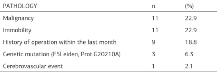
The average age of the 48 patients enrolled in the study was 62.68 ± 15.88 (30-84), of whom 27 (56.2%) patients were male. The average duration of hospitalization was 9.2 ± 4.64 (1-22) days. When frequencies of risk factors and comorbidities were analyzed, 35 (62.6%) patients were determined to have at least one pathology (Table 1).
When grouped in terms of early mortality, 30 patients were in the low-risk group (Group 1), 13 patients were in the interme-diate-risk group, and ive patients were in the high-risk group for embolism (Group 2) (Table 2). No statistically signiicant dif-ferences were found between the two groups in terms of NLR, MPV, and WBC values at the time of diagnosis (Table 3).
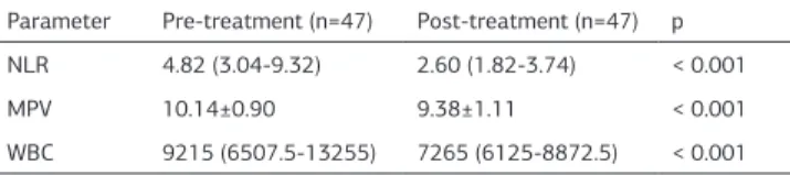
The median values of NLR, MPV, and WBC were 4.82 (3.04-9.32), 10.2 (7.40-11.8) , and 9215 (6507.5-13255), respectively, at the time of diagnosis. Since one patient died at the second hour of treatment, the results of 47 patients were available fol-lowing treatment. Post-treatment median values of NLR, MPV, and WBC were 2.60 (1.82-3.74), 9.3 (7-12.9), and 7265 (6125-8872.5), respectively; these were statistically signiicantly lower when compared to the results at the time of diagnosis (Table 4). Table 1. The Frequency of Risk Factors and Comorbidities in Patients with Pul-monary Embolism
PATHOLOGY n (%)
Malignancy 11 22.9
Immobility 11 22.9
History of operation within the last month 9 18.8 Genetic mutation (F5Leiden, Prot.G20210A) 3 6.3
Cerebrovascular event 1 2.1
Table 2. Classiication of Early Mortality
Mortality Risk n %
High 5 10.4
Intermediate - High 4 8.3
Intermediate - Low 9 18.8
Low 30 62.5
Table 3. The Comparison of Hematological Parameters According to Risk Groups
Parameter Grup 1 (n=30) Grup 2 (n=18) p NLR 6.04 (3.14-9.85) 4.51 (2.64-7.31) 0.277
MPV 10.03±0.90 10.38±0.87 0.186
WBC 9215 (6662.5-12815) 9125 (5695-13820) 0.766
| Journal of Clinical and Analytical Medicine 208
Role of Inlammation in Pulmonary Embolism
Discussion
Pulmonary embolism is a disorder with high mortality when let untreated. Establishment of the prognosis and the choice of treatment are made according to the classiication based on early mortality risk 1. Until now, clinical scorings such as Ge-neva scoring, and pulmonary embolism severity index or cardi-ac markers such as troponin and natriuretic peptide have been used to determine prognosis [7-9 ].
Although the role of systemic inlammation in PE has been known, its association with prognosis has not been clearly identiied. In conducted studies, leucocytes were shown to par-ticipate in venous thrombosis by creating endothelial injury [9]. Additionally, NLR is considered to be a simple indicator of sub-clinical inlammation and is used for prediction of mortality in diseases such as coronary artery disease and cancer [10]. Our study investigated not only clinical scoring systems but also the relationships of laboratory tests, such as NLR, MPV, and WBC, simply determined by complete blood count, with the PE prog-nosis. When classiication was made according to early mortal-ity, no signiicant diferences were found between the low-mor-tality PE group and the intermediate-high morlow-mor-tality PE group at the time of diagnosis, in terms of NLR, MPV, and WBC. The study conducted by Ermiş et al. on 209 patients with acute pul-monary embolism (APE) and 162 healthy controls, investigated whether MPV was a prognosticindicator in high-risk pulmonary embolism; MPV was found to be unrelated to the severity of embolism (massive, submassive, nonmassive) [11]. Similarly, in our study, a relationship between severity of embolism and MPV was not found. In the study conducted by Kostrubiec et al. on 192 APE patients and 100 healthy controls, no diference was found between the two groups in terms of MPV value. However, when the patients were classiied according to low, intermedi-ate and high mortality risks, the MPV values were signiicantly higher in the intermediate and high-risk groups when compared to the low-risk group. In addition, there was no signiicant difer-ence between the MPV values of the intermediate and high-risk group and those of healthy controls [12]. Similar to the study by Ermiş et al., they determined that MPV value was higher in patients who died when compared to patients who survived. In both studies, this situation was explained by the possible rela-tionship of MPV with the right ventricular dysfunction and myo-cardial injury [11,12]. Varol et al., in their study on 107 APE that patients and 70 healthy controls, showed that MPV was higher in APE and this was correlated with the diameter of the right ventricle [13]. We had no healthy control group in our study and therefore, comparisons with a control group were not possible. However, when patients were grouped according to the mortal-ity risk, no signiicant diferences were found between groups in terms of NLR, MPV, and WBC. This situation might be explained by the small number of patients in our study and the fact that the groups were nonhomogeneous.
A study by Kayrak et al. on 359 APE patients, investigated whether NLR was a prognostic indicator for early mortality in APE. In the group that died at the end of the irst month of treatment, NLR and WBC were found to be signiicantly higher when compared to patients who survived. For this reason, it was emphasized that NLR and WBC were simple and cheap tests that predict early mortality (4). In another study conduct-ed by Yeşildağ et al., the relationships of computerizconduct-ed tomog-raphy obstruction score (Qanadli obstruction score) with NLR and MPV were investigated in 95 patients, who were diagnosed with PE by computerized tomography pulmonary angiography (CTPA). In this study, when right ventricle / let ventricle short axis ratios and NLR were compared with survival rate and mor-tality, mortality was signiicantly related to NLR and MPV [14]. In our study, radiological indings were not evaluated. Since mortality had occurred in only one patient at the second hour of treatment, evaluation was not possible in terms of mortality. Currently, the issues of whether NLR, MPV, and WBC are in-creased in APE and are indicators of mortality is being investi-gated; however, there have not been suicient studies evaluat-ing post-treatment response. In the study conducted by Eren et al. on 209 patients who received treatment for acute coronary syndrome, poor cardiovascular results of NLR and its charac-teristics for mortality prediction were investigated. The cut-of value of NLR was taken as 4.7 in terms of mortality and the patients were grouped as low (<3.0), intermediate (3.0-4.7) and high (>4.7). Their in-admission, in-hospital, 6th month, and follow-up NLR values were compared to the risk scores named GRACE (Global Registry of Acute Coronary Events) and TIMI (Thrombolysis in Myocardial Infarction); no diferences were found between groups in cardiac-related hospitalizations [15]. In our study, it was determined that following treatment, the values of NLR, MPV, and WBC were signiicantly reduced when compared to the pre-treatment values.
The Limitations of the Study
The retrospective characteristics of our study and the relatively small number of patients when group analysis was performed constituted the limitations of the study. Also, we didn’t include healthy control groups in the study; instead, we compared the before-treatment and ater-treatment values of NLR, MPV, and WBC in patients with an indication of pulmonary embolism.
Conclusion
The values of NLR, MPV, and WBC were found to be signii-cantly reduced following treatment. We suggest that these pa-rameters are simple, cheap, and easily accessible indicators for demonstrating the response to the treatment of the disease. Since the number of patients is small in our study, more com-prehensive, prospective studies are required in order to support our suggestion.
Competing interests
The authors declare that they have no competing interests.
References
1. Arseven O, Okumus NG, Ongen G, Müsellim B. Turkish Thoracic Society Con-sensus Report on Diagnosis and Treatment of Pulmonary Thromboembolism in 2015. Istanbul, 2015.
Table 4. The Comparison of Pre-treatment and Post-treatment Hemogram Pa-rameters
Parameter Pre-treatment (n=47) Post-treatment (n=47) p NLR 4.82 (3.04-9.32) 2.60 (1.82-3.74) < 0.001 MPV 10.14±0.90 9.38±1.11 < 0.001 WBC 9215 (6507.5-13255) 7265 (6125-8872.5) < 0.001
Journal of Clinical and Analytical Medicine | 209
Role of Inlammation in Pulmonary Embolism
2. Bhat T, Teli S, Rijal J, Bhat H, Raza M, Khoueiry G, et al. Neutrophil to lympho-cyte ratio and cardiovascular diseases: a review. Expert Review of Cardiovascular Therapy 2013;11:55-9.
3. Celik A, Ozcan IT, Gündes A, Topuz M, Pektas I, Yesil E, et al. Usefulness of admission hematologic parameters as diagnostic tools in acute pulmonary embo-lism. Kaohsiung Journal of Medical Sciences 2015;31:145-9.
4. Kayrak M, Erdoğan HI, Solak Y, Akilli H, Gül EE , Yildirim O, et al. Prognostic Value of Neutrophil to Lymphocyte Ratio in Patients with Acute Pulmonary Embolism: A Restrospective Study. Heart Lung Circ 2014;23:56-62.
5. Varol E, Icli A, Uysal BA, Ozaydin M. Platelet indices in patients with acute pul-monary embolism. Scand J Clin Lab Invest 2011;71:163-7.
6. Talay F, Ocak T, Alcelik A, Erkuran K, Akkaya A, Duran A, et al. A New Diagnostic Marker For Acute Pulmonary Embolism In Emergency Department: Mean Platelet Volume. African Health Sciences 2014;14:94-9.
7. Aujesky D, Perrier A, Roy PM, Stone RA, Cornuz J, Meyer G, et al. Validation of a clini¬cal prognostic model to identify low-risk patients with pulmonary embolism. J Intern Med 2007;261:597-604.
8. Wicki J, Perrier A, Perneger TV, Bounameaux H, Junod AF. Predicting ad¬verse outcome in patients with acute pulmonary em¬bolism: a risk score. ThrombHae-most 2000;84:548-552.
9. Jo JY, Lee MY, Lee JV, Rho BH, Choi W. Leukocytes and systemic inlammatory response syndrome as prognostic factors in pulmonary embolism patients. BMC Pulm Med 2013;13:74-81.
10. AlkhouriN, Stif GM, Campbell C, Lopez R, Tamimi TAR, Yerian L, et al. Neutro-phil to lymphocyte ratio: a new marker for predicting steatohepatitis and ibrosis in patients with nonalcoholic fatty liver disease. Liver International 2012;32:297-302.
11. Ermis H, Yucel N, Gulbas G, Turkkan S, Aytemur ZA. Does the mean platelet volume have any importance in patients with acute pulmonary embolism? Wien Klin Wochenschr 2013;125:381-5.
12. Kostrubiec M, Labyk A, Pedowska-Wlosek J, Hrynkiewicz-Szymanska A, Pacho S, Jankowski K, et al. Mean platelet volume predicts early death in acute pulmo-nary embolism. Heart 2010;96:460-5.
13. Varol E, Icli A, Uysal BA, Ozaydin M. Platelet indices in patients with acute pulmonary embolism. Scandinavian Journal of Clinical and Laboratory Investiga-tion 2011;71:163-7.
14. Yesildag M, Keskin S, Güler I, Keskin Z. Correlation of Computerized Tomogra-phy Angiographic Pulmonary Artery Obstruction Score with Hematologic Outcome and Mortality in Patients with Acute Pulmonary Embolism. TurkiyeKlinikleri J Med Sci 2013;33:952-7.
15. Eren M, Ozpelit E, Aytemiz F, Güngör H, Güneri S. Neutrophil to Lympho-cyte Ratio on Admission: Is a Predictor of Cardiovascular Outcome in Patients with Acute Coronary Syndrome as it Predicts Mortality? Koşuyolu Heart Journal 2014;17:153-8.
How to cite this article:
Gülen ŞT, Yazıcı O, Ömürlü İK. The Prognostic Value of Hematological Parameters in Patients with Pulmonary Embolism. J Clin Anal Med 2017;8(3): 207-10.
| Journal of Clinical and Analytical Medicine 210