D
GAG Knock-In and Purkinje Cell-Specific
Dyt1
Conditional Knockout Mice
Lin Zhang1, Fumiaki Yokoi1, Yuan-Hu Jin1, Mark P. DeAndrade1, Kenji Hashimoto2, David G. Standaert1, Yuqing Li1*
1Center for Neurodegeneration and Experimental Therapeutics, Department of Neurology, School of Medicine, University of Alabama at Birmingham, Birmingham, Alabama, United States of America,2Division of Clinical Neuroscience, Chiba University Center for Forensic Mental Health, Chiba, Japan
Abstract
Background: DYT1 early-onset generalized dystonia is a neurological movement disorder characterized by involuntary muscle contractions. It is caused by a trinucleotide deletion of a GAG (DGAG) in theDYT1(TOR1A) gene encoding torsinA; the mouse homolog of this gene isDyt1(Tor1a). Although structural and functional alterations in the cerebellum have been reported in DYT1 dystonia, neuronal morphology has not been examinedin vivo.
Methodology/Principal Findings:In this study, we examined the morphology of the cerebellum inDyt1DGAG knock-in (KI) mice. Golgi staining of the cerebellum revealed a reduction in the length of primary dendrites and a decrease in the number of spines on the distal dendrites of Purkinje cells. To determine if this phenomenon was cell autonomous and mediated by a loss of torsinA function in Purkinje cells, we created a knockout of theDyt1gene only in Purkinje cells of mice. We found the Purkinje-cell specificDyt1conditional knockout (Dyt1pKO) mice have similar alterations in Purkinje cell morphology, with shortened primary dendrites and decreased spines on the distal dendrites.
Conclusion/Significance:These results suggest that the torsinA is important for the proper development of the cerebellum and a loss of this function in the Purkinje cells results in an alteration in dendritic structure.
Citation:Zhang L, Yokoi F, Jin Y-H, DeAndrade MP, Hashimoto K, et al. (2011) Altered Dendritic Morphology of Purkinje cells inDyt1DGAG Knock-In and Purkinje Cell-SpecificDyt1Conditional Knockout Mice. PLoS ONE 6(3): e18357. doi:10.1371/journal.pone.0018357
Editor:Takeo Yoshikawa, Rikagaku Kenkyu¯sho Brain Science Institute, Japan
ReceivedJanuary 29, 2011;AcceptedFebruary 28, 2011;PublishedMarch 29, 2011
Copyright:ß2011 Zhang et al. This is an open-access article distributed under the terms of the Creative Commons Attribution License, which permits unrestricted use, distribution, and reproduction in any medium, provided the original author and source are credited.
Funding:This work was supported by National Institutes of Health grants (NS37409, NS47466, NS47692, NS54246, NS57098, NS65273, NS72872, and NS74423), the Department of Neurology (UAB), and Tyler’s Hope for a Dystonia Cure, Inc. The funders had no role in study design, data collection and analysis, decision to publish, or preparation of the manuscript.
Competing Interests:The authors have declared that no competing interests exist.
* E-mail: [email protected]
Introduction
Dystonia is a neurological syndrome characterized by involun-tary contractions of both agonist and antagonist muscles of affected regions that cause twisting and abnormal movements or postures [1]. DYT1 dystonia is a genetically determined form of generalized early-onset dystonia, with an age of onset between childhood and adolescence. Symptoms usually first affect the lower limbs and eventually progress to the entire body [2]. DYT1 dystonia is caused by a trinucleotide deletion of a GAG (DGAG) codon in theDYT1(TOR1A) gene, which results in the loss of a glutamic acid residue in the C-terminal region of the torsinA protein [3]. We previously generated Dyt1DGAG knock-in (KI) mice, a mouse model of DYT1 dystonia, showed impairments of motor coordination and balance in the beam-walking test and hyperactivity in the open-field test [4]. The function of torsinA is largely unknown, but it is a member of the AAA+ ATPase superfamily and is believed to have a chaperone-like function [5,6,7,8,9,10].
Biochemical and cellular studies show that torsinA localizes to the endoplasmic reticulum [6], protects against oxidative stress,
and prevents protein aggregate formation [5,7,8,9]. In situ hybridization studies have also revealed that torsinA mRNA is highly expressed in the dopaminergic neurons of the substantia nigra pars compacta, granule and pyramidal neurons of the hippocampus, Purkinje and dentate nucleus neurons of the cerebellum, and cholinergic neurons of the neostriatum in humans [11,12]. Furthermore, ultrastructural studies of the striatum of humans and macaques have revealed an association of torsinA immunostaining with small vesicles within axons and presynaptic terminals forming symmetric synapses [13].
genecaytaxin, cerebellectomy eliminates the motor symptoms and rescues the juvenile lethality [20,21]. Electronic lesions of dorsal portions of the lateral vestibular nuclei (dLV), which receive input from the Purkinje cells, are associated with the greatest improvement in this rat [22]. The Purkinje cells send the major inhibitory signal from the cerebellum to the deep cerebellar nuclei, which is mediated by the neurotransmitter c-aminobutyric acid (GABA). Pharmacological disruption of the cerebellar signaling is also shown to induce dystonia in mice [23]. Lastly, the tottering mouse, which has a recessive mutation of a calcium channel gene, shows ataxia and paroxysmal dystonia, but this phenotype can be eliminated by surgical removal of the cerebellum or introduction into a Purkinje cell-specific degenerative background [24,25].
Despite these findings, few studies have sought to examine the potential role of the Purkinje cells in the pathogenesis of DYT1 dystonia and other dystonias. In this study, we examined the morphology of the cerebellum inDyt1DGAG knock-in (KI) mice. Golgi staining of the cerebellum of KI mice revealed a reduction in the length of primary large dendrites and a decrease in the number of spines on the distal dendrites of Purkinje cells. Since it has been reported that theDGAG mutation causes a reduction of torsinA in the striatum and the entire brain [26,27,28], we sought to determine if this phenomenon was mediated by a loss of function of torsinA in Purkinje cells by creating a knockout of theDyt1gene only in Purkinje cells (Dyt1pKO) of mice. We previously reported the making of Dyt1 loxP mice [29]. In the present study, we produced Dyt1 pKO mice by crossing the Dyt1 loxP mice with Pcp2-cre mice, which restricts lox-mediated recombination to Purkinje cells [30]. We found that the Purkinje cells in theDyt1 pKO mice have a similar morphology to that of the KI mice, with shortened primary large dendrites and decreased spines on the distal dendrites.
Results
Golgi staining of Purkinje cells in KI mice
To examine the morphological structures of the Purkinje cells in KI mice, we used Golgi staining of cerebellar sections. First, the sizes of the Purkinje cell soma were measured and no significant difference was found between the KI and control (CT) mice (means6standard errors; CT: 10062.22%; KI: 99.1961.35%; p.0.05, Figure 1A–1B). However, the length of the large primary dendrite in the KI mice was approximately 20% shorter than those in the control mice (CT: 10067.84%; KI: 80.1562.95%; p,0.01, Figure 1C). Furthermore, the number of spines in the quaternary dendrite branch of KI mice was approximately 27% percent less than those in control mice (CT: 10061.9%; KI: 73.1162.17%; p,0.01, Figure 1D–1E). These results suggest the important role of torsinA in the dendritic structure and morphology of Purkinje cells.
Generation of the Purkinje-cell specificDyt1 conditional knockout (Dyt1pKO) mice
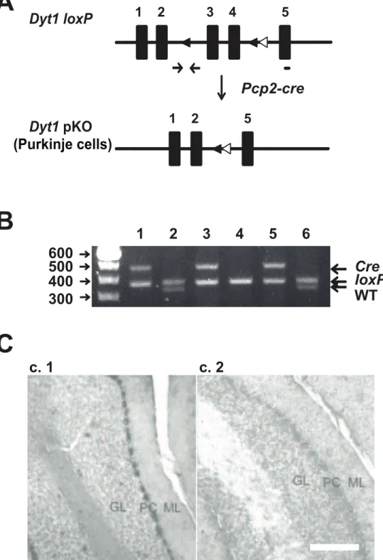
To examine whether the morphological alterations in the KI mice was caused by a loss of torsinA function in Purkinje cells, we generated a Purkinje cell-specific knockout of theDyt1.Dyt1 loxP mice [29] were crossed with a line of mice with thecrerecombinase gene driven by the promoter ofPcp2, a Purkinje cell-specific gene [30]. Mice double heterozygous for both theDyt1 loxPandPcp2-cre were then crossed withDyt1 loxPhomozygous mice to deriveDyt1 pKO mice and their control littermates (Figure 2A). Genotyping forDyt1pKO and control littermates was performed by multiplex PCR (Figure 2B). We confirmed Purkinje cell specific knockout of Dyt1byin situhybridization. TorsinA mRNA was highly expressed
in the Purkinje cells in control littermates (Figure 2C.1). In contrast, torsinA mRNA was not detected in Purkinje cells inDyt1 pKO mice (Figure 2C.2), suggestingDyt1was specifically knocked out in the Purkinje cells.
Golgi staining of Purkinje cells inDyt1pKO mice To examine whether the Dyt1 pKO mice recapitulate the dendritic morphology of the KI mice, we performed Golgi staining on cerebellar sections fromDyt1pKO mice at approximately 8 months old. Similar to the KI mice, no significant difference in the size of the Purkinje cell soma in Dyt1 pKO mice compared to control mice was observed (CT: 10063.20%; Dyt1 pKO: 94.2762.82%; p.0.05, Figure 3A–3B). Furthermore, the length of the large primary dendrite in the Dyt1 pKO mice was approximately 37% shorter than those in control mice (CT: 10065.65%; Dyt1 pKO 63.3661.73%; p,0.001, Figure 3C). Lastly, the number of spines in the quaternary dendrite branch of the Dyt1pKO mice was approximately 33% less than those in control mice (CT: 10063.45%;Dyt1pKO: 67.3662.2%; p,0.01, Figure 3D–3E). These results suggest that torsinA plays an important role in the Purkinje cell dendritic development.
To examine whether the morphological alteration in Purkinje cells was developmentally regulated, we analyzed the Purkinje cells inDyt1pKO mice at 2–3 months old. No significant difference in the size of the Purkinje cell soma inDyt1pKO mice was observed compared to control mice (CT: 10063.71%; Dyt1 pKO: 98.0162.52%; p.0.05). There was also no significant difference in the length of the large primary dendrite in Dyt1 pKO mice compared to control mice (CT: 10063.81%; Dyt1 pKO 90.6863.37%; p.0.05). However, the number of spines in the quaternary dendrite branch of the Purkinje cells was reduced in Dyt1pKO mice compared to control mice (CT: 10060.55%;Dyt1 pKO: 93.5860.85%; p,0.001). The results suggest that the spine numbers reduced in advance of the reduction of the length of the large primary dendrite by the loss of torsinA function in the Purkinje cells.
Discussion
First, we examined the morphology of Purkinje cells in the cerebellum in KI mice. We found that the primary large dendrites of the Purkinje cells in KI mice were significantly shorter than that of wild type mice and there was a marked reduction in the number of spines. Next, to determine if this morphological change was mediated by a cell-autonomous effect of loss of torsinA function, we generated a line of mice in which torsinA was conditionally knocked out only in Purkinje cells. TheDyt1pKO mice showed a similar decrease in the length of primary large dendrites and a reduction in the number of spines. The results also suggest that the spine numbers reduced in advance of the reduction of the length of the large primary dendrite by the loss of torsinA function in the Purkinje cells. These results suggest that torsinA plays an important role in the development of the cerebellum, and that a loss of this function in the Purkinje cells results in a cell autonomous effect leading to an alteration in dendritic structure. If the same alterations are present in patients with DTY1 dystonia, this change in synaptic associations between the parallel fibers and the Purkinje cells may contribute to the pathogenesis of the dystonic symptoms.
the cortico-striatal-pallido-thalamocortical and the cerebellar-thalamo-cortical pathways [32]. Anatomical studies have pro-posed an interaction between the cerebellum and the basal ganglia through a disynaptic pathway originating in the cerebellum and projecting to the striatum via the thalamus [33]. A deficiency in this connectivity was identified in patients with DYT1 human carriers [34]. Additionally, rats that have
undergone a hemicerebellectomy were found to have a complete loss of striatal long-term depression (LTD) [35], which was also compromised in mutant human torsinA transgenic mice [36]. Since the Purkinje cells are the sole output from the cerebellum, the abnormal function of Purkinje cells, as represented by our results, may be responsible for the change in striatal LTD in mutant torsinA animals.
Figure 1. Purkinje cells in KI mice.(A) A representative Purkinje cell trace (left). A representative Purkinje cell from CT and KI mice at 406 magnification (right). (B) There was no significant difference in size of the Purkinje cell soma between CT and KI mice. (C) However, the large primary dendrite of the Purkinje cells in the KI mice was significantly shorter than those in CT mice. (D) Next, the quaternary dendrite branch of CT and KI mice was examined at 1006magnification. (E) The number of spines on the quaternary dendrite branch in the KI mice was significantly reduced compared to CT mice. Scale bars in Panel A represent 10mm. Scale bars in panel D represent 1mm. Bars in Panels B, C and E represent means with standard errors. ** p,0.01.
Figure 2. Generation ofDyt1pKO mice.(A) Schematic diagram of the generation of theDyt1pKO mice. Filled boxes represent exons. Filled triangles indicateloxPsites. Open triangles indicate theFRTsites that were incorporated to remove theneocassette.Dyt1 loxPmice were crossed withPcp2-cremice to obtain double heterozygotes. The double heterozygotes were crossed withDyt1 loxPhomozygotes to obtainDyt1pKO mice. The primer sites for genotyping ofDyt1locus were shown by an arrow pairs. The short bar under exon 5 is the site of probe used forin situ
hybridization.Dyt1exons 3 and 4 were removed in Purkinje cells ofDyt1pKO mice. (B) An agarose gel showing the various PCR products that were used to genotype mice. The top band indicates the presence of thePcp2-crelocus, the middle band represents theDyt1 loxPlocus, and the bottom band represents theDyt1wild-type locus. Lanes 4:Dyt1 loxPhomozygous mice. Lanes 2, 6:Dyt1 loxPheterozygous mice. Lanes 1, 3, 5:Dyt1pKO mice. (C)In situhybridization was used to confirm the Purkinje cell-specific knockout of theDyt1gene. CT (C.1) andDyt1pKO (C.2) mice. GL: granule cell layer; PC: Purkinje cell layer; ML: molecular layer. Scale bar represents 100mm in C.2.
doi:10.1371/journal.pone.0018357.g002
The Purkinje cells play an important role in motor coordination and motor learning by integrating two types of excitatory inputs: climbing fibers and parallel fibers. Climbing fibers originate from the inferior olivary nucleus and convey the motor signals to the parallel fibers. Parallel fibers are the T-shaped axons of cerebellar granule cells, and convey the sensory and motor information carried through the pontocerebellar and spinocerebellar mossy fiber pathways. It is also known that the parallel fiber-Purkinje cell (PF-PC) synapse plays an important role in the adaptive learning process [37]. Glutamate receptord2 subunit (GluRd2) is selectively expressed in the Purkinje cells of the cerebellum [38]. Impairment
of motor coordination, Purkinje cell synapse formation, and cerebellar LTD was reported in GluRd2 mutant mice [39]. GluRd2 mutant mice were unable to stabilize PF-PC synapses and resulted in a reduction in the number of PF-PC synapses along with impaired CF synapse elimination [40]. We previously reported motor deficits in KI mice [4] and in this study found morphological alterations of Purkinje cells in KI mice. The results suggest that functional alterations of the cerebellum may associate with the pathogenesis of DYT1 dystonia.
Furthermore, torsinA is known to interact with kinesin 1 [41], a motor protein involved in cellular transport and the cytoskeleton Figure 3. Purkinje cells inDyt1pKO mice.(A) A representative Purkinje cell from control andDyt1pKO mice, produced at 406magnification. (B) The size of the Purkinje cell soma inDyt1pKO mice was not significantly different than those of CT mice. (C) The large primary dendrite of the Purkinje cells in theDyt1pKO mice was significantly shorter than those in CT mice. (D) A representative quaternary dendrite branch of CT andDyt1
pKO mice was examined at 1006magnification. (E) The number of spines on the quaternary dendrite branch in theDyt1pKO mice were significantly reduced compared to those of CT mice. Scale bars in Panel A represent 10mm. Scale bars in Panel D represent 1mm. Bars in Panels B, C and E represent means with standard errors. ** p,0.01, *** p,0.001.
of the cell.In vitrostudies have shown that suppression of kinesin leads to decreased neurite extension in hippocampal neurons [42]. Furthermore, in human neuroblastoma cells, it was shown that overexpression of mutant torsinA also leads to decreased neurite extension [43]. A decrease in neurite extension possibly through kinesin could explain the decrease in primary dendritic length. In addition to the shortened primary dendrite length, however, the KI mice also showed a decrease in spine number. This decrease in neurite extension would not explain the decrease in number of spines. Recent reports have shown that overex-pression of mutant human torsinA inDrosophilaresults in altered synaptic morphology at the neuromuscular junction [44]. It is, therefore, likely that this decrease in spine number results in altered synaptic plasticity, possibly leading to decreased parallel fiber connectivity.
Lastly, we have created a novel Purkinje cell-specific knockout ofDyt1to compare its morphology to the KI and wild-type mice. Several genetic studies suggest that a loss-of-function of torsinA contributes to the pathology of dystonia [28,29,45]. We have found theDyt1pKO mice replicates the KI dendritic morphology of Purkinje cells, in that they have decreased primary dendrite length and decreased spine number. These findings suggest that theDGAG mutation in the KI mice results in a loss of function of torsinA and provide further evidence of the important role of torsinA in the cerebellum.
In conclusion, these results add to the growing body of evidence of the importance of the cerebellum in the pathogenesis of dystonia and this being the first reported morphological alteration in the cerebellum. Furthermore, these results suggest that torsinA plays an important role in the regulations of dendritic length and spine number in the cerebellum. Finally, the
DGAG mutation in theDyt1 may result in a loss of function of torsinA in the Purkinje cells. These findings will further the understanding of the pathophysiology that underlies not only DYT1 dystonia but also possibly other neurological movement disorders.
Materials and Methods
Animals
All experiments were carried out in compliance with the USPHS Guide for Care and Use of Laboratory Animals and approved by the Institutional Animal Care and Use Committee at the University of Alabama at Birmingham with Animal Protocol Number 10008918. All experiments were performed by investigators blind to the genotype of the mice. Dyt1 loxP mice were generated as previously described [29]. To generate the Purkinje cell-specific knockout,Dyt1 loxPmice were first crossed with Pcp2-cre mice [30]. The double heterozygous mice were then crossed with Dyt1 loxP homozygous mice to derive Dyt1 pKO mice and their control littermates. Genotyping for Dyt1 pKO and control littermates was performed by multiplex PCR using F (59-ATTCAAAAATGTTGTCATAGCCAGG-39) and T (59-CTACAGTGACCTGAATCATGTGGC-39) primer sets [29] for Dyt1 loxP and creA (59 -ATCTCCGGTATTGAAA-CTCCAGCGC-39) and cre6 (59 -CACTCATGGAAAATAGC-GATC-39) primer sets forcre[46]. KI mice were prepared and genotyped as previously described [4]. Mice were housed under a 12-h-light/dark cycle withad libitumaccess to food and water.
In SituHybridization
Purkinje-cell specific knockout of torsinA was confirmed byin situ hybridization. To prepare the Digoxigenin (DIG) -labeled probe, a DNA fragment corresponding to 39-UTR of Dyt1 was
amplified by PCR with a primer sets of Dyt1insitu2 (59 -CACCAAGCTGGACTACTACCTGGA-39) and Dyt1insitu3 (59 -GAAAGCTTCTTATAGTATTAAAACC-39) and Dyt1 DNA plasmid template. The amplified PCR fragment was then ligated into a pGEM-T Easy vector (Promega). Next, the construct was transformed inE. coliJM109 competent cells (Promega) and single colonies were isolated. An appropriate clone that had the DNA fragment in the correct direction was confirmed by PCR using T7 and Dyt1insitu3 primer sets. The plasmid DNA was purified and cut with the restricted enzyme NcoI. The DNA fragment was purified and dissolved in diethyl pyrocarbonate (DEPC)-treated water. DIG-labeled probe for Dyt1 was prepared by using digoxingenin RNA labeling kit with the SP6 promoter (Roche Applied Science, Indianapolis, IN).In situhybridization to sagittal sections of the cerebellum was performed as previously described [47].
Golgi staining
Adult KI mice (CT: n = 4; KI: n = 7, approximately 8 months old),Dyt1pKO mice (CT: n = 3;Dyt1pKO: n = 3, approximately 8 months old), and another batch ofDyt1pKO mice (CT: n = 4; Dyt1pKO: n = 4, 2–3 months old) were used in this experiment. After mice were deeply anesthetized with pentobarbital (1 ml/kg, intraperitoneally), the cerebellum was quickly removed and prepared for Golgi staining using the FD Rapid Golgi Stain Kit (FD NeuroTechnologies, Ellicot City, MD). After staining, the cerebellum was frozen with dry ice and sectioned parasagittally (150mm) using a sliding microtome (Histoslide 2000,
Reichert-Jung). The sections were then mounted on slide, dehydrated with xylene and then cover-slipped with permount (Fisher Scientific). Images of individual Purkinje cells were captured using a Nikon ECLIPSE E800M microscope with a 406Plan Fluor objective
lens. The size of each Purkinje cell soma and the length of the large primary dendrite (5 to 16 cells from each mouse) were measured using ImageJ software (NIH, Ver. 1.42 g). Quaternary dendrite branches from the soma as shown in Figure 1A were chosen at random and the spines were counted. The numbers of spines located on randomly selected quaternary dendrite branches (5 to 16 cells from each mouse), 10mm in length, were counted
manually using a 406and 1006Plan Fluor objective lens and a
106CFIUW ocular lens. The size of Purkinje cell soma, the
length of the large primary dendrite, and the spine numbers were measured by an investigator blind to the genotypes and the data were analyzed by another investigator.
Statistical analysis
The area (mm2) of each Purkinje cell soma and the length of the large primary dendrite (mm) were measured and the data were normalized to control mice and expressed as percentage. The number of spines were counted and normalized to that of control mice. All results were analyzed using Student’s t-test. Significance was assigned by aP-value less than 0.05.
Acknowledgments
We thank Alena Samal and Miki Jinno for their technical assistance, and Andrea McCullough, Kevin Feng, and their staff for animal care.
Author Contributions
Conceived and designed the experiments: YL LZ. Performed the experiments: LZ FY YHJ. Analyzed the data: LZ MPD. Contributed reagents/materials/analysis tools: LZ FY YHJ YL. Wrote the paper: LZ KH DGS YL.
References
1. Breakefield XO, Blood AJ, Li Y, Hallett M, Hanson PI, et al. (2008) The pathophysiological basis of dystonias. Nat Rev Neurosci 9: 222–234. 2. Fahn S (1988) Concept and classification of dystonia. Adv Neurol 50: 1–8. 3. Ozelius LJ, Hewett JW, Page CE, Bressman SB, Kramer PL, et al. (1997) The
early-onset torsion dystonia gene (DYT1) encodes an ATP-binding protein. Nat Genet 17: 40–48.
4. Dang MT, Yokoi F, McNaught KS, Jengelley TA, Jackson T, et al. (2005) Generation and characterization of Dyt1 DeltaGAG knock-in mouse as a model for early-onset dystonia. Exp Neurol 196: 452–463.
5. Caldwell GA, Cao S, Sexton EG, Gelwix CC, Bevel JP, et al. (2003) Suppression of polyglutamine-induced protein aggregation in Caenorhabditis elegans by torsin proteins. Hum Mol Genet 12: 307–319.
6. Hewett J, Gonzalez-Agosti C, Slater D, Ziefer P, Li S, et al. (2000) Mutant torsinA, responsible for early-onset torsion dystonia, forms membrane inclusions in cultured neural cells. Hum Mol Genet 9: 1403–1413.
7. Kuner R, Teismann P, Trutzel A, Naim J, Richter A, et al. (2003) TorsinA protects against oxidative stress in COS-1 and PC12 cells. Neurosci Lett 350: 153–156.
8. McLean PJ, Kawamata H, Shariff S, Hewett J, Sharma N, et al. (2002) TorsinA and heat shock proteins act as molecular chaperones: suppression of alpha-synuclein aggregation. J Neurochem 83: 846–854.
9. Shashidharan P, Paris N, Sandu D, Karthikeyan L, McNaught KS, et al. (2004) Overexpression of torsinA in PC12 cells protects against toxicity. J Neurochem 88: 1019–1025.
10. Burdette AJ, Churchill PF, Caldwell GA, Caldwell KA (2010) The early-onset torsion dystonia-associated protein, torsinA, displays molecular chaperone activity in vitro. Cell Stress Chaperones 15: 605–617.
11. Augood SJ, Martin DM, Ozelius LJ, Breakefield XO, Penney JB, Jr., et al. (1999) Distribution of the mRNAs encoding torsinA and torsinB in the normal adult human brain. Ann Neurol 46: 761–769.
12. Augood SJ, Penney JB, Jr., Friberg IK, Breakefield XO, Young AB, et al. (1998) Expression of the early-onset torsion dystonia gene (DYT1) in human brain. Ann Neurol 43: 669–673.
13. Augood SJ, Keller-McGandy CE, Siriani A, Hewett J, Ramesh V, et al. (2003) Distribution and ultrastructural localization of torsinA immunoreactivity in the human brain. Brain Res 986: 12–21.
14. Delmaire C, Vidailhet M, Elbaz A, Bourdain F, Bleton JP, et al. (2007) Structural abnormalities in the cerebellum and sensorimotor circuit in writer’s cramp. Neurology 69: 376–380.
15. Draganski B, Thun-Hohenstein C, Bogdahn U, Winkler J, May A (2003) ‘‘Motor circuit’’ gray matter changes in idiopathic cervical dystonia. Neurology 61: 1228–1231.
16. Obermann M, Yaldizli O, De Greiff A, Lachenmayer ML, Buhl AR, et al. (2007) Morphometric changes of sensorimotor structures in focal dystonia. Mov Disord 22: 1117–1123.
17. Carbon M, Ghilardi MF, Argyelan M, Dhawan V, Bressman SB, et al. (2008) Increased cerebellar activation during sequence learning in DYT1 carriers: an equiperformance study. Brain 131: 146–154.
18. Le Ber I, Clot F, Vercueil L, Camuzat A, Viemont M, et al. (2006) Predominant dystonia with marked cerebellar atrophy: a rare phenotype in familial dystonia. Neurology 67: 1769–1773.
19. Rumbach L, Barth P, Costaz A, Mas J (1995) Hemidystonia consequent upon ipsilateral vertebral artery occlusion and cerebellar infarction. Mov Disord 10: 522–525.
20. LeDoux MS, Lorden JF, Ervin JM (1993) Cerebellectomy eliminates the motor syndrome of the genetically dystonic rat. Exp Neurol 120: 302–310. 21. Xiao J, Ledoux MS (2005) Caytaxin deficiency causes generalized dystonia in
rats. Brain Res Mol Brain Res 141: 181–192.
22. LeDoux MS, Lorden JF, Meinzen-Derr J (1995) Selective elimination of cerebellar output in the genetically dystonic rat. Brain Res 697: 91–103. 23. Pizoli CE, Jinnah HA, Billingsley ML, Hess EJ (2002) Abnormal cerebellar
signaling induces dystonia in mice. J Neurosci 22: 7825–7833.
24. Campbell DB, North JB, Hess EJ (1999) Tottering mouse motor dysfunction is abolished on the Purkinje cell degeneration (pcd) mutant background. Exp Neurol 160: 268–278.
25. Neychev VK, Fan X, Mitev VI, Hess EJ, Jinnah HA (2008) The basal ganglia and cerebellum interact in the expression of dystonic movement. Brain 131: 2499–2509.
26. Cao S, Hewett JW, Yokoi F, Lu J, Buckley AC, et al. Chemical enhancement of torsinA function in cell and animal models of torsion dystonia. Dis Model Mech 3: 386–396.
27. Yokoi F, Yang G, Li J, DeAndrade MP, Zhou T, et al. (2010) Earlier onset of motor deficits in mice with double mutations in Dyt1 and Sgce. J Biochem 148: 459–466.
28. Goodchild RE, Kim CE, Dauer WT (2005) Loss of the dystonia-associated protein torsinA selectively disrupts the neuronal nuclear envelope. Neuron 48: 923–932.
29. Yokoi F, Dang MT, Mitsui S, Li J, Li Y (2008) Motor deficits and hyperactivity in cerebral cortex-specific Dyt1 conditional knockout mice. J Biochem 143: 39–47.
30. Barski JJ, Dethleffsen K, Meyer M (2000) Cre recombinase expression in cerebellar Purkinje cells. Genesis 28: 93–98.
31. Yokoi F, Dang MT, Miller CA, Marshall AG, Campbell SL, et al. (2009) Increased c-fos expression in the central nucleus of the amygdala and enhancement of cued fear memory in Dyt1 DeltaGAG knock-in mice. Neurosci Res 65: 228–235.
32. Carbon M, Eidelberg D (2009) Abnormal structure-function relationships in hereditary dystonia. Neuroscience 164: 220–229.
33. Hoshi E, Tremblay L, Feger J, Carras PL, Strick PL (2005) The cerebellum communicates with the basal ganglia. Nat Neurosci 8: 1491–1493.
34. Argyelan M, Carbon M, Niethammer M, Ulug AM, Voss HU, et al. (2009) Cerebellothalamocortical connectivity regulates penetrance in dystonia. J Neurosci 29: 9740–9747.
35. Rossi S, Mataluni G, De Bartolo P, Prosperetti C, Foti F, et al. (2008) Cerebellar control of cortico-striatal LTD. Restor Neurol Neurosci 26: 475–480. 36. Martella G, Tassone A, Sciamanna G, Platania P, Cuomo D, et al. (2009)
Impairment of bidirectional synaptic plasticity in the striatum of a mouse model of DYT1 dystonia: role of endogenous acetylcholine. Brain 132: 2336–2349. 37. Blazquez PM, Hirata Y, Highstein SM (2004) The vestibulo-ocular reflex as a
model system for motor learning: what is the role of the cerebellum? Cerebellum 3: 188–192.
38. Araki K, Meguro H, Kushiya E, Takayama C, Inoue Y, et al. (1993) Selective expression of the glutamate receptor channel delta 2 subunit in cerebellar Purkinje cells. Biochem Biophys Res Commun 197: 1267–1276.
39. Kashiwabuchi N, Ikeda K, Araki K, Hirano T, Shibuki K, et al. (1995) Impairment of motor coordination, Purkinje cell synapse formation, and cerebellar long-term depression in GluR delta 2 mutant mice. Cell 81: 245–252. 40. Ichikawa R, Miyazaki T, Kano M, Hashikawa T, Tatsumi H, et al. (2002) Distal extension of climbing fiber territory and multiple innervation caused by aberrant wiring to adjacent spiny branchlets in cerebellar Purkinje cells lacking glutamate receptor delta 2. J Neurosci 22: 8487–8503.
41. Kamm C, Boston H, Hewett J, Wilbur J, Corey DP, et al. (2004) The early onset dystonia protein torsinA interacts with kinesin light chain 1. J Biol Chem 279: 19882–19892.
42. Ferreira A, Niclas J, Vale RD, Banker G, Kosik KS (1992) Suppression of kinesin expression in cultured hippocampal neurons using antisense oligonucle-otides. J Cell Biol 117: 595–606.
43. Hewett JW, Zeng J, Niland BP, Bragg DC, Breakefield XO (2006) Dystonia-causing mutant torsinA inhibits cell adhesion and neurite extension through interference with cytoskeletal dynamics. Neurobiol Dis 22: 98–111.
44. Koh YH, Rehfeld K, Ganetzky B (2004) A Drosophila model of early onset torsion dystonia suggests impairment in TGF-beta signaling. Hum Mol Genet 13: 2019–2030.
45. Dang MT, Yokoi F, Pence MA, Li Y (2006) Motor deficits and hyperactivity in Dyt1 knockdown mice. Neurosci Res 56: 470–474.
46. Campos VE, Du M, Li Y (2004) Increased seizure susceptibility and cortical malformation in beta-catenin mutant mice. Biochem Biophys Res Commun 320: 606–614.