Available on line at
Association of the Chemical Engineers AChE
www.ache.org.rs/CICEQ
Chemical Industry & Chemical Engineering Quarterly 15 (1) 33−35 (2009) CI&CEQ
33 RADA PJANOVIĆ1
RADOSLAVA STOJANOVIĆ1
MILANA ŠAJBER2
JELENA VELJKOVIĆ3
NEVENKA BOŠKOVIĆ-VRAGOLOVIĆ1
SRDJAN PEJANOVIĆ1
1Faculty of Technology and
Metallurgy, Department of Chemical Engineering, Belgrade, Serbia
2Hemofarm a.d., Institute
Hemofarm, Vršac, Serbia
3Galenika a.d., Belgrade, Serbia
SHORT COMMUNICATION
UDC 66.021.3:577.115+615.211
DIFFUSION OF LIDOCAINE HYDROCHLORIDE
FROM LIPID MICROPARTICLES
Lipid particles, as drug carriers, are of increasing research interest, because a sustained drug release and avoidance of side effects could be achieved by using them.
Lidocaine hydrochloride is a very efficient local anesthetic, but it has a short effect. The objective was to prolong the effect of the drug by encapsulating li-docaine hydrochloride in phospholipids microparticles.
Two different procedures were used for the preparation of phospholipids micro-particles. In both cases, phosphatidylcholine and lysophosphatidylcholine were used for the preparation of microparticles in which 5 % w/w lidocaine hydro-chloride solution was encapsulated.
The standard Franz diffusion cell was used for the experimental diffusion rate determination. The obtained results show that diffusion rate from micropar-ticles is significantly lower than from the lidocaine hydrochloride solution, which means that this kind of microparticles could be used for the prolonged drug release. There was no much difference in diffusion rates from microparticles obtained by different procedures. That indicates that only the composition of the particles membrane has an influence on the lidocaine hydrochloride re-lease rate.
Key words: microparticles; diffusion; phospholipids; lidocaine; Franz cell.
Phospholipids particles for drug-delivery appli-cations are now the most widely investigated area of their practical application [1-3]. These drug vesicles are self-assembled particles that even occur naturally [4].
Phospholipids posses a polar and a non-polar group on the same molecule and they represent am-phiphiles. Adding a small water amount to phosphor-lipids will cause their self-organization into a lamellar phase. In this self-assembled structure the molecules are oriented in such a way that the polar part of the molecule is in contact with the water and shields the non-polar portion. The additional dilution in the ex-cess water and a mechanical energy input will cause the formation of stable colloidal microparticles. These microparticles are hollow spheres which could be used as drug carriers, for hydrophilic and hydrophobic molecules [5,6].
Lidocaine hydrochloride was chosen as a drug which will be encapsulated in phospholipids
Corresponding author: R. Pjanović, Faculty of Technology and Metallurgy, Department of Chemical Engineering, Karnegijeva 4, 11000 Belgrade, Serbia.
E-mail: [email protected] Paper received: October 16, 2008. Paper revised: October 31, 2008. Paper accepted: November 5, 2008.
ticles. Lidocaine hydrochloride is a local anesthetic the use of which is limited by the relatively short ac-tion of the drug compared with the potential duraac-tion of the pain. This drug is water soluble and could be incorporated into the aqueous phase of phospholipids microparticles. By encapsulation of lidocaine hydro-chloride a slow release could be achieved and the ac-tion of the drug will be prolonged [7].
EXPERIMENTAL
Microparticles were prepared by two different methods.
hydan-R. PJANOVIĆet al.: DIFFUSION OF LIDOCAINE HYDROCHLORIDE… CI&CEQ 15 (1) 33−35 (2009)
34
toin (preservative) were added in the concentrated dispersion and it was agitated on 505 rpm for 10 min. The homogeneous dispersion with 5 % w/w encapsu-lated lidocaine hydrochloride was obtained.
The second method of the microparticle prepa-ration was a thin film method [9]. Lipoid S 75, which was kindly donated by Lipoid AG (Germany), was dis-solved in chloroform and transferred into a round-bot-tom flask. The solvent was evaporated in a rotary evaporator at 40 °C and 350 mbar. The thin film for-med was hydrated with 5 % w/w solution of lidocaine hydrochloride in water and then hand shaken. Small PET particles were added to intensify the mixing.
In both cases, a microparticles membrane con-sists mostly of phosphatidylcholine, with small amounts of lysophosphatidylcholine present. Prolipo S contents 30 % w/w of phosphatidylcholine and 0.5 % w/w lyso-phosphatidylcholine, while Lipoid S75 has 73 % w/w of phosphatidylcholine, 9 % w/w of phosphatidyletha-nolamine and 1.5 % w/w lysophosphatidylcholine. All phospholipids are unsaturated.
Particles were analyzed using an optical micro-scopy with magnification of 1500×.
In order to study the release of lidocaine hydro-chloride from the prepared phospholipids micropar-ticles a jacketed Franz-type cell 25 mm in diameter (donation of PermeGear, Inc., USA) was used (Figure 1). A static diffusion cell comprised two compartments with a membrane between the donor and the receptor sections. The receptor compartment had a fixed vo-lume (20 ml) and it was kept at 37 °C with the water bath. The receptor fluid (distilled water) was mixed with a magnetic bar stirrer.
Figure 1. Jacketed Franz diffusion cell.
Three different diffusion experiments were done in the same way. The first experiment endured 4.5 h and the second and the third 12 h. For all the runs a Franz cell with the receptor solution in both
compart-ments first was thermostated in the water bath at 37 °C for 30 min.
Between the donor and the receptor compart-ments the acetate-cellulose membrane with a pore size of 0.45 µm was placed.
In the first experiment, 5 % w/w solution of lido-caine hydrochloride in water was used as a donor phase. In the second and the third experiment, the obtained liposomal dispersions with encapsulated lido-caine hydrochloride were used as a donor phase.
The samples were withdrawn periodically, first two after 15 min, next three after 30 min and then af-ter 60 min. In all cases, the receptor compartment was fully discharged, the sample(s) weight was measured and a new receptor solution, preheated at 37 °C, was added to the receptor compartment.
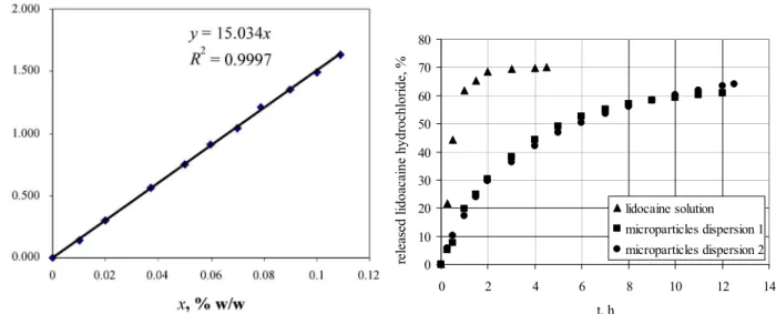
The concentration of lidocaine hydrochloride in the water solution was determined spectrophotometri-cally measuring the absorbance at 262.2 nm. The correlation between the absorbance and the lidocaine hydrochloride concentration was determined measu-ring the absorbance at 262.2 nm for concentrations of lidocaine hydrochloride from 0.01 to 0.11 % w/w. This correlation was linear:
x A=15.034~
where A is the absorbance and x~ is w/w concentra-tion of lidocaine hydrochloride expressed as a percent mass concentration. This correlation with experiment-tally obtained points was shown in Figure 2.
RESULTS AND DISCUSSION
The mean diameter of microparticles was about 800 nm in both cases. The largest particles were with diameter of 17.4 µm and they were lonely in disper-sion. Both particles dispersions were stored at 4 °C for 90 days and they did not change their structures and that was the evidence of their stability.
On the bases of the measured samples weight and the concentration of lidocaine hydrochloride in the receptor compartment, a cumulative weight of lido-caine hydrochloride which permeated the membrane was calculated. The results of the diffusion across the membrane of lidocaine hydrochloride from the water solution and microparticles dispersions obtained by two different methods were shown in Figure 3.
R. PJANOVIĆet al.: DIFFUSION OF LIDOCAINE HYDROCHLORIDE… CI&CEQ 15 (1) 33−35 (2009)
35
Therefore, phospholipids microparticles could successfully be used for a prolonged release of lido-caine hydrochloride. The microparticles preparation method has no influence on the release rate of an ac-tive, especially because a mean particle diameter was almost the same in both experiments.
CONCLUSION
Phospholipids microparticles are a promising device for the prolonged release of local anesthetics. The diffusion from this kind of particles is significantly slower than from the drug solution. This finding is in accordance with the literature data for lidocaine hy-drochloride [10].
Microparticles dispersions are stable for at least 3 months.
The diffusion rate is determined by the particle size and the membrane composition. For both prepa-ration method the membrane of particles was mainly of phosphatidylcholine, and the diffusion of an active could be further modified only by using hydrogenated
phospholipids and cholesterol as the membrane con-stituents.
Nomenclature
A Absorbance
x
~
Lidocaine concentration w/w, %.REFERENCES
[1] M. J. Choi, H. I. Maibach, Int. J. Cosmetic Sci. 27 (2005) 211-221
[2] F. Maestrelli, M. L. Gonzalez – Rodriguez, A. M. Rabasco, P. Mura, Int. J. Pharmaceutics 312 (2006) 53-60
[3] T. Nii, F. Ishii, Int. J. Pharmaceutics 298 (2005) 198-205
[4] A. D. Bangham, Liposome Letters, Academic Press, 1983
[5] G. Storm, D. J. A. Crommelin, PSTT 1 (1998) 19-31
[6] D. D. Basic, TIBTECH 16 (1998) 307-321
[7] A. Eidelman, J. M. Weiss, J. Lau, D. B. Carr, Ann. Emerg. Med. 46 (2005) 343-351
[8] Lucas Meyer, Brochure "Pro-Lipo", 1998
[9] V. Torchilin, V. Weissig, Liposomes – A Practical Ap-proach, Oxford University Press, 2003
[10] M. Glavas-Dodov, K. Gorcinova, K. Mladenovska, E. Fre-dro-Kumbaradzi, Int. J. Pharmaceutics 242 (2002) 81-384. Figure 2. The absorbance of lidocaine hydrochloride
water solution of different concentrations.
0 10 20 30 40 50 60 70 80
0 2 4 6 8 10 12 14
t, h
re
leas
ed
l
id
o
aca
in
e hy
d
roch
lor
id
e, %
lidocaine solution microparticles dispersion 1 microparticles dispersion 2