Iranian Journal of Basic Medical Sciences
ijbms.mums.ac.ir
The effects of nano-silver and garlic administration during
pregnancy on neuron apoptosis in rat offspring hippocampus
Maryam Lale Ataei
1, Ali Reza Ebrahimzadeh-bideskan
2*
1 Department of Anatomy and Cell biology, School of Medicine, Mashhad University of Medical Sciences, Mashhad, Iran
2 Department of Anatomy and Cell biology, School of Medicine, Mashhad University of Medical Sciences, Mashhad, Iran
A R T I C L E I N F O A B S T R A C T
Article type: Original article
Objective(s): The aim of this study was to investigate the effects of nano-silver and garlic administration during pregnancy on neuron apoptosis in rat offspring hippocampus.
Materials and Methods: Fifty pregnant wistar rats were randomly divided into five groups: 1- nano- silver (N.S) group; 30 mg/kg of N.S treated via gavage. 2- Control (C) group, administrated with distilled water via gavage. 3- N.S and garlic (N.S+G) group; N.S (30 mg/kg) and garlic juice (1 ml/100 g) treated via gavage simultaneously. 4- Garlic group (G); garlic juice (1 ml/100 g) administrated via gavage, 5- normal (N) without any intervention. All the interventions were done during pregnancy (21 days). Finally, the brains of rat offspring were removed to use for nano-silver level measurement and TUNEL staining. The mean of TUNEL positive cell numbers per unit area
(NA) in different regions of hippocampus were compared in all animal groups.
Results: The results revealed a significant increase of hippocampus nano-silver level in N.S and
N.S+G groups comparing to N group (P<0.05) and a significant decrease in nano-silver level in
N.S+G group comparing to N.S group (P<0.01). The number of TUNEL positive cells in the CA1, CA3,
and DG fields of rat offspring hippocampus increased in N.S and N.S+G groups comparing to other
ones, and also reduced significantly in N.S+G group comparing to N.S group (¥ P< 0.01).
Conclusion: Our results showed that co-administration of nano-silver and garlic during pregnancy may lead to reduce nano-silver induced apoptotic cells in their offspring hippocampus.
Article history: Received: Jul 11, 2013 Accepted: Dec 9, 2013
Keywords: Apoptosis Garlic Hippocampus Nano-silver
►
Please cite this paper as:Lale Ataei M, Ebrahimzadeh-bideskan AR. The effects of nano-silver and garlic administration during pregnancy on neuron apoptosis in rat offspring hippocampus. Iran J Basic Med Sci 2014; 17:411-418.
Introduction
Today, the wide spread use of nanoparticles increases the risk of nanoparticle induced toxic effects in the environment and humans. The rate of exposure to nanoparticles increased progressively over the years as they were used extensively in a variety of industries. Intentional manipulation of nanoparticle surfaces with biomolecules and chemicals to cater various applications resulted in nano-materials with unforeseeable activity. Large scale production and improper waste disposal may elevate human exposure to them and subsequent accumulation of these nano-materials in nature.
Nanoparticles are defined as particles of 1-100 nm at least in one dimension that widely used due to their physicochemical properties. Nano-silver is one of the new nanotechnology materials has been recently made which could be used in different areas such as antimicrobial products, air and water filtration, wound dressings, burn treatment and surgical instruments (1). In addition, nowadays
nano-silver are used in laundry detergent, paint walls, disinfectants, water pipes as well as the fabric for making clothes, underwear and socks. Although, this material is used widely, but it has some adverse effects on human health. The results of some studies performed on nano-silver, proved that they may have cytotoxic properties and are able to cause apoptosis (2, 3). The suggested mechanism for silver nanoparticles induced apoptosis is cytochrome C release into the cytosol and Bax protein transport into mitochondria, in which induce mitochondria-dependent apoptosis (4). The results of several studies have been indicated that the toxicity effects of nano-silver on the nervous system are
dose-dependent (5). nano-silver are able to cross the placental membrane (6) and could affect on the embryos cranial development and even cause
embryo death (7). It also pass through the blood - brain barrier, and induce cerebral edema, neuroblastoma as well as inflammatory responses in the brain (8). In addition, there are evidences
that nano-silver, accumulates in various organs including the brain(9 ,10). At the cellular level, silver nanoparticles can penetrate the cell membrane and various organelles, leading to cessation of proliferation, and apoptosis.
Apoptosis is a regular sequence of biological events in response to stress stimuli, including oxidative stress which is caused by exposure to metals. Apoptotic cells are characterized by distinct morphological features including chromatin condensation, nuclear shrinkage and oligonucleosomal DNA fragmentation that can be detected by means of TUNEL method in which apoptotic nuclei identify by the presence of dark brown staining (11).
Allium sativum, as a phytomedicine, commonly
known garlic, is a species in the onion genus, allium. Garlic contains many sulfur, phosphorus, potassium and zinc ions, moderate amounts of selenium, vitamin A, vitamin C and smaller amounts of calcium, magnesium, sodium, iron, magnesium, B-complex vitamins and allicin. The antioxidant effects of garlic are due to allicin, a compound to trap free radicals (12, 13). Recently, it founded that the sulfur-containing compounds of garlic have mutagenesis and anti-carcinogenesis effects. In other hand, these components decrease the incidence of tumors such as stomach, colorectal and prostate cancer (14). Garlic extract components such as S-allyl-L-cysteine (SAC) have neuroprotective effects against reactive oxygen species (ROS), H2O2-mediated neuronal cell damages (12, 15).
Hippocampus is a part of the cerebral cortex; located in the medial temporal lobe, underneath the cortical surface. It contains two main parts: Ammon's horn (CA1, CA2, CA3, and CA4) and the dentate gyrus in which visible during the first week of life (16,17). Hippocampus development continues through adolescence, with a distinct developmental time regulated with long-term potentiation (LTP), which is a presumed measure of plasticity, emerging during the second postnatal week in CA1, and during the third postnatal week in DG (18, 19).
Materials and Methods
This experimental research was done according to Ethics Committee Guidelines of Mashhad University of Medical Sciences, Mashhad, Iran, and all protocols of animal experiments have been approved by the Institution's Animal Care Committee.
Animals
This research was carried out on 50 virgin female Wistar rats weighing about 180-220 g. The animals were maintained at the animal house under controlled conditions (12 hr light and dark cycle, 22°C and 60%
relative humidity) with laboratory chow and water provided ad libitum.
Study groups
Female wistar rats were mated overnight with
fertile males of the same strain (1 male + 2 female in each cage). The day on which spermatozoa were found in the vaginal smear was designated as embryonic day 0 (E0). All the pregnant wistar rats
were divided randomly in to 5 groups as follow: 1- Nano-silver (N.S) group: the animals were administrated with 30 mg/kg of N.S via gavage. Nano-silver dispersed in distilled water. The N.S exposure regimen was chosen based on a previous study (9).
2- Control (C) group: the animals were administrated with distilled water via gavage without any other intervention.
3- Nano-silver + Garlic (N.S+G) group: the animals were received N.S (30 mg/kg) and after that at the same time the animals were administrated with 1 ml/100 g of body weight garlic juice via gavage (20). 4- Garlic (G): the rats received 1 ml/100 g of body weight garlic juice via gavage (21).
5- Normal (N): without any intervention.
All the interventions were done in each day during pregnancy (21 day).
Preparation of garlic juice
Fresh garlic bulbs were purchased from a local store and identified by botanists in Ferdowsi University of Mashhad, Iran and a voucher number deposited (FUMH: 39493). To prepare garlic juice, garlic bulbs were separated, peeled and washed with distilled water. After drying in a shed, the clean garlic bulbs were crushed with an electric grinder and the juice was decanted carefully through muslin (21).
Nano- silver preparation
Nano-silver (Ag-NPs) used in this study was purchased from Sigma-Aldrich, USA (Cat. No. 576832), It is a kind of nano-powder with particle size <100 nm (confirmed by using transmission electron microscopy), purity of 99.98%. Ag-NPs were suspended with distilled water and were fed to rats with feeding needles (gavage). Every day, the suspensions were prepared freshly before the administration.
Hippocampus silver measurement
At the end of experimental interventions, after removing of the rats offspring brains, in some cases, hippocampal tissue were digested with 70% nitric acid in a microwave oven digestion system (MARS 230/60, CEM) for 15 min at 180°C. The
concentration of silver in digested fluid was analyzed with a flameless method using an atomic absorption spectrophotometer equipped with a Zeeman graphite furnace (Perkin Elmer 5100ZL, Zeeman Furnace Module, USA) based on the NIOSH 7300 method. The concentration of silver in the tissue was expressed as µg/g wet weight (22).
Tissues preparation
0 0,5 1 1,5 2 2,5 3 3,5 4 4,5
N.S N.S+G C G N
M
e
a
n o
f H
ippo
ca
m
pus
na
no
si
lv
e
r
lev
el
(m
cg
/g
)
Series1
¥ *
***
anesthetized with chloroform. The brains were removed immediately, washed in normal saline and fixed in normalized fixative consisting 10% formaldehyde in 0.01 M phosphate buffered saline (PBS) overnight at room temperature. Then, the tissue blocks were dehydrated with an ascending ethanol series, cleared with xylene and then embedded in paraffin. The paraffin blocks were cut into transverse serial sections of 5 µm thickness (23). Next, ten sections including hippocampus tissue from each animal were randomly chosen and mounted on poly-L-lysine coated slides for TUNEL technique(21, 23).
Apoptotic cell detection
To detect apoptotic cells, DNA fragmentation in apoptotic cell nuclei was determined using terminal deoxynucleotidyl transferase-mediated dUTP nick end-labelling (TUNEL) reaction by means of TUNEL Kit (Roche, Germany). First, tissue sections were deparaffinized with xylene, rehydrated through descending concentrations of ethanol and rinsed for 10 min in 0.1 M PBS and then treated with 20 µg/ml proteinase K for 20 min at room temperature. The specimens were treated with 3% H2O2 in methanol for
10 min to inactivate endogenous peroxidase. After washing with PBS, specimens were incubated in the labelling reaction mixture containing terminal deoxynucleotidyl transferase and the deoxynucleotide at 4°C for overnight. After incubation, all the sections were rinsed in PBS and incubated with horseradish peroxidase (POD, 1:500) for 30 min at room temperature. Then, the sections were washed extensively with PBS for 3 min and treated with DAB solution (30 mg DAB and 200 µl H2O2/100 ml PBS) for 15 min at room temperature in dark. After being washed under running water, all the sections were counterstained with haematoxylin for 1 min. Finally the sections were dehydrated in increasing graded ethanol, cleared in xylene and mounted with cover slip. In this method, apoptotic nuclei were identified by the presence of dark brown staining (23).
For materials and method procedures confirmation, positive and negative controls were done as follow: positive control; sections were incubated with DNase I (3000 U/ml in 50 mM Tris-HCl, pH 7.5, 1 mg/ml BSA) for 10 min at 15-25°C
to induce DNA strand breaks, then TUNEL reaction was applied. Negative Control; sections were incubated with label solution only (without terminal transferase) instead of TUNEL reaction mixture (23).
Quantification and statistical analysis of apoptotic
cells
All the sections were examined using a light microscope by a ×40 objective lens (UPlan FI, Japan), images transferred to computer using a high-resolution camera (BX51, Japan), photographed and morphometerical method was used to count TUNEL
positive cells per unit area (NA) in CA1, CA3 and DG
different subdivisions of the hippocampus. The numbers of TUNEL positive cells were counted using a 10000 μm2 counting frame. The mean numbers of
neurons per unit area (NA) in different regions of
hippocampus were calculated using the formula as follow:
In this formula " " is the summation of counted TUNEL positive cells appeared in sections, "a/f" is
the area associated with each frame (10000 μm2),
"∑P" is the sum of frames associated points hitting
the reference (24).
Statical analysis
To compare differences between samples, one-way analysis of variance (ANOVA), Tukey Post hoc
statistical tests and SPSS 11.5 statistical software were used.
Effects of garlic administration during pregnancy on rat offspring hippocampus silver level
Silver levels inhippocampus of rat offspring after treatment were evaluated in all groups. The results revealed a significant increase in silver level in two groups including nano-silver and nano-silver plus garlic groups comparing to other ones (P <0.05) and a significant decrease in silver level in nano-silver plus garlic group comparing to nano-silver treated group (P<0.01)(Figure 1).
Figure1. Comparison of hippocampus rat offspring silver levels in
different groups (mean ± SEM), Silver levels in nano-silver treated
group (group1) and nano-silver plus garlic treated group
(group 3) were higher comparing to normal group (***P< 0.001
and *P< 0.05 respectively). But silver level in nano-silver plus
garlic treated group (group 3) was decrease comparing to
nano-silver treated group (¥P< 0.01)
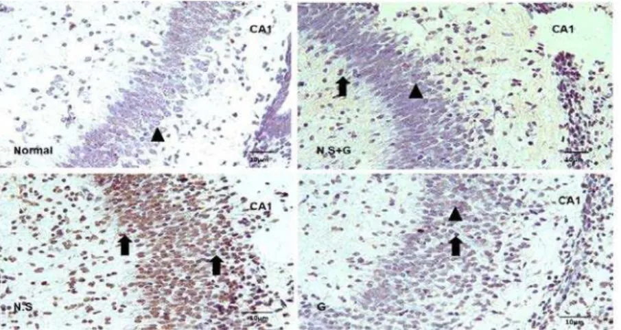
Figure 2A. Coronal section of CA1 hippocampal subdivision in rat offspring, prepared by using TUNEL technique. Some cells reacted with TUNEL (TUNEL positive) which pointed by arrow and no reacted cells pointed by with head arrow)
N.S= nano-silver treated group, N.S +G= nano- silver plus garlic treated group, G= garlic group
0 2 4 6 8 10
N.S N.S+G C G N
Me
a
n
o
f
Ap
o
p
to
ti
c
ce
ll
n
um
b
e
r pe
r un
it
a
re
a
(n
/m
m
²)
CA1
٭٭
¥
٭٭٭
Figure 2B. Comparison of apoptotic cell per unit area in CA1 hippocampal subdivision in different groups (mean±SEM). Apoptotic cell
numbers in N.S and N.S+G groups increased significantly comparing to normal and control groups (***P<0.001 and ** P<0.01). Apoptotic
cell numbers in N.S+G groups decreased significantly comparing to nano-silver treated group (¥P<0.05)
N.S= nano-silver treated group, N.S+G= nano- silver plus garlic treated group, C= control group, G= garlic group, N= normal group
Results
Effects of garlic on the number of nano-silver-induced apoptotic cells per unit area in the rat offspring hippocampus
TUNEL-positive cells were counted in CA1, CA3 and DG of hippocampal subdivision using camera equipped microscope (Olympus BH-51) at × 400 magnifications. These cells detected by the morphological features of apoptosis such as cell and nuclear shrinkage, chromatin condensation, and DNA fragmentation demonstrated with TUNEL methods. The number of TUNEL positive cells in the CA1 were significantly higher in the nano- silver and nano- silver plus garlic treated groups comparing to control group (P< 0.001and P< 0.01 respectively). The number of TUNEL-positive cells in group 3 (nano-silver + garlic) were reduced significantly in the CA1comparing to the nano-silver group (P<0.01) (Figure 2A and B). There was no significant difference in TUNEL-positive cell numbers in the CA1 between control, garlic and normal groups.
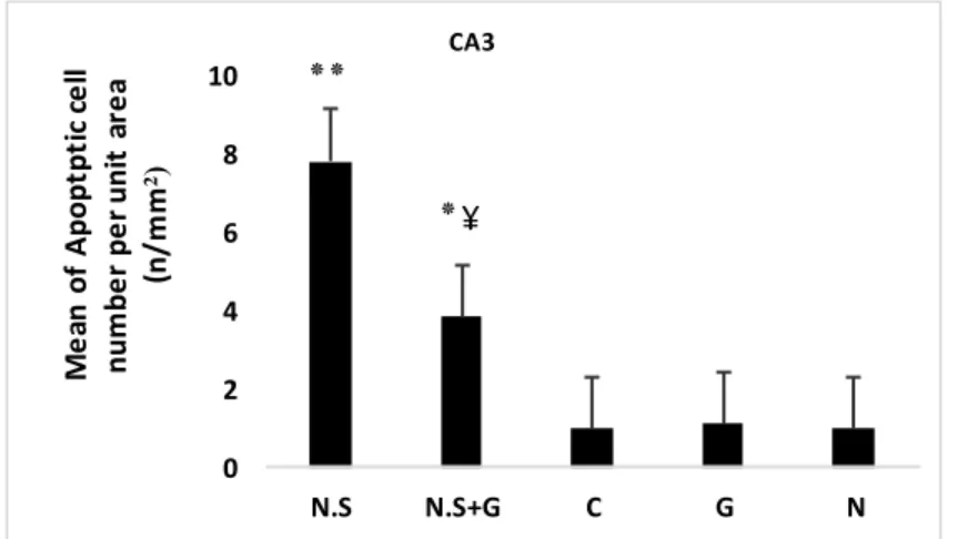
TUNEL positive cell numbers in the CA3 were higher significantly in the nano- silver
and
nano- silver plus garlic treated groups comparing to control group (P<0.001and P<0.05 respectively). The number of TUNEL-positive cells in group 3 were reduced significantly in the CA3 comparing to the nano-silver group (P<0.01). There was no significant difference in TUNEL-positive cell number in the CA3 region between control, garlic and normal groups (Figure 3A and B).Figure 3A. Coronal section of CA3 hippocampal subdivision in rat offspring, prepared by using TUNEL technique. Some cells reacted with TUNEL (TUNEL positive) which pointed by arrow and no reacted cells pointed with head
N.S= nano-silver treated group, N.S+G= nano- silver plus garlic treated group, G= garlic group
0 2 4 6 8 10
N.S N.S+G C G N
M
e
a
n
o
f A
p
o
p
tp
ti
c
ce
ll
n
u
m
b
e
r
p
e
r u
n
it
a
re
a
(n
/
m
m
²)
CA3
٭٭
٭
¥
Figure 3B. Comparison of apoptotic cell per unit area in CA3 hippocampal subdivision in different groups (mean±SEM). Apoptotic cell
numbers in N.S and N.S+G groups increased significantly comparing to normal and control groups (**P<0.001 and * P<0.05). Apoptotic cell
numbers in N.S+G groups decreased significantly comparing to nano-silver treated group (¥P<0.01)
N.S= nano-silver treated group, N.S+G= nano- silver plus garlic treated group, C= control group, G= garlic group, N= normal group
Discussion
Nanotechnology that deals with materials typically less than 100 nanometer in size is contributed to the fields of computer storage, semiconductors, biotechnology, manufacturing and energy. Nano-materials can enter to the human body through several ports such as lungs after inhalation, digestive system and possibly through the skin (25, 26). It has been demonstrated that the nanoparticles cross through the blood–brain barrier and accumulate in different part of the brain (27).
Although applications of nanoparticles have increased, few toxicology studies are available. So some researchers have shown that the exposure to nano-silver is toxic under certain experimental conditions (28). Other researchers have shown that nano-silver is non-toxic under similar experimental conditions. Our results demonstrated that the animal with 30 mg/kg of nano-silver administration via gavage during the pregnancy induced apoptosis in their offspring hippocampus subdivisions. According to a review of the
toxicological studies, the main difference in the outcome of these studies is due to variations in physicochemical features of the nano-silver being used in various studies (9).
The toxicity of nanoparticles to humans and mammals depends on various factors such as the size, their composition, ease of aggregation, physical and chemical (such as crystallinity, electromagnetic properties, presence of functional groups and dose) surface characteristics, (29). The toxicity of the nanoparticle is also heavily dependent on the mammal’s genetic complement, its susceptibility and ability to adapt to the changes in the environment, and also to fight with toxic substances (30). Some studies showed that single silver 12 nm nanoparticles affected early development of embryos (30). In addition, more nano-silver In vitro
and In vivo toxicity studies have been performed in
Figure 4A. Coronal section of dentate gyrus (DG) hippocampal subdivision in rat offspring, prepared by using TUNEL technique. Some cells reacted with TUNEL (TUNEL positive) which pointed by arrow and no reacted cells pointed with head. N.S= nano-silver treated group, N.S+G= nano- silver plus garlic treated group, G= garlic group
0 5 10 15 20 25 30 35 40
N.S N.S+G C G N
M
e
a
n
o
f A
p
o
p
to
tic
c
e
ll
n
u
m
b
e
r
p
e
r u
n
it
a
re
a
(n
/
m
m
²)
DG
٭٭
٭
Figure 4B. Comparisons of apoptotic cell per unit area in dentate gyrus of hippocampal subdivision in different groups (mean±SEM).
Apoptotic cell numbers in N.S and N.S+G groups increased significantly comparing to normal and control groups (**P<0.01 and * P<0.05).
Apoptotic cell numbers in N.S+G groups decreased significantly comparing to nano-silver treated group (¥P<0.01)
N.S= nano-silver treated group, N.S +G= nano- silver plus garlic treated group, C= control group, G= garlic group, N= normal group
stress, inflammation, genetic damage, and the inhibition of cell division and cell death (33). Most work to date has suggested that ROS generation (which can be either protective or harmful during biological interactions) and consequent oxidative stress are frequently observed with nanoparticle toxicity (29). The physicochemical characteristics of nanoparticle including particle size, surface charge, and chemical composition are the key indicators for the resulting ROS response and nanoparticle-induced injury since many of these nanoparticles intrinsic properties can catalyze the ROS production. Some nanoparticle have been shown to activate inflammatory cells such as macrophages and neutrophils which can result in the increased production of ROS (34). Other nanoparticles including titanium dioxide (TiO2), zinc oxide (ZnO),
cerium oxide (CeO2), and silver nanoparticle
accumulate on the cell surface or in the cell organelles and induce oxidative stress to the cell (35).
According to above mentioned subjects, it is concluded that there are some mechanisms to express nano-silver toxicity and cytotoxic effects,
inducing apoptosis: 1- Nano-silver induces the release of cytochrome C into the cytosol and translocation of Bax to mitochondria, indicating that nano-silver mediated apoptosis is mitochondria-dependent. 2-Nano-silver induces apoptosis via generation of ROS and JNK activation. According the above mentioned mechanism, it is possible to protect the nano-silver induced apoptosis by using antioxidant or antioxidant candidate materials including garlic as a traditional remedy (36).
Identification and characterization of new medicinal plants to cure neurodegenerative diseases and brain injuries resulting from stroke is the major and increasing scientific interest in recent years (37). Garlic juice as an antioxidant candidate, scavenges the ROS, enhance cellular antioxidant enzymes superoxide dismutase. Besides these properties, the other efficiency of garlic is perhaps due to the presence of sulfur-containing amino acids and compounds having free carboxyl (C=0) and amino (NH2) groups in their structures. The garlic
nanoparticles, reducing their intestinal absorption, and enhanced its excretion from the body to reduce nano-silver accumulation in soft tissues (21).
Our results also showed that the hippocampus silver level in nano-silver plus garlic treated group (group 3) was decrease comparing to nano-silver treated group (¥ P< 0.01, Figure 1).
Conclusion
In conclusion, this study results indicated that the nano-silver administration during pregnancy, can lead to product apoptotic cells in rat offspring hippocampus. Moreover, the fresh garlic juice prescription during pregnancy showed preventive and beneficial effects on nano-silver induced apoptotic cells production in hippocampus.
Acknowledgment
The presented data in this article is from an MSc student thesis results and research protocol which was supported financially by the vice chancellor for research, Mashhad University of Medical Sciences, Mashhad, Iran. In addition, the authors would like to thank Ms. Motejaded for her technical assistance.
References
1. Chen X, Schluesener HJ. Nanosilver: a nanoproduct in medical application. Toxicol Lett 2008; 176:1-12. 2. Miura N, Shinohara Y. Cytotoxic effect and apoptosis induction by silver nanoparticles in HeLa cells. Biochem Biophys Res Commun 2009; 390:733-737.
3. Greulich C, Kittler S, Epple M, Muhr G, Koller M. Studies on the biocompatibility and the interaction of silver nanoparticles with human mesenchymal stem cells (hMSCs). Langenbecks Arch Surg 2009; 394:495-502.
4. Hsin Y-H, Chen C-F, Huang S, Shih T-S, Lai P-S, Chueh PJ. The apoptotic effect of nanosilver is mediated by a ROS- and JNK-dependent mechanism involving the mitochondrial pathway in NIH3T3 cells. Toxicol Lett 2008; 179:130-139.
5. Asharani P, Sethu S, Lim HK, Balaji G, Valiyaveettil S, Hande MP. Differential regulation of intracellular factors mediating cell cycle, DNA repair and inflammation following exposure to silver nanoparticles in human cells. Genome Integr 2012; 3:2.
6. Christensen FM, Johnston HJ, Stone V, Aitken RJ,
Hankin S, Peters S, et al. Nano-silver - feasibility and
challenges for human health risk assessment based on open literature. Nanotoxicology 2010; 4:284-295. 7. Vega-Villa KR, Takemoto JK, Yanez JA, Remsberg CM, Forrest ML, Davies NM. Clinical toxicities of nanocarrier systems. Adv Drug Deliv Rev 2008; 60:929-938.
8. Sharma HS, Hussain S, Schlager J, Ali SF, Sharma A. Influence of nanoparticles on blood-brain barrier permeability and brain edema formation in rats. Acta Neurochir Suppl 2010; 106:359-364.
9. Kim YS, Kim JS, Cho HS, Rha DS, Kim JM, Park JD, et
al. Twenty-eight-day oral toxicity, genotoxicity, and
gender-related tissue distribution of silver
nanoparticles in Sprague-Dawley rats. Inhal Toxicol 2008; 20:575-583.
10. Park EJ, Bae E, Yi J, Kim Y, Choi K, Lee SH, et al.
Repeated-dose toxicity and inflammatory responses in mice by oral administration of silver nanoparticles. Environ Toxicol Pharmacol 2010; 30:162-168. 11. Rojo MC, Gonzalez ME. In situ detection of apoptotic cells by TUNEL in the gill epithelium of the developing brown trout (Salmo trutta). J Anatomy 1998; 193:391-398.
12. Massadeh AM, Al-Safi SA, Momani IF, Alomary AA, Jaradat QM, AlKofahi AS. Garlic (Allium sativum L.) as a potential antidote for cadmium and lead intoxication: cadmium and lead distribution and analysis in different mice organs. Biol Trace Elem Res 2007; 120:227-234. 13. El-Demerdash FM, Yousef MI, El-Naga NI. Biochemical study on the hypoglycemic effects of onion and garlic in alloxan-induced diabetic rats. Food Chem Toxicol 2005; 43:57-63.
14. Shaarawy SM, Tohamy AA, Elgendy SM, Elmageed
ZY, Bahnasy A, Mohamed MS, et al. Protective effects
of garlic and silymarin on NDEA-induced rats hepatotoxicity. Int J Biol Sci 2009; 5:549-557.
15. Ray B, Chauhan NB, Lahiri DK. Oxidative insults to neurons and synapse are prevented by aged garlic extract and S-allyl-L-cysteine treatment in the neuronal culture and APP-Tg mouse model. J Neurochem 2011; 117:388-402.
16. Eriksson PS, Perfilieva E, Bjork-Eriksson T, Alborn
AM, Nordborg C, Peterson DA, et al. Neurogenesis in
the adult human hippocampus. Nat Med 1998; 4:1313-1317.
17. Convit A, Wolf OT, Tarshish C, de Leon MJ. Reduced glucose tolerance is associated with poor memory performance and hippocampal atrophy among normal elderly. Proc Natl Acad Sci USA 2003; 100:2019-2022.
18. Raineki C, Holman PJ, Debiec J, Bugg M, Beasley A, Sullivan RM. Functional emergence of the hippocampus in context fear learning in infant rats. Hippocampus 2010; 20:1037-1046.
19. Muller RU, Stead M, Pach J.The hippocampus as a cognitive graph. J Gen Physiol 1996; 107(6):663-94. 20. Mittal AK, Chisti Y, Banerjee UC. Synthesis of metallic nanoparticles using plant extracts. Biotechnol Adv 2013; 31:346-356.
21. Sadeghi A, Ebrahimzadeh-bideskan, Alipour F, Fazel A, Haghir H. The effect of ascorbic acid and garlic administration on lead-induced neural damage in rat offspring’s hippocampus. Iran J Basic Med Sic 2013; 16:157-164.
22. Kim YS, Song MY, Park JD, Song KS, Ryu HR, Chung
YH, et al. Subchronic oral toxicity of silver
nanoparticles. Part Fibre Toxicol 2010; 7:20.
23. Khordad E, Fazel F, Ebrahimzadeh-bideskan AR. The effect of ascorbic acid and garlic administration on lead-induced apoptosis in rat offspring’s eye retina. Iran Biomed J 2013; 17:206-213.
25. Jani P, Halbert GW, Langridge J, Florence AT. Nanoparticle uptake by the rat gastrointestinal mucosa: quantitation and particle size dependency. J Pharm Pharmacol 1990; 42:821-826.
26. Warheit DB, Hoke RA, Finlay C, Donner EM, Reed KL, Sayes CM. Development of a base set of toxicity tests using ultrafine TiO2 particles as a component of nanoparticle risk management. Toxicol Lett 2007; 171:99-110.
27. Oberdorster G, Sharp Z, Atudorei V, Elder A,
Gelein R, Kreyling W, et al. Translocation of inhaled
ultrafine particles to the brain. Inhal Toxicol 2004; 16:437-445.
28. Shanmugasundaram T, Radhakrishnan M, Gopikrishnan V, Pazhanimurugan R, Balagurunathan R. A study of the bactericidal, anti-biofouling, cytotoxic and antioxidant properties of actinobacterially synthesised silver nanoparticles. Colloids Surf B Biointerfaces 2013; 111C:680-687. 29. Suliman YA, Ali D, Alarifi S, Harrath AH, Mansour L, Alwasel SH. Evaluation of cytotoxic, oxidative stress, proinflammatory and genotoxic effect of silver nanoparticles in human lung epithelial cells. Environ
Toxicol 2013.View at Publisher· View at Google Scholar.
30. Chua KB, Chua BH, Lee CS, Chem YK, Ismail N,
Kiyu A, et al. Genetic diversity of enterovirus 71
isolated from cases of hand, foot and mouth disease
in the 1997, 2000 and 2005 outbreaks, Peninsular Malaysia. Malays J Pathol 2007; 29:69-78.
31. Piepenbrink MS, Hussain I, Marsh JA, Dietert RR. Developmental immunotoxicology of Di-(2-Ethylhexyl)phthalate (DEHP): age-based assessment in the female rat. J Immunotoxicol 2005; 2:21-31. 32. Ji Q, Williams HJ, Roessner CA, Scott AI. Biomimetic self-condensation of malonates mediated by nucleosides. Tetrahedron Lett 2007; 48:8026-8028.
33. Grosse S, Evje L, Syversen T. Silver nanoparticle-induced cytotoxicity in rat brain endothelial cell
culture. Toxicol In Vitro 2013; 27:305-313.
34. Risom L, Moller P, Loft S. Oxidative stress-induced DNA damage by particulate air pollution. Mutat Res 2005; 592:119-137.
35. Manke A, Wang L, Rojanasakul Y. Mechanisms of nanoparticle-induced oxidative stress and toxicity. Biomed Res Int 2013;2013:942916.
36. Hsin YH, Chen CF, Huang S, Shih TS, Lai PS, Chueh PJ. The apoptotic effect of nanosilver is mediated by a ROS- and JNK-dependent mechanism involving the mitochondrial pathway in NIH3T3 cells. Toxicol Lett 2008; 179:130-139.