Metagenomic Survey of Viral Diversity
Obtained from Feces of Subantarctic and
South American Fur Seals
Mariana Kluge
1, Fabrício Souza Campos
1, Maurício Tavares
2, Derek Blaese de Amorim
2,
Fernanda Pedone Valdez
3, Adriana Giongo
4, Paulo Michel Roehe
1, Ana Claudia Franco
1*
1Virology Laboratory, Department of Microbiology, Immunology and Parasitology, Institute of Basic Health Sciences, UFRGS (Federal University of Rio Grande do Sul), Porto Alegre, Rio Grande do Sul, Brazil,
2CECLIMAR (Center for Coastal, Limnology and Marine Studies), UFRGS (Federal University of Rio Grande do Sul), Imbé, Rio Grande do Sul, Brazil,3Genomic and Molecular Biology Laboratory, PUCRS (Pontifical Catholic University of Rio Grande do Sul), Porto Alegre, Rio Grande do Sul, Brazil,4IPR (Institute of Petroleum and Natural Resources), PUCRS (Pontifical Catholic University of Rio Grande do Sul), Porto Alegre, Rio Grande do Sul, Brazil
*anafranco.ufrgs@gmail.com
Abstract
The Brazilian South coast seasonally hosts numerous marine species, observed
particu-larly during winter months. Some animals, including fur seals, are found dead or debilitated
along the shore and may harbor potential pathogens within their microbiota. In the present
study, a metagenomic approach was performed to evaluate the viral diversity in feces of fur
seals found deceased along the coast of the state of Rio Grande do Sul. The fecal virome of
two fur seal species was characterized: the South American fur seal (
Arctocephalus
austra-lis
) and the Subantarctic fur seal (
Arctocephalus tropicalis
). Fecal samples from 10
speci-mens (
A
.
australis
, n = 5;
A
.
tropicalis
, n = 5) were collected and viral particles were purified,
extracted and amplified with a random PCR. The products were sequenced through Ion
Tor-rent and Illumina platforms and assembled reads were submitted to BLASTx searches.
Both viromes were dominated by bacteriophages and included a number of potentially
novel virus genomes. Sequences of picobirnaviruses, picornaviruses and a hepevirus-like
were identified in
A
.
australis
. A rotavirus related to group C, a novel member of the
Sakobu-virus
and a sapovirus very similar to
California sea lion sapovirus 1
were found in
A
.
tropica-lis
. Additionally, sequences of members of the
Anelloviridae
and
Parvoviridae
families were
detected in both fur seal species. This is the first metagenomic study to screen the fecal
vir-ome of fur seals, contributing to a better understanding of the complexity of the viral
commu-nity present in the intestinal microbiota of these animals.
Introduction
Every year, hundreds of marine species arrive at the coast of Rio Grande do Sul, the
southern-most state in Brazil. Among these species, which include birds, turtles and mammals, fur seals
OPEN ACCESSCitation:Kluge M, Campos FS, Tavares M, de Amorim DB, Valdez FP, Giongo A, et al. (2016) Metagenomic Survey of Viral Diversity Obtained from Feces of Subantarctic and South American Fur Seals. PLoS ONE 11(3): e0151921. doi:10.1371/ journal.pone.0151921
Editor:Ulrike Gertrud Munderloh, University of Minnesota, UNITED STATES
Received:November 26, 2015
Accepted:March 7, 2016
Published:March 17, 2016
Copyright:© 2016 Kluge et al. This is an open access article distributed under the terms of the
Creative Commons Attribution License, which permits unrestricted use, distribution, and reproduction in any medium, provided the original author and source are credited.
Data Availability Statement:All sequence data is available from the NIH Sequence Read Archive (SRA —http://www.ncbi.nlm.nih.gov/sra/) database (accession number SRP070196).
Funding:The research leading to these results has received funding from the National Council for the Improvement of Higher Education (CAPES—http://
www.capes.gov.br/), the National Council for Scientific and Technological Development (CNPq—
are regular visitors that can be observed near or on-shore. These animals are driven to this
region by the Malvinas current, particularly during winter months [
1–3
]. Although some fur
seals may reach the coast to rest, several are found dead or debilitated along the shore and the
cause of their weakness or death cannot always be determined [
4
,
5
]. Few studies have
attempted to identify the pathogens that infect these populations and their roles as etiological
agents of diseases and as potential zoonotic agents, especially those concerned with viruses [
6–
10
]. While the virome of marine mammals has already been investigated [
11
], these studies
have been restricted to species native to the northern hemisphere. Little is known about the
viruses that infect marine mammals limited to the southern hemisphere and the effects of this
geographical difference on their virome profiles.
Here, we evaluated the viral diversity of two species of pinnipeds from the Otariidae family
from the southern hemisphere: the South American fur seal (Arctocephalus australis) and the
Subantarctic fur seal (Arctocephalus tropicalis). While the South American fur seal is found
along the Pacific and Atlantic coast of South America, the Subantarctic fur seal has a broader
range that extends from the South Atlantic to Indian ocean islands. The South American fur
seal is more frequently sighted in Rio Grande do Sul coast, mostly juveniles, due to the
proxim-ity of its closest breeding colony, located in the neighboring country of Uruguay. By contrast,
the closest Subantarctic fur seals colonies are located at more than 4,000 km away at the south
Atlantic islands of Gough and Tristan da Cunha [
3
,
12
]. Juveniles and adults specimens of
Sub-antarctic fur seals reach the Atlantic coast with the help of ocean currents, and it is known that
juveniles do not stay in the colonies during breeding seasons, while adults can travel long
dis-tances after mating [
1
,
13
].
The aim of this study was to examine the fecal virome of two species of fur seals whose
cadavers were found along the shore of Rio Grande do Sul state. Anelloviruses, parvoviruses
and picornaviruses were identified, as well as potential new members of
Sakobuvirus,
Picobir-navirus
and
Rotavirus. A sapovirus very similar to
California sea lion sapovirus 1
was found in
the Subantarctic fur seal, and a hepevirus-like sequence was identified. The data provides a
pre-liminary characterization of the viruses that occur within fur seals populations of the southern
hemisphere.
Materials and Methods
Sample Collection
Fecal samples from 10 specimens (A.
australis, n = 5;
A.
tropicalis, n = 5) were collected directly
from the intestines of deceased fur seals found along shores between August 2012 and
Septem-ber 2013 by the Center for Coastal, Limnology and Marine Studies (CECLIMAR) team.
Sam-ples for each species were pooled and kept at -80°C until processing. All samSam-ples from this
study were collected in strict accordance with the Brazilian law, and the license for collecting
zoological material was granted by SISBIO/Ministry of the Environment (License number:
20185
–
4). The location and information about the specimens are provided in
Table 1
and
Fig 1
.
Viral Particle Purification and Nucleic Acid Extraction
Fecal samples were suspended in Hank's balanced salt solution, vigorously vortexed and then
centrifuged at 2500 × g for 90 min at 4°C. The supernatant was again centrifuged for 10 min at
maximum speed and then filtered through a 0.45
μm syringe filter (MF-Millipore). The viral
particles were harvested and pelleted on a 25% sucrose cushion by ultracentrifugation at
190000 × g for 4h at 4°C. The pellet was resuspended in TE buffer and clarified by emulsifying
with 1/1 (v/v) chloroform and centrifugation. In order to remove nucleic acids not protected
collection and analysis, decision to publish, or preparation of the manuscript.
by the capsid, the purified samples were treated with 100 U of DNase I (Roche) and 20 U of
RNase (Invitrogen) at 37°C for 2h, as similar to other studies [
16
,
17
].
Viral genomes were extracted via commercial kits (PureLink
1
Viral RNA/DNA Invitrogen
for DNA extraction; RNeasy
1
Mini Kit Qiagen for RNA) and processed as described by [
18
]
with minor modifications. Briefly, a complementary strand of extracted DNA (5
μl) was
syn-thesized using the Klenow fragment DNA polymerase (New England Biolabs) and primer
K-randoms (GAC CAT CTA GCG ACC TCC ACM NN MNM) designed by [
19
]. For the
extracted RNA (10
μl), a reverse transcription using the primer K-randoms was carried out
prior the second strand synthesis using Klenow fragment DNA polymerase.
Library Construction for Metagenomic Sequencing
A random PCR was performed in a final volume of 50
μL, containing 5
μL of template, 0.8
μM
of the fixed portion of primer K-randoms (GAC CAT CTA GCG ACC TCC AC), 0.2 mM of
each dNTP, 1X PCR Buffer, 2.5 mM MgCl
2, and 1 U of
Taq
DNA polymerase. Amplification
conditions consisted of an initial denaturation cycle at 95°C for 5 min, followed by 35 cycles for
amplification (95°C for 1 min, 53°C for 1 min and 72°C for 1 min), and final extension at 72°C
Table 1. Samples used in this study.
Pool no.
No. Species Length
(cm)
Weight (kg)
Sex Carcass
classification code* Date of collection (dd/ mm/yyyy) Collection location Geolocation latitude/ longitude (decimal)
1 G1529 South American fur sealArctocephalus
australis
94.2 10.6 Male 2 02/08/2012 Osório, RS
-29.878119/-50.073224
1 G1560 South American fur sealArctocephalus
australis
92 9.5 Male 2 09/08/2012 Cidreira, RS
-30.165946/-50.197728
1 G1574 South American fur sealArctocephalus
australis
89.4 10.8 Male 2 16/08/2012 Imbé, RS
-29.94758/-50.105842
1 G1604 South American fur sealArctocephalus
australis
92 15.5 Male 2 31/08/2012 Capão da
Canoa, RS
-29.657469/-49.954338
1 G1657 South American fur sealArctocephalus
australis
88 12 Male 2 11/09/2013 Imbé, RS
-29.94579/-50.10498
2 G1535 Subantarctic fur seal Arctocephalus
tropicalis
91.9 8.4 Male 2 02/08/2012 Capão da
Canoa, RS
-29.730323/-49.995557
2 G1537 Subantarctic fur seal Arctocephalus
tropicalis
90.6 9 Male 2 02/08/2012 Capão da
Canoa, RS
-29.665492/-49.959170
2 G1561 Subantarctic fur seal Arctocephalus
tropicalis
91.9 8.8 Male 2 09/08/2012 Cidreira, RS
-30.174591/-50.200809
2 G1577 Subantarctic fur seal Arctocephalus
tropicalis
80.5 7.1 Male 3 16/08/2012 Osório, RS
-29.917806/-50.091768
2 G1640 Subantarctic fur seal Arctocephalus
tropicalis
157.5 40.9 Male 2 25/07/2013 Tramandaí,
RS
-30.13273/-50.18535
*Code for carcass classification according to Geraci & Lounsbury (1993)[14]: freshly dead, edible(2); and decomposed, but organs basically intact(3).
for 7 min. The products were visualized by 1% agarose gel electrophoresis, purified and
pro-cessed for Ion Torrent (Life Technologies, USA) using a 316 Ion chip, which was performed by
the Genomic and Molecular Biology Laboratory from the Pontifical Catholic University of Rio
Grande do Sul. The same process, including the random PCR, was repeated for Illumina MiSeq
platform sequencing using Kit v2 in the 300-cycles (2x150) format performed by Fepagro
Ani-mal Health Institute of Veterinary Research Desidério Finamor (IPVDF), Eldorado do Sul,
Brazil.
Bioinformatics
Ion Torrent reads were trimmed using PRINSEQ (
prinseq.sourceforge.net
) and the quality of
the sequences was analyzed with FastQC (
www.bioinformatics.babraham.ac.uk/projects/
fastqc/
). Trimmed reads were assembled
de novo
by using MetaVelvet v1.2.01 (metavelvet.dna.
bio.keio.ac.jp) with a k-mer of 51. Illumina reads were trimmed for primers using Geneious
Fig 1. Sample location map.Map indicating the location of where the samples were collected along the coast of the State of Rio Grande do Sul, Brazil (shaded). The map was extracted from the Open Street Map[15] database.
8.1.3 and
de novo
assembled with St. Petersburg genome assembler (SPAdes) 3.5.0 (
bioinf.spbau.
ru/spades
). The resulting contigs (
>
100bp) were submitted to BLASTx search against the
National Center for Biotechnology Information (NCBI,
www.ncbi.nlm.nih.gov
) non-redundant
database (nr) and its viral database by using an E-value cutoff of 1e-05. The contigs were classified
into eukaryotic viruses, bacteriophages, bacterium, eukaryotes and unknown based on lowest
E-value. Contigs of eukaryotic viruses were used for sequence and phylogenetic analyses and
bacteriophage sequences were not further analyzed. The GenBank accession numbers for the
sequences derived in this study are: KR261062, KR261063, KR261065, KR816222, KR816223
(fur seal anelloviruses); KR261066-KR261068, KR261070-KR261075, KR261077-KR261079,
KR816217, KR816218, KR816220, KR816221 (fur seal parvoviruses); KR106199-KR106202,
KR816213, KR816215, KR337994 (fur seal picornaviruses); KR072975-KR072979, KR072981,
KR072982, KR072984 (fur seal sakobuvirus); KR106194-KR106196, KR106198, KR816216 (fur
seal picobirnavirus); KR072985-KR072990 (fur seal rotavirus), KR827461 (fur seal hepevirus);
KR072992, KR072994, KR072995 (fur seal sapovirus). The sequence data obtained from this
study is available at the NIH Sequence Read Archive (SRA) under the study accession number
SRP070196.
Phylogenetic Analysis
Nucleotide or translated amino acid sequences from the contigs of anellovirus, parvovirus,
picornavirus, picobirnavirus, rotavirus, sapovirus and hepevirus-like were aligned with
MUS-CLE (
www.drive5.com/muscle
) and phylogenetic trees were built using MEGA6 [
20
]. Trees
were constructed by the neighbor-joining (NJ) method [
21
] with a bootstrap of 1000 replicates,
p-distance model, and gaps were treated as pairwise deletion. The contig sequences from this
study were compared with other selected gene sequences available in the GenBank.
Results
Overview
A substantial proportion of the assembled reads detected in both fur seals species have no
sig-nificant similarity to any of the sequences deposited to date at GenBank. About 70% of the
assembled reads from the Ion Torrent platform had no significant hits, whereas in Illumina
NGS apparatus the sequences with no identified matches reached 35% (cutoff for significant as
<
1e-05 BLASTx E score). The same divergence was observed with the number of bacterial hits,
however, Ion Torrent had the lowest number of hits (about 25%) when compared to Illumina
(about 60%).
The viral component detected in either of the sequencing platforms represented 4
–
5% of
total sequences, regardless of the fur seal species analyzed. Most of the viral hits were from
bac-teriophages, in agreement with previous studies of bats and dromedary fecal viromes [
22–24
].
Some of the contigs from eukaryotic viruses displayed low similarity to currently known
viruses and, as such, may represent novel viruses. The Subantarctic fur seal was found to carry
a larger proportion of identifiable sequences of eukaryotic viruses (95 hits, corresponding to
33% of total assembled reads assigned to viruses) when compared to the South American fur
seal (53 hits, corresponding to 11% of total assembled reads assigned to viruses). The
propor-tional taxonomic composition of the assembled reads is shown in
Fig 2
.
South American Fur Seal (Arctocephalus australis)
Fig 2. Taxonomic classification of assembled reads (>100bp).(A) Pie charts of assembled reads based on BLASTx best E-scores (cutoff: 10e-05) against the GenBank non-redundant and viral databases. (B) Taxonomic distribution of viruses for each fur seal species.
trimmed reads resulted in 10,801 contigs (
>
100 bp). Illumina sequencing generated a total of
496,016 paired-end reads (average length of 149 bp) which were trimmed for primers and
assembled
de novo
with St. Petersburg genome assembler (SPAdes) into 2,053 contigs (
>
100
bp). BLASTx results from the Ion Torrent contigs revealed sequences with similarity to the
eukaryotic virus families
Parvoviridae
(11 contigs),
Anelloviridae
(5),
Picornaviridae
(10),
Pico-birnaviridae
(5) and invertebrate virus (1). Illumina contigs displayed similarity to genomes of
members of the families
Parvoviridae
(3),
Anelloviridae
(3),
Picornaviridae
(5),
Picobirnaviri-dae
(3) and
Hepeviridae
(1), among other viruses that infect fish, small invertebrates and
insects (7). Contigs with significant BLASTx hits and their GenBank accession numbers are
shown in
Table 2
.
Subantarctic Fur Seal (Arctocephalus tropicalis)
Ion Torrent sequencing generated a total of 784,917 reads with an average length of 184 bp
which were trimmed into 288,611 reads. Trimmed reads were
de novo
assembled with
Meta-Velvet into 6,690 contigs (
>
100 bp). Illumina sequencing generated a total of 1,253,988
paired-end reads (average length of 144 bp) which were trimmed for primers and
de novo
assembled
with SPAdes into 628 contigs (
>
100 bp). For Ion Torrent contigs, the eukaryotic virus families
with significant similarity to results from BLASTx searches were
Parvoviridae
(24 contigs),
Anelloviridae
(2),
Picornaviridae
(19),
Reoviridae
(13),
Caliciviridae
(18), other insect viruses
(2) and a circovirus-like hit (1). Illumina contigs had similarity with
Parvoviridae
(9),
Anello-viridae
(1),
Picornaviridae
(3),
Caliciviridae
(3) and
Reoviridae
(1). Contigs with significant
BLASTx hits and their GenBank accession numbers are shown in
Table 3
.
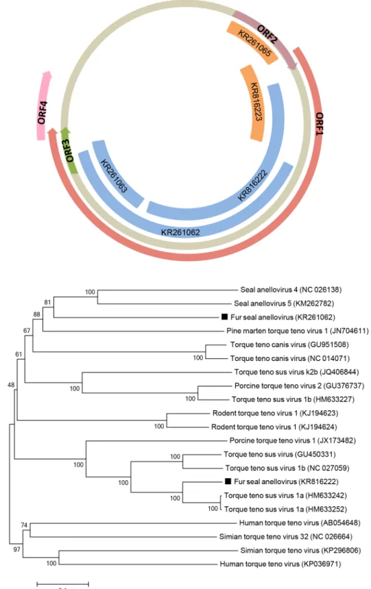
Anellovirus
Anelloviruses are small, non-enveloped, circular ssDNA viruses belonging to the
Anelloviridae
family [
25
]. Anellovirus genome sequences were detected in both fur seal species (
Fig 3A
). Few
sequences from the South American fur seal had the closest similarity to
Seal anellovirus 5,
with an amino acid identity ranging from 35
–
45% and covering up to 65% of ORF1. Another
sequence from the Subantarctic fur seal had the closest similarity to
Torque teno zalophus
virus1
(TTZV), with 78% amino acid identity and covering 69% of ORF2 of TTZV. Both fur
seal species had sequences with the closest similarity to
Torque teno sus virus 1a, with an amino
acid identity ranging from 84
–
88% and coverage of up to 56% of ORF1.
Due to the high divergence within anelloviruses, ORF1 sequences are the most indicative to
phylogenetic analyses [
25
]. Phylogenetic trees of partial ORF1 amino acid sequences obtained
from South American fur seals showed that distinct anelloviruses grouped in different clusters:
one most closely related to seal anelloviruses, whereas the other sequence was placed on the
same clade as swine torque teno viruses (
Fig 3B
).
Parvovirus
Parvoviruses are non-enveloped linear ssDNA viruses, members of the
Parvoviridae
family. In
this study, both fur seals species had sequences (
Fig 4A
) more closely related to mammal
par-voviruses of the
Parvovirinae, a subfamily that infects vertebrates and is currently divided into
eight genera [
26
,
27
]. The amino acid identity of those sequences with members of the
Parvovir-inae
ranged from 36
–
82%. Phylogenetic analysis of partial NS1 sequence, conserved within
Picornavirus
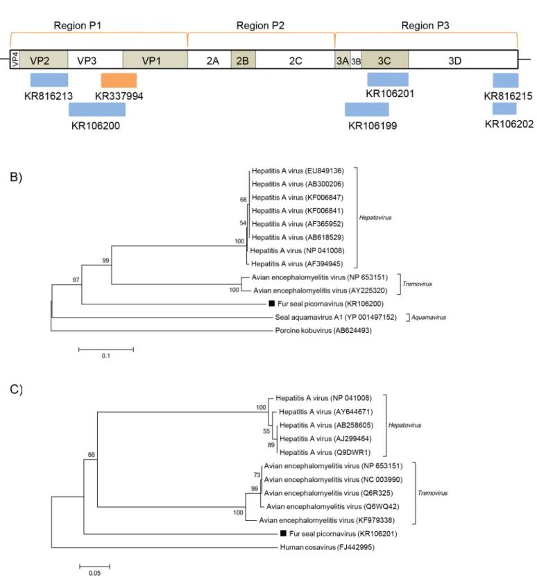
Picornaviruses are small, non-enveloped, positive sense ssRNA viruses of the
Picornaviridae
family, which has, to date, 29 recognized genera, though often increasing [
28–30
]. Picornavirus
sequences more related to Hepatitis A (HAV) and Avian encephalomyelitis viruses (AEV)
were detected in both fur seal species examined (
Fig 5A
). HAV belongs to
Hepatovirus
and
AEV to
Tremovirus, which are closely related genera [
31
]. The polyprotein sequences obtained
here displayed between 32
–
39% of amino acid identity to both
Hepatovirus
and
Tremovirus
Table 2. Contigs (>200bp) with significant BLASTx hits to known eukaryotic viruses obtained from the South American fur seals (Arctocephalus australis). Contig ID Accession number Length (nt)Family/Genus Genome Product Best hit Amino acid
identity (%) E-value
58 KR261062 1292 Anelloviridae ssDNA putative ORF1 ORF1 [Seal anellovirus 5]
(KM262782)
35 5e-61
59 KR261063 480 Anelloviridae ssDNA putative ORF1 ORF1 [Seal anellovirus 5]
(KM262782)
45 1e-14
62 KR816222 1080 Anelloviridae ssDNA putative ORF1 ORF1 [Torque teno sus virus 1a] (HM633252)
84 0.0
53 KR261066 616 Parvoviridae ssDNA capsid protein VP2 [Tusavirus 1] (KJ495710) 46 8e-46
54 KR261067 334 Parvoviridae ssDNA capsid protein capsid protein [Canine parvovirus 2a](HM042734)
50 2e-29
55 KR261068 460 Parvoviridae ssDNA NS1 NS1 [Solwezi bufavirus]
(LC011438)
43 1e-23
57 KR261070 237 Parvoviridae ssDNA capsid protein VP2 [Fox parvovirus] (KC692368) 46 3e-11
63 KR816220 344 Parvoviridae ssDNA NS1 NS1 [Tusavirus 1] KJ495710) 82 1e-65
34 KR106199 561 Picornaviridae +ssRNA polyprotein polyprotein [Hepatitis A virus] (FJ360731)
36 3e-15
35 KR106200 707 Picornaviridae +ssRNA polyprotein capsid protein [Hepatitis A virus] (AF365952)
37 1e-39
36 KR106201 519 Picornaviridae +ssRNA polyprotein putative 3C [Avian
encephalomyelitis virus] (NP_653151)
39 2e-26
37 KR106202 285 Picornaviridae +ssRNA polyprotein polyprotein [Bat picornavirus] (KJ641684)
38 5e-12
65 KR816213 466 Picornaviridae +ssRNA polyprotein 1B VP2 mature peptide [Hepatitis A virus] (NP_041008)
52 2e-50
67 KR816215 318 Picornaviridae +ssRNA polyprotein hypothetical protein [Avian encephalomyelitis virus]
(AJ006950)
32 2e-09
29 KR106194 217 Picobirnaviridae
Picobirnavirus
dsRNA RNA-dependent RNA polymerase
RNA dependent RNA polymerase [Human picobirnavirus]
(AB517735)
52 3e-13
30 KR106195 968 Picobirnaviridae
Picobirnavirus
dsRNA RNA-dependent RNA polymerase
RNA-dependent RNA polymerase [Fox picobirnavirus] (KC692366)
71
1e-169
31 KR106196 240 Picobirnaviridae
Picobirnavirus
dsRNA RNA-dependent RNA polymerase
putative RNA-dependent RNA polymerase [Dromedary picobirnavirus] (KM573806)
77 3e-34
33 KR106198 293 Picobirnaviridae
Picobirnavirus
dsRNA capsid protein hypothetical protein [Human picobirnavirus] (GU968923)
35 1e-08
64 KR816216 330 Picobirnaviridae
Picobirnavirus
dsRNA RNA-dependent RNA polymerase
putative RNA-dependent RNA polymerase [Dromedary picobirnavirus] (KM573806)
82 3e-61
73 KR827461 661 Hepevirus-like +ssRNA polyprotein nonstructural protein [Hepatitis E virus] (JQ026407)
27 4e-07
Table 3. Contigs (>200bp) with significant BLASTx hits to known eukaryotic viruses obtained from the Subantarctic fur seals (Arctocephalus tropicalis). Contig ID Accession number Length (nt)
Family/Genus Genome Product Best hit Amino acid
identity (%) E-value
52 KR261065 347 Anelloviridae ssDNA putative ORF2 ORF2 [Torque teno zalophus
virus 1] (NC_012126)
78 5e-18
72 KR816223 467 Anelloviridae ssDNA putative ORF2
and ORF1
ORF1 [Torque teno sus virus 1a] (HM633252)
88 4e-39
40 KR261071 1519 Parvoviridae ssDNA capsid protein VP2 [Tusavirus 1] (KJ495710) 39 1e-85
41 KR261072 1648 Parvoviridae ssDNA NS1 NS1 [Miniopterus schreibersii
parvovirus] (KC154061)
57
7e-131
42 KR261073 628 Parvoviridae ssDNA NS1 nonstructural protein NS1 [Tumor
virus X] (KJ631100)
44 2e-43
43 KR261074 565 Parvoviridae ssDNA NS1 NS1 [Turkey parvovirus
TP1-2012/HUN] (KF925531)
36 2e-13
44 KR261075 612 Parvoviridae ssDNA capsid protein putative VP1 [Tusavirus 1] (KJ495710)
39 2e-18
46 KR261077 349 Parvoviridae ssDNA capsid protein VP protein [Canine parvovirus] (KM235293)
55 2e-26
47 KR261078 957 Parvoviridae ssDNA NS1 non-structural protein 1
[Chipmunk parvovirus] (U86868)
37 1e-26
48 KR261079 301 Parvoviridae ssDNA capsid protein capsid protein [Canine parvovirus 2b] (JQ730016)
53 6e-25
68 KR816217 438 Parvoviridae ssDNA capsid protein putative VP1 [Tusavirus 1] (KJ495710)
42 1e-18
69 KR816218 322 Parvoviridae ssDNA capsid protein capsid protein VP2 [Mpulungu bufavirus] (NC_026815)
36 1e-07
70 KR816221 319 Parvoviridae ssDNA NS1 NS1 [Miniopterus schreibersii
parvovirus] (KC154061)
41 1e-14
61 KR337994 438 Picornaviridae +ssRNA polyprotein AEV polyprotein [Avian
encephalomyelitis virus] (NC_003990)
34 4e-15
12 KR072975 1271 Picornaviridae
Sakobuvirus
+ssRNA polyprotein polyprotein [Feline sakobuvirus A] (NC_022802)
58
6e-126
13 KR072976 477 Picornaviridae
Sakobuvirus
+ssRNA polyprotein polyprotein [Kobuvirus SZAL6-KoV/2011/HUN]
(KJ934637)
52 4e-12
14 KR072977 289 Picornaviridae
Sakobuvirus
+ssRNA polyprotein polyprotein [Feline sakobuvirus A] (NC_022802)
66 3e-22
15 KR072978 273 Picornaviridae
Sakobuvirus
+ssRNA polyprotein VP3 [Feline sakobuvirus A] (YP_008802588)
66 1e-34
16 KR072979 227 Picornaviridae
Sakobuvirus
+ssRNA polyprotein VP1 [Feline sakobuvirus A] (YP_008802588)
59 2e-12
18 KR072981 466 Picornaviridae
Sakobuvirus
+ssRNA polyprotein 2C [Feline sakobuvirus A] (YP_008802588)
59 3e-58
20 KR072982 767 Picornaviridae
Sakobuvirus
+ssRNA polyprotein 3D [Feline sakobuvirus A] (YP_008802588)
65
3e-117
22 KR072984 430 Picornaviridae
Sakobuvirus
+ ssRNA polyprotein 3D [Feline sakobuvirus A] (YP_008802588)
73 1e-67
23 KR072985 469 Reoviridae
Rotavirus
dsRNA NSP2 nonstructural protein 2 [Bovine rotavirus C] (AB874653)
69 3e-66
24 KR072986 412 Reoviridae
Rotavirus
dsRNA NSP3 nonstructural protein 3 [Bovine rotavirus C] (AB874654)
45 6e-33
25 KR072987 928 Reoviridae
Rotavirus
dsRNA VP1 VP1 [Bovine rotavirus C]
(AB738412)
69
members, and one sequence shared 52% amino acid identity with HAV VP2. Phylogenetic
analyses were based on the picornavirus polyprotein functional regions: P1, which encodes for
structural proteins, and P2-P3, which encode for proteins involved in replication [
28
]. The
analysis of partial P1 sequences identified here showed the fur seal picornavirus forming a
monophyletic group with the
Hepatovirus
and
Tremovirus
genera, but on a different branch
(
Fig 5B
). When partial sequences of P3 region were analyzed they still shared the same root,
with the fur seal picornavirus placed in a similar way (
Fig 5C
).
In addition to the above-mentioned picornaviruses, distinct members of this family were
found only in Subantarctic fur seals. With the exception of one sequence whose best BLASTx
hit had 52% of amino acid identity to a kobuvirus, all other contigs displayed the highest
amino acid identity to
Feline sakobuvirus A
(FSVA), which ranged from 58
–
73% with a total
coverage of 59% of its polyprotein (
Fig 6A
). One sequence displayed 59% of amino acid
iden-tity to FSVA VP1, and the amino acid ideniden-tity of VP3 was of 66%. The sequence covering the
2C region had 59% of amino acid identity, while the two 3D sequences here identified ranged
from 65
–
73% of FSVA 3D region. Phylogenetic analyses of partial P2 and P3 regions (
Fig 6B
and 6C
, respectively) showed the fur seal picornavirus, temporarily named Fur seal
sakobu-virus (FSSV), clustered with FSVA, member of the
Sakobuvirus
genus.
Picobirnavirus
Picobirnaviruses are small, non-enveloped, bisegmented dsRNA viruses of the
Picobirnaviridae
family. These highly variable viruses are classified in a sole genus,
Picobirnavirus, which on its
turn is divided into two genogroups (I and II), based on sequence similarities of the
RNA-dependent-RNA-polymerase gene (RdRp) [
32
,
33
]. Sequences of picobirnavirus RdRp and
cap-sid protein were detected in the South American fur seal samples (
Fig 7A
), having the highest
similarity with members of genogroup I, with an amino acid identity ranging from 35
–
82%.
Phylogenetic analyses of the partial RdRp gene (743 bp, which corresponds to 44% of the RdRp
gene) confirmed that the fur seal picobirnavirus identified here clustered with members of
gen-ogroup I, with a nucleotide identity ranging from 60
–
68% (
Fig 7B
).
Rotavirus
Rotaviruses are non-enveloped segmented dsRNA viruses from the
Reoviridae
family. They
belong to the
Rotavirus
genus and their genomes contain 11 segments. Based on sequence and
Table 3. (Continued)
Contig ID Accession number Length (nt)
Family/Genus Genome Product Best hit Amino acid
identity (%) E-value
26 KR072988 442 Reoviridae
Rotavirus
dsRNA VP3 VP3 [Human rotavirus C]
(HQ185645)
51 5e-41
27 KR072989 357 Reoviridae
Rotavirus
dsRNA VP3 viral protein 3 [Bovine rotavirus C] (AB874621)
65 6e-46
28 KR072990 360 Reoviridae
Rotavirus
dsRNA VP7 outer capsid protein VP7 [Human rotavirus C] (JQ177070)
59 9e-42
02 KR072992 2932 Caliciviridae
Sapovirus
+ ssRNA polyprotein polyprotein [California sea lion sapovirus 1] (JN420370)
98 0.0
06 KR072994 1400 Caliciviridae
Sapovirus
+ ssRNA polyprotein polyprotein [California sea lion sapovirus 1] (JN420370)
98 0.0
07 KR072995 2408 Caliciviridae
Sapovirus
+ ssRNA polyprotein and VP2
polyprotein [California sea lion sapovirus 1] (JN420370)
96 0.0
serological analyses of the structural protein VP6, there are seven species, also known as
groups, of rotaviruses (A-G), and recently a new group H has been proposed [
34
]. Rotavirus
sequences were detected in the Subantarctic fur seal (
Fig 8A
) with an amino acid identity from
45
–
69% to group C rotaviruses. The phylogenetic analysis, performed with a partial VP1
sequence, covering 30% of the complete gene, confirmed closer relatedness to group C
rotavi-ruses (
Fig 8B
). The VP1 gene, which encodes the RdRp, is well conserved within the genus and
may be also used for to differentiate rotavirus species [
35
,
36
].
Fig 4. Phylogenetic analysis of fur seal parvovirus.(A) Schematic representation of the genome of parvoviruses using as example the tusavirus (~4.4 kb). The blue bars represent the contigs from South American fur seal and the orange bars represent the contigs from Subantarctic fur seal. (B) Neighbor-joining phylogenetic tree based on the alignment of partial amino acid sequences (261 aa) from the NS1 protein of 17 parvoviruses.Seal parvovirusand California sea lion sesaviruswere used as outgroup. The parvovirus sequence from Subantarctic fur seal identified in this study is labeled with a black square. The GenBank accession numbers of the viral sequences are shown in parentheses.
Fig 5. Phylogenetic analysis of fur seal picornavirus.(A) Schematic representation of the genome of picornaviruses using as an example the hepatits A virus (~7.4 kb). The blue bars represent the contigs from South American fur seal and the orange bars represent the contigs from Subantarctic fur seal. (B) Neighbor-joining phylogenetic tree based on the alignment of partial amino acid sequences (219 aa) from the P1 region of the polyprotein of 13
picornaviruses.Porcine kobuviruswas used as outgroup. (C) Neighbor-joining phylogenetic tree based on the alignment of partial amino acid sequences (122 aa) from the P3 region of the polyprotein of 12 picornaviruses.Human cosaviruswas used as outgroup. The picornavirus sequences from South American fur seal identified in this study are labeled with a black square. The GenBank accession numbers of the viral sequences are shown in parentheses.
Hepevirus
Hepeviruses are non-enveloped, positive sense ssRNA viruses from the
Hepeviridae
family,
which is divided in two genera:
Orthohepevirus
and
Piscihepevirus
[
37
,
38
]. In this study, a
sequence of 661bp with low amino acid identity (27%) to the polyprotein gene of
hepe-viruses was detected in the South American fur seal (
Fig 9A
). Phylogenetic analysis of
par-tial sequences of the polyprotein of hepeviruses and hepevirus-like viruses was performed
(
Fig 9B
).
Fig 6. Phylogenetic analysis of fur seal sakobuvirus.(A) Schematic representation of the sakobuvirus genome usingFeline sakobuvirus A(~7.8 kb— NC_022802) as a reference. The orange bars represent the contigs from Subantarctic fur seal. (B) Neighbor-joining phylogenetic tree based on the alignment of partial amino acid sequences (409 aa) from the P2 region of the polyprotein of 11 picornaviruses.Sicinivirus 1was used as outgroup. (C) Neighbor-joining phylogenetic tree based on partial amino acid sequences (255 aa) from the 3D region of the polyprotein of 13 picornaviruses.California sea lion sapelovirus 2was used as outgroup. The sakobuvirus sequences from the Subantarctic fur seal from this study used in phylogenetic analyses are labeled with a black square. The GenBank accession numbers of the viral sequences are shown in parentheses.
Sapovirus
The genus
Sapovirus
consists of non-enveloped, positive sense ssRNA viruses of the
Caliciviri-dae
family. At present, five genogroups have been recognized based on VP1 sequence analyses
[
39
]. Sapovirus sequences were detected in the Subantarctic fur seal samples and results include
contigs covering over 90% of the
California sea lion sapovirus 1
(CslSaV1) genome (
Fig 10A
)
while sharing 98% amino acid identity and 89% nucleotide sequence identity. Phylogenetic
analysis of complete VP1 gene and nearly complete VP2 are shown in
Fig 10B and 10C
,
respectively.
Fig 7. Phylogenetic analysis of fur seal picobirnavirus.(A) Schematic representation of the genome of picobirnaviruses using as an example the human picobirnavirus (~4.2 kb). The blue bars represent the contigs from South American fur seal. (B) Neighbor-joining phylogenetic tree based on the alignment of partial nucleotide sequences (743 bp) from the RdRp gene of 17 picobirnaviruses. Human picobirnavirus GII was used as outgroup. The picobirnavirus sequence from South American fur seal identified in this study is labeled with a black square. The GenBank accession numbers of the viral sequences are shown in parentheses.
Discussion
This study has detected enteric viruses in the fecal samples of two species of fur seals that occur
in the coast of Rio Grande do Sul, South of Brazil. Such viruses belong to families whose
Fig 8. Phylogenetic analysis of fur seal rotavirus.(A) Schematic representation of the genome of rotaviruses using as an example the group C rotavirus (~17.9 kb). The orange bars represent the contigs from Subantarctic fur seal. (B) Neighbor-joining phylogenetic tree based on the alignment of partial amino acid sequences (307 aa) from the RpRd (segment 1) of 19 rotaviruses. Sequences of groups B, G and H were used as outgroup. The rotavirus sequence from Subantarctic fur seal identified in this study is labeled with a black square. The GenBank accession numbers of the viral sequences are shown in parentheses.
members are either apathogenic or known to cause disease in mammals. Anelloviruses were
detected in both species of fur seals examined. Based on anelloviruses demarcation criteria,
ORF1 sequences must have a divergence higher than 56 and 35% for genus and species,
respec-tively [
25
].The sequences with the highest similarity to seal anelloviruses displayed a 60%
divergence, suggesting that we found a new genus of
Anelloviridae. For the sequences with
higher similarity to
Torque teno sus virus 1a, the divergence was of 26%, which may indicate
they belong to the same genus,
Iotatorquevirus. Anelloviruses, which in most cases are not
associated to any particular disease, have been detected in seals and sea lions involved in
mor-tality events and virus-specific seroconversion of seals has been demonstrated, suggesting that
such animals are indeed susceptible to a productive infection following natural contact with
the virus [
11
,
40–42
].
Parvovirus sequences were detected in the two species of fur seals. Members of a same
genus within the
Parvoviridae
should share at least 30% amino acid identity in the predicted
NS1 sequence, and less than 30% identity when compared to other genera [
26
]. Although only
partial sequences from NS1 were detected in both species, all of them share more than 30%
amino acid identity with members of the
Protoparvovirus
genus, which was also shown by
phy-logenetic analysis, suggesting a new species within the genus. Parvoviruses have been detected
in pinnipeds [
43
,
44
], and members of different genera, including bocaviruses, dependoviruses
and a novel parvovirus named
Sesavirus
have been detected in California sea lions [
11
,
45
].
Fig 9. Phylogenetic analysis of fur seal hepevirus-like.(A) Schematic representation of the genome of hepeviruses using as an example the hepatitis E virus (~7.2 kb). The blue bar represents the contig from South American fur seal. (B) Neighbor-joining phylogenetic tree based on the alignment of partial amino acid sequences (182 aa) from the polyprotein of 15 hepeviruses.HepelivirusandFesavirus 2were used as outgroup. The hepevirus-like virus sequence from South American fur seal identified in this study is labeled with a black square. The GenBank accession numbers of the viral sequences are shown in parentheses.
Parvoviruses cause infections that can manifest through a variety of illnesses including
leuko-penia, myocarditis, gastroenteritis, as well as asymptomatically, and have been detected in
healthy and debilitated pinnipeds [
44–48
]. Parvoviruses are also known to be transmitted
between wild and domestic species [
49
,
50
].
Picornaviruses, which have been found in marine mammals [
51
], have also been detected in
this study. Here, two distinct picornaviruses were detected: one more similar to HAV and AEV
and other similar to FSVA. According to the
Picornaviridae
genus demarcation criteria,
differ-ent genera should share less than 40%, 40% and 50% amino acid iddiffer-entity in P1, P2 and P3,
Fig 10. Phylogenetic analysis of fur seal sapovirus.(A) Schematic representation of the genome of sapoviruses usingCalifornia sea lion sapovirus 1 (~7.5 kb—JN420370.2) as a reference. The orange bars represent the contigs from Subantarctic fur seal. (B) Neighbor-joining phylogenetic tree based on complete nucleotide sequences from the VP1 gene of 9 caliciviruses.Human noroviruswas used as outgroup. (C) Neighbor-joining phylogenetic tree based on the alignment of nearly-complete nucleotide sequences from the VP2 gene of 9 caliciviruses.Human noroviruswas used as outgroup. The sapovirus sequences from Subantarctic fur seal from this study used in phylogenetic analyses are labeled with a black square. The GenBank accession numbers of the viral sequences are shown in parentheses.
respectively [
30
]. Analyses of partial sequences of the polyprotein of the picornavirus similar to
HAV and AEV showed that the amino acids identities to members of
Hepatovirus
and
Tremo-virus
genera were below these cut-offs. The only exception was one sequence that shared 52%
amino acid identity with HAV VP2. A higher identity in this region, however, can be expected
within members of
Hepatovirus
and
Tremovirus
according to previous studies [
31
]. Based on
these values, a possible novel picornavirus more closely related to HAV and AEV was detected
in both fur seal species.
In addition, a new putative species within the recently recognized genus
Sakobuvirus
was
identified in the Subantarctic fur seal, here named
Fur seal sakobuvirus
(FSSV). These share an
amino acid identity of at least 50% with
Feline sakobuvirus A
when comparing all polyprotein
coding regions, indicating that FSSV belongs to the same genus. To date, FSVA was the sole
member of the genus; the genome reported here corresponds to the first description of a
sako-buvirus in another animal species, which was first found in cat feces [
52
].
Other potential novel enteric virus was also identified. A genogroup I picobirnavirus was
detected in South American fur seals. This fur seal picorbirnavirus is distinct from the
Otarine
picobirnavirus
previously found in California sea lions [
53
] and may represent a new species
within the genus. Picobirnaviruses have been detected both in asymptomatic and symptomatic
animals, including humans, and an etiologic association with diarrhea is not fully established.
However, coinfections of picobirnaviruses with other enteric viruses are not uncommon, can
be opportunistic, and may also have a synergistic effect [
54–57
].
A rotavirus related to group C was found in Subantarctic fur seals. Group C rotaviruses
have been associated with sporadic outbreaks in humans and other animals such as pigs and
bovines [
58–61
]. Other studies have detected rotaviruses in marine mammals: anti-group A
rotavirus antibodies were first found in Galapagos sea lions and fur seals [
62
] and RNA
sequences from rotaviruses related to lineage B were also detected in California sea lions [
11
].
Here, results show that more groups of rotaviruses, other than groups A and B, can circulate in
marine mammals.
A hepevirus-like sequence was detected in South American fur seals. Hepeviruses, such as
Hepatitis E virus (HEV), have been detected in mammals and birds [
37
,
63
]. HEVs can cause
asymptomatic infections to acute hepatitis and are known zoonotic agents [
64
]. Recently, a
new member of the
Hepeviridae
family was identified in cutthroat trout, a fish that occurs in
the Pacific ocean in North America [
65
] and unclassified hepevirus-like sequences named
hepelivirus and fesavirus-2 were detected in untreated sewage and cat feces, respectively
[
66
,
67
]. Phylogenetic analysis of the predicted partial polyprotein sequences of the
hepevirus-like identified here showed closer relatedness to other members of the
Hepeviridae
family than
to the unclassified hepelivirus and fesavirus-2. Although its low amino acid identity (
<
30%)
might indicate a novel member of the
Hepeviridae
family, more parts of the polyprotein would
need to be sequenced to better taxonomically allocate it.
A sapovirus was detected in samples from Subantarctic fur seals, which is genetically closely
related to CslSaV1 that was previously detected in a California sea lion with severe
osteomyeli-tis and nephrolithiasis [
11
]. Subantarctic fur seals are found in South Atlantic and Indian
oceans and our data shows that a very similar sapovirus circulates among fur seals from the
southern hemisphere, in addition to the ones that occur in the northern hemisphere.
Calici-viruses have been isolated from marine mammals and are known to cause vesicular lesions and
diarrhea in those animals [
10
,
44
,
68
]. Sapoviruses can cause gastroenteritis and have been
asso-ciated with diarrhea in animals [
69–71
].
occasions be exposed to humans, farm animals or pets, which may represent a risk of
cross-spe-cies transmission of pathogens and zoonoses [
10
]. Such risk of transmission to humans, for
example, was reported with an avian influenza virus isolated from harbor seals [
72
]. Besides, it
is not uncommon to find dogs in contact with carcasses of these animals found ashore, which
may give rise to emerging infectious diseases and transmission of known viruses, as already
reported with morbilliviruses [
73
]. An historical example of cross-species transmission
occurred with
San Miguel sea lion virus
that infects marine mammals. This calicivirus is nearly
identical to
Vesicular exanthema of swine virus, eradicated in swine since 1956, and was able to
cause an identical disease in pigs fed with infected carcasses of pinnipeds [
74
]. Sequences of
viruses belonging to viral families known to be transmitted between wild, domestic and farm
animals were detected in the present study: parvoviruses, hepeviruses and caliciviruses.
Indisputably, factors such as diet, age and different geographical distributions factors could
have contributed to the virome profile of both fur seal species [
75
]. Based on their lengths and
weight, most of the fur seals were classified as juveniles
—
only one Subantarctic fur seal was an
adult animal
–
and one can expect that juvenil animals are more susceptible to viral infections
than adults [
76
]. Fur seals are carnivores and can feed on a variety of preys. Subtantarctic fur
seals mostly feed on fish and cephalopods whereas South American fur seals main diet consists
of fish and crustaceans [
12
,
77
,
78
]. Their diet can impact on the virome of each fur seal species
and could explain, for example, the detection of eukaryotic viruses that do not infect mammals.
The use of fecal samples can allow the detection of sequences that may be originated from
dif-ferent hosts rather than the fur seals. Rotaviruses and sapoviruses were only detected in
Sub-antarctic fur seals whereas picobirnaviruses and a hepevirus-like were only found in South
American fur seals. Sequences of anelloviruses, parvoviruses and picornaviruses were detected
in both species of fur seals. These have also been reported in seals from the northern
hemi-sphere, indicating the widespread distributions of viruses of such families in pinnipeds
[
11
,
41
,
42
,
51
]. Furthermore, the occurrence of a very genetically closely related sapovirus that
infects California sea lions in Subantarctic fur seals shows that viruses previously isolated in
the North can also circulate in the South, infecting pinniped populations over a large
geograph-ical range.
Although the nucleotide sequences reported in this study do not comprise full genomes, this
initial characterization contributes to the knowledge of the viral populations that occur in fur
seals, and has identified potential novel viruses that may be of interest for future studies. This is
the first study to use next generation sequencing to explore the viral diversity of southern
hemi-sphere marine mammals. The findings presented here are expected to help to understand how
viral infections in pinnipeds may impact the health of the pinniped population and its potential
as sources of viruses which may potentially infect other animal species.
Acknowledgments
We would like to thank CECLIMAR for kindly providing the samples used in this study and
the laboratories from PUCRS and IPVDF for performing next-generation sequencing. We also
thank the Federal University of Rio Grande do Sul (UFRGS) Supercomputing National Center
(CESUP) for allowing access to run the metagenomic analyses.
Author Contributions
References
1. Ferreira JM, De Oliveira LR, Wynen L, Bester MN, Guinet C, Moraes-Barros N, et al. Multiple origins of vagrant Subantarctic fur seals: A long journey to the Brazilian coast detected by molecular markers. Polar Biol. 2008; 31: 303–308. doi:10.1007/s00300-007-0358-z
2. Moura JF, Siciliano S. Straggler subantarctic fur seal (Arctocephalus tropicalis) on the coast of Rio de Janeiro State, Brazil. Lat Am J Aquat Mamm. 2007; 6: 103–107. doi:10.5597/lajam00114
3. Pinedo MC. Ocorrência de pinípedes na costa brasileira. Gracia Orla Série Zool. 1990; 15: 37–48.
4. Oliveira A, Kolesnikovas CKM, Serafini PP, Moreira LMP, Pontalti M, Simões-Lopes PC, et al. Occur-rence of pinnipeds in Santa Catarina between 2000 and 2010. Lat Am J Aquat Mamm. 2011; 9: 145– 149.
5. Simões-Lopes PC, Drebmer CJ, Ott PH. Nota sobre os otariidae e phocidae (mammalia: carnivora) da costa norte do Rio Grande do Sul e Santa catarina, Brasil. Biociências. 1995. pp. 173–181.
6. Arbiza J, Blanc A, Castro-Ramos M, Katz H, León AP de, Clara M. Uruguayan Pinnipeds (Arctocepha-lus australis and Otaria flavescens): Evidence of Influenza Virus and Mycobacterium pinnipedii Infec-tions. New Approaches to Study Mar Mamm. 2012; 2: 151–182. doi:10.5772/54214
7. de Bruyn PJN, Bastos ADS, Eadie C, Tosh CA, Bester MN. Mass mortality of adult male subantarctic fur seals: Are alien mice the culprits? PLoS One. 2008; 3: e3757. doi:10.1371/journal.pone.0003757
PMID:19018284
8. Katz H, Morgades D, Castro-Ramos M. Pathological and Parasitological Findings in South American Fur Seal Pups (Arctocephalus australis) in Uruguay. ISRN Zool. 2012; 2012: 1–7. doi:10.5402/2012/
586079
9. Sikorski A, Dayaram A, Varsani A. Identification of a Novel Circular DNA Virus in New Zealand Fur Seal (Arctocephalus forsteri) Fecal Matter. Genome Announc. 2013; 1: 2012–2013. doi:10.1099/vir.0.
052654–0.17
10. Waltzek TB, Cortés-Hinojosa G, Wellehan JFX, Gray GC. Marine mammal zoonoses: a review of dis-ease manifestations. Zoonoses Public Health. 2012; 59: 521–535. doi:10.1111/j.1863-2378.2012.
01492.xPMID:22697432
11. Li L, Shan T, Wang C, Côté C, Kolman J, Onions D, et al. The fecal viral flora of california sea lions. J Virol. 2011; 85: 9909–9917. doi:10.1128/JVI.05026-11PMID:21795334
12. Vaz-Ferreira R. Arctocephalus australis (Zimmermann), South American fur seal. In: Ser FF, editor. Mammals in the seas. Rome: FAO; 1982. pp. 497–508.
13. Riedman M. The Pinnipeds: Seals, Sea Lions, and Walruses. Berkeley: University of California Press; 1990.
14. Geraci JR, Lounsbury VJ. Marine Mammals Ashore: A Field Guide for Strandings. 1st ed. Galveston, TX: Texas A&M Sea Grant; 1993.
15. Open Street Map [Internet]. p.https://www.openstreetmap.org.
16. Donaldson EF, Haskew AN, Gates JE, Huynh J, Moore CJ, Frieman MB. Metagenomic analysis of the viromes of three North American bat species: viral diversity among different bat species that share a common habitat. J Virol. 2010; 84: 13004–13018. doi:10.1128/JVI.01255-10PMID:20926577
17. Victoria JG, Kapoor A, Li L, Blinkova O, Slikas B, Wang C, et al. Metagenomic analyses of viruses in stool samples from children with acute flaccid paralysis. J Virol. 2009; 83: 4642–4651. doi:10.1128/JVI.
02301-08PMID:19211756
18. Allander T, Tammi MT, Eriksson M, Bjerkner A, Tiveljung-Lindell A, Andersson B. Cloning of a human parvovirus by molecular screening of respiratory tract samples. Proc Natl Acad Sci U S A. 2005; 102: 12891–12896. doi:10.1073/pnas.0504666102PMID:16118271
19. Stang A, Korn K, Wildner O, Uberla K. Characterization of virus isolates by particle-associated nucleic acid PCR. J Clin Microbiol. 2005; 43: 716–720. doi:10.1128/JCM.43.2.716–720.2005PMID: 15695669
20. Tamura K, Stecher G, Peterson D, Filipski A, Kumar S. MEGA6: Molecular Evolutionary Genetics Anal-ysis version 6.0. Mol Biol Evol. 2013; 30: 2725–2729. doi:10.1093/molbev/mst197PMID:24132122
21. Saitou N, Nei M. The neighbor-joining method: a new method for reconstructing phylogenetic trees. Mol Biol Evol. 1987; 4: 406–425. PMID:3447015
22. He B, Li Z, Yang F, Zheng J, Feng Y, Guo H, et al. Virome Profiling of Bats from Myanmar by Metage-nomic Analysis of Tissue Samples Reveals More Novel Mammalian Viruses. PLoS One. 2013; 8: e61950. doi:10.1371/journal.pone.0061950PMID:23630620
23. Wu Z, Ren X, Yang L, Hu Y, Yang J, He G, et al. Virome Analysis for Identification of Novel Mammalian Viruses in Bat Species from Chinese Provinces. J Virol. 2012; 86: 10999–11012. doi:10.1128/JVI.
24. Woo PCY, Lau SKP, Teng JLL, Tsang AKL, Joseph M, Wong EYM, et al. Metagenomic analysis of vir-omes of dromedary camel fecal samples reveals large number and high diversity of circoviruses and picobirnaviruses. Virology. 2014; 473: 117–125. doi:10.1016/j.virol.2014.09.020
25. Biagini P, Bendinelli M, Hino S, Kakkola L, Mankertz A, Niel C, et al. Anelloviridae. In: King A, Adams M, Carstens E, Lefkowitz E, editors. Virus taxonomy: classification and nomenclature of viruses: ninth report of the International Committee on Taxonomy of Viruses. 1st ed. San Diego: Elsevier; 2011. pp. 326–341.
26. Cotmore SF, Agbandje-McKenna M, Chiorini JA, Mukha D V, Pintel DJ, Qiu J, et al. The family Parvo-viridae. Arch Virol. 2014; 159: 1239–1247. doi:10.1007/s00705-013-1914-1PMID:24212889
27. Tijssen P, Agbandje-McKenna M, Almendral J, Bergoin M, Flegel T, Hedman K, et al. Parvoviridae. In: King A, Lefkowitz E, Adams M, Carstens E, editors. Virus taxonomy: classification and nomenclature of viruses: ninth report of the International Committee on Taxonomy of Viruses. 1st ed. San Diego: Else-vier; 2011. pp. 405–425.
28. Knowles N, Hovi T, Hyypiä T, King A, Lindberg A, Pallansch M, et al. Picornaviridae. In: King A, Adams M, Carstens E, Lefkowitz E, editors. Virus taxonomy: classification and nomenclature of viruses: ninth report of the International Committee on Taxonomy of Viruses. 1st ed. San Diego: Elsevier; 2011. pp. 855–880.
29. Adams MJ, Lefkowitz EJ, King AMQ, Bamford DH, Breitbart M, Davison AJ, et al. Ratification vote on taxonomic proposals to the International Committee on Taxonomy of Viruses (2015). Arch Virol. 2015; 160: 1837–50. doi:10.1007/s00705-015-2425-zPMID:25913692
30. The Pirbright Institute. The Picornavirus Pages [Internet]. 2015. Available:http://www.picornaviridae. com
31. Marvil P, Knowles NJ, Mockett AP, Britton P, Brown TD, Cavanagh D. Avian encephalomyelitis virus is a picornavirus and is most closely related to hepatitis A virus. J Gen Virol. 1999; 80: 653–662. PMID:
10092005
32. Delmas B. Picobirnaviridae. In: King A, Adams M, Carstens E, Lefkowitz E, editors. Virus taxonomy: classification and nomenclature of viruses: ninth report of the International Committee on Taxonomy of Viruses. 1st ed. San Diego: Elsevier; 2011. pp. 535–539.
33. Ganesh B, Masachessi G, Mladenova Z. Animal picobirnavirus. Virus Dis. 2014; 25: 223–238. doi:10.
1007/s13337-014-0207-y
34. Matthijnssens J, Otto PH, Ciarlet M, Desselberger U, van Ranst M, Johne R. VP6-sequence-based cut-off values as a criterion for rotavirus species demarcation. Arch Virol. 2012; 157: 1177–1182. doi:10.
1007/s00705-012-1273-3PMID:22430951
35. Attoui H, Mertens P, Becnel J, Belaganahalli S, Bergoin M, Brussaard C, et al. Reoviridae. In: King A, Adams M, Carstens E, Lefkowitz E, editors. Virus taxonomy: classification and nomenclature of viruses: ninth report of the International Committee on Taxonomy of Viruses. 1st ed. San Diego: Elsevier; 2011. pp. 541–637.
36. Ogden KM, Johne R, Patton JT. Rotavirus RNA polymerases resolve into two phylogenetically distinct classes that differ in their mechanism of template recognition. Virology. 2012; 431: 50–57. doi:10.1016/
j.virol.2012.05.011PMID:22687427
37. Meng XJ. Recent advances in Hepatitis E virus. J Viral Hepat. 2010; 17: 153–161. doi:
10.1111/j.1365-2893.2009.01257.xPMID:20040046
38. Smith DB, Simmonds P, Jameel S, Emerson SU, Harrison TJ, Meng X-J, et al. Consensus proposals for classification of the family Hepeviridae. J Gen Virol. 2014; 95: 2223–32. doi:10.1099/vir.0.068429– 0PMID:24989172
39. Clarke I, Estes M, Green K, Hansman G, Knowles N, Koopmans M, et al. Caliciviridae. In: King A, Adams M, Carstens E, Lefkowitz E, editors. Virus taxonomy: classification and nomenclature of viruses: ninth report of the International Committee on Taxonomy of Viruses. 1st ed. San Diego: Elsevier; 2011. pp. 977–986.
40. Fahsbender E, Rosario K, Cannon JP, Gulland F, Dishaw LJ, Breitbart M. Development of a Serological Assay for the Sea Lion (Zalophus californianus) Anellovirus, ZcAV. Sci Rep. 2015; 5: 9637. doi:10. 1038/srep09637PMID:25965294
41. Ng TFF, Suedmeyer WK, Wheeler E, Gulland F, Breitbart M. Novel anellovirus discovered from a mor-tality event of captive California sea lions. J Gen Virol. 2009; 90: 1256–1261. doi:10.1099/vir.0.
008987–0PMID:19264590
43. Bodewes R, García AR, Wiersma LCM, Getu S, Beukers M, Schapendonk CME, et al. Novel B19-like parvovirus in the brain of a harbor seal. PLoS One. 2013; 8: 1–9. doi:10.1371/journal.pone.0079259
44. Burek K a, Gulland FMD, Sheffield G, Beckmen KB, Keyes E, Spraker TR, et al. Infectious disease and the decline of Steller sea lions (Eumetopias jubatus) in Alaska, USA: insights from serologic data. J Wildl Dis. 2005; 41: 512–524. doi:10.7589/0090-3558-41.3.512PMID:16244061
45. Phan TG, Gulland F, Simeone C, Deng X, Delwart E. Sesavirus: prototype of a new parvovirus genus in feces of a sea lion. Virus Genes. 2014; 50: 134–136. doi:10.1007/s11262-014-1123-3PMID:
25272961
46. Ikeda Y, Nakamura K, Miyazawa T, Tohya Y, Takahashi E, Mochizuki M. Feline host range of Canine parvovirus: Recent emergence of new antigenic types in cats. Emerg Infect Dis. 2002; 8: 341–346. doi:
10.3201/eid0804.010228PMID:11971764
47. Steinel a, Parrish CR, Bloom ME, Truyen U. Parvovirus infections in wild carnivores. J Wildl Dis. 2001; 37: 594–607. doi:10.7589/0090-3558-37.3.594PMID:11504234
48. Hoelzer K, Parrish CR. The emergence of parvoviruses of carnivores. Vet Res. 2010; 41: 39. doi:10. 1051/vetres/2010011PMID:20152105
49. Allison a. B, Harbison CE, Pagan I, Stucker KM, Kaelber JT, Brown JD, et al. Role of Multiple Hosts in the Cross-Species Transmission and Emergence of a Pandemic Parvovirus. J Virol. 2012; 86: 865– 872. doi:10.1128/JVI.06187-11PMID:22072763
50. Allison AB, Kohler DJ, Fox K a, Brown JD, Gerhold RW, Shearn-Bochsler VI, et al. Frequent Cross-Species Transmission of Parvoviruses among Diverse Carnivore Hosts. J Virol. 2013; 87: 2342–2347. doi:10.1128/jvi.02428-12PMID:23221559
51. Kapoor a, Victoria J, Simmonds P, Wang C, Shafer RW, Nims R, et al. A highly divergent picornavirus in a marine mammal. J Virol. 2008; 82: 311–320. doi:10.1128/JVI.01240-07PMID:17942560
52. Ng TFF, Mesquita JR, Nascimento MSJ, Kondov NO, Wong W, Reuter G, et al. Feline fecal virome reveals novel and prevalent enteric viruses. Vet Microbiol. 2014; 171: 102–111. doi:10.1016/j.vetmic.
2014.04.005PMID:24793097
53. Woo PCY, Lau SKP, Bai R, Teng JLL, Lee P, Martelli P, et al. Complete Genome Sequence of a Novel Picobirnavirus, Otarine Picobirnavirus, Discovered in California Sea Lions. J Virol. 2012; 86: 6377– 6378. doi:10.1128/JVI.00686-12PMID:22570247
54. Giordano MO, Masachessi G, Martinez LC, Barril PA, Ferreyra LJ, Isa MB, et al. Two instances of large genome profile picobirnavirus occurrence in Argentinian infants with diarrhea over a 26-year period (1977–2002). J Infect. 2008; 56: 371–375. doi:10.1016/j.jinf.2008.02.017PMID:18403022
55. Bhattacharya R, Sahoo GC, Nayak MK, Rajendran K, Dutta P, Mitra U, et al. Detection of Genogroup I and II human picobirnaviruses showing small genomic RNA profile causing acute watery diarrhoea among children in Kolkata, India. Infect Genet Evol. 2007; 7: 229–238. doi:10.1016/j.meegid.2006.09.
005PMID:17049316
56. Carruyo-Núñez GM, Alcalá-Aristiguieta AC, Liprandi-Fraire F, Ludert-Leon JE. Porcine picobirnavirus infection in venezuelan farms. Rev Cient. 2014; XXIV: 125–131.
57. Malik YS, Kumar N, Sharma K, Dhama K, Shabbir MZ, Ganesh B, et al. Epidemiology, Phylogeny, and Evolution of Emerging Enteric Picobirnaviruses of Animal Origin and Their Relationship to Human Strains. Biomed Res Int. Hindawi Publishing Corporation; 2014; 2014: 780752. doi:10.1155/2014/ 780752PMID:25136620
58. Gabbay YB, Jiang B, Oliveira CS, Mascarenhas JD, Leite JP, Glass RI, et al. An outbreak of group C rotavirus gastroenteritis among children attending a day-care centre in Belém, Brazil. J Diarrhoeal Dis Res. 1999; 17: 69–74. PMID:10897889
59. Esona MD, Humphrey CD, Dennehy PH, Jiang B. Prevalence of group C rotavirus among children in Rhode Island, United States. J Clin Virol. 2008; 42: 221–224. doi:10.1016/j.jcv.2008.02.002PMID:
18374629
60. Kim Y, Chang KO, Straw B, Saif LJ. Characterization of group C rotaviruses associated with diarrhea outbreaks in feeder pigs. J Clin Microbiol. 1999; 37: 1484–1488. 0095-1137/99 PMID:10203510
61. Mawatari T, Taneichi A, Kawagoe T, Hosokawa M, Togashi K, Tsunemitsu H. Detection of a bovine group C rotavirus from adult cows with diarrhea and reduced milk production. J Vet Med Sci. 2004; 66: 887–890. doi:10.1292/jvms.66.887PMID:15297766
62. Coria-Galindo E, Rangel-Huerta E, Verdugo-Rodríguez A, Brousset D, Salazar S, Padilla-Noriega L. Rotavirus infections in Galapagos sea lions. J Wildl Dis. 2009; 45: 722–728. doi:
10.7589/0090-3558-45.3.722PMID:19617482
64. Bihl F, Negro F. Hepatitis E virus: A zoonosis adapting to humans. J Antimicrob Chemother. 2010; 65: 817–821. doi:10.1093/jac/dkq085PMID:20335188
65. Batts W, Yun S, Hedrick R, Winton J. A novel member of the family Hepeviridae from cutthroat trout (Oncorhynchus clarkii). Virus Res. Elsevier B.V.; 2011; 158: 116–123. doi:10.1016/j.virusres.2011.03.
019PMID:21458509
66. Ng TFF, Marine R, Wang C, Simmonds P, Kapusinszky B, Bodhidatta L, et al. High Variety of Known and New RNA and DNA Viruses of Diverse Origins in Untreated Sewage. J Virol. 2012; 86: 12161– 12175. doi:10.1128/JVI.00869-12PMID:22933275
67. Zhang W, Li L, Deng X, Kapusinszky B, Pesavento P a., Delwart E. Faecal virome of cats in an animal shelter. J Gen Virol. 2014; 95: 2553–2564. doi:10.1099/vir.0.069674–0PMID:25078300
68. Smith AW, Akers TG, Madin SH, Vedros NA. San Miguel sea lion virus isolation, preliminary characteri-zation and relationship to vesicular exanthema of swine virus. Nature. 1973; 244: 108–110. Available:
http://www.ncbi.nlm.nih.gov/pubmed/4583480PMID:4583480
69. Li L, Pesavento P a., Shan T, Leutenegger CM, Wang C, Delwart E. Viruses in diarrhoeic dogs include novel kobuviruses and sapoviruses. J Gen Virol. 2011; 92: 2534–2541. doi:10.1099/vir.0.034611–0 PMID:21775584
70. Soma T, Nakagomi O, Nakagomi T, Mochizuki M. Detection of Norovirus and Sapovirus from diarrheic dogs and cats in Japan. Microbiol Immunol. 2015; 59: 123–128. doi:10.1111/1348-0421.12223PMID:
25545754
71. Wang QH, Souza M, Funk J a., Zhang W, Saif LJ. Prevalence of noroviruses and sapoviruses in swine of various ages determined by reverse transcription-PCR and microwell hybridization assays. J Clin Microbiol. 2006; 44: 2057–2062. doi:10.1128/JCM.02634-05PMID:16757598
72. Karlsson EA, Ip HS, Hall JS, Yoon SW, Johnson J, Beck MA, et al. Respiratory transmission of an avian H3N8 influenza virus isolated from a harbour seal. Nat Commun. 2014; 5: 4791. doi:10.1038/ ncomms5791PMID:25183346
73. Kennedy S, Kuiken T, Jepson PD, Deaville R, Forsyth M, Barrett T, et al. Mass die-Off of Caspian seals caused by canine distemper virus. Emerg Infect Dis. 2000; 6: 637–639. doi:10.3201/eid0606.000613 PMID:11076723
74. Gelberg HB, Dieterich RA, Lewis RM. Vesicular Exanthema of Swine and San Miguel Sea Lion Virus: Experimental and Field Studies in Otarid Seals, Feeding Trials in Swine. Vet Pathol. 1982; 19: 413– 423. doi:10.1177/030098588201900407PMID:6283713
75. Dierauf L, Gulland FMD. CRC Handbook of Marine Mammal Medicine: Health, Disease, and Rehabili-tation, Second Edition. 2nd ed. Boca Raton, FL: CRC Press; 2001.
76. Barry AF, Ribeiro J, Alfieri AF, van der Poel WHM, Alfieri AA. First detection of kobuvirus in farm ani-mals in Brazil and the Netherlands. Infect Genet Evol. 2011; 11: 1811–1814. doi:10.1016/j.meegid.
2011.06.020PMID:21763785
77. Bester M. Subantarctic fur seal, Arctocephalus tropicalis, at Gough Island (Tristan da Cunha group). In: Croxall J, Gentry R, editors. Status, biology, and ecology of fur seals: proceedings of an international symposium and workshop Cambridge, England: 23–27 April 1984. Springfield: NOAA Tech Rep NMFS 51; 1987. pp. 57–60.