UNIVERSIDADE FEDERAL DO CEARÁ
CENTRO DE CIÊNCIAS
DEPARTAMENTO DE BIOQUÍMICA E BIOLOGIA MOLECULAR
PROGRAMA DE PÓS-GRADUAÇÃO EM BIOQUÍMICA
ANTÔNIO JOSÉ ROCHA
CLONAGEM DE cDNAS CODIFICANDO GLOBULINAS 7S (VICILINAS) DE DOIS
GENÓTIPOS DE CAUPI [Vigna unguiculata (L.) Walp] COM RESPOSTA
CONTRASTANTE AO CARUNCHO (Callosobruchus maculatus): SIMULAÇÕES
COMPUTACIONAIS REVELAM COMO AS VICILINAS DO CAUPI INTERAGEM
COM QUITINA.
FORTALEZA
ANTÔNIO JOSÉ ROCHA
CLONAGEM DE cDNAS CODIFICANDO GLOBULINAS 7S (VICILINAS) DE DOIS
GENÓTIPOS DE CAUPI [Vigna unguiculata (L.) Walp] COM RESPOSTA
CONTRASTANTE AO CARUNCHO (Callosobruchus maculatus): SIMULAÇÕES
COMPUTACIONAIS REVELAM COMO AS VICILINAS DO CAUPI INTERAGEM COM
QUITINA.
Tese de doutorado apresentado à coordenação do Programa de Pós-Graduação em Bioquímica como requisito obrigatório para a obtenção do título de Doutor em Bioquímica pela Universidade Federal do Ceará-UFC.
Orientador: Prof. Dr. Thalles Barbosa
Grangeiro
CLONAGEM DE cDNAS CODIFICANDO GLOBULINAS 7S (VICILINAS) DE DOIS
GENÓTIPOS DE CAUPI [Vigna unguiculata (L.) Walp] COM RESPOSTA
CONTRASTANTE AO CARUNCHO (Callosobruchus maculatus): SIMULAÇÕES
COMPUTACIONAIS REVELAM COMO AS VICILINAS DO CAUPI INTERAGEM COM
QUITINA.
Tese de doutorado apresentado à coordenação do Programa de Pós-Graduação em Bioquímica como requisito obrigatório para a obtenção do título de Doutor em Bioquímica pela Universidade Federal do Ceará-UFC.
Aprovada em: / /
BANCA EXAMINADORA
__________________________________________________________ Prof. Dr. Thalles Barbosa Grangueiro (Orientador)
Universidade Federal do Ceará-UFC
__________________________________________________________ Prof. Dr. Celso S. Nagano
Universidade Federal do Ceará-UFC
__________________________________________________________ Prof.Dr. José Hélio Costa
Universidade Federal do Ceará-UFC
__________________________________________________________ Profa. Dra. Vânia M. Ceccatto
Universidade Estadual do Ceará-UECE
__________________________________________________________ Prof. Dr. Daniel F. Feijó
Ao Professor Dr. Thalles Barbosa Grangeiro Agradeço por ser um orientador presente, um cientista admirável e por ter me possibilitado trabalhar em um projeto que me deu bastante satisfação. Professor, obrigado pelas oportunidades.
Aos professores Drs. Celso N. Nagano, Bruno A. Mathias Rocha, José Tadeus A. Oliveira e Valder Nogueira por suas contribuições essenciais para conclusão do manuscrito, a quem os admiro não somente por serem profissionais exemplares, mas também por serem pessoas altamente capacitados. Obrigado pela disponibilidade de contribuição para o trabalho e pela oportunidade de ter feito a parceria. Foi uma honra tê-los como colaboradores.
Aos prof. Drs. Bruno Lopes, Ito L. Barroso-Neto, Matheus Girão e ao Dr. Rômulo Farias pela contribuição no trabalho
Aos amigos do Laboratório de Genética e Biologia Molecular-LABGEM: Dr.Ednésio, Dr. Juscelino, Simone, Suelen, Jéssica Moura, e principalmente ao Dr. Edvar Monteiro -Júnior que contribui bastante para que esse trabalho fosse concluído. Agradeço a todos diretamente e indiretamente àqueles que contribuíram para esse trabalho e também pelos momentos de descontração, desabafos e pelo bom convívio.
Aos amigos do departamento: prof. Dr. Márcio Viana Ramos, Dr. Rafael de Sousa Miranda, Dra. Kátia Daniella da Cruz, saraiva, Ms. Mauro, Wallace, Débora, Eilton, Júnior, Sandro, etcc..
Às minhas amigas zeladoras do departamento de Biologia: Dna. Terezinha e Eliene, e da Bioquímica: Nega e Teca polos cafezinhos e brincadeiras!
É bom que posso sempre contar com vocês todos!
A todos os professores e os integrantes do Departamento de Bioquímica e Biologia Molecular da Universidade Federal do Ceará.
Aos meus pais Raimunda Aquiles Rocha e José Eudes Rocha (In memoriam) pelo apoio,
dedicação e amor incondicional, vocês são o meu melhor presente de Deus e os responsáveis por todas as minhas conquistas.
À Coordenação de Aperfeiçoamento de Pessoal de Nível Superior (CAPES), Fundação
e Biologia Molecular da Universidade Federal do Ceará, Departamento de Biologia,
departamento de Física, Laboratório de Genética e Biologia Molecular-LABGEM,
departamento de Engenharia de pesca e Centro Nacional de Processamento de Alto
Desempenho-Universidade Federal do Ceará (CENAPAD-UFC) em cujos laboratórios esta
A mente que se abre a uma nova idéia, jamais voltará ao seu tamanho original
O feijão caupi (Vigna unguiculata) é uma leguminosa que tem grande importância
socioeconômica no Nordeste brasileiro. Entretanto, na fase de pós-colheita, um dos problemas enfrentados pelos agricultores é a infestação pelo Calosobruchus maculatus (Coleoptera:
Bruchidae). Essa praga pode causar danos significativos às sementes, diminuindo seu teor nutricional e valor econômico. Diversos trabalhos correlacionam a resistência de alguns cultivares de caupi a uma ou mais variantes de vicilinas, globulinas 7S que são uma das principais proteínas de reserva dessas sementes. Essas variantes se ligam fortemente à quitina da membrana peritrófica de larvas de C. maculatus, prejudicando a absorção de nutrientes e,
por consequência, causando mortalidade das mesmas. Por outro lado, variantes de vicilinas de cultivares susceptíveis ao C. maculatus se ligam fracamente à quitina. Mas até o momento, as
diferenças estruturais que estão na origem da interação diferenciada entre as variantes de vicilinas e a quitina ainda são desconhecidas. Por essa razão, o objetivo principal do presente estudo foi investigar a base estrutural desse comportamento diferenciado das variantes de vicilinas 7S que poderiam explicar, pelo menos em parte, a resistência de sementes de algumas cultivares de caupi ao C. maculatus. Neste trabalho, as sequências parciais de cDNA que
codificam vicilinas de caupi foram obtidas a partir de sementes em desenvolvimento dos genótipos EPACE-10 (suscetível ao C. maculatus) e IT81D-1053 (resistente ao bruquídeo),
mediante amplificação por PCR, clonagem e sequenciamento. As sequências de aminoácidos deduzidas das sequências dos cDNAs foram então comparadas para se verificar as possíveis diferenças que poderiam existir entre as variantes de vicilinas 7S. Modelos moleculares tridimensionais das vicilinas de caupi, obtidos por modelagem por homologia, mostraram as características típicas do domínio dos membros da superfamília das cupinas. Simulações computacionais usando técnicas de docking e dinâmica molecular revelaram que cada trímero
ao oligômero β-vignina R3). Estes resultados concordam com trabalhos anteriores, que demonstraram que as vicilinas de caupi purificadas a partir de sementes de genótipos suscetíveis ou resistentes a C. maculatus têm a capacidade de se ligar invitro à quitina e invivo a estruturas
quitinosas do intestino médio das larvas. Além disso, a conservação em outras globulinas 7S de muitos dos resíduos que constituem o sítio de ligação a quitina de β-vignina, como previsto neste trabalho, também suporta as hipóteses anteriores que mostraram que as vicilinas de outras espécies de leguminosas também podem se ligar à quitina. Os resultados do presente trabalho fornecem a primeira descrição do mecanismo molecular envolvido na interação entre vicilinas de caupi e quitina.
Palavras-chaves: Proteínas de armazenamento de sementes. Leguminosae. Callosobruchus
globulins of many of the residues constituting the β-vignin chitin binding site as predicted in this paper also supports previous hypotheses that have shown that the vicillins of other legume species can also bind to chitin. The results of the present work provide the first description of the molecular mechanism involved in the interaction between cowpea and chitin vicillins.
Keywords: Seed storage proteins. Leguminosae. Callosobruchus maculatus.
Figure 1-Multiple sequence alignment of the amino acid sequences of β-vignin with the primary structures of representative vicilin-like 7S globulins. Amino acid sequences of β-vignin obtained from V. unguiculata genotypes EPACE-10 (sequence S2) and IT81D-1053
(sequence R2) were aligned with those of V. angularis (adzuki bean) 7S globulin-3
(Adzuki 7S3; UniProtKB accession number: A0A0S3SX36), V. radiata 8S globulin
(UniProtKB accession number: Q198W3), β-conglycinin (from Glycine max;
UniProtKB accession number: P25974) and canavalin (from Canavalia ensiformis;
UniProtKB accession number: P50477). Segments in the primary structures of β-vignins that were shown to contribute to their chitin-binding site (ChBS), as evidenced by computational simulations, are indicated. The alignment was edited using the program ALINE (Bond and Schüttelkopf, 2009).
Figure 2-Three-dimensional molecular model of β-vignin. N- and C-terminal cupin_1 domains are colored red and blue, respectively. (A) Ribbon diagram of the β-vignin monomer structure, as obtained by homology modeling (the structure modeled from sequence R2 is shown). (B) Superposition of the β-vignin model (blue) over the x-ray crystallographic structure of the V. angularis 7S globulin-1 (orange), which was used as
template (PDB ID: 2EA7).
Figure 3-Ribbon diagrams of the β-vignin homotrimers. Homotrimers of the models S2 (A) and R2 (B) are shown. Subunits are colored pink, green and cyan. Chito-oligosaccharide molecules [(GlcNAc)4] docked in the chitin-binding sites of each oligomer are also
shown as stick models (carbon, nitrogen and oxygen atoms are colored yellow, blue and red, respectively).
Figure 4-Close-up view of a (GlcNAc)4 molecule docked in the chitin-binding site of β-vignin.
The panels show a ligand molecule docked in the ChBS of models S2 (A), S3 (B), R2 (C) and R3 (D). Water molecules are represented as red spheres, hydrogen bonds are shown as green dotted lines (the distance cut-off is 3.5 Å) and the side chains of interacting residues are depicted as sticks. Nitrogen and oxygen atoms are colored blue and red, respectively.
Figure 5-Two-dimensional diagram of the interaction model between a (GlcNAc)4 molecule
depicting the interaction models obtained for the other complexes are shown in Figs. S25-S27. The diagrams were generated using the program LigPlot+ (Laskowski and Swindells, 2011).
Figure 5 (S1)-1% agarose gel electrophoresis of cDNA fragments encoding cowpea vicilins amplified by RT-PCR from total mRNA, which was purified from developing seeds from genotypes IT81D-1053 and EPACE-10. Lane 1: DNA size markers; lane 2: PCR products from genotype IT81D-1053; lane 3: PCR products from genotype EPACE-10. The major amplified band from each sample, which was cloned into the pGEM-T Easy vector, is indicated by an arrow.
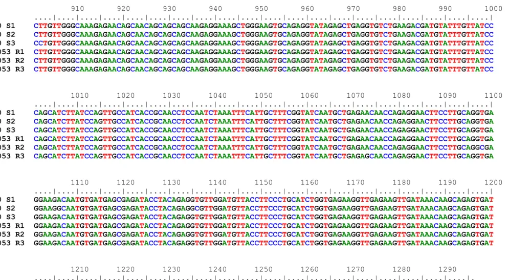
and the last amino acid residue in each row are shown on the left and right, respectively Figure 9 (S6)-Partial cDNA sequence and deduced amino acid sequence of cowpea vicilin from genotype IT81D-1053 isoform R2 (GenBank accession number: MG973245). The sequences of the oligonucleotide primers, which were used to amplify the cDNA fragment, are underlined. The amino acids matching the N-terminal sequence determined from β-vignin subunits is shaded in gray. Numbers for the first nucleotide and the last amino acid residue in each row are shown on the left and right, respectively. Figure 10 (S7)-Partial cDNA sequence and deduced amino acid sequence of cowpea vicilin from genotype IT81D-1053 isoform R3 (GenBank accession number: MG973246). The sequences of the oligonucleotide primers, which were used to amplify the cDNA fragment, are underlined. The amino acids matching the N-terminal sequence determined from β-vignin subunits is shaded in gray. Numbers for the first nucleotide and the last amino acid residue in each row are shown on the left and right, respectively. Figure 11 (S8)-Alignment of partial cDNA sequences encoding cowpea vicilins, which were cloned from genotypes EPACE-10 (sequences S1, S2 and S3) and IT81D-1053 (sequences R1, R2 and R3).
Figure 12 (S9)-Alignment of the amino acid sequences of cowpea vicilins, which were deduced from partial cDNA sequences obtained from genotypes EPACE-10 and IT81D-1053. Sites containing identical residues are shaded in black, whereas positions with chemically similar residues are shaded in gray. The alignment was rendered using BOXSHADE v3.21 through the web server embnet.vital-it.ch/software/BOX_form.html.
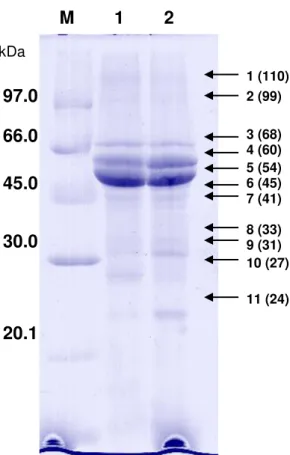
Figure 13 (S10)-SDS-polyacrylamide gel electrophoresis of seed vicilins from cowpea (V.
unguiculata). SDS-PAGE was performed as described by Laemmli (Laemmli, 1970)
S-200 HR coupled to an ÄKTA prime plus chromatography system. Vicilins from genotypes IT81D-1053 (A) and EPACE-10 (B) were resuspended in 50 mM Tris-HCl buffer, pH 8.0 (5 mg/mL), and 10 mg of each sample were loaded onto the column. The column was equilibrated and eluted with 50 mM Tris-HCl buffer, pH 8.0, at a constant flow rate (0.5 mL/min) and 3 mL fractions were collected.
Figure 15 (S12)-Ion exchange chromatography of cowpea vicilins on DEAE-Sepharose. Vicilins from cowpea genotypes IT81D-1053 (A) and EPACE-10 (B) were loaded onto a DEAE Sepharose Fast Flow (GE Healthcare) column (6 mL) equilibrated with 50 mM Tris-HCl buffer pH 8.0. The chromatography was performed at a constant flow rate (2 mL/min) and 3 mL fractions were collected.
Figure 16 (S13)-SDS-polyacrylamide gel electrophoresis of seed vicilins from cowpea (V.
unguiculata) purified by ion exchange chromatography. Peaks I (lane 1; 20 µg) and II
(lane 2; 20 µg) from the ion exchange chromatography of seed vicilins from genotypes IT81D-1053 (A) and EPACE-10 (B) were subjected to SDS-PAGE (15% polyacrylamide) and protein bands were stained with 0.2% (w/v) Coomassie Brilliant Blue R250 in 50% methanol/10% acetic acid for 16 h. Destaining was carried out with 12.5% isopropanol/10% acetic acid. Lane M: molecular weight markers.
Figure 17 (S14). MALDI-TOF mass spectra of tryptic digest of cowpea seed vicilins from genotype IT81D-1053. Spectra are from tryptic digestion of proteins from peak I (A) and peak II (B), as obtained by ion exchange chromatography. Assignment of masses to appropriate amino acid sequences is shown in Table S3.
Figure 17 (S15)-MALDI-TOF mass spectra of tryptic digest of cowpea seed vicilins from genotype EPACE-10. Spectra are from tryptic digestion of proteins from peak I (A) and peak II (B), as obtained by ion exchange chromatography. Assignment of masses to appropriate amino acid sequences is shown in Table S4.
Figure 18 (S16)-MS/MS spectra of the ion at m/z 1391. Amino acid sequence specific y-ions are indicated.
Figure 19 (S17)-MS/MS spectra of the ion at m/z 1960. Amino acid sequence specific y-ions are indicated.
Figure 20 (S18)-MS/MS spectra of the ion at m/z 1177. Amino acid sequence specific y-ions are indicated
2EA7) from adzuki bean (V. angularis). Identical and chemically-similar residues are
shaded in black and gray, respectively. The alignment was rendered using BOXSHADE v3.21 through the web server embnet.vital-it.ch/software/BOX_form.html.
Figure 22 (S20)-Alignment of the amino acid sequence of cowpea vicilin from genotype EPACE-10 (sequence S3) with the primary structure of the 7S globulin-1 (PDB ID: 2EA7) from adzuki bean (V. angularis). Identical and chemically-similar residues are
shaded in black and gray, respectively. The alignment was rendered using BOXSHADE v3.21 through the web server embnet.vital-it.ch/software/BOX_form.html.
Figure 23 (S21)-Alignment of the amino acid sequence of cowpea vicilin from genotype IT81D-1053 (sequence R2) with the primary structure of the 7S globulin-1 (PDB ID: 2EA7) from adzuki bean (V. angularis). Identical and chemically-similar residues are
shaded in black and gray, respectively. The alignment was rendered using BOXSHADE v3.21 through the web server embnet.vital-it.ch/software/BOX_form.html.
Figure 24 (S22)-Alignment of the amino acid sequence of cowpea vicilin from genotype IT81D-1053 (sequence R3) with the primary structure of the 7S globulin-1 (PDB ID: 2EA7) from adzuki bean (V. angularis). Identical and chemically-similar residues are
shaded in black and gray, respectively. The alignment was rendered using BOXSHADE v3.21 through the web server embnet.vital-it.ch/software/BOX_form.html.
Figure 25 (S23)-Superposition of the three-dimensional molecular models of β-vignin. The superposed models, shown as ribbon diagrams, are colored pink (model S2), yellow (model S3), green (model R2) and cyan (model R3). The modeled structures were superposed by overlapping 354 Cα-atoms of structurally equivalent residues with an RMSD of 1.479 Å.
Figure 26 (S24)-Root-mean-square deviations (RMSD; Å) of the backbone atoms of a tetra-N
-acetyl-chitotetraose molecule [(GlcNAc)4] docked in the chitin-binding site of cowpea
vicilins, during the 40 ns MD simulations. The graphics show the backbone RMSD plots of (GlcNAc)4 docked in the three-dimensional molecular models generated from
sequences S2 (A), S3 (B), R2 (C) and R3 (D).
Figure 27 (S25)-Two-dimensional diagram of the interaction model between a (GlcNAc)4
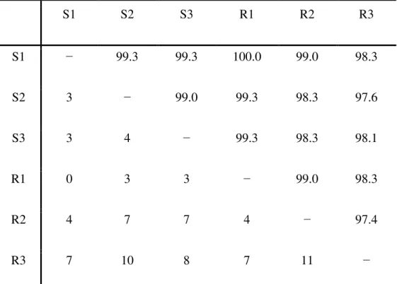
Table S1-Matrix of pairwise comparisons of cDNA sequences encoding cowpea vicilins, which were cloned from genotypes EPACE-10 (S1, S2 and S3) and IT81D-105 (R1, R2 and R3). For each pair of compared sequences, the percentage of sequence identity (above the diagonal) and the number of different nucleotides (below the diagonal) between them are shown. These numbers were calculated based on the multiple sequence alignment shown in Fig. S8.
Table S2-Matrix of pairwise comparisons of vicilin amino acid sequences from cowpea genotypes EPACE-10 (S1, S2 and S3) and IT81D-105 (R1, R2 and R3). For each pair of compared sequences, the percentage of sequence identity (above the diagonal) and the number of different amino acid residues (below the diagonal) between them are shown. These numbers were calculated based on the multiple sequence alignment shown in Fig. S9.
Table S3-Calculated molecular masses and theoretical isoelectric point (pI) values of
cowpea β-vignins. The values were calculated by submitting the amino acid sequences to ExPASy’s Compute pI/Mw tool (web.expasy.org/compute_pi/)
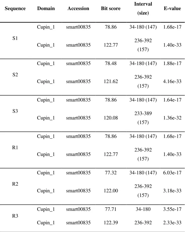
Table S4-Results of the BLAST searches against the Conserved Domain Database (https://www.ncbi.nlm.nih.gov/Structure/cdd/wrpsb.cgi) using as query the cowpea vicilin sequences from genotypes EPACE-10 (sequences S1, S2 and S3) and IT81D-1053 (R1, R2 and R3)
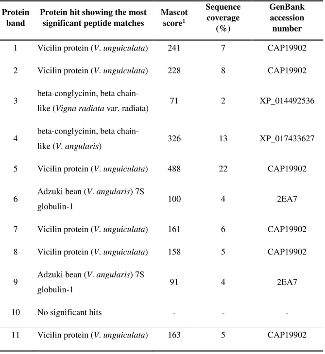
Table S5-Identification of the protein bands from the vicilin fraction of cowpea (V.
unguiculata) genotype IT81D-1053 by ESI-MS/MS. MS/MS ions searches against
the NCBIprot database were performed using Mascot through the software’s web server.
Table S6-Identification of the protein bands from the vicilin fraction of cowpea (V.
unguiculata) genotype EPACE-10 by ESI-MS/MS. MS/MS ions searches against the
NCBIprot database were performed using Mascot through the software’s web server. Table S7-Tryptic peptides from cowpea seed vicilins, purified from genotype IT81D-1053, as identified by MALDI-TOF mass spectrometry
model of cowpea vicilin from sequence S2. The analysis was performed by submitting the three-dimensional molecular model to the MolProbity web server (http://molprobity.biochem.duke.edu/)
Table S10-Summary statistics of the MolProbity analysis of the three-dimensional molecular model of cowpea vicilin from sequence S3. The analysis was performed by submitting the three-dimensional molecular model to the MolProbity web server (http://molprobity.biochem.duke.edu/)
Table S11-Summary statistics of the MolProbity analysis of the three-dimensional molecular model of cowpea vicilin from sequence R2. The analysis was performed by submitting the three-dimensional molecular model to the MolProbity web server (http://molprobity.biochem.duke.edu/)
Table S12-Summary statistics of the MolProbity analysis of the three-dimensional molecular model of cowpea vicilin from sequence R3. The analysis was performed by submitting the three-dimensional molecular model to the MolProbity web server (http://molprobity.biochem.duke.edu/)
Table S13-Calculated binding free energies (kcal/ml) of (GlcNAc)4 molecules bound to
R-Resistent
S-Susceptível
cDNA- complementar ao DNA
GlcNAc - N-acetil-D-glucosamina
2- BLAST- Basic Local Alignment Search Tool
LC-ESI-MS/MS- Liquid Chromatography Electrospray Ionization Tandem Mass Spectrometric
UPLC-ultra-high-performance liquid chromatography
MALDI-TOF/TOF- Matrix-assisted laser desorption/ionization time-of-flight tandem
PMF-peptide mass fingerprint
NCBI- National Center for Biotechnology Information
ESI-MS/MS- Electrospray Ionization Tandem Mass
RMSD- root-mean-square deviation
SUMÁRIO
1 INDRODUÇÃO GERAL 22
2 REFERENCIAL TEÓRICO 24
3 HIPÓTESES 32
4 OBJETIVOS 33
5 CLONING OF cDNA SEQUENCES ENCODING COWPEA (Vigna
unguiculata) VICILINS: MOLECULAR MODELING, DOCKING
CALCULATIONS AND MOLECULAR DYNAMICS SIMULATIONS SUGGEST A BINDING MODE OF COWPEA VICILINS TO CHITIN OLIGOMERS
34
6 CLONING OF cDNA SEQUENCES ENCODING COWPEA (Vigna unguiculata) VICILINS: MOLECULAR MODELING, DOCKING
CALCULATIONS AND MOLECULAR DYNAMICS SIMULATIONS SUGGEST A BINDING MODE OF COWPEA VICILINS TO CHITIN OLIGOMERS
35
7 CONSIDERAÇÕES FINAIS 110
1. INTRODUÇÃO GERAL
O feijão caupi (Vigna unguiculata) possui um importante papel sócio-econômico em
regiões tropicais e subtropicais da África, Ásia e Américas (SINGH et al., 2003). No Brasil, principalmente nas regiões Norte e Nordeste, as sementes de caupi são uma das principais fontes de proteínas na nutrição humana. Na fase de pós-colheita, um dos problemas enfrentados pelos produtores é a infestação das sementes pelo caruncho do caupi, Callosobruchus
maculatus Fabricius, 1775 (Coleoptera: Chrysomelidae). Essa praga causa danos às sementes,
reduzindo o valor comercial do produto destinado ao consumo humano.
Na década de 70, a coleção de germoplasma de caupi do Instituto Internacional de Agricultura Tropical (IITA, Nigéria) foi avaliada com o objetivo de se identificar cultivares naturalmente resistentes ao C. maculatus. De oito mil cultivares analisadas, três apresentaram resistência ao caruncho do caupi: TVu 2027, TVu 11952 e TVu 11953 (SINGH et al., 1985).
Gatehouse e colaboradores (1979) atribuíram a resistência das sementes da cultivar resistente TVu 2027 ao ataque de C. maculatus a níveis elevados de inibidores de proteases do tipo tripsina em relação a oito cultivares suscetíveis, que apresentaram baixos níveis desse inibidor. Entretanto, Xavier-Filho e colaboradores (1989), utilizaram cultivares suscetíveis e resistentes ao ataque do caruncho do feijão caupi e mostraram que não existia correlação significativa entre os níveis de inibidores de tripsina e a resistência/suscetibilidade das mesmas, contrariando as sugestões de Gatehouse e colaboradores (1979). De fato, larvas de C. maculatus
não usam majoritariamente proteases do tipo tripsina para sua digestão proteica, mas sim proteinases aspárticas e principalmente proteinases cisteínicas (Campos et al., 1989).
Posteriormente, Macedo e colaboradores (1993) sugeriram que a resistência ao C.
maculatus poderia ser atribuída a uma forma variante de vicilina 7S, que era deletéria ao
desenvolvimento das larvas. Estudos posteriores revelaram que as vicilinas dos cultivares resistentes se ligavam fortemente à quitina (in vitro) e à camada de quitina que reveste a membrana peritrófica do trato intestinal (SALES et al., 1996; SALES et al., 2001). Sales e colaboradores (1996; 2001) sugeriram que essa interação limitava a assimilação dos nutrientes no intestino das larvas de C. maculatus, dificultando assim a sobrevivência e desenvolvimento
do inseto. Mais recentemente, Aquino (2009) realizou um trabalho in silico e modelou a proteína vicilina 7S de sementes de feijão caupi usando uma vicilina 7S de sementes de Glycine
max como modelo comparativo. Em seguida, através de ferramentas de docking molecular
Diante desses dados, algumas lacunas precisam ser elucidadas. Embora Aquino (2009) tenha identificado um domínio de ligação à quitina, este achado foi baseado apenas em dados in silico. Ademais, Silva (2014) evidenciou diferenças nas sequências de aminoácidos deduzidas a partir do sequenciamento parcial do cDNA que codifica vicilinas 7S de dois genótipos de feijão caupi contrastantes quanto à resistência ao C. maculatus: IT81D-1053
(resistente) e EPACE 10 (susceptível). As sequências parciais de aminoácidos das vicilinas 7S de ambas cultivares divergiram especificamente em regiões ricas em glutamina. Tal análise encontrou locais potencialmente ligantes a radicais fosfato e radicais eletronegativos como grupamentos amínicos e carboxílicos, sendo que a região variável de glutaminas foi indicada como potencial ligante a esses grupos eletronegativos. Apesar dos achados descritos estes dados foram obtidos com base nas sequências parciais das vicilinas 7S e não com base nas sequências completas. Em resumo, até o momento ainda não se sabe com clareza o mecanismo estrutural de interação das vicilinas 7S de feijão caupi com quitina.
Tendo em vista essas importantes lacunas à respeito das vicilina 7S do feijão caupi, além dos prejuízos pós-colheita ocasionados por C. maculatus, é de suma importância que um estudo que objetive elucidar o mecanismo de interação de vicilinas 7S de feijão caupi com a quitina de C. maculatus seja realizado, a fim de que os prejuízos ocasionados por esta praga sejam minimizados.
2-REFERENCIAL TEÓRICO
2. 1. Feijão caupi (Vigna unguiculata)
O feijão caupi [Vigna unguiculata (L.) Walpers], é uma planta dicotiledônea,
pertencente à ordem Fabales, família Fabaceae, subfamília Faboideae, tribo Phaseoleae e à subtribo Phaseolinae. O gênero Vigna apresenta cerca de 160 espécies (PADULOSI; NG., 1997).
Essa cultura é originária do oeste da África, sendo cultivada nas principais regiões tropicais em todo o mundo (SINGH et al, 1985). O maior centro de diversidade fica também
no oeste da África, localizado no Instituto Internacional de Agricultura Tropical (IITA, Nigéria) (PANT et al., 1982; NG). Entretanto, a maior diversidade de genótipos selvagens ocorre, principalmente, na região sudeste da África (PADULOSI; NG., 1997).
No Brasil, existe uma grande diversidade de cultivares de feijão caupi, entretanto, essa cultura recebe vários nomes dependendo da região do Brasil, como feijão de corda, massacar, feijão de vagem, feijão de vara, feijão de moita. Na Bahia é conhecido como feijão catador; feijão trepa-pau no Maranhão; feijão gurutuba no norte de Minas Gerais. Já no Rio de Janeiro é conhecido como feijão fradinho e no Sul como feijão miúdo. Já no Norte é conhecido como feijão da praia. (CAJAZEIRAS, 2000). Diante de todas as diversidades desses nomes populares a Embrapa cientificamente a denominou de feijão caupi em referência ao nome em inglês Cowpea.
O grão seco é o principal produto do consumo humano, sendo este uma das principais fontes de proteínas, carboidratos e lipídios para a população brasileira, principalmente a região Nordeste, e também na África (SINGH et al, 1985).
O teor proteico das sementes varia de 19,5 a 26,1% (MAIA et al, 2000). As proteínas totais podem ser divididas em quatro grupos de acordo com sua solubilidade: albuminas, globulinas, glutelinas e prolaminas, sendo as globulinas, as principais proteínas de reversa (cerca de 70% do conteúdo total de nitrogênio (AREMU 1990; OSBORN, 1988). Todos os aminoácidos essenciais encontram-se nas sementes de feijão caupi, sendo bastante rico em lisina e pobre em aminoácidos sulfurados (AREMU, 1990).
outros (OLIVEIRA, 1981; RIOS, 1988). Já no período pós-colheita a principal praga que ataca seu armazenamento é Callosobruchus maculatus, conhecido popularmente como caruncho ou gorgulho (CREDLAND, 1986).
2. 2. Callosobruchus maculatus e sua interação com o feijão caupi (Vigna unguiculata)
Dentre os insetos que atacam o feijão caupi no período de armazenamento, o
Callosobruchus maculatus tem sido seu principal predador, entretanto, esse bruquídeo não
ataca às sementes do feijão-comum (Phaseolus vulgaris), devido à presença de inibidores de α
-amilases. O principal predador do feijão comum é o bruquídeo Zabrotes subfasciatus. O C.
maculatus pertence à classe Insecta, ordem Coleóptera, subordem Poliphaga, subfamília
Chrysomeloidea, família Chrysomelidae, gênero Calosobruchus e à espécie C. maculatus. Essa
espécie é originária da África, no entanto, hoje é frequentemente encontrada nas regiões tropicais e subtropicais do mundo, uma vez que foi propagada através do comércio (SOUTHGATE, 1979).
As larvas desses insetos só se alimentam e crescem no interior das sementes de leguminosas (Fabaceae). Já os adultos desta espécie, não se alimentam ou necessitam de água, são chamados insetos afagos (OLIVEIRA, 2013). Além disso, o tempo de desenvolvimento é de aproximadamente duas a três semanas, passando boa parte desse tempo acasalando-se e realizando a postura de ovos nos feijões (OLIVEIRA, 2013). Esse inseto possui um desenvolvimento holometabólico descrito por BASTOS (1968), passando pelas fases de ovo, larva, pupa e adulto.
O ataque do C. maculatus ao feijão caupi se inicia quando as fêmeas depositam seus
2. 3. Cultivares de Vigna unguiculata resistentes ao Callosobruchus maculatus
A cultivar TVu 2027 identificada pelo IITA na Nigéria deu origem a várias outras, como IT81D-1032, IT81D-1064 e IT81D-1045, mostrando diferentes níveis de resistência ao caruncho do feijão caupi (SINGH et al, 1985, SINGH et al, 2003). A resistência conferida a estes feijões variantes foi atribuída a vários fatores, que na sua maioria, são de natureza química.
Inicialmente Gatehouse e colaboradores (1979) propuseram que a resistência observada estava associada a altos níveis de inibidores de tripsina nessas sementes. Entretanto, uma hipótese proposta por Xavier-Filho e colaboradores (1989) descartou a relação dos inibidores de proteases com a resistência ao caruncho proposta por Gatehouse e colaboradores (1979). Um experimento usando três cultivares suscetíveis (CE-11, CE-12 e CE-524) e três cultivares resistentes (ITB1D-1045, ITB1D-1064 e TVu 2027) ao caruncho do caupi, mostraram que não existia correlação significativa entre os níveis de inibidores de tripsina e a resistência/suscetibilidade das mesmas. Nesse experimento, as cultivares resistentes IT81D-1045 e IT81D-1064 mostraram baixas concentrações de inibidores de tripsina, refutando a hipótese de Gatehouse e colaboradores (1979), muito embora uma das cultivares resistentes, TVu 2027, tenha apresentado altos níveis de inibidor de tripsina. Entretanto, três cultivares suscetíveis (CE-11, CE-12 e CE-524) também apresentaram altos níveis desse inibidor, o que descartou totalmente a teoria de Gatehouse e colaboradores (1979).
Atrelado a isso, Campos e colaboradores (1989) demonstraram em seus experimentos que as larvas de C. maculatus usam proteinases aspárticas e principalmente proteinases cisteínicas para sua digestão proteica e não proteases do tipo tripsina.
Ademais, Sales e colaboradores (1992) submeteram amostras de vicilinas de cultivares suscetíveis [CE-11 e CE-31 (Pitiuba)] e resistentes (IT81D-1032 e IT81D-1045) ao inseto à digestão proteica com pepsina, papaína e extratos do intestino de larvas de C. maculatus, contendo as proteases nativas. Os autores observaram que as vicilinas das cultivares resistentes foram mais refratárias à ação das proteases do que as vicilinas das cultivares suscetíveis.
Mais tarde, Macedo e colaboradores (1993) realizaram um experimento com duas cultivares resistentes ao C. maculatus, IT81D-1032 e IT81D-1045 que os levaram a sugerir que
a forma variante da vicilina (globulina 7S) foi deletéria ao desenvolvimento das larvas.
aderindo-se nos ápices epiteliais das células intestinas das larvas (SALES et al., 1996; SALES et al., 2001).
Além disso, Sales e colaboradores (2001) mostraram também que as vicilinas das cultivares resistentes e susceptíveis se ligavam fortemente e fracamente, respectivamente, à quitina (in vitro) e à camada da membrana peritrófica do trato intestinal. Diante disso, os autores sugeriram que essa interação da vicilina 7S com a camada de quitina que compõe o intestino do C. maculatus não deixava que as larvas assimilassem os nutrientes necessários para o seu desenvolvimento, dificultando sua sobrevivência ou levando à morte do inseto.
Portanto, os autores postularam que a suscetibilidade diferenciada das vicilinas 7S às proteases do inseto constitui um aspecto do mecanismo de resistência das sementes ao ataque pelo C. maculatus.
2. 4. Proteínas de reserva de sementes
As proteínas de reserva foram primeiramente classificadas por Osborne (1988), com base na sua extração e solubilidade em água (albuminas), solução salina (globulinas), solução alcoólica (prolaminas) e solução ácida ou básica (glutelinas) (SHEWRY et al., 1995).
A maioria das proteínas em sementes de leguminosas consiste de globulinas, proteínas de reserva sintetizadas durante o desenvolvimento da semente. Elas estão presentes não somente em dicotiledôneas, mas também em monocotiledôneas, incluindo cereais e palmeiras e em esporos de samambaias (TEMPLEMAN et al., 1987).
Durante a germinação da semente as globulinas são estocadas em corpos proteicos, ou esqueletos de nitrogênio e carbono que fornecem energia para o desenvolvimento das plântulas e são hidrolisadas durante o processo germinativo. As demais proteínas são principalmente albuminas, que incluem proteínas de reserva e de manutenção, lectinas, lipoxigenases, entre outras (SHEWRY et al., 1995; DODEMAN et al., 1997; MARUYAMA et al., 2003; WANG et al., 2003).
2. 5. Vicilinas (Globulinas 7S)
As vicilinas (7S) representam aproximadamente 80% das proteínas totais de sementes maduras. As globulinas de sementes que são tipicamente classificadas como vicilinas consistem em uma combinação de subunidades com massa molecular que varia de 150 a 190 kDa. Nestas subunidades de vicilinas, não há presença de pontes dissulfeto e a proteína é estabilizada por forças não covalentes ( PAES et al., 2008; OLIVEIRA, 2013).
As vicilinas 7S Possuem dois domínios cupin_1 (superfamília cupinas: bicupinas) e são constituídas majoritariamente por β-vigninas e Proteína trimérica e possui 55 e 60 kDa em suas subunidades de polipeptídeos (Salles et al,1992; 2001). Elas consistem em uma combinação de 3 subunidades com massa molecular que varia de 156 a 177 kDa formando um trímero em formato de triângulo e apresentam uma composição de aminoácidos com altas concentrações de ácido aspártico, ácido glutâmico, arginina, fenilalanina e leucina, mas têm, no entanto, concentrações mínimas de aminoácidos sulfurados como metionina e cisteína (MACEDO et al.,1995). A ausência de resíduos de cisteínas nas globulinas do tipo vicilina 7S impede que as mesmas possam realizar pontes dissulfeto. O tamanho final da proteína madura varia consideravelmente devido aos processamentos pós-traducionais, como proteólise e glicosilação (SHEWRY et al., 1995; SHUTOV et al., 1995).
6-REFERÊNCIAS
1. Aremu, C. Y. Proximate and amino acid composition of cowpea (vigna unguiculata, walp)
protein concentrate prepared by isoeletric point precipitation. Food chemistry, v. 8, p. 61-68, 1990.
2. Aquino, R.erminação de motivos de ligação à quitina em vicilinas de canavalia ensiformis
e vigna unguiculata através de métodos in silico e relação com suas toxicidades para o
bruquídeo callosobruchus (coleoptera: bruchidae). 2009. Dissertação (mestrado em
bioquímica) -centro de biociências, universidade federal do rio grande do norte, natal, 2009. 3. Bastos, J. A. M. Principais pragas das culturas e seus controles. 3.ed. São paulo: nobel, 1985, 329p.
4. Cajazeiras, J. B. Identificação de genótipos de caupi [vigna unguiculata (l.) Walp.]
Resistentes ao caruncho (Callosobruchus maculatus fabricius, 1792), 2000. Dissertação
(mestrado em bioquímica) - centro de ciências, universidade federal do ceará, fortaleza, 2000. 5. Campos, F. A. P.; Xavier-Filho, J.; Silva, C. P.; Ary, M. B. Resolution and partial
characterization of proteinases and aamylases from midguts of larvae of the bruchid beetle callosobruchus maculatus (f.). Comp. Biochemistry physiology, v. 92, p. 51-57, 1989.
6. Oliveira, G. B. Comparação entre a absorção de vicilinas de sementes suscetíveis e resistentes do feijão-de-corda, vigna unguiculata pelo epitélio intestinal de larvas do
caruncho Callosobruchus maculatus e o processamento de vicilinas no corpo gorduroso após
absorção – 2013. Dissertação (mestrado em bioquímica) - centro de ciências biológicas, universidade federal de santa catarina, florianópolis, 2013.
7. Credland, P.F. Effect of host availability on reproductive perfomance in Callosobruchus maculatus (f.) (coleoptera: bruchidae). Journal of stored products research, v. 22, p. 49-54,
1986.
8. Souza, S. M. Absorção e compartimentalização de vicilinas de vigna unguiculata por callosobruchus maculatus e zabrotes subfasciatus (coleoptera: chrysomelidae: bruquinae).
2009. Dissertação (mestrado em biociências e biotecnologia) - centro de biociências e biotecnologia, universidade estadual do norte fluminense, campos dos goytacazes, 2009. 9. Dick, K. M.; Credland, P. F. Chances in response of Callosobruchus maculatus (f.)
(coleoptera: bruchidae) to a resistant variety of cowpea.journal of stored products research, v. 22, p. 227-233, 1986.
10. Dodeman, V. L.; Ducreux, G.; Kreis, M. Zygotic embryogenesis versus somatic
embryogenesis.journal of experimental botany, v. 48, p.1493-1509, 1997.
11. Duranti, M.; Gius, C. Legume seeds: protein content and nutritional value. Field crops research, v. 53, p.31-45, 1997.
12. Edvardsson, M., Tregenza, T. Why do male callosobruchus maculatus harm their mates. Behavioural ecology, v.16, p. 788-793, 2005.
13. Gatehouse, A. M. R.; Gatehouse, J. A.; Dobie, P.; kilminster, a. M.; boulter, d.
Biochemical basis of insect resistance in vigna unguiculata. Journal science food agriculture,
v. 30, p. 948-958, 1979.
14. Macedo, M. R. L.; Andrade, L. B. S.; Moraes, R. A.; Xavier-Filho, J. Vicilin variants and the resistance of cowpea (vigna unguiculata) seeds to the cowpea weevil (callosobruchus maculatus).comparative biochemistry and physiology, v.105, p. 89-94, 1993.
activities of some brazilian vigna unguiculata (l) walp cultivars. Journal of the science of food and agriculture, v. 80, p. 453-458, 2000.
16. Maruyama, N.; Fukuda, T.; Saka, S.; Inui, N.; kotoh, J.; Miyagawa, M.; Hayashi, M.; Sawda, M.; Moriyama, T.; Utsumi, S. Molecular and structural analysis of electrophoretic variants of soybean seed storage proteins. Phytochemistry, v. 64, p. 701-708, 2003.
17. Oliveira, M. Z. A. Fungos associados à semente de caupi: identificação, patogenicidade e controle. 1981. Dissertação (mestrado em biologia molecular). Centro de ciências biológicas, universidade de brasília, brasília, 1981.
18. Osborn, T. C.; Alexander, D. C.; Sun, S. S.; Cardona, C.; Bliss, F. A. Insecticidal activity and lectin homology of arcelin seed protein. Science, p. 240, v. 207-210, 1988.
19. Padulosi, S.; NG, Q. N. Cowpea gene pool distribution and crop improvement. 1988. In: (ed.) Ehlers Id, hall, A. E. Cowpea (vigna unguiculata l. Walp.).fields crop res, v. 53, p.
187-204, 1997.
20. Paes, E. V.; Uchoa, A. F.; pinto, M. S. T.; Silva, C. P.; Fernandes, K. V. S.; Oliveira, A. E. A.; Xavier-Filho, J. Binding of vigna unguiculata vicilins to the peritrophicmembrane of tenebrio molitor affects larval development. Entomologia experimentalis et applicata, v. 129,
p.11-17, 2008.
21. Pant, K. C.; Chandel, K. P. S.; Joshi, B. S. Analysis of diversity in indian cowpea genetic resourch, v. 14, p. 103-111, 1982.
22. Pedalino, M.; paino-d'urzo, M.; Delle Done, G.; Grillo, S.; Rao, R. The structure of cowpea (vigna unguiculata l. Walp.) Seed storage proteins.seed science and technology, v.
20, p. 223-231, 1992.
23. Rios, G.P. doenças fúngicas e bacterianas do caupi. In: Araujo, J.P.P; Watt E.E. o caupino brasil. Brasília: iita/embrapa, v. 1, p. 547-589, 1988.
24. Sales, M. P.; Fernandes, K. V. S.; Gomes, V. M.; Xavier-Filho, J. Chitin-binding proteins from cowpea (vigna unguiculata) seeds.brazilian journal of medical and biological research,
v. 29, p. 319-326, 1996.
25. Sales, M. P.; Macedo, M. R. L.; Xavier-Filho, J. Digestibility of cowpea (vigna unguiculata) vicilins by pepsin, papain and bruchid midgut proteinases. Comparative
biochemistry and physiology, v.103, p. 945-950, 1992.
26. Sales, M. P.; Pimenta, P. P.; Paes, N. S.; Grossi-de-Sá, M. F.; Xavier-Filho, J. Vicilins (7S storage globulins) of cowpea (vigna unguiculata) seeds bind to chitinious structures of
the midgut of callosobruchus maculatus (coleoptera: bruchidae) larvae. Brazilian journal
medical biology research, v. 34, p. 27–34, 2001.
27. Shewry, P. R.; Napier, J. A.; Tatham, A. S. Seed storage proteins: structures andbiosynthesis. The plant cell, v. 7, p. 945-956, 1995.
28. Shutov, A. D., Karhovskaya, I. A., Braun, H., Baümlein, H., Müntz, K. Legumin like and vicilin-like seed storage proteins: evidence for a common single-domain ancestral gene. Journal of molecular evolution, v. 41, p. 1057-1069, 1995.
29. Singh, B. B.; Ajeigbe, H. A.; Tarawali, S. A.; Fernandez-Rivera, S.; Abubakar, M. Improving the production and utilization of cowpea as food and fodder. Field crops research, v. 84, p. 169-177, 2003.
30. Singh, B. B.; singh, S. R.; Adjadi, O. Bruchid resistance in cowpea. Crop science, v. 25,
p. 736-739, 1985.
31. Southgate, B. J. Biology of the bruchidae. Annual review of entomology, v. 24, p.
449-473, 1979.
32. Templematn, S.; Demaggioa, E.; Stetlerd, A. Biochemistry of fern spore germination: globulin storage proteins in matteuccia struthiopteris l. Plant physiology, v. 85, p. 343-349,
3. HIPÓTESES
I. Vicilinas (β-vigninas) de caupi devem possuir um ou mais sítios na sua estrutura, sítio esse envolvido na interação entre a proteína e cadeias de quitina;
II. Vicilinas de cultivares resistentes possivelmente possuem diferenças na sua estrutura em relação às vicilinas de cultivares susceptíveis;
4. OBJETIVOS
5. Cloning of cDNA sequences encoding cowpea (
Vigna
unguiculata
) vicilins:
molecular modeling, docking calculations and molecular dynamics
simulations suggest a binding mode of cowpea vicilins to chitin oligomers
Antônio J. Rochaa, Bruno L. Sousab, Matheus S. Girãoa, Ito L. Barroso-Netoc, José E.
Monteiro-Júniord, José T. A. Oliveiraa, Celso S. Naganoe, Rômulo F. Carneiroe, Ana C. O.
Monteiro-Moreiraf, Bruno A. M. Rochaa, Valder N. Freireg, Thalles B. Grangeirod,*
a Departamento de Bioquímica e Biologia Molecular, Universidade Federal do Ceará (UFC),
Fortaleza, Ceará, Brazil
b Faculdade de Filosofia Dom Aureliano Matos, Universidade Estadual do Ceará, Av. Dom
Aureliano Matos, 2060, Limoeiro do Norte, CE, 62930-000, Brazil
c Departamento de Química Analítica e Físico-química, UFC, Fortaleza, Ceará, 60455-760,
Brazil
d Departamento de Biologia, UFC, Fortaleza, Ceará, 60440-900, Brazil
e Departamento de Engenharia de Pesa, UFC, Fortaleza, Ceará, Brazil
f Núcleo de Biologia Experimental, Universidade de Fortaleza, Fortaleza, Ceará, Brazil
g Departamento de Física, UFC, Fortaleza, Ceará, 60440-760, Brazil
*To whom all correspondence should be addressed Phone: +558533669809; Fax: +558533669806 E-mail: tbgrangeiro@gmail.com
6. Cloning of cDNA sequences encoding cowpea (
Vigna
unguiculata
) vicilins:
molecular modeling, docking calculations and molecular dynamics
simulations suggest a binding mode of cowpea vicilins to chitin oligomers
Antônio J. Rochaa, Bruno L. Sousab, Matheus S. Girãoa, Ito L. Barroso-Netoc, José E.
Monteiro-Júniord, José T. A. Oliveiraa, Celso S. Naganoe, Rômulo F. Carneiroe, Ana C. O.
Monteiro-Moreiraf, Bruno A. M. Rochaa, Valder N. Freireg, Thalles B. Grangeirod,*
a Departamento de Bioquímica e Biologia Molecular, Universidade Federal do Ceará (UFC),
Fortaleza, Ceará, Brazil
b Faculdade de Filosofia Dom Aureliano Matos, Universidade Estadual do Ceará, Av. Dom
Aureliano Matos, 2060, Limoeiro do Norte, CE, 62930-000, Brazil
c Departamento de Química Analítica e Físico-química, UFC, Fortaleza, Ceará, 60455-760,
Brazil
d Departamento de Biologia, UFC, Fortaleza, Ceará, 60440-900, Brazil e Departamento de Engenharia de Pesa, UFC, Fortaleza, Ceará, Brazil
f Núcleo de Biologia Experimental, Universidade de Fortaleza, Fortaleza, Ceará, Brazil g Departamento de Física, UFC, Fortaleza, Ceará, 60440-760, Brazil
Abstract
Vicilins are 7S globulins which constitute the major seed storage proteins in leguminous
species. The ability of cowpea vicilins to bind chitin has been implicated in the resistance of
some genotypes to the cowpea weevil (Callosobruchusmaculatus). Vicilins belong to the cupin
superfamily, a conserved -barrel fold that is found in a wide variety of enzymes as well as in
non-catalytic seed storage proteins. The cupin fold does not share similarity with any known
chitin-biding domain, like those found in hevein-like proteins and chitin-binding lectins.
Therefore, it is poorly understood how these storage proteins bind to chitin oligomers and hence
affect the larval development of bruchid beetles. In this work, partial cDNA sequences encoding
cowpea vicilins were obtained from developing seeds from genotypes EPACE-10 and
IT81D-1053. Three-dimensional molecular models of cowpea vicilins, obtained by homology
modeling, showed the characteristic cupin fold. Computational simulations revealed that each
vicilin trimer contained 3 chitin-binding sites, each site located at the vertex of the
triangle-shaped oligomer. Interaction models predicted that docked carbohydrate molecules were
stabilized mainly by hydrogen bonds, a common structural feature observed in a wide variety
of carbohydrate-binding proteins. Furthermore, many of the residues involved in the
chitin-binding sites of cowpea vicilins are conserved in other 7S globulins. The results of the present
work support previous experimental evidences on the ability of vicilin-like proteins from
cowpea and other leguminous species to bind in vitro to chitin as well as invivo to chitinous
structures of C. maculatus larvae’s midgut.
Keywords
1. Introduction
Cowpea [Vignaunguiculata (L.) Walp.] is an important food crop that is cultivated in arid and
semi-arid regions of Africa, Asia and the Americas (Ehlers and Hall, 1997). This leguminous
species is adapted to high temperatures, drought and other abiotic stresses, and their dry seeds
constitute a valuable source of proteins and calories for human consumption, especially for
smallholder farmers (Singh et al., 2003). One major constraint to cowpea production in
developing countries is the attack by the bruchid beetle Callosobruchus maculatus (F.),
commonly known as the cowpea weevil, which is a cosmopolitan pest of stored cowpeas.
Fertilized females of C. maculatus lay their eggs on the surface of the seeds, usually a single
egg per seed. Eggs hatch after 5-6 days of oviposition and the first-instar larvae burrow directly
into the seed. Each developing larva feeds within a single seed, excavating a chamber as it
grows. All larval stages and pupation occur within a single seed. At 25 °C and 70% relative
humidity, adults emerge approximately 36 days after the eggs were laid (Howe and Currie,
1964). Adults live on average 12-14 days, and during this time, they mate and the fertilized
females lay eggs on undamaged seeds. Infestation of this bruchid species starts in the field and
continues in storage, causing sometimes the complete destruction of seeds within a period of
3-4 months through secondary infestation (Murdock et al., 2003). Damaged seeds are unsuitable
for human or animal consumption, have a decreased germination potential, lower nutritional
qualities and a reduction in commercial value (Ojimelukwe and Ogwumike, 1999) (Melo et al.,
2010). Due to financial and technical limitations, insect-resistant varieties would be the most
effective method of controlling the cowpea weevil in northeastern Brazil and other
underdeveloped regions of the world.
In the 1970ʼs, researchers at the International Institute of Tropical Agriculture (IITA) in Ibadan,
Nigeria, initiated a systematic screening of over 8,000 germplasm accessions of cowpea, aiming
to develop bruchid resistant varieties. These efforts allowed the identification of three cowpea
C. maculatus (Singh et al., 1985). These accessions showed similar levels of resistance, in
which the percentage of adult emergence was delayed, staggered and lower in comparison to
the same parameters obtained for insects reared on susceptible seeds. Using these resistant
genotypes, especially TVu 2027, several improved breeding lines combining resistance to C.
maculatus with other desirable traits have been developed (Singh and Singh, 1990). Gatehouse
et al. were the first to hypothesize that resistance to cowpea weevil in TVu 2027 was due to
elevated levels of trypsin inhibitors (Gatehouse et al., 1979) (Gatehouse and Boulter, 1983).
However, this claim was not supported by further experimental evidences, which have shown
that the levels of proteinase inhibitors targeting different types of C. maculatus midgut proteases
or even -amylase inhibitors are not correlated with the resistance or susceptibility to bruchid
infestation (Baker et al., 1989) (Xavier-Filho et al., 1989) (Fernandes et al., 1993) (Reis et al.,
1997). On the other hand, some authors have shown that cowpea vicilins purified from resistant
lines derived from TVu 2027 are detrimental to C. maculatus larvae, and these vicilin variants
are more refractory to digestion by the bruchid’s midgut proteinases in comparison to vicilins
from susceptible lines (Sales et al., 1992) (Macedo et al., 1993). Furthermore, it has been shown
that cowpea vicilins bind invitro to chitin (Sales et al., 1996) and invivo to chitinous structures
of the midgut of C. maculatus larvae (Firmino et al., 1996) (Sales et al., 2001). Binding of
cowpea vicilins to larvae’s midgut epithelium cell surface leads to the absorption of intact
molecules and their transport into the haemolymph, fat body cells and malpighian tubules
(Uchôa et al., 2006). Vicilin molecules are detected in these organs in all larval stages and
pupae, and vicilin-derived peptides are also found in fat bodies of male and female adults, even
10 days after emergence (Souza et al., 2010). Recent data have demonstrated that internalization
of vicilin molecules in the enterocytes of C. maculatus larvae is accomplished predominantly
through receptor-mediated, clathrin-independent endocytosis and vesicular trafficking in the
cytoplasm (Oliveira et al., 2014) (Kunz et al., 2017). These findings have lead to the hypothesis
enterocytes, adversely affecting the larval development. In this proposed mechanism, binding
to chitin-containing structures in the larvae midgut is a crucial step, that ultimately provokes
the toxic effects of cowpea vicilins on developing insects.
Vicilins are 7S seed storage globulins that contain two copies of the cupin superfamily domain.
The cupin domain is characterized by a double-stranded -helix fold, which is found in diverse
enzymes as well as in non-catalytic seed storage proteins (Dunwell et al., 2004). However, there
is no structural similarity between the cupin domain and known chitin-binding proteins, such
as hevein and chitin-binding lectins, for example. These canonical chitin-binding proteins have
typical carbohydrate-binding sites that recognize and bind one or more residues of N
-acetyl-D-glucosamine, the repeating unit of chitin. Therefore, the molecular mechanism involved in the
interaction between cowpea vicilins and chitin oligomers remains poorly understood. In the
present work, partial cDNA sequences encoding cowpea vicilins were obtained from genotypes
EPACE-10 and IT81D-1053. Three-dimensional molecular models were generated, validated
and subjected to molecular docking calculations and molecular dynamics simulations, aiming
2. Materials and methods
2.1 Plant material
Cowpea seeds (genotypes IT81D-1053 and EPACE-10) were kindly provided by F. R.
Freire-Filho (Embrapa Meio-Norte, Teresina-PI, Brazil) and E. M. Teofilo (Centro de Ciências
Agrárias, UFC, Fortaleza-CE, Brazil), respectively.
2.2 Plasmid, bacterial strain and reagents
The plasmid pGEM-T Easy and cells of Escherichiacoli strain TOP10Fʹ were purchased from
Promega (Madison, WI, USA) and Invitrogen (Carlsbad, CA, USA), respectively. All other
reagents were of analytical grade.
2.3 RNA purification, cDNA synthesis, amplification and cloning of PCR products
Total RNA was purified from developing seeds (harvested 12 days after pollination), using the
method described by Chang et al. (Chang et al., 1993). Conversion of total RNA to DNA was
performed as previously described (Maranhão et al., 2017). First-strand cDNA products were
then amplified by polymerase chain reaction (PCR) using the following oligonucleotide
primers: 5ʹ-ATTGTACACCGGGAGCACCAAG-3ʹ (forward) and 5ʹ
-GTAGARASTGYCCAAAATWGAAGATAA-3ʹ (reverse). These primers were designed
according to expressed sequence tags (ESTs) from cowpea, deposited in the NCBIʼs EST
database (www.ncbi.nlm.nih.gov/dbEST/). The ESTs that encoded amino acid sequences
matching the N- and C-terminal ends of characterized vicilins from related leguminous species
were used to design the forward and reverse primers, respectively. Amplification reactions and
cloning of PCR products into the pGEM-T Easy vector were performed as previously described
2.4 DNA sequencing and sequence analysis
DNA sequencing was performed at the Macrogen Inc. (Seoul, South Korea) using the Sangerʼs
dideoxy chain termination method. DNA sequencing and sequence assembly were done as
described elsewhere (Maranhão et al., 2017). Nucleotide sequences were translated to amino
acid sequences using the ExPASy Translate tool (web.expasy.org/translate/). Manipulation,
edition and alignment of sequences were performed using the program BioEdit v. 7.2.5 (Hall,
1999). Searches for homologous sequences in public databases were performed using BLAST
(Altschul et al., 1990).
2.5 Purification of cowpea vicilins
Cowpea vicilins were extracted from seed flour according to the protocol first described by
Samour et al. (Sammour et al., 1984) and including the modifications described by Macedo et
al. (Macedo et al., 1993). The preparations were further purified by size exclusion
chromatography on Sephacryl S-200 and ion exchange chromatography on DEAE-Sepharose.
Protein fractions were analyzed by SDS-polyacrylamide gel electrophoresis according to the
method of Laemmli (Laemmli, 1970), using 15% slab gels. Staining and destaining of protein
bands were performed as described by Lobo et al. (Lobo et al., 2013). N-terminal amino acid
sequencing of protein bands resolved by SDS-PAGE was performed as described by Landim et
al. (Landim et al., 2017).
2.6 Mass spectrometry analysis
Proteins were resolved by SDS-PAGE and electrophoretic bands were subjected to in-gel
digestion with trypsin (Promega), according to the protocol previously described (Shevchenko
et al., 2006). Identification of tryptic peptides by tandem mass spectrometry (LC-ESI-MS/MS)
was performed using a Synapt G1 HDMS Q-ToF mass spectrometer (Waters Co., Milford, MA,
processing, MS/MS ions search and peptide identification were done as described in detail by
Freire et al. (Freire et al., 2015). Tryptic peptides were also subjected to MALDI-TOF/TOF MS
analysis (AutoFlex III mass spectrometer, Bruker Daltonics, USA). The spectrometer was
operated in the reflector mode for MALDI-TOF MS peptide mass fingerprint (PMF), at m/z
range of 700 to 4000, and in the “LIFT™” mode for MALDI-TOF/TOF MS/MS fragmentation
experiments (at m/z range of precursor). Spectra were processed with Flex Analysis 3.4
software. PMF spectra were compared with in silico digestions of vicilin sequences using
PEPTIDEMASS through the ExPASy server (Gasteiger et al., 2005). MS/MS spectra were
interpreted manually. The ambiguities for isobaric amino acids were approached by amino acid
sequences deduced from cDNA. Sequenced peptides were searched online against NCBI and
UniProt databases.
2.7 Molecular modeling
Three-dimensional molecular models were generated using MODELLER (Sali et al., 1995)
through the MPI Bioinformatics Toolkit (Alva et al., 2016) web service
(toolkit.tuebingen.mpg.de). The crystal structure of the 7S globulin-1 (PDB ID: 2EA7) from
adzuki bean (V. angularis) was used as template (Fukuda et al., 2008). Models generated using MODELLER were refined using GalaxyRefine (Heo et al., 2013)through the program’s web server. Refined models were validated concerning their stereo-chemical properties (Ramachandran plots, steric overlaps, Cβ deviation parameters, rotamers, and bond angle
quality) using MolProbity (Chen et al., 2010) through the software’s web server (molprobity.biochem.duke.edu/).
2.8 Molecular docking calculations
A standard docking procedure was performed with AutoDock Vina v1.1.2 (Trott and Olson,
2010). Initially, a blind docking strategy was performed using a search space defined by a 40
thus covering the whole protein surface. The ligands were (GlcNAc)4 and (GlcNAc)5, which
were built using the program SWEET (Bohne et al., 1999). All of the torsional bonds of the
ligands were free to rotate while the protein atoms were held rigid. Polar hydrogen atoms were
added using the AutoDock Tools version 1.5.6, and Kollman united atom partial charges were
assigned (Morris et al., 2009). Afterwards, a refined search with the same ligands was
performed using a search space defined by a 20 Å × 20 Å × 20 Å cube, based on the
oligosaccharides length, centered on the top-ranked chitin binding site. Exhaustiveness was set
to 15, and for all other parameters, default values were used. Selection of the best results was
done as previously described (Maranhão et al., 2017).
2.9 Molecular dynamics (MD) simulations
MD simulations (30 ns) were performed using the program NAMD v2.10 (Nanoscale
Molecular Dynamics) (Phillips et al., 2005), with the force fields CHARMM27 (Best et al.,
2012) and CHARMM36 (Guvench et al., 2011). The software VMD (Visual Molecular
Dynamics) was used for file preparation (Humphrey et al., 1996). All MD simulations were
performed essentially as described in detail by Maranhão et al. (Maranhão et al., 2017).
2.10 Binding energy calculations
Binding energy calculations were done using a dispersion-corrected density functional theory
(DFT-D) method (Delley, 2000), using the procedure described in detail by Maranhão et al.
3. Results and discussion
3.1 Cloning of partial cDNA fragments encoding -vignin, the major vicilin from cowpea
PCR products with approximately 1,200 bp that presumptively encoded vignin were amplified
from total cDNA, which was obtained from developing seeds of cowpea genotypes EPACE-10
and IT81D-1053 (Fig S1). Several clones were sequenced and 3 unique partial cDNA sequences
from each genotype were identified. These sequences are herein referred to as S1, S2 and S3
from EPACE-10, which is susceptible to C. maculatus, and R1, R2 and R3 from IT81D-1053,
which is resistant to C. maculatus. All cDNA sequences were 1,296 nucleotides long, except
sequence S3, which had a length of 1,287 nucleotides (Figs. S2-S7). Sequence identity ranged
from 99.9% (1 different nucleotide), between S1 and R1, to 98.6% (17 distinct nucleotides),
between R2 and R3 (Table S1). Sequence S3 encoded a polypeptide chain with 429 amino acid
residues, whereas the other 5 sequences encoded proteins with 432 residues. The shorter
sequence S3 was due to a deletion of 9 nucleotides (Fig. S8), encoding a stretch of 3 amino acid
residues (193QDE195 in the other 5 sequences) (Fig. S9). Pairwise comparisons revealed that the
amino acid sequences of S1 and R1 were identical, whereas the other pairs of compared
structures had differences, ranging from 3 to 11 residues (Table S2). Molecular masses
calculated from these amino acid sequences varied from 49347.02 (S3) to 49827.45 Da (S1 and
R1) (Table S3). BLASTp searches against the NCBI protein database revealed that the 6 amino
acid sequences had highest similarity with 7S globulins from Leguminosae species, such as
adzuki bean (V. angularis; ~86%), mung bean (V. radiata; ~86%), common bean (Phaseolus
vulgaris; ~64%) and soybean (Glycine max; ~68%). Moreover, searches against the CDD
showed that each protein contained two domains, both belonging to the cupin_1 family
(SMART accession number: SM00835) of the cupin superfamily (Table S4). In the 6 cowpea
proteins, the N- and C-terminal cupin_1 domains were 147 and 157 residues long, respectively.
The cupin_1 family, which represents the conserved barrel domain of the cupin superfamily,
Legumins and vicilins are two-domain proteins (bicupins), whereas germins are single-domain
molecules (monocupins). This analysis showed that the cDNA sequences from cowpea encoded
6 proteins that have typical structural features of 7S seed storage globulins (Fig. 1).
To verify the relationship between the products encoded by the cDNA sequences and cowpea
seed storage proteins, a fraction enriched in 7S globulins was obtained from mature seeds of
both genotypes. When subjected to SDS-PAGE, the vicilin fractions from EPACE-10 and
IT81D-1053 showed very similar profiles (Fig. S10). Each pattern was characterized by 11
protein bands, with apparent molecular masses ranging from 110 to 24 kDa, in which the bands
with 60 and 54 kDa were the most abundant ones in each genotype. Almost all resolved bands
(21 out of 22) were identified as 7-8S globulins by ESI-MS/MS (Tables S5 and S6). These
results agree with previous works, which have demonstrated that under denaturing and reducing
conditions, cowpea vicilins are a heterogeneous mixture of polypeptides of various sizes
(Cerdeira et al., 1985) (Khan et al., 1980) (Murray et al., 1983). When these vicilin fractions
were subjected to size exclusion chromatography, one main peak was obtained (PII; Fig. S11),
which was further resolved in two peaks (PI and PII) by ion exchange chromatography (Fig.
S12). Each major peak from the ion exchange chromatography showed a similar pattern of
polypeptides, when analyzed by SDS-PAGE, and both were enriched in the 60 and 54 kDa
bands, which are characteristic of β-vignin, the major component of cowpea globulins (Freitas
et al., 2004). When these 2 major bands were subjected to Edman degradation, the same
N-terminal amino acid sequence (26 residues) was obtained from both β-vignin bands of each
genotype: IVHREHQESQEESEPRGQNNPFYFDS. This sequence matched exactly the first
26 amino acid residues deduced from the 6 partial cDNA fragments, which were obtained from
developing seeds of cowpea. The average molecular mass calculated from these 6 sequences
was, approximately, 49.7 kDa, which is closer to the faster migrating β-vignin subunit. The
differences between the values calculated from the sequences and those determined by