Baseline and Trend of
Lymphocyte-to-Monocyte Ratio as Prognostic Factors in
Epidermal Growth Factor Receptor Mutant
Non-Small Cell Lung Cancer Patients Treated
with First-Line Epidermal Growth Factor
Receptor Tyrosine Kinase Inhibitors
Yu-Mu Chen1, Chien-Hao Lai1, Huang-Chih Chang1, Tung-Ying Chao1, Chia-Cheng Tseng1, Wen-Feng Fang1,2, Chin-Chou Wang1, Yu-Hsiu Chung1, Yi-Hsi Wang1, Mao-Chang Su1, Kuo-Tung Huang1, Hung-Chen Chen1, Ya-Chun Chang1, Meng-Chih Lin1*
1Division of Pulmonary and Critical Care Medicine, Department of Internal Medicine, Chang Gung Memorial Hospital-Kaohsiung Medical Center, Chang Gung University College of Medicine, Kaohsiung, Taiwan,
2Department of Respiratory Care, Chang Gung Institute of Technology, Chiayi, Taiwan
*mengchih@adm.cgmh.org.tw
Abstract
Background
Patients with early-stage lung cancer who have a high baseline lymphocyte-to-monocyte ratio (LMR) have a favorable prognosis. However, the prognostic significance of LMR in patients with advanced-stageEGFR-mutant non-small cell lung cancer (NSCLC) receiving
first-line epidermal growth factor receptor (EGFR)-tyrosine kinase inhibitors (TKIs) has not been established. We conducted a retrospective analysis to investigate the influence of LMR on clinical outcomes including progression-free survival (PFS) and overall survival (OS) inEGFR-mutant patients with NSCLC.
Materials and Methods
Of 1310 lung cancer patients diagnosed between January 2011 and October 2013, 253 patients receiving first-line EGFR-TKIs forEGFR-mutant NSCLC were included. The cut-off values for baseline and the 1-month-to-baseline ratio of LMR (MBR), determined by using receiver operating characteristic curves, were 3.29 and 0.63, respectively. Patients were divided into 3 prognostic groups: high LMR and MBR, high LMR or MBR, and low LMR and MBR.
Results
The mean patient age was 65.2 years, and 41% were men. The median PFS and OS were 10.3 and 22.0 months, respectively. The PFS in patients with high LMR and MBR, high
OPEN ACCESS
Citation:Chen Y-M, Lai C-H, Chang H-C, Chao T-Y, Tseng C-C, Fang W-F, et al. (2015) Baseline and Trend of Lymphocyte-to-Monocyte Ratio as Prognostic Factors in Epidermal Growth Factor Receptor Mutant Non-Small Cell Lung Cancer Patients Treated with First-Line Epidermal Growth Factor Receptor Tyrosine Kinase Inhibitors. PLoS ONE 10(8): e0136252. doi:10.1371/journal. pone.0136252
Editor:Armin Gerger, Medical University of Graz, AUSTRIA
Received:June 22, 2015
Accepted:August 2, 2015
Published:August 27, 2015
Copyright:© 2015 Chen et al. This is an open access article distributed under the terms of the Creative Commons Attribution License, which permits unrestricted use, distribution, and reproduction in any medium, provided the original author and source are credited.
Data Availability Statement:All relevant data are within the paper and its Supporting Information files.
Funding:The authors have no support or funding to report.
LMR or MBR, and low LMR and MBR were 15.4, 7.1, and 2.0 months, respectively (p<
0.001), whereas the OS were 32.6, 13.7, and 5.1 months, respectively (p<0.001).
Conclusion
A combination of baseline and trend of LMR can be used to identify patients with a high mor-tality risk inEGFR-mutant NSCLC patients receiving first-line EGFR-TKIs.
Introduction
Lung cancer is the leading cause of cancer-related death worldwide and in Taiwan, and the incidence of lung cancer in Taiwan is increasing.[1,2] Epidermal growth factor receptor (EGFR) mutations are more common in Asian patients with non-small cell lung cancer (NSCLC) compared with non-Asians, in non-smokers compared with current or ex-smokers, and in adenocarcinoma compared with other cancer histologies.[3–5]
InEGFR-mutant NSCLC patients, EGFR-tyrosine kinase inhibitors (TKIs) can improve
progression-free survival (PFS), overall survival (OS), and quality of life, and they are less toxic when compared with platinum-based doublet chemotherapy.[6–8]
Although presence ofEGFRmutation is a robust predictor of EGFR-TKIs responsiveness,
17–29% of TKI-naïve patients do not respond to first-line TKIs.[9,10] EGFR-TKIs response could be influenced by clinical characteristics; it is therefore reasonable to determine the signif-icance of these characteristics, which might also affect patient survival.
Because lymphocytes play an important role in tumor eradication[11] and macrophages are associated with tumor progression[12,13], we presumed that patients with higher lymphocyte-to-monocyte ratio (LMR) might have better prognosis inEGFR-mutant NSCLC patients
receiving first-line EGFR-TKIs. The LMR was found to be a prognostic factor in hematological cancer [14,15] and in several types of solid tumors. [16–18] In addition, elevated LMR was found to be an independent prognostic factor in patents with early-stage lung cancer after com-plete resection[19] and in patients with advanced-stage lung cancer who were undergoing plat-inum-based chemotherapies.[20] However, to the best of our knowledge, the prognostic significance of baseline and trend of LMR inEGFR-mutant NSCLC patients receiving first-line
EGFR-TKIs has not been established. We conducted a retrospective analysis to investigate the influence of baseline and trend of LMR on PFS and OS.
Material and Methods
Patient and clinical characteristics
We conducted a retrospective study between January 2011 and October 2013 at Kaohsiung Chang Gung Memorial Hospital in Taiwan. Patients were followed-up until March 2015. Adult patients aged18 years with histologically or cytologically confirmed stage IIIB or IV NSCLC withEGFRmutations who were undergoing first-line cancer therapy with EGFR–TKIs
were included. Patients were excluded if they had received other chemotherapies, targeted ther-apy, or immunological therapies.
Baseline assessments including clinical parameters, hematological variables, biochemistry, chest radiography, chest computed tomography, bone scintigraphy, and brain magnetic reso-nance imaging were performed within 4 weeks of treatment initiation.
Clinical parameters included age, sex, smoking status, Eastern Cooperative Oncology Group (ECOG) performance status (PS), and history of diabetes mellitus. Hematological parameters included neutrophil, lymphocyte, and monocyte counts at baseline and 1-month after treatment initiation. Baseline LMR was obtained by dividing baseline lymphocyte count by monocyte count. The 1-month to baseline LMR (MBR) was obtained by dividing the 1-month LMR by the baseline LMR. This study was approved by the Institutional Review Board of Kaohsiung Chang Gung Memorial Hospital. The need for informed consent was waived.
EGFR
mutation testing
Tumor specimens were obtained by bronchoscopy, CT-guided biopsy, pleural effusion cytol-ogy, or surgical procedures. TheEGFRmutational analyses was performed using SCORPIONS
and ARMS polymerase chain reaction from fragments amplified from genomic DNA extracted from paraffin-embedded tissues (QIAGEN EGFR RGQ PCR KIT). Exon 19 deletion and L858R mutations were defined as common mutations. Other mutations or compound muta-tions were defined as uncommon mutamuta-tions.
EGFR-TKI treatment response evaluation
Patients underwent routine chest radiography every 2–4 weeks, and chest computed tomogra-phy every 2–3 months to evaluate tumor response. Disease progression was determined by the clinician according to Response Evaluation Criteria In Solid Tumors criteria 1.1 [21]. The pri-mary endpoint was PFS defined as the first day of EGFR-TKI administration until disease pro-gression, death before documented propro-gression, or the last visit during the follow-up period. The secondary endpoint of OS was defined as the first day of EGFR-TKIs administration until death, loss to follow-up, or last follow-up.
Statistical analyses
Statistical analyses were performed using MedCalc (version 14.10.2). Receiver operating char-acteristic (ROC) curves, Youden's index were used to determine the best cut-off value for LMR as a prognostic factor. PFS and OS analyses were performed using the Kaplan-Meier method and the log-rank test. Cox proportional hazards regression test were used to evaluate indepen-dent factors. Cox regression proportional hazard test were also used to determine continuous variables including lymphocyte count, monocyte count, baseline LMR, one month LMR, MBR and their association with PFS and OS. Spearman’s-Rho analysis was used to determine associ-ations between LMR, clinical factors, PFS, and OS. Kruskal-Wallis test was used for assessing the relationship between LMR and ECOG PS. P value<0.05 was considered significant in sta-tistical tests.
Results
Patient characteristics
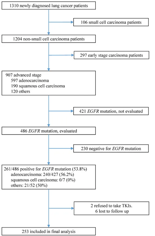
Among 1310 lung cancer patients diagnosed between January 2011 and October 2013, 486 patients with advanced NSCLC were screened forEGFRmutations (Fig 1). Of these, 261
(53.7%) patients hadEGFR-mutant NSCLC. Two patients refused to undergo treatment with
follow-up, 217 (85.8%) patients showed disease progression and 135 (53.4%) had died. The best cut-off point of LMR, MBR determined by ROC curve and Youden’s Index was 3.29, 0.63 respec-tively. Patients were divided into high or low LMR and MBR based on above cut-off value. Fig 1. Inclusion, screening, and group assignment of patients.Among 1310 non-small-cell lung cancer patients diagnosed between January 2011 and October 2013, 253 patients were included into final analysis.
doi:10.1371/journal.pone.0136252.g001
There were 153 (60.5%) patients with high LMR, 100 patients (39.5%) with low LMR; 118 (78.1%) patients with high MBR and 33 (21.9%) patients with low MBR.
Survival analysis of clinical factors
For PFS, clinical factors significant in univariable analysis included high LMR (p = 0.003) (Fig 2A), high MBR (p<0.001) (Fig 2B), commonEGFRmutations (p = 0.001), less distant organ
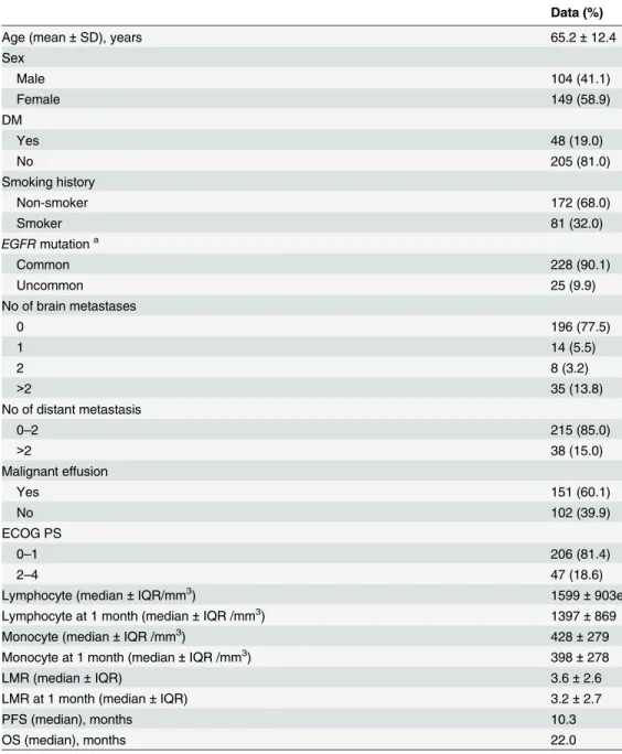
Table 1. Clinical characteristics and therapy responses of all 253 patients.
Data (%)
Age (mean±SD), years 65.2±12.4
Sex
Male 104 (41.1)
Female 149 (58.9)
DM
Yes 48 (19.0)
No 205 (81.0)
Smoking history
Non-smoker 172 (68.0)
Smoker 81 (32.0)
EGFRmutationa
Common 228 (90.1)
Uncommon 25 (9.9)
No of brain metastases
0 196 (77.5)
1 14 (5.5)
2 8 (3.2)
>2 35 (13.8)
No of distant metastasis
0–2 215 (85.0)
>2 38 (15.0)
Malignant effusion
Yes 151 (60.1)
No 102 (39.9)
ECOG PS
0–1 206 (81.4)
2–4 47 (18.6)
Lymphocyte (median±IQR/mm3) 1599±903e
Lymphocyte at 1 month (median±IQR /mm3) 1397±869
Monocyte (median±IQR /mm3) 428±279
Monocyte at 1 month (median±IQR /mm3) 398±278
LMR (median±IQR) 3.6±2.6
LMR at 1 month (median±IQR) 3.2±2.7
PFS (median), months 10.3
OS (median), months 22.0
a
Exon 19 deletion and L858R mutations were defined as common mutations. Other mutations or compound mutations were defined as uncommon mutations.
DM, diabetes mellitus; EGFR, epidermal growth factor receptor; ECOG, Eastern Cooperative Oncology Group; PS, performance status; LMR, lymphocyte-to-monocyte ratio; PFS, progression-free survival; OS, overall survival
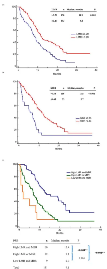
Fig 2. Progression-free survival (PFS) of epidermal growth factor receptor mutant non-small-cell lung cancer patients treated with first-line tyrosine kinase inhibitors therapy.(A) PFS between high and low baseline lymphocyte-to-monocyte ratio (LMR) patients; (B) PFS between high and low 1-month-to-baseline ratio of LMR (MBR) patients; (C) PFS between“high LMR and MBR”,“high LMR or MBR”,“low LMR and MBR”patients.
doi:10.1371/journal.pone.0136252.g002
metastases (p<0.001), no malignant effusion (p = 0.007), and good ECOG PS (p<0.001) (Table 2).
Age, sex, DM history, smoking history, and tumor histology had no significant influence on PFS. In the multivariable analysis, independent predictive factors for a longer PFS were high LMR (p = 0.009), high MBR (p = 0.004), commonEGFRmutations (p = 0.032), and having
less distant organ metastases (p = 0.044) (Table 2.).
For OS, clinical factors significant in univariable analysis included high LMR (p<0.001) (Fig 3A), less distant organ metastases (p<0.001), no malignant effusion (p = 0.010), and
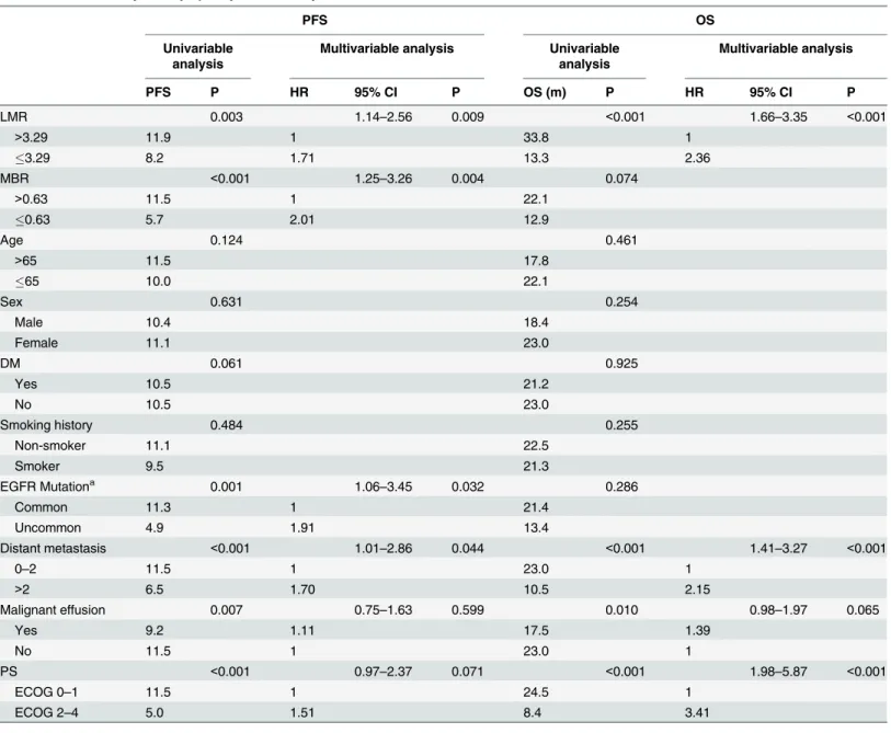
Table 2. Survival analysis of lymphocyte-to-monocyte ratio and clinical factors.
PFS OS
Univariable analysis
Multivariable analysis Univariable analysis
Multivariable analysis
PFS P HR 95% CI P OS (m) P HR 95% CI P
LMR 0.003 1.14–2.56 0.009 <0.001 1.66–3.35 <0.001
>3.29 11.9 1 33.8 1
3.29 8.2 1.71 13.3 2.36
MBR <0.001 1.25–3.26 0.004 0.074
>0.63 11.5 1 22.1
0.63 5.7 2.01 12.9
Age 0.124 0.461
>65 11.5 17.8
65 10.0 22.1
Sex 0.631 0.254
Male 10.4 18.4
Female 11.1 23.0
DM 0.061 0.925
Yes 10.5 21.2
No 10.5 23.0
Smoking history 0.484 0.255
Non-smoker 11.1 22.5
Smoker 9.5 21.3
EGFR Mutationa 0.001 1.06–3.45 0.032 0.286
Common 11.3 1 21.4
Uncommon 4.9 1.91 13.4
Distant metastasis <0.001 1.01–2.86 0.044 <0.001 1.41–3.27 <0.001
0–2 11.5 1 23.0 1
>2 6.5 1.70 10.5 2.15
Malignant effusion 0.007 0.75–1.63 0.599 0.010 0.98–1.97 0.065
Yes 9.2 1.11 17.5 1.39
No 11.5 1 23.0 1
PS <0.001 0.97–2.37 0.071 <0.001 1.98–5.87 <0.001
ECOG 0–1 11.5 1 24.5 1
ECOG 2–4 5.0 1.51 8.4 3.41
a
Exon 19 deletion and L858R mutations were defined as common mutations. Other mutations or compound mutations were defined as uncommon mutations.
PFS, progression-free survival; OS, overall survival; LMR, lymphocyte-to-monocyte ratio; MBR, 1-month-to-baseline ratio of LMR; DM, diabetes mellitus; EGFR, epidermal growth factor receptor; PS, performance status; ECOG, Eastern Cooperative Oncology Group
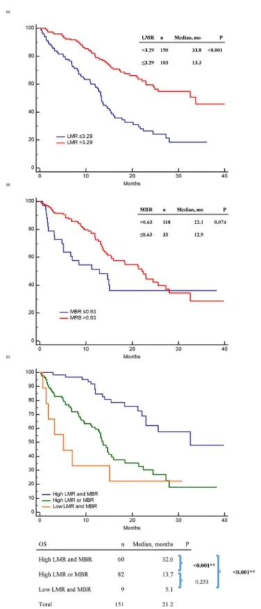
Fig 3. Overall survival (OS) of epidermal growth factor receptor mutant non-small-cell lung cancer patients treated with first-line tyrosine kinase inhibitors therapy.(A): OS between high and low baseline LMR patients; (B): OS between high and low 1-month-to-baseline ratio of LMR (MBR) patients; (C) OS between“high LMR and MBR”,“high LMR or MBR”,“low LMR and MBR”patients.
doi:10.1371/journal.pone.0136252.g003
good ECOG PS (p<0.001). MBR (Fig 3B), age, sex, smoking history, and tumor histology had no significant influence on OS. In the multivariable analysis, high LMR (p<0.001), less distant organ metastases (p<0.001), and good ECOG PS (p<0.001) were independent predictive factors for a longer OS.
Combination of LMR and MBR for survival analysis
The PFS in patients with high LMR and MBR, high LMR or MBR, and low LMR and MBR were 15.4, 7.1, and 2.0 months, respectively (p<0.001) (Fig 2C). The OS in the above three subgroups were 32.6, 13.7, and 5.1 months, respectively (p<0.001) (Fig 3C).
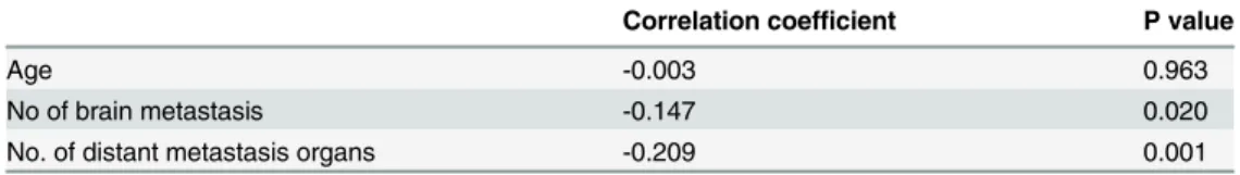
Correlations between LMR and clinical factors
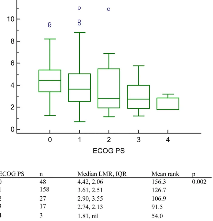
Significant correlations were noted between LMR and several clinical factors including the number of brain metastases, and the number of distant organ metastases. However, the correla-tion coefficient was low. The LMR correlacorrela-tion coefficients for the number of brain metastases, and the number of distant organ metastases were -0.147 (p = 0.020), and -0.209 (p = 0.001), respectively. (Table 3.) As to relationship between LMR and ECOG PS, significance was noted in Kruskal-Wallis test (p = 0.002). (Fig 4)
Discussion
Our study demonstrated that combination of LMR and MBR could predict prognosis in
EGFR-mutant NSCLC patients receiving first-line EGFR-TKIs. Our study also revealed that a
higher LMR correlated with a better ECOG PS, and a lower incidence of brain and distant metastases. Previous studies have demonstrated that Cytotoxic T lymphocytes play an impor-tant role in the anticancer response[11], and tumor-associated macrophages remodel the tumor extracellular matrix to promote proliferation, progression, and neovascularization[12,
13]. Based on the above pathophysiology, patients with a lower LMR most likely have a higher tumor burden and less cytotoxic T lymphocytes, which may, in part, explain why LMR acts as a prognostic factor in patients with advanced lung cancer who are receiving EGFR-TKIs. Besides being a prognostic factor, we hypothesize that peripheral blood LMR has the potential to act as a predictive factor for immunotherapy response because a low LMR would indicate less recruitable lymphocyte after the initiation of immunotherapy, although this concept requires further studies for confirmation.
A previous study indicated that LMR was a prognostic factor in hematological cancers such as diffuse large B-cell lymphoma, and postoperatively in several early-stage malignancies such as lung cancer[19], colorectal cancer[18], and non-metastatic nasopharyngeal carcinoma [16]. LMR has also been suggested as a prognostic factor in advanced-stage lung cancer patients receiving platinum-based chemotherapies[20], and in patients with breast cancer following neo-adjuvant chemotherapies.[17] To the best of our knowledge, this is the first study demon-strating that LMR is a prognostic factor in patients with advanced-stage,EGFR-mutant
NSCLC receiving EGFR-TKIs.
Table 3. Correlation between lymphocyte-to-monocyte ratio and clinical parameters.
Correlation coefficient P value
Age -0.003 0.963
No of brain metastasis -0.147 0.020
No. of distant metastasis organs -0.209 0.001
Recently study revealed that CEA level change during EGFR-TKIs therapy can be used to a predictor of survival.[22] However, LMR change (MBR) during EGFR-TKIs was seldom men-tioned before. Our study revealed patients with higher MBR had better PFS (11.5 vs 5.7 months, p<0.001). Although patients with high MBR had almost twice longer OS than those with low MBR (22.1 vs. 12.9 months), no significant was noted in log-rank test. (p = 0.074) It needs more studies to prove if lack of significance in MBR on OS were due to small study popu-lation or a true negative result.
Fig 4. The lymphocyte-to-monocyte ratio in patients with different Eastern Cooperative Oncology Group performance status.Patients with better ECOG PS had higher Median LMR. (p = 0.002).
doi:10.1371/journal.pone.0136252.g004
However, our study had several limitations. First, tumor programmed death-ligand 1 expression, which can provide direct information about the degree of immune paralysis in tumor microenvironment, was not available. Second, we do not have immune contexture, the amount of immune cell infiltration, in tumors, which can more precisely reflect the immune response in tumor microenvironment.[23] Further studies are required to determine whether add-on immunotherapy or anti-angiogenesis agents, in addition to first line EGFR-TKIs, could prolong survival in NSCLC patients, especially in those with low LMR. Finally, because our study was a retrospective study with a small patient population, a prospective trial is needed to validate these results.
Conclusion
A combination of baseline and trend of LMR can be used to predict survival inEGFR-mutant
NSCLC patients who treated with first-line EGFR-TKI therapy.
Supporting Information
S1 Table. Immune cell counts, ratios and their association with survival.As a continuous variable, monocyte count and lymphocyte-to-monocyte ratio had significant association with progression-free survival. Lymphocyte, monocyte count, and lymphocyte-to-monocyte ratio had significant association with overall survival.
(DOC)
Acknowledgments
We thank Tsui-Ping Tang for data collection.
Author Contributions
Conceived and designed the experiments: YMC CHL HC Chang TYC CCT WFF MCL. Ana-lyzed the data: YMC CCW YHC. Contributed reagents/materials/analysis tools: YMC YHW MCS KTH. Wrote the paper: YMC HC Chen YCC.
References
1. Henley SJ, Richards TB, Underwood JM, Eheman CR, Plescia M, McAfee TA, et al. Lung cancer inci-dence trends among men and women—United States, 2005–2009. MMWR Morbidity and mortality weekly report. 2014; 63(1):1–5. PMID:24402465.
2. Wang BY, Huang JY, Cheng CY, Lin CH, Ko J, Liaw YP. Lung cancer and prognosis in taiwan: a popu-lation-based cancer registry. Journal of thoracic oncology: official publication of the International Asso-ciation for the Study of Lung Cancer. 2013; 8(9):1128–35. doi:10.1097/JTO.0b013e31829ceba4
PMID:23945383.
3. Pao W, Miller V, Zakowski M, Doherty J, Politi K, Sarkaria I, et al. EGF receptor gene mutations are common in lung cancers from "never smokers" and are associated with sensitivity of tumors to gefitinib and erlotinib. Proceedings of the National Academy of Sciences of the United States of America. 2004; 101(36):13306–11. doi:10.1073/pnas.0405220101PMID:15329413; PubMed Central PMCID: PMC516528.
4. Tokumo M, Toyooka S, Kiura K, Shigematsu H, Tomii K, Aoe M, et al. The relationship between epider-mal growth factor receptor mutations and clinicopathologic features in non-sepider-mall cell lung cancers. Clin-ical cancer research: an official journal of the American Association for Cancer Research. 2005; 11 (3):1167–73. PMID:15709185.
5. Kosaka T, Yatabe Y, Endoh H, Kuwano H, Takahashi T, Mitsudomi T. Mutations of the epidermal growth factor receptor gene in lung cancer: biological and clinical implications. Cancer research. 2004; 64(24):8919–23. doi:10.1158/0008-5472.CAN-04-2818PMID:15604253.
patients with advanced EGFR mutation-positive non-small-cell lung cancer (NSCLC). Annals of oncol-ogy: official journal of the European Society for Medical Oncology / ESMO. 2013; 24(6):1615–22. doi:
10.1093/annonc/mdt012PMID:23456778.
7. Thongprasert S, Duffield E, Saijo N, Wu YL, Yang JC, Chu DT, et al. Health-related quality-of-life in a randomized phase III first-line study of gefitinib versus carboplatin/paclitaxel in clinically selected patients from Asia with advanced NSCLC (IPASS). Journal of thoracic oncology: official publication of the International Association for the Study of Lung Cancer. 2011; 6(11):1872–80. doi:10.1097/JTO. 0b013e31822adaf7PMID:22011650.
8. Yang JC, Wu YL, Schuler M, Sebastian M, Popat S, Yamamoto N, et al. Afatinib versus cisplatin-based chemotherapy for EGFR mutation-positive lung adenocarcinoma (LUX-Lung 3 and LUX-Lung 6): analy-sis of overall survival data from two randomised, phase 3 trials. The Lancet Oncology. 2015; 16 (2):141–51. doi:10.1016/S1470-2045(14)71173-8PMID:25589191.
9. Zhou C, Wu YL, Chen G, Feng J, Liu XQ, Wang C, et al. Erlotinib versus chemotherapy as first-line treatment for patients with advanced EGFR mutation-positive non-small-cell lung cancer (OPTIMAL, CTONG-0802): a multicentre, open-label, randomised, phase 3 study. The Lancet Oncology. 2011; 12 (8):735–42. doi:10.1016/S1470-2045(11)70184-XPMID:21783417.
10. Fukuoka M, Wu YL, Thongprasert S, Sunpaweravong P, Leong SS, Sriuranpong V, et al. Biomarker analyses and final overall survival results from a phase III, randomized, open-label, first-line study of gefitinib versus carboplatin/paclitaxel in clinically selected patients with advanced non-small-cell lung cancer in Asia (IPASS). Journal of clinical oncology: official journal of the American Society of Clinical Oncology. 2011; 29(21):2866–74. doi:10.1200/JCO.2010.33.4235PMID:21670455.
11. Aerts JG, Hegmans JP. Tumor-specific cytotoxic T cells are crucial for efficacy of immunomodulatory antibodies in patients with lung cancer. Cancer research. 2013; 73(8):2381–8. doi:10.1158/0008-5472. CAN-12-3932PMID:23580578.
12. Yang J, Liao D, Chen C, Liu Y, Chuang TH, Xiang R, et al. Tumor-associated macrophages regulate murine breast cancer stem cells through a novel paracrine EGFR/Stat3/Sox-2 signaling pathway. Stem cells. 2013; 31(2):248–58. doi:10.1002/stem.1281PMID:23169551.
13. Lin EY, Li JF, Gnatovskiy L, Deng Y, Zhu L, Grzesik DA, et al. Macrophages regulate the angiogenic switch in a mouse model of breast cancer. Cancer research. 2006; 66(23):11238–46. doi:10.1158/ 0008-5472.CAN-06-1278PMID:17114237.
14. Porrata LF, Ristow K, Habermann TM, Ozsan N, Dogan A, Macon W, et al. Absolute monocyte/lympho-cyte count prognostic score is independent of immunohistochemically determined cell of origin in pre-dicting survival in diffuse large B-cell lymphoma. Leukemia & lymphoma. 2012; 53(11):2159–65. doi:
10.3109/10428194.2012.690605PMID:22551474.
15. Lin B, Chen C, Qian Y, Feng J. Prognostic role of peripheral blood lymphocyte/monocyte ratio at diag-nosis in diffuse large B-cell lymphoma: a meta-analysis. Leukemia & lymphoma. 2015:1–6. doi:10. 3109/10428194.2015.1014367PMID:25686648.
16. Li J, Jiang R, Liu WS, Liu Q, Xu M, Feng QS, et al. A large cohort study reveals the association of ele-vated peripheral blood lymphocyte-to-monocyte ratio with favorable prognosis in nasopharyngeal carci-noma. PloS one. 2013; 8(12):e83069. doi:10.1371/journal.pone.0083069PMID:24386144; PubMed Central PMCID: PMC3873908.
17. Ni XJ, Zhang XL, Ou-Yang QW, Qian GW, Wang L, Chen S, et al. An elevated peripheral blood lympho-cyte-to-monocyte ratio predicts favorable response and prognosis in locally advanced breast cancer following neoadjuvant chemotherapy. PloS one. 2014; 9(11):e111886. doi:10.1371/journal.pone. 0111886PMID:25372468; PubMed Central PMCID: PMC4221197.
18. Stotz M, Pichler M, Absenger G, Szkandera J, Arminger F, Schaberl-Moser R, et al. The preoperative lymphocyte to monocyte ratio predicts clinical outcome in patients with stage III colon cancer. British journal of cancer. 2014; 110(2):435–40. doi:10.1038/bjc.2013.785PMID:24357796; PubMed Central PMCID: PMC3899781.
19. Hu P, Shen H, Wang G, Zhang P, Liu Q, Du J. Prognostic significance of systemic inflammation-based lymphocyte- monocyte ratio in patients with lung cancer: based on a large cohort study. PloS one. 2014; 9(9):e108062. doi:10.1371/journal.pone.0108062PMID:25275631; PubMed Central PMCID: PMC4183469.
20. Lin GN, Peng JW, Xiao JJ, Liu DY, Xia ZJ. Prognostic impact of circulating monocytes and lymphocyte-to-monocyte ratio on previously untreated metastatic non-small cell lung cancer patients receiving plati-num-based doublet. Medical oncology. 2014; 31(7):70. doi:10.1007/s12032-014-0070-0PMID:
24927957.
21. Eisenhauer EA, Therasse P, Bogaerts J, Schwartz LH, Sargent D, Ford R, et al. New response evalua-tion criteria in solid tumours: revised RECIST guideline (version 1.1). European journal of cancer. 2009; 45(2):228–47. doi:10.1016/j.ejca.2008.10.026PMID:19097774.
22. Facchinetti F, Aldigeri R, Aloe R, Bortesi B, Ardizzoni A, Tiseo M. CEA serum level as early predictive marker of outcome during EGFR-TKI therapy in advanced NSCLC patients. Tumour biology: the journal of the International Society for Oncodevelopmental Biology and Medicine. 2015. doi: 10.1007/s13277-015-3269-6PMID:25731731.