PSDRS, BDI, MoCA and MMSE as screening tools
for the evaluation of mood and cognitive functions
in patients at the early stage of cerebral stroke
Dorota Anita Przewoźnik, Anna Maria Rajtar-Zembaty, Bogusława
Bober-Płonka, Anna Starowicz-Filip, Ryszard Nowak, Ryszard Przewłocki
Dorota Anita Przewoźnik1, Anna Maria Rajtar-Zembaty1, Bogusława Bober-Płonka2,3, Anna Starowicz-filip4, Ryszard Nowak3, Ryszard Przewłocki2,5
: 1Jagiellonian University Medical
College, Department of Psychiatry; 2Jagiellonian University Insti
-tute of Applied Psychology, Department of Neurobiology and Neu-ropsychology; 3Department of Neurology and Cerebral Strokes
with a Subdivision for Cerebral Strokes, Ludwik Rydygier Spe-cialist Hospital, Krakow; 4Jagiellonian University Medical
Col-lege, Department of Psychiatry, Institute of Medical Psychology;
5 Jagiellonian University Institute of Applied Psychology, Department
of Molecular Neuropharmacology, Institute of Pharmacology. Corre-spondence address: [email protected]
Acknowledgments: The research was carried out as part of
a Demeter research project, with the approval of the Bioethics Committee at the local Chamber of Krakow, Poland, no. 12/KBL/2010, 26 January 2011.
Summary
Aims. To evaluate the suitability of the Post-Stroke Depression Scale (PSDRS) for detecting affective
dis-orders, to examine the correlation of depressed mood states with cognitive disorders in patients at an ear-ly stage of cerebral stroke, and to attempt a comparison of the effectiveness of detecting depressive and cognitive disorders with the selected clinical scales.
Material and methods. The examination involved 43 patients within the first week after cerebral stroke. It
was carried out with the application of two screening scales, the Mini-Mental State Examination (MMSE) and the Montreal Cognitive Assessment (MoCA), and two scales for the evaluation of the degree of de-pressiveness: PSDRS and Beck Depression Inventory (BDI).
Results. A significant negative correlation of the results of the PSDRS and MoCA scales was shown.
De-pressed mood in patients post-cerebral stroke was statistically significantly correlated with the disorders in selected cognitive skills: visual and spatial functions, memory, attention functions and abstracting ability.
Conclusions. The PSDRS and MoCA scales proved to be more effective tools for the evaluation of
de-pressive and cognitive disorders in patients at an early stage after cerebral stroke than the conventionally applied MMSE and BDI scales. The examination results additionally show a significant dependence be-tween mood and the cognitive impairment in this group of patients. With the weakening of cognitive func-tioning, the patients’ mood also became depressed.
stroke / post-stroke depression / cognitive functions
INTRODuCTION
study group. In a randomly selected community sample, this proportion ranges between 23 and 44%, but it increases to 35–72% in hospitalised patients [2]. As research shows, post-stroke de-pression is a key factor in further convalescence of the patient after stroke. Its intensity is associ-ated with worse results of motor rehabilitation and more difficult recovery [3]. What is more, post-stroke depression affects the reduction of the efficiency of patients’ cognitive functioning [4,5]. However, the vast majority of studies in-clude persons who experienced stroke at least a month before the examination. This limits the possibility of exploring the dynamics of the de-velopment of post-stroke depression, as well as its influence on patients’ cognitive functioning.
MATERIAl AND METHOD
The group under study consisted of patients ofthe Ludwik Rydygier Specialist Hospital in Krakow. The study included in-patients at the Department of Neurology and Cerebral Strokes, on the seventh day after the cerebralvascular ac-cident. Patients in an acute state were not able to undergo tests and patients with considera-ble paresis and deep aphasia were also exclud-ed from the study. The study did not include persons addicted to psychoactive substances or persons with coexisting neurological and men-tal disorders.
Overall, 43 persons aged 42 to 87 participated in the study (mean age 65.58 years (±10.63). Age distribution in the study group was close to the normal distribution and 58% of the group were men. The majority of patients in the study had secondary school education (51%), 9% had pri-mary education, 30% vocational education and 7% higher education. No education data were available for one person.
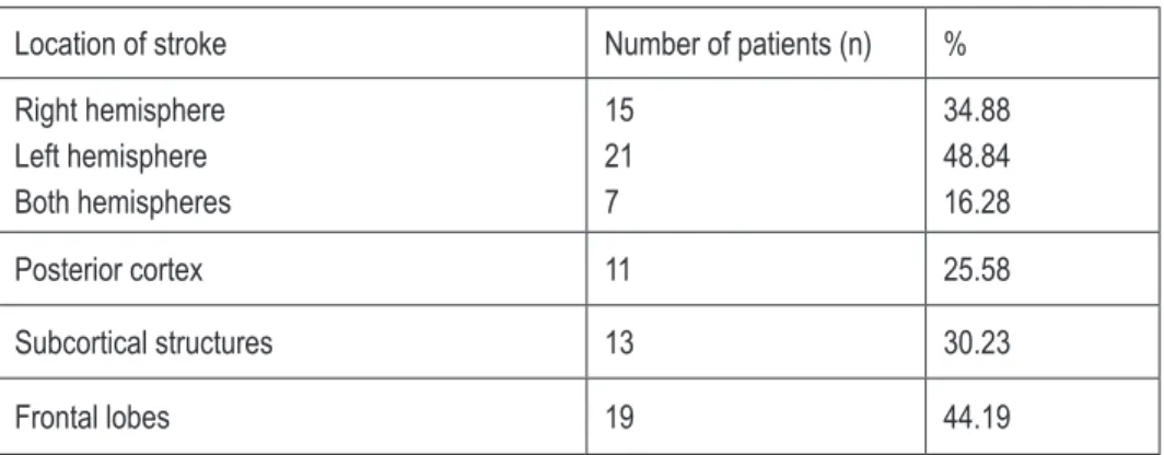
The location of the stroke focus varied (Ta-ble 1). As the study focused on patients with left cerebral stroke, only those were included in whom aphasia was only vestigial and did not hinder the questionnaire test performance and the application of the cognitive functions scales. In about one quarter of the patients dysarthria was found, but the remainder did not have any speech defects. Paresis occurred in the majority of patients in the study (58%).
INTRODuCTION
Cerebral stroke is one of the most frequent health problems affecting modern society. In about 60–70% of patients after cerebral stroke the reduction of both motor and cognitive abil-ity is noted, while after a year approximate-ly 50% of patients do not regain full ability [1]. The most frequent complications of mental na-ture that occur after a vascular incident include post-stroke depression (PSD). Depending on the criteria adopted and research tools applied, it is assumed that it occurs in about 16–72% per-sons after cerebral stroke. These differences can result, among others, from the selection of the study group. In a randomly selected community sample, this proportion ranges between 23 and 44%, but it increases to 35–72% in hospitalised patients [2]. As research shows, post-stroke de-pression is a key factor in further convalescence of the patient after stroke. Its intensity is associ-ated with worse results of motor rehabilitation and more difficult recovery [3]. What is more, post-stroke depression affects the reduction of the efficiency of patients’ cognitive functioning [4,5]. However, the vast majority of studies in-clude persons who experienced stroke at least a month before the examination. This limits the possibility of exploring the dynamics of the de-velopment of post-stroke depression, as well as its influence on patients’ cognitive functioning.
MATERIAl AND METHOD
The group under study consisted of patients ofthe Ludwik Rydygier Specialist Hospital in Krakow. The study included in-patients at the Department of Neurology and Cerebral Strokes, on the seventh day after the cerebralvascular ac-cident. Patients in an acute state were not able to undergo tests and patients with considera-ble paresis and deep aphasia were also exclud-ed from the study. The study did not include persons addicted to psychoactive substances or persons with coexisting neurological and men-tal disorders.
men. The majority of patients in the study had secondary school education (51%), 9% had pri-mary education, 30% vocational education and 7% higher education. No education data were available for one person.
The location of the stroke focus varied (Ta-ble 1). As the study focused on patients with left cerebral stroke, only those were included in whom aphasia was only vestigial and did not hinder the questionnaire test performance and the application of the cognitive functions scales. In about one quarter of the patients dysarthria was found, but the remainder did not have any speech defects. Paresis occurred in the majority of patients in the study (58%).
completed by the researcher on the basis of what the patient says and how they behave, assign-ing to each group a value rangassign-ing from 0 to 5 points. The maximum test score is 45 points. Sec-tion 10 is not counted towards the sum of points. Its purpose is to distinguish whether depressive symptoms occurred only after cerebral stroke or have a different background [7]. PSDRS differs from the other, default measurement tools in its adjustment for the specific nature of mood dis-orders occurring in persons after cerebral stroke. An additional value of PSDRS is a possibili-ty of analyzing the specific subscales separate-ly, thus obtaining a qualitative profile of the pa-tient’s mood disorders. It is also possible to
ap-Table 1. Location of stroke
Location of stroke Number of patients (n) %
Right hemisphere Left hemisphere Both hemispheres
15 21
7
34.88 48.84 16.28
Posterior cortex 11 25.58
Subcortical structures 13 30.23
Frontal lobes 19 44.19
METHOD
The following scales were used in the study: Post-Stroke Depression Rating Scale (PSDRS,
translated by Łucja Domańska [6, 7] ), Beck De
-pression Inventory (BDI), Mini-Mental State Ex-amination (MMSE) and Montreal Cognitive As-sessment (MoCA).Due to a lower prevalence and popularity of MoCA and PSDRS, they will be discussed in greater detail below.
Post-Stroke Depression Rating Scale
The scale [6, 7]consists of 10 sections. The first 9 study various components of post-stroke de-pression, such as: depressed mood, sense of guilt, thoughts about death/suicidal thoughts, vegetative symptoms, apathy, loss of interests, level of anxiety, catastrophic reactions, emo-tional hyperactivity and anhedonia. The scale is
ply the total score in order to carry out a screen-ing evaluation of depressed mood in post-stroke patients, as has been done in this study.
Montreal Cognitive Assessment (MoCA)
memo-ry (5 points) and orientation in time and space (6 points). Education level and age can influence test scores [8].
Statistical methods
Results were analysed using the PQStat sta-tistical package version 1.4.2.324. The normality of distributions was examined with the Szapiro-Wilk test. The relationship between age, MMSE
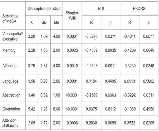
tions were significantly negatively correlat-ed (p=0.0035; R=-0.4359) with mood disorders measured both with the BDI and PSDRS scales (p=0.0049; R=-0.4209). The BDI scale was not sig-nificantly correlated with any additional dimen-sion of the MoCA test. On the other hand, post-stroke depression measured with the PSDRS scale also showed significant correlations with the subscales measuring attention functions (p=0.0346; R=-0.3230), including attention
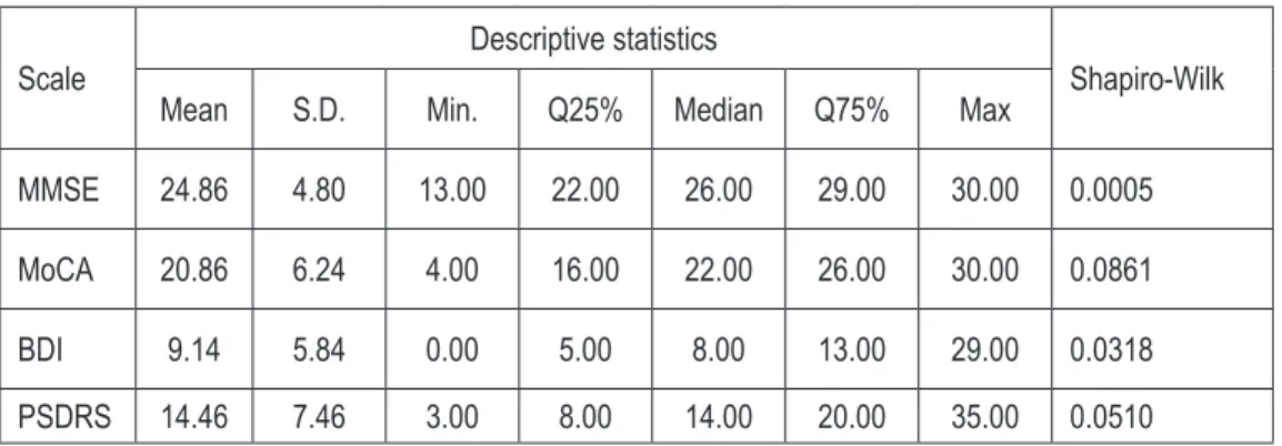
shift-Table 2. Descriptive statistics and distribution of the results of MMSE, MoCA, BDI and PSDRS
Scale
Descriptive statistics
Shapiro-Wilk
Mean S.D. Min. Q25% Median Q75% Max
MMSE 24.86 4.80 13.00 22.00 26.00 29.00 30.00 0.0005
MoCA 20.86 6.24 4.00 16.00 22.00 26.00 30.00 0.0861
BDI 9.14 5.84 0.00 5.00 8.00 13.00 29.00 0.0318
PSDRS 14.46 7.46 3.00 8.00 14.00 20.00 35.00 0.0510
Table 3. The correlations of MMSE and MoCA with BDI and PSDRS scales
Spearman rank order correlations Pearson correlations
MMSE MoCA MMSE MoCA
R p R p r p r p
BDI -0.1449 0.3538 -0.3551 0.0195 -0.0690 0.6600 -0.2660 0.0850
PSDRS -0.1998 0.1988 -0.4233 0.0047 -0.1385 0.3760 -0.3094 0.0430
and MoCA scales and BDI and PSDRS was ana-lyzed estimating the Spearman rank correlation and Pearson linear ratios. The results of the BDI and PSDRS scales, gender dependent, were com-pared with the Mann–Whitney U-test and t -stu-dent test. For the examination of the distribution of the results in BDI and PSDRS scales, depending on education level and the location of brain dam-age, the Kruskal-Wallis and ANOVA tests were applied. On the other hand, the relationship be-tween the individual components of the MoCA test with the results of BDI and PSDRS question-naires was analyzed estimating the correlations of the Spearman rank order. Test probability at the level of p<0.05 was assumed as significant.
ability (‘sequence’ with p=0.0205; R=0.3522) and the ability to abstract (p=0.0311; R=-0.3292).
DISCuSSION
Table 4.Correlations of MoCA sub-scales with the BDI and PSDRS scores
Sub-scale of MoCA
Descriptive statistics
Shapiro-Wilk
BDI PSDRS
X SD Me R p R p
Visuospatial/
executive 3.28 1.59 4.00 0.0001 -0.3293 0.0311 -0.4011 0.0077
Memory 2.28 1.68 2.00 0.0023 -0.4359 0.0035 -0.4209 0.0049
Attention 3.79 1.87 4.00 0.0010 -0.2608 0.0911 -0.3230 0.0346
Language 1.56 0.98 2.00 0.0001 0.1184 0.4495 0.0913 0.5602
Abstraction 1.40 0.62 1.00 <0.0001 -0.2569 0.0963 -0.3292 0.0311
Orientation 5.42 1.28 6.00 <0.0001 0.0375 0.8112 -0.1069 0.4950
Attention
shiftability 2.05 1.72 2.00 0.0006 0.2830 0.0659 0.3522 0.0205
RESulTS
The distribution of the MMSE (p=0.0005) and BDI (p=0.0318) results differs significantly from the normal distribution, while the MoCA and PSDRS scales are consistent with it (Table 2). Due to the fact that some of the scales signifi-cantly diverge from the theoretical normal distri-bution and some are consistent with it, the anal-yses of the correlations were carried out apply-ing both parametric (p1) and non-parametric (p2)
approach.
In Table 3 the results of the correlation between the MMSE and MoCA scales and the BDI and PSDRS are presented. No significant (p>0.05) correlations were found between the cogni-tive impairment measured with the MMSE and mood measured with the BDI and PSDRS. On the other hand, in the case of the cognitive im-pairment measured with the MoCA scale, the dependence was significantly negatively cor-related in rank (p=0.0195, R=-0.3551) with the mood level measured with the BDI test and sig-nificantly negatively correlated (p=0.0047, R=-0.4233) with the depressed mood measured with the PSDRS scale. This means that with the
increase of the results of the MoCA scale (bet-ter cognitive functioning) the results of the BDI and PSDRS scales become lower (less depressed mood). Those correlations considered in a linear fashion are significant (p=0.0430) only in the case of the relations between the MoCA and PSDRS scales (R=-0.3094), and it is the only pair of var-iables to have a normal distribution and a justi-fied linear correlation.
No significant correlations were found be-tween mood disorders measured with the BDI and PSDRS scales and age (p1=0.6980; p2=0.9573);
gender (for BDI p1=0.5848, p2=0.8923; for PSDRS
p1=0.7253, p2=0.7489) and education (for BDI
p1=0.6973, p2=0.7686; for PSDRS p1=0.3801,
p2=0.6336). There were also no significant
differ-ences whether the right, left or both hemispheres were affected (for BDI p1=0.2007, p2=0.1198; for
PSDRS p1=0.2013, p2=0.1235) and between brain
damage location (for BDI p1=0.3960, p2=0.5192;
for PSDRS p1=0.3791, p2=0.3251).
func-the trend of func-the influence of func-the factors described on the basis of the examinations carried out. The depressed mood can deepen the already exist-ing cognitive dysfunctions, whereas the aware-ness of those mental limitations certainly con-tributes to the worsening of the mood, sadness and depression. Cognitive functioning can also be affected by the person’s IQ and education. It has also to be taken into account that depressed mood can occur before cerebral stroke.
The present study found a significant relation-ship concerning memory disorders, attention functions and visual and spatial functions and the occurrence of depressed mood in persons af-ter cerebral stroke; the occurrence of executive dysfunctions can also be similarly significant. The association between post-stroke depression and cognitive functioning has been documented in the literature [4, 5, 10]. Some indicate a partic-ularly strong correlation between the decrease in the efficiency of executive functions [11-13] and post-stroke depression. One of the papers sug-gests that persons after cerebral stroke who ex-perience disorders of executive functions suffer from depression more frequently than persons without such disorders. Over half (53.3%) of people suffering from both depression and disor-ders of executive functions were still experiencing them 2 years after the stroke, and forecasts con-cerning their recovery were much worse than for patients with either disorders of executive func-tions or depression [14]. Considerably fewer stud-ies focus on a more precise division of executive functions and their influence on the occurrence of depression. The study carried out on healthy per-sons indicate however that memory disorders [15, 16] co-occur with the emergence of depression. Similar conclusions were reached by Hommel et al., but in the case of persons who had cerebral stroke [11]. Disorders in attention functions can also affect the development of depression. Lock-wood and colleagues [17] estimate that about a third of otherwise healthy elderly persons with depression have co-existing attention and execu-tive functions disorders. Nys in turn [18] indicates an association between depression post-cerebral stroke and visual memory disorders, disorders of higher-order visual and spatial functions and hemispatial neglect. They are at the same time de-scribed as the best predictors of the retention of post-stroke depression at 6 months.
There are few studies involving patients in the first 3 weeks after the cerebral stroke that show the early interdependence of depressive symp-toms and the worsening of cognitive functioning [13], and scant data indicating the influence of the latter upon the development of post-stroke depression within 6 months post-stroke [18]. However, a trend is emerging that tends to the earliest possible detection of depression, which was pointed out in the guidelines of European Stroke Organisation [19]. Fuentes et al. [20] ex-amined patients before the 10th day after cere-bral stroke and followed them up after 3 months. They noted that depression in ‘the acute phase’ of the stroke is maintained in the later period. They stress the importance of early detection of this disorder.
The present paper concerns persons in the first week after cerebral stroke, that is in the ‘acute phase’. The results indicate co-occurrence of de-pressive symptoms and the worsening of cogni-tive functioning as early as in the first week after the vascular incident, whereby unlike other au-thors, it was decided not to use the term “post-stroke depression” in relation to the examination carried out due to the great dynamics of chang-es occurring in this period.
Post-stroke depression can be assessed us-ing various scales, with the BDI beus-ing one of the most popular [12, 21]. However, scientific progress encourages one to search for new meas-ures. That is why we have decided to introduce the Post-Stroke Depression Scale (PSDRS), aimed at the specific examination of patients who have experienced cerebral stroke. Although no stud-ies were found that would compare both scales mentioned, the juxtaposition of the PSDRS with Hamilton Depression Scale which is also fre-quently applied post-cerebral stroke [20] indi-cates the diagnostic validity of the PSDRS [7]. Gainotti suggests, however, the possibility of the total comparison of the results of both scales (at the test author’s consent). When cognitive func-tions need to be examined, the MMSE scale is normally used [13, 22].
treated mainly as an initial diagnosis of the pa-tient’s functioning facilitating a selection of more precise tools for the examination of specific cog-nitive functions. The results of the examinations presented in this paper additionally suggest the clinical prevalence of the PSDRS scale over the commonly applied BDI scale, due to, among oth-ers, its greater relevance and diagnostic efficien-cy in this group of patients.
REfERENCES
1. Wiebers DO, Feigin VL, Brown RD. Udar mózgu [Stroke: Handbook]. Warszawa: Medipage; 2006.
2. Tateno A, Robinson RG. The effect of poststroke depres-sion on recovery from stroke. Psychogeriatrics. 2002; 2(2): 73-84.
3. Heruti R, Lusky A, Dankner R. Rehabilitation outcome of el-derly patients after a first stroke: effect of cognitive status at admission on the functional outcome. Arch Phys. 2002; 83(6): 742-749.
4. Kauhanen M-L, Korpelainen JT, Hiltunen P, Brusin E, Monon-en H, Maatta R, et al. Poststroke depression correlates with cognitive impairment and neurological deficits. Stroke. 1999; 30(9): 1875-1880.
5. Leeds L, Meara R, Woods R, Hobson J. A comparison of the new executive functioning domains of the CAMCOG-R with existing tests of executive function in elderly stroke survivors. Age Ageing. 2001; 30(3): 251-254.
6. Domańska Ł. Ocena depresji u osób ze schorzeniami nac-zyniowymi mózgu [Evaluation of depression in persons with vascular illnesses of the brain]. In: Leszek J, editor. Choroby otępienne. Teoria i praktyka [Dementia ilnesses. Theory and
practice]. Wrocław: Continuo; 2003. p. 331-344.
7. Quaranta D, Marra C, Gainotti G. Mood disorders after stroke: diagnostic validation of the poststroke depression rating scale. Cerebrovasc Dis. 2008; 26(3): 237-243.
8. Talarowska M, Florkowski A, Zboralski K, Gałecki P. Skala MoCA oraz MMSE w diagnozie łagodnych zaburzeń funkcji poznawczych. Psychiatr Psychoter. 2011; 7(1): 13-20. 9. Pąchalska M. Neuropsychologia kliniczna: urazy mózgu.
Warszawa: Wydawnictwo Naukowe PWN; 2012.
10. Hackett M, Anderson C. Predictors of depression after stroke: a systematic review of observational studies. Stroke. 2005; 36(10): 2296-2301.
11. Hommel M, Carey L, Jaillard A. Depression: Cognition rela-tions after stroke. Int J Stroke. 2013; 1-4.
12. Nys G. Early depressive symptoms after stroke: neuropsy-chological correlates and lesion characteristics. J Neurol Sci. 2005; 247(2): 149-156.
with the scales for depression. Significant corre-lations were however noted in the case of com-paring them with the MoCA. This can result from the fact that MoCA appears to be more ef-fective, particularly in detecting disorders with-in executive functions and workwith-ing memory, which, as mentioned before, indicate a strong-er association with the occurrence of depressive symptoms. It is worth mentioning that the cor-relation was strongest in comparing the two less frequently applied scales, namely the MoCA and the PSDRS.
This study had certain limitations, such as a small and non-homogenous sample. Due to the changing dynamics of the course of cerebral stroke and great fatigability of the patients, only certain individuals were able to fully participate in the examination. However, despite the obsta-cles encountered, the results of the study can be an indication for diagnosticians. Owing to the early application of the scales suggested, a fast-er and more precise detection and countfast-eraction against the worsening of cognitive functions as well as the co-occurring depressive symptoms will be possible.
findings
The results of this study indicate that with the worsening of cognitive functioning, meas-ured with the MoCA screening scale, depressive symptoms as measured by the BDI and PSDRS tests are more severe. This is true when meas-uring visual and spatial functions, memory, at-tention (including atat-tention shiftability) and the ability to abstract. It can be therefore assumed that in the group of persons in the study who did not suffer from severe aphasia or paresis, the depressed mood which manifests as early as in the first week post-cerebral stroke is related to the worsening of the quality of the patient’s cog-nitive functioning.
13. Nys G, Van Zandvoort M. Cognitive disorders in acute stroke: prevalence and clinical determinants. Cerebrovasc Dis. 2006; 23(5-6): 408-416.
14. Bour A, Rasquin S, Limburg M, Verhey F. Depressive symp-toms and executive functioning in stroke patients: a follow-up study. Int J Geriatr Psychiatry. 2011; 26(7): 679-686. 15. Singh-Manoux A, Akbaraly TN, Marmot M, Melchior M, Ankri
J, Sabia S, et al. Persistent depressive symptoms and cogni-tive function in late midlife: the Whitehall II study. J Clin Psy-chiatry. 2010; 71(10): 1379-1385.
16. Tam CWC, Lam LCW. Cognitive function, functional perfor-mance and severity of depression in Chinese older persons with late-onset depression. East Asian Arch Psychiatry. 2012; 22(1): 12-17.
17. Lockwood K. Executive dysfunction in geriatric depression. Am J Psychiatry. 2002; 159(7): 1119-1126.
18. Nys GMS, van Zandvoort MJE, van der Worp HB, de Haan EHF, de Kort PLM, Jansen BPW, et al. Early cognitive impair-ment predicts long-term depressive symptoms and quality of life after stroke. J Neurol Sci. 2006; 247(2): 149-156. 19. European Stroke Organisation (ESO) Executive Committee,
and ESO Writing Committee. Guidelines for management of ischaemic stroke and transient ischaemic attack 2008. Cere-brovasc Dis. 2008; 25(5): 457-507.
20. Fuentes B, Ortiz X, Sanjose B, Frank A, Díez-Tejedor E. Post-stroke depression: can we predict its development from the acute stroke phase? Acta Neurol Scand. 2009; 120(3): 150-156. 21. Aben I, Verhey F, Lousberg R, Lodder J, Honig A. Validity of
the Beck Depression Inventory, Hospital Anxiety and Depres-sion Scale, SCL-90, and Hamilton DepresDepres-sion Rating Scale as screening instruments for depression in stroke patients. Psychosomatics. 2002; 43(5): 386-393.
22. Borkowska A, Warwas I, Wiłość M, Drożdż W. Neuropsy-chologiczna ocena dysfunkcji poznawczych w depresji po udarze mózgu [Neuropsychological assessment of cognitive dysfunctions in poststroke depression]. Psychiatria. 2007; 2: 39-44.
23. Nasreddine Z, Collin I, Chertkow H. Sensitivity and specificity of the Montreal Cognitive Assessment (MoCA) for detection of mild cognitive deficits. Can J Neurol Sci. 2003; 30(2): 30. 24. Dong Y, Sharma VK, Chan BP-L, Venketasubramanian N,
Teoh HL, Seet RCS, et al. The Montreal Cognitive Assess-ment (MoCA) is superior to the Mini-Mental State Examination (MMSE) for the detection of vascular cognitive impairment af-ter acute stroke. J Neurol Sci. 2010; 299(1-2): 15-18. 25. Pendlebury ST, Cuthbertson FC, Welch SJ V, Mehta Z,