C
Carolina Simonetti L odi
AVALIAÇÃO DO POTENCIAL CARIOGÊNICO
DE LEITES FERMENTADOS CONTENDO
PROBIÓTICOS
C
Carolina Simonetti L odi
AVALIAÇÃO DO POTENCIAL CARIOGÊNICO
DE LEITES FERMENTADOS CONTENDO
PROBIÓTICOS
Tese apresentada à Faculdade de
Odontologia da Universidade Estadual
Paulista “Júlio de Mesquita Filho”,
Campus de Araçatuba, para obtenção de
título de Doutor em Odontopediatria.
Orientadora: Profª Drª Cleide Cristina Rodrigues Martinhon
Co-orientador: Prof. Adj. Alberto Carlos Botazzo Delbem
Catalogação na Publicação (CIP)
Serviço Técnico de Biblioteca e Documentação – FOA / UNESP
Lodi, Carolina Simonetti.
L823a Avaliação do potencial cariogênico de leites fermentados contendo probióticos / Carolina Simonetti Lodi. - Araçatuba : [s.n.], 2011
115 f. : il. ; tab. + 1 CD-ROM
Tese (Doutorado) – Universidade Estadual Paulista, Faculdade
de Odontologia
Orientadora: Profa. Cleide Cristina Rodrigues Martinhon Co-orientador: Prof. Alberto Carlos Botazzo Delbem
1. Produtos fermentados do leite 2. Probióticos 3. Cárie dentária 4. Desmineralização do dente 5. Streptococcus mutans 6. Lactoba- cillus
Black D27
Carolina Simonetti L odi – Tese de Doutorado 2011 4
Dados Curriculares
Carolina Simonetti Lodi
Nascimento 25.10.1978 – Bauru - SP
Filiação Antônio Roberto Correa Lodi
Vera Lígia Simonetti Lodi
2000/2003 Curso de Graduação em Odontologia pela Faculdade de Odontologia de Bauru, FOB-USP – Campus Bauru.
2004-2006 Curso de Especialização em Odontopediatria pelo Hospital de Reabilitação de Anomalias Craniofaciais,
HRAC-USP
2006/2008
2008/2011
Curso de Pós Graduação em Odontopediatria, nível de
Mestrado, na Faculdade de Odontologia de Araçatuba –
UNESP
Curso de Pós-Graduação em Ciência Odontológica, área
de concentração Odontopediatria, nível de Doutorado,
na Faculdade de Odontologia de Araçatuba.
Associações CROSP - Conselho Regional de Odontologia de São Paulo
SBPqO - Sociedade Brasileira de Pesquisa Odontológica
Carolina Simonetti L odi – Tese de Doutorado 2011 5
D
Carolina Simonetti L odi – Tese de Doutorado 2011 6
Dedico este trabalho,
Aos meus pais ROBERTO e VERA,
P
Pela confiança que depositam em mim... Exemplos de dedicação,
honestidade, simplicidade, felicidade e amor. Agradeço por todos os
momentos em que podemos estar juntos e pelas palavras de conforto que
sempre trazem segurança e tranqüilidade.
Nada teria acontecido se eu não tivesse o apoio constante de vocês.
M uitas vezes não fisicamente, mas em pensamentos e palavras;
A vocês dedico este trabalho, pois sem vocês eu nada seria.
Carolina Simonetti L odi – Tese de Doutorado 2011 7
A
Carolina Simonetti L odi – Tese de Doutorado 2011 8
À Deus,
Presente em todos os momentos da minha vida, protegendo-me e guiando
meus passos nesse longo caminho rumo a grandes realizações. Devo a Ele
todos os momentos de alegria e sucesso até aqui conquistado.
Carolina Simonetti L odi – Tese de Doutorado 2011 9
À Roberta e Rodrigo, minha irmã e meu irmão,
A
Agradeço à Deus e meus pais por me concederem a bênção de ter dois
irmãos.
Pessoas que fazem meu coração sorrir...
...Que não encontro todos os dias, mas que tenho a chance de dizer agora...
Para mim o que importa não é o que eu tenho na vida, mas quem eu tenho
na vida... Por isso...
Guardo vocês dentro do meu coração....
Amo vocês!
Carolina Simonetti L odi – Tese de Doutorado 2011 10
Ao João Eduardo, Meu marido...
““Quando encontrar alguém e esse alguém fizer seu coração parar de
funcionar por alguns segundos, preste atenção: pode ser a pessoa mais
importante da sua vida.
Se os olhares se cruzarem e, neste momento, houver o mesmo brilho intenso
entre eles, fique alerta: pode ser a pessoa que você está esperando desde o
dia em que nasceu.
Se o toque dos lábios for intenso, se o beijo for apaixonant e, e os olhos se
encherem d’água neste momento, perceba: existe algo mágico entre vocês.
Se o primeiro e o último pensamento do seu dia for essa pessoa, se a vontade
de ficar juntos chegar a apertar o coração, agradeça: Deus te mandou um
presente.”
Se um dia tiverem que pedir perdão um ao outro por algum motivo e, em
troca, receber um abraço, um sorriso, um afago nos cabelos e os gestos
valerem mais que mil palavras, entregue-se: vocês foram feitos um pro
outro.
Se por algum motivo você estiver triste, se a vida te deu uma rasteira e a
outra pessoa sofrer o seu sofrimento, chorar as suas lágrimas e enxugá-las
com ternura, que coisa maravilhosa: você poderá contar com ela em qualquer
momento de sua vida.
Se você tiver a certeza que vai ver a outra envelhecendo e, mesmo assim,
tiver a convicção que vai continuar sendo louco por ela...
...é o amor que chegou na sua vida.”
Carolina Simonetti L odi – Tese de Doutorado 2011 11
A
Agradeço à Deus todos os dias por ter colocado você em minha vida....
Obrigada pelo companheirismo e apoio constante.
Carolina Simonetti L odi – Tese de Doutorado 2011 12
À minha orientadora,
Profª. Drª. Cleide Cristina Rodrigues Martinhon,
A
Agradecer é admitir que houve um momento em que se precisou de alguém; é
reconhecer que ninguém jamais poderá lograr para si o dom de ser
auto-suficiente. Ninguém e nada cresce sozinho; sempre é preciso um olhar de
apoio, uma palavra de incentivo, um gesto de compreensão, uma atitude de
amor...
Obrigada pela paciência, compreensão e carinho que dedicou à mim e a este
trabalho. Pela confiança que depositou em mim e pela amizade eterna...
M inha imensa gratidão.
Ao meu co-orientador,
Prof. Dr. Alberto Carlos Botazzo Delbem,
Admiro sua competência e dedicação ao trabalho e à pesquisa.
Obrigada pelas orientações sábias e práticas. Pela ajuda, disponibilidade e
pela companhia agradável em vários momentos.
Aos Prof. Dr. Carlos Gonzalez Cabezas e Profa Margherita Fontana,
Toda minha admiração. Agradeço por toda a sabedoria transmitida durante
minha estada em Ann Arbor e por todos os momentos de alegria e
descontração.
Carolina Simonetti L odi – Tese de Doutorado 2011 13
Ao meu avô Raphael,
P
Pela presença constante em minha vida e pelas palavras sempre doces e
carinhosas.
Pela maneira invejável que se dedica aos seus filhos e netos.
Amo você.
À Mariana, Donizeti, Alessandra e Marcelo,
M eus cunhados e irmãos de coração...
Pela convivência sempre alegre e feliz.
Vocês também são minha família.
Carolina Simonetti L odi – Tese de Doutorado 2011 14
À aluna Lidiane Viana,
Pela ajuda indispensável no desenvolvimento da parte experimental deste
trabalho, pelos momentos alegres e pela amizade.
M uito Obrigada!
À Susan Flannagan,
Obrigada pelas orientações sábias, pela ajuda, disponibilidade, amizade e
pela companhia sempre agradável em Ann Arbor.
Carolina Simonetti L odi – Tese de Doutorado 2011 15
Carolina Simonetti L odi – Tese de Doutorado 2011 16
Faculdade de Odontologia de Araçatuba, na pessoa dos professores Dr.
Pedro Felício Estrada Bernabé, digníssimo Diretor e Dra. Ana M aria Pires
Soubhia, digníssima Vice-Diretora.
Ao Curso de Pós-Graduação em Odontopediatria da Faculdade de
Odontologia de Araçatuba –U NESP, na pessoa do coordenador Prof. Dr.
Alberto Carlos Botazzo Delbem.
Aos docentes da Disciplina de Odontopediatria da Faculdade de
Odontologia de Araçatuba - U NESP, Prof. Dr. Alberto Carlos Botazzo
Delbem, Prof. Dr. Célio Percinoto,
Prof. Dr. Robson Frederico Cunha,
Profª. Dr
a. Rosângela dos Santos Nery, Profª. Dr
a. Sandra M . H. C. Ávila
de Aguiar pelo carinho e atenção em todos os momentos.
Aos funcionários da Disciplina de Odontopediatria, M aria dos Santos
Ferreira Fernandes e M ário L uis da Silva, pela amizade, auxílio
indispensável e convivência tão especiais.
Aos queridos amigos da minha turma de Doutorado, Adelisa, Renata e
Vanessa, pelos anos de convivência, amizade e respeito.
À todos os colegas do curso de Pós-Graduação em Odontopediatria
(M estrado e Doutorado) da Faculdade de Odontologia de Araçatuba –
Carolina Simonetti L odi – Tese de Doutorado 2011 17
Aos amigos do L aboratório de pesquisa do Departamento de
Odontopediatria pela convivência durante todo o doutorado: Adelisa,
Amanda, Ana Paula, Ana Elisa, Ana Carolina (Tuca), Carla, Dani Câmara,
Dani Oliveira, Emilene, Eliana Takeshita, Gabriel, Jackeline, José Antonio,
I sabelle, L idiane, M arcelle, M aria Daniela, M arcelo, M ichele.
Aos amigos da U niversidade de M ichigan – Ann Arbor: Chip, M att, Susan,
L etícia, Renata, Sudha, Tatiana Botero, Zhihong, Kristy, Zhaocheng,
Atsushi pela amizade e pelos momentos de descontração.
Aos funcionários da Biblioteca da Faculdade de Odontologia de Araçatuba
- U NESP, Ana Cláudia, Cláudio, I vone, I zamar, L uzia, M aria Cláudia e
M arina pela atenção e disponibilidade com que sempre me receberam.
Aos funcionários da Seção de Pós-Graduação da Faculdade de Odontologia
de Araçatuba - U NESP, pelo profissionalismo, carinho e paciência.
Aos demais professores e funcionários da Faculdade de Odontologia de
Araçatuba – U NESP.
À Fernanda Brighenti, por ter colaborado em todos os momentos da
Carolina Simonetti L odi – Tese de Doutorado 2011 18
À Prof. Dr
aCristiane I to por me dar a oportunidade de acompanhar os
trabalhos do laboratório e assim aprender as metodologias necessárias para o
desenvolvimento do meu trabalho.
Aos voluntário deste trabalho, Dani Oliveira, Fernanda, I sabelle, Jackeline,
José Antônio, L idiane, M arcelle, M arcelo, M archelo e M ichele pela imensa
colaboração e compreensão durante a realização deste trabalho.
Às professoras e amigas do curso de Especialização do HRAC-U SP, que de
alguma forma contribuíram para que eu chegasse até aqui e que apesar da
distância continuam guardadas na lembrança e no coração.
Ao diretor do hemocentro de Araçatuba, Dr. Wolney Gois Barreto, e aos
funcionários pela coleta e doação das bolsas de sangue.
Ao Frigorífico Friboi, em especial, ao veterinário Henrique Borges, pela
atenção e por ter permitido a coleta dos dentes bovinos.
À Coordenadoria de Aperfeiçoamento de Pessoal de Nível Superior
(CAPES), pelo apoio financeiro para a realização do Curso de Doutorado.
A todos aqueles que, direta ou indiretamente, contribuíram para a
realização deste trabalho,
Carolina Simonetti L odi – Tese de Doutorado 2011 19
Posso ter defeitos, viver ansioso e ficar irritado algumas vezes mas não
esqueço de que minha vida é a maior empresa do mundo, e posso evitar que
ela vá à falência.
Ser feliz não é ter um céu sem tempestade, caminhos sem acidentes,
trabalhos sem fadigas, relacionamentos sem desilusões.
Ser feliz é encontrar força no perdão, esperança nas batalhas, segurança no
palco do medo, amor nos desencontros.
Ser feliz não é apenas valorizar o sorriso, mas refletir sobre a tristeza. Não
é apenas comemorar o sucesso, mas aprender lições nos fracassos. Não é
apenas ter júbilo nos aplausos, mas encontrar alegria no anonimato.
Ser feliz é reconhecer que vale a pena viver apesar de todos os desafios,
incompreensões e períodos de crise.
Ser feliz é deixar de ser vítima dos problemas e se tornar um autor da
própria história. É atravessar desertos fora de si, mas ser capaz de encontrar
um oásis no recôndito da sua alma. É agradecer a Deus a cada manhã pelo
milagre da vida.
Ser feliz é não ter medo dos próprios sentimentos. É saber falar de si mesmo.
É ter coragem para ouvir um "não". É ter segurança para receber uma
crítica, mesmo que injusta.
E, quando você errar o caminho, recomece. Pois assim você descobrirá que
ser feliz não é ter uma vida perfeita. M as usar as lágrimas para irrigar a
tolerância. U sar as perdas para refinar a paciência. U sar as falhas para
lapidar o prazer. U sar os obstáculos para abrir as janelas da inteligência.
Pedras no caminho?
Guardo todas, um dia vou construir um castelo…
Fernando Pessoa
E
Carolina Simonetti L odi – Tese de Doutorado 2011 20
Carolina Simonetti L odi – Tese de Doutorado 2011 21 Lodi CS. Avaliação do potencial cariogênico de leites fermentados contendo
probióticos. [Tese]. Araçatuba: Universidade Estadual Paulista; 2011.
Um número crescente de produtos contendo probióticos está disponível
no mercado e vem sendo utilizados pelos consumidores. Diante disso, o
objetivo deste trabalho foi avaliar in situ, in vivo e in vitro a relação da bactéria
probiótica com a cárie dentária. No estudo in situ investigou-se a
cariogenicidade do leite fermentado contendo probióticos através da
quantificação dos açúcares totais e redutores presente no produto, análise do
seu efeito na desmineralização do esmalte dental bovino, análise microbiológica
da saliva antes e após o período experimental, análise microbiológica e
quantificação dos carboidratos álcalis-solúveis presente no biofilme. Para isso,
dez voluntários utilizaram um dispositivos contendo 4 blocos de esmalte dental
bovino. O experimento consistiu de 3 etapas de 14 dias cada onde os
voluntários gotejaram solução de sacarose 20% ou a solução de tratamento
(Tratamento A - Yakult® ou Tratamento B - Batavito®) 8X/dia. Decorrido o
período experimental, o biofilme e a saliva foram analisados quanto a
quantidade de microrganismos totais (MT), Streptococcus totais (ST) e
Streptococcus do grupo mutans (SM), Lactobacillus (L). Para os dados de dureza
foram calculados a porcentagem de variação de dureza superficial e a perda
integrada de dureza de subsuperfície. Após o tratamento B foi observado
menor quantidade de MT no biofilme quando comparado com o tratamento A,
mas não diferiu da solução de sacarose 20%. Na saliva, o tratamento com
solução de sacarose 20% diminuiu a quantidade de MT e ST, e aumento a
quantidade de SM. O tratamento A provocou uma diminuição na quantidade
de MT, ST e SM e o tratamento B diminuiu a quantidade de MT. Para os dados
de dureza, foi observado que no tratamento B maior dureza superficial final,
menor porcentagem de variação de dureza de superfície e menor perda
mineral. No estudo in vivo, investigou-se a capacidade do leite fermentado
contendo probióticos de alterar a microbiota bucal após a sua ingestão. Para
Carolina Simonetti L odi – Tese de Doutorado 2011 22 fermentado A – Yakult®, leite fermentado B – Batavito®). O experimento
consistiu de 2 etapas (Tratamento C – Yakult® e tratamento D – Batavito®) de 14
dias cada. As amostras de saliva foram coletadas no início e final de cada fase
para avaliar a contagem de MT, ST e SM e L. Após o tratamento D foi
observado uma diminuição de todos os microrganismos avaliados. E por fim,
no estudo in vitro, foi avaliada a capacidade da bactéria probiótica em prevenir
o desenvolvimento da lesão de cárie utilizando biofilme multiespécie. O estudo
foi dividido em 4 grupos onde os blocos de esmalte dental bovino foram
inoculados com uma mistura em quantidades iguais de culturas overnight de
Streptococcus mutans, Streptococcus salivarius, Streptococcus gordonii, A ctinomyces
naeslundii e um Lactobacillus que foi alterado em cada grupo: G1 - Lactobacillus
casei; G2 - Lactobacillus rhamnosus GG; G3 - Lactobacillus reuteri e G4 -
Lactobacillus casei Shirota. Todos os grupos foram expostos 3X/dia durante 30
minutos ao TSBS (caldo triptona de soja) suplementado com sacarose a 5% e a
uma solução de saliva artificial contendo flúor o restante do dia, durante 7 dias.
Ao final do período o biofilme foi analisado quanto à quantidade de MT, ST,
SM e L. Os blocos foram avaliados em relação à porcentagem de variação de
dureza de superfície, área e profundidade da lesão. Foi observado maior
quantidade de ST e SM no G3. O G3 apresentou dureza final maior quando
comparado com os outros grupos e menor porcentagem de alteração de dureza
de superfície quando comparados com os grupos G1 e G4.
Com base nos resultados obtidos e considerando as limitações dos
modelos utilizados, pode-se concluir nos estudos in situ e in vivo que o leite
fermentado contendo probiótico foi capaz de promover alterações na
microbiota bucal além de desmineralização no esmalte dental; e o Batavito® foi
o leite fermentado que menos favoreceu o desenvolvimento da lesão de cárie; e
no estudo in vitro que apenas o grupo contendo a bactéria probiótica L. reuteri
Carolina Simonetti L odi – Tese de Doutorado 2011 23
A
Carolina Simonetti L odi – Tese de Doutorado 2011 24 Lodi CS. Probiotic-containing fermented milk cariogenicity evaluation. [Thesis]. Araçatuba: Univ. Estadual Paulista; 2011.
Probiotics are live microorganisms, which when administered in
adequate amounts, confer a health benefit on the host. An increasing number of
probiotic-containing products are available, and these products have been
orally consumed. However, the objective of this study was to evaluate in situ, in
vivo and in vitro the relation between probiotic bacteria and dental caries. In the
in situ and in vivo study it was evaluate the effect of 2 probiotic-containing
fermented milk on biofilm and saliva microorganisms and on enamel surface.
The in situ study was performed in 3 phases: 20% sucrose, treatment A
(Yakult®) and treatment B (Batavito®). Salivary microorganisms were counted at
baseline and after and biofilm was analyzed just after the trial period. In vivo
study was performed in 2 phases: treatment C (Yakult®) and treatment D
(Batavito®). The saliva was collected at baseline and at the end of the trial
period for microbiological analysis. In the in situ study, biofilm data showed
less total microorganisms (TM) after treatment B than treatment A but similar to
20% sucrose. In the saliva, 20% sucrose decreased TM and total streptococci
(TS), and increased mutans streptococci (MS). Treatment A significantly
decreased TM, TS and MS. Treatment B decreased TM. It was observed less MS
in treatment B when the final data were compared among the treatments.
Treatment B differed from the other treatments in relation to final
microhardness, percentage change of surface hardness and integrated loss of
subsurface hardness (p<0.05). In vivo study showed that just treatment D
decreased all microorganisms. It was observed higher lactobacilli (L) in
treatment D when the baseline data were compared among the groups. In the in
vitro study, it was determined the ability of probiotic bacteria to prevent
primary caries development using a multi-species biofilm and microbial
artificial-mouth model. Four groups were inoculated with mixed overnight
cultures of Streptococcus mutans, Streptococcus salivarius, Streptococcus gordonii,
A ctinomyces naeslundii and one Lactobacillus strain that was changed for each
Carolina Simonetti L odi – Tese de Doutorado 2011 25
reuteri and G4 - Lactobacillus casei Shirota. All groups were exposed to
circulating trypticase soy broth supplemented with 5% sucrose (TSBS), 30 min,
3 times per day and a mineral wash solution for the rest of the day during 7
days. At the end of the study, biofilm bacterial colonies counts, surface
microhardness change, caries lesion area and lesion depth were determined.
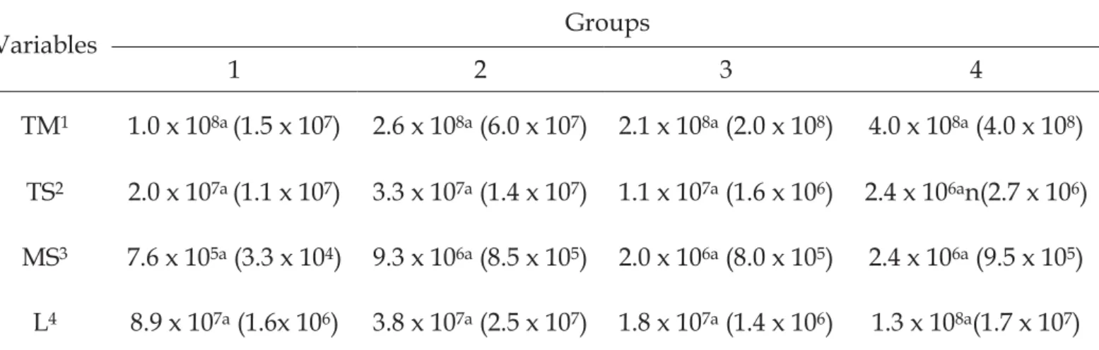
There were no significant differences in the number of TM and L among the
groups. However, TS and MS were significantly higher in the G3 compared to
the other groups. The drainage fluid pH and caries vessel fluid pH at the end of
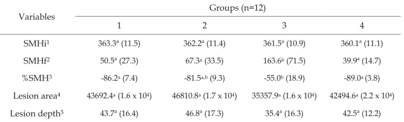
the experiment were very similar for all groups. There were no significant
difference in the initial microhardness, lesion area and lesion depth among the
groups. But, the G3 had significantly higher final microhardness than the other
groups, and a significantly smaller surface hardness change than the G1 and G4
groups. Based on these results and considering the limitations of the models
used, it was possible to conclude in the in situ and in vivo study that
probiotic-containing fermented milks were able to promote changes in the oral
microbiota and demineralization in enamel; and Batavito® was the fermented
milk that less favored the caries lesion development; and in the in vitro study
that only the L. reuteri mix-culture biofilm group had a reduced mineral loss as
Carolina Simonetti L odi – Tese de Doutorado 2011 26
Lista de figuras Capítulo 2
Figure 1. Representative confocal microscopy images of caries lesions
Carolina Simonetti L odi – Tese de Doutorado 2011 27
Lista de Tabelas Capítulo 1
Table 1.Variables analyzedin the treatment solutions 53
Table 2. Mean and standard deviation (SD) of the variables analyzed in the
enamel surface according to the treatments 54
Table 3. The distribution of microorganisms in the biofilm (log CFU/mg) after
14 days according to the treatments (in situ study) 55
Table 4. The distribution of microorganisms in saliva (log CFU/mL) at the
baseline and after 14 days according to the treatments (in situ study) 56
Table 5. The distribution of microorganisms in saliva (log CFU/mL) after 14
days according to the treatments (in situ study) 57
Table 6. The distribution of microorganisms in saliva (log CFU/mL) at the
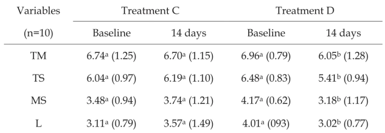
baseline and after 14 days according to the treatments (in vivo study) 58
Capítulo 2
Table 1. Study design and bacterial strain 78
Table 2. The distribution of microorganisms in the bacterial mixture inoculated in each group 79
Table 3. The distribution of microorganisms in the biofilm after 7 days in the artificial caries model 80
Carolina Simonetti L odi – Tese de Doutorado 2011 28
Lista de Abreviaturas
ATCC = American Type Culture Collection
BHI = Brain heart infusion
°C = Graus Celsius
CFU = Colony-forming units
dNTP = Desoxirribonucleotídeos trifosfatados
EPS = Extracellular Polysaccharide
g = Gramas
h = Horas
kb = Kilo base (= 1000 base pair)
Kg/mm2 = Kilogramas por milímetro quadrado
KHN = Knoop hardness number
ΔKHN = Integrated loss of subsurface hardeness KCl = Cloreto de potássio
K2HPO4 = Fosfato de potássio dibásico
KH2PO4 = Fosfato de potássio monobásico
L = Lactobacillus
Log = Logarítimo
min = Minutos
mg = Miligramas
MgCl2 = Cloreto de magnésio
mL = Mililitros
mm = Milímetros
μL = microlitros
mol L-1 = Mol por litro (Molar)
MRS = Man, Rogosa and Sharpe
MS = Mutans streptococci
MT = Micorganismo total
MW = Mineral washing
NaCl = Cloreto de sódio
Carolina Simonetti L odi – Tese de Doutorado 2011 29 PBS = Phosphate buffered saline
PCR = Polymerase Chain Reaction
pH = Potencial hidrogeniônico
p = Significância
ppm = Partes por milhão
s = segundos
SD = Standard deviation
SM = Streptococcus do grupo mutans
SMH = Surface microhardness
SMHi = Initial surface microhardness
SMHf = Final surface microhardness
%SMH = Percentage change in surface microhardness
ST = Streptococcus total
TBE buffer = Tris/Borate/EDTA buffer
TM = Total microorganisms
TS = Total streptococci
TSBS = Caldo triptona de soja suplementado com 5% de sacarose
UFC = Unidades formadoras de colônias
U = Units
V = volt
W = Watt
Carolina Simonetti L odi – Tese de Doutorado 2011 30
SUMÁRIO
Introdução Geral 31
Capítulo 1 – Probiotic-containing fermented milk effects on biofilm, oral
microbiota and dental enamel 36
Abstract 37
Introduction 38
Material and methods 39
Results 44
Discussion 46
Acknowledgments 51
Tables 53
References 59
Capítulo 2 - In vitro evaluation of probiotic bacteria efficacy in preventing
primary caries development using a multi-species microbial artificial-mouth
model 62
Abstract 63
Introduction 64
Material and methods 65
Results 70
Discussion 71
Acknowledgments 76
Tables 78
Figure 82
References 83
I ntrodução Geral
Carolina Simonetti L odi – Tese de Doutorado 2011 32
Introdução Geral
Probiótico é definido pela Organização Mundial da Saúde como sendo
microrganismos vivos que quando administrados em quantidades adequadas
conferem benefícios à saúde do hospedeiro [Food and Agriculture Organization
of the United Nations, World Health Organization, 2001]. As espécies mais
comumente utilizadas e pesquisadas pertencem ao gênero dos Lactobacillus e
Bifidobacterium [Ouwehand et al., 2002], microrganismos estes comumente
encontrados na cavidade bucal, inclusive nas lesões de cárie [Chhour et al.,
2005].
O conhecimento atual a respeito do importante papel da microflora
intestinal conduziu a estratégias para promover saúde através da manipulação
dessa comunidade microbiana [Fooks e Gibson, 2002]. A ação do probiótico no
trato gastrointestinal é baseada na aderência da bactéria probiótica à mucosa
intestinal, inibindo dessa forma a adesão de patógenos. Semelhantemente na
cavidade oral, os probióticos devem aderir aos tecidos dentais como parte do
biofilme e competir com o crescimento de bactérias cariogênicas ou patógenos
peridontais [Comelli et al, 2002].
A produção eficiente de ácidos orgânicos, que é uma característica
comum tanto dos Lactobacillus como dos Bifidobacterium pode ser prejudicial à
saúde bucal. Matsumoto et al. [2005], avaliaram a cariogenicidade da bactéria
probiótica Lactobacillus salivarius em ratos e puderam observar que a inoculação
destes microrganismos promoveu um incremento da atividade cariogênica
aumentando a aderências dos S. muttans à superfície dental. Por outro lado,
esses mesmos microrganismos têm sido relacionados com benefícios à saúde
bucal, como a produção de substâncias inibitórias do crescimento de
Streptococcus sobrinus [Meurman et al, 1995; Meurman, 2005; Meurman e
Stamatova, 2007; Yli-Knuuttila et al, 2006], bem como a redução do risco de
cárie em crianças de 3-4 anos de idade após a ingestão de leite contendo
probiótico [Näse et al, 2001]. Além disso, o consumo de iogurte contendo
Lactobacillus reuteri reduziu ligeiramente a contagem de Streptococcus mutans
I ntrodução Geral
Carolina Simonetti L odi – Tese de Doutorado 2011 33 cepas de Bifidobacterium ou Lactobacillus reuteri [Çaglar et al, 2005; 2006; 2007;
2008]. Já os resultados de Montalto et al. [2004] demonstraram que não houve
nenhum aumento na contagem de Streptococcus mutans quando se administrou
uma mistura de Lactobacillus probióticos na forma de cápsulas ou líquida.
A sobrevivência das bactérias probióticas no produto alimentício é de
fundamental importância, devendo o alimento conter pelo menos uma
população de 107 UFC/g de bactérias probióticas viáveis no momento da
compra do produto. Esta concentração é recomendada por alguns autores
[Rybka e Fleet, 1997; Vinderola e Reinheimer, 2000]. Entretanto, tem sido
proposto que a dose mínima diária de cultura probiótica considerada
terapêutica seja de 108 e 109 UFC, o que corresponde ao consumo diário de 100 g
de um produto contendo 106 a 107 UFC/g [Lee e Salminen, 1995; Blanchette et
al, 1996]. Tem sido mostrado que a rotina de consumo [Cabana et al, 2006] e o
processamento do alimento pode comprometer a viabilidade da bactéria
probiótica e interferir no benefício terapêutico advindo desse consumo
[Twetman e Stecksén-Blicks, 2008].
Para que o probiótico seja capaz de exercer um efeito anticariogênico,
primeiramente a bactéria deve ser capaz de aderir à superfície dental; segundo,
ela deve se tornar parte do biofilme dental; e finalmente, ela deve competir com
as bactérias cariogênicas reduzindo o nível de colonização destas [Comelli et al,
2002].
Além dos gêneros bacterianos, a maioria dos leites fermentados contém
açúcares e pH ácido que podem facilitar a aderência de microrganismos
patogênicos à superfície dentária e consequentemente o desenvolvimento de
cárie [Matsumoto et al, 2005], por isso alguns veículos como leites, iogurtes,
queijos, gomas de mascar e comprimidos [Çaglar et al, 2005; 2006; 2007; 2008]
têm sido estudados na tentativa de se estabelecer a maneira mais eficaz de se
administrar os probióticos.
Twentman e Stecksén-Blicks [2008] realizaram um trabalho de revisão de
literatura mostrando a escassez de estudos que relacionam a ação dos
I ntrodução Geral
Carolina Simonetti L odi – Tese de Doutorado 2011 34 enfoque, mas todos avaliaram a instalação da bactéria probiótica ou a
diminuição/aumento dos microrganismos patogênicos após a ingestão de
produtos contendo probióticos em amostras de saliva, não sendo encontrados
trabalhos que avaliaram a instalação desses microorganismos no biofilme e
segundo Meurman [2005] amostras de saliva podem subestimar a verdadeira
situação do biofilme.
Em estudo realizado por Lodi et al [2010a, 2010b] foi analisado o
potencial cariogênico de leites fermentados contendo probióticos (Parmalat®
-uva, Chamyto®, Paulista®, Batavito®, Yakult®, Vigor Club®). O estudo consistiu
de 2 etapas, sendo primeiramente realizado um estudo in vitro [Lodi et al.
2010a] para analisar algumas propriedades desses produtos como pH,
capacidade tampão, conteúdo de flúor, cálcio e fósforo. Ficou demonstrado que
todos os produtos possuem pH baixo e capacidade tampão elevada,
características estas que podem auxiliar na desmineralização do esmalte dental.
Além disso, pode ser observado também a presença dos íons flúor, cálcio e
fósforo nos leites fermentados possibilitando uma diminuição no potencial
cariogênico dos produtos.
Posteriormente, foram selecionadas 2 marcas dentre as citadas
anteriormente (Yakult®, Batavito®) para a realização de um estudo in situ onde
foram avaliados a concentração dos íons flúor, cálcio, fósforo e carboidratos
álcalis-solúveis presentes no biofilme dental, bem como a desmineralização do
esmalte dental bovino através do teste de microdureza superficial [Lodi et al.,
2010b]. Ficou demonstrado que ambos os produtos promoveram queda no pH
do biofilme dental e desmineralização na superfície do esmalte dental bovino,
embora o tratamento com o leite fermentado Batavito® tenha apresentado perda
mineral significantemente menor quando comparado com os outros grupos
(Yakult® e Sacarose 20%), bem como, a maior concentração de íons flúor, cálcio
e fósforo e menor concentração de carboidratos álcalis-solúveis no biofilme
dental. Neste estudo não pode ser determinado o motivo de uma perda mineral
menor após a utilização de uma determinada marca de leite fermentado
I ntrodução Geral
Carolina Simonetti L odi – Tese de Doutorado 2011 35 tipo e a quantidade de bactéria probiótica e a quantidade de carboidratos
presente nos produtos.
Por outro lado, acredita-se que quanto mais cedo ocorrer à colonização
bucal por bactérias probióticas maior é seu efeito em longo prazo [Twentman e
Stecksén-Blicks, 2008], portanto, diante da escassez de trabalhos relacionando
probióticos e saúde bucal, o objetivo deste trabalho foi avaliar in situ, in vivo e in
vitro a relação da bactéria probiótica com a cárie dentária a fim de elucidar se a
indicação do consumo precoce desses produtos poderá trazer prejuízos à saúde
bucal das crianças.
Carolina Simonetti L odi – Tese de Doutorado 2011 36
Capítulo 1
Carolina Simonetti L odi – Tese de Doutorado 2011 37 Probiotic-containing fermented milk effects on biofilm, oral microbiota, and
dental enamel
Abstract
The aim of this study was to evaluate the effect of 2 probiotic-containing
fermented milks on biofilm and salivary microorganisms and on enamel
surface. In situ study was performed in 3 phases: 20% sucrose, treatment A
(Yakult®), and treatment B (Batavito®). Salivary microorganisms were counted
at baseline and after, and biofilm was analyzed just after the trial period. In vivo
study was performed in 2 phases: treatment C (Yakult®) and treatment D
(Batavito®). Saliva was collected at baseline and at the end of the trial period for
microbiological analysis. In the in situ study, biofilm data showed less total
microorganisms (TM) after treatment B than after treatment A, but similar to
20% sucrose. In saliva, 20% sucrose decreased TM and total streptococci (TS)
and increased mutans streptococci (MS). Treatment A significantly decreased
TM, TS, and MS. Treatment B decreased TM. It was observed less MS in
treatment B when the final data were compared among the treatments.
Treatment B differed from the other treatments in relation to the final
microhardness, percentage change in the surface hardness, and integrated loss
of subsurface hardness (p < 0.05). In vivo study showed that just treatment D
decreased all microorganisms. It was observed higher lactobacilli (L) in
treatment D when the baseline data were compared among the groups. In
conclusion, probiotic-containing fermented milks were able to promote changes
in the biofilm, oral microbiota, and demineralization of the enamel; Batavito®
was the fermented milk that less favored the caries lesion development.
Key words: Cultured milk products; Probiotics; Tooth demineralization; Dental caries;
Streptococcus mutans; Lactobacillus
Capítulo 1
Carolina Simonetti L odi – Tese de Doutorado 2011 38
Introduction
Limitations in traditional disease management strategies have been
overcome, and a number of researchers are developing probiotic methods to
treat the dental caries [Anderson and Shi, 2006]. Research focusing on probiotics
has progressed considerably in the last 20 years, and significant advances have
been made in the selection and characterization of specific probiotic cultures
and the substantiation of health claims relating to their consumption [Teughels
et al., 2008].
The mechanism of probiotic action in the oral cavity is not fully
understood but is commonly explained by a combination of local and systemic
immune responses as well as non-immunologic defense mechanisms [Çaglar et
al., 2005a; Meurman and Stamatova, 2007]. The principal health-promoting
effects are ascribed to the enhancement of the mucosal immune defense and
macrophage activity as well as elevations in the number of killer cells, T-cells,
and interferons [Fuller and Gibson, 1997]. To be effective against oral infections,
probiotic bacteria need to adhere to the oral mucosa and dental tissues as part
of the biofilm and compete with the growth of dental pathogens [Comelli et al.,
2002]. The most widely used species belong to the genera Lactobacillus and
Bifidobacterium once these organisms are already produced in the dairy industry
and because they are very rarely implicated in infections of humans [Teughels
et al., 2008].
Probiotics are provided into food items in 1 out of 4 basic ways: as a
culture concentrate added to beverages (such as fruit juices); inoculated into
prebiotic fibers, which promote the probiotic bacterial growth; inoculated into
milk and milk-based foods (such as milk drinks, yoghurt, and cheese); and as
lyophilized cells packaged as dietary supplements (tablets, chewing gums, and
straws). The archetypical probiotic food is yoghurt, and the daily consumption
of dairy products seems to be the most natural way to ingest probiotic bacteria
[Çaglar et al., 2005a]. Another advantage is that milk products contain basic
Capítulo 1
Carolina Simonetti L odi – Tese de Doutorado 2011 39 possible beneficial effects on the salivary microbial composition and inhibition
of caries development due to their natural content of casein, calcium, and
phosphorous [Levine, 2001; Petti et al., 2001].
However, the best vehicle for probiotic delivery has yet to be identified.
Çaglar et al. [2006] investigated the effect of the probiotic bacterium L. reuteri
ATCC 55730 on the levels of salivary mutans streptococci and lactobacilli in
young adults, when ingested via 2 different non-dairy delivery systems (straws
and tablets). A significant reduction in the mutans streptococci levels was
recorded after the ingestion of the probiotic bacteria via both straws and tablets,
which was in contrast to the placebo controls. Another study [Montalto et al.,
2004] evaluated whether there was any difference between taking probiotic
lactobacilli in the liquid form or in capsules on S. mutans counts in a 45-day
double-blind, randomized, placebo-controlled intervention study. The oral
administration of the probiotic strains significantly increased the salivary
counts of the lactobacilli. The effect occurred irrespective of whether the
lactobacilli were administered in liquid or in capsule form, indicating that
probiotics ingested in capsule form might result in a temporary increase in oral
lactobacilli.
Recently, our research group evaluated some properties of some
fermented milk brands [Lodi et al., 2010a] and their effect on biofilm inorganic
composition [Lodi et al., 2010b]. It was observed that although all the products
were milk-based beverages and presented in their composition fluoride,
calcium, and phosphate, they promoted caries lesion in bovine enamel blocks
[Lodi et al., 2010a; Lodi et al., 2010b]. In this context, the aim of this study was
evaluate the effect of the short-term using of 2 brands of probiotic-containing
fermented milks on biofilm, salivary microorganisms, and dental enamel.
Material and Methods
Capítulo 1
Carolina Simonetti L odi – Tese de Doutorado 2011 40 The fermented milks used in this study were Yakult® (fermented milk A)
and Batavito® (fermented milk B) that have fluoride, calcium, and phosphate in
their composition [Lodi et al., 2010a] also contains probiotic bacteria.
According to the manufacturers, Yakult® contains in its composition a
single probiotic bacteria L. casei Shirota, and Batavito® a mix of 4 probiotic
bacteria (L. acidophilus, bifidobacteria, Streptococcus salivariusthermophilus, and L.
paracasei).
Fermented Milk Total and Reducing Sugar A nalysis
The determination of total (alkali-soluble carbohydrates) and reducing
sugars was done using the colorimetric spectrophotometer (HITACHI U-2000,
Hitachi High-Technologies Corporation, Toronto, ON, Canada), as described,
respectively, by Dubois et al. [Dubois et al., 1956] and Somogyi-Nelson [Nelson,
1944].
Fermented Milk M icrobial A nalysis
Both the fermented milks used in this study were serial tenfold diluted in
phosphate-buffered saline (PBS) and double plated in Brain Heart Infusion agar
(Himedia, Himedia Laboratories Pvt. Ltd., Mumbai, Maharashtra, India), Mitis
Salivarius agar (Himedia, Himedia Laboratories Pvt. Ltd., Mumbai,
Maharashtra, India), Mitis Salivarius Sucrose Bacitracin agar (Sigma-Aldrich
Co., St. Louis, MO, USA), and Rogosa agar (Himedia, Himedia Laboratories
Pvt. Ltd., Mumbai, Maharashtra, India) to analyze total microorganisms (TM),
total streptococci (TS), mutans streptococci (MS), and lactobacilli (L),
respectively. The plates for TM, TS, and MSwere incubated at 37ºC for 48 h in
an anaerobic chamber. The plates for L were incubated aerobically at 37ºC for
72 h. Colonies were counted with a colony counter (CP 600 Plus, Phoenix
Indútria e Comércio de Equipamentos Científicos Ltda., Araraquara, SP, Brazil).
The identification of the mutans streptococci colonies was performed using
Capítulo 1
Carolina Simonetti L odi – Tese de Doutorado 2011 41 expressed as colony-forming units by milligrams of fermented milk (CFU/mg
of fermented milk).
Enamel Block Preparation and A nalysis
Enamel blocks measuring 4 × 4 × 2 mm were obtained from bovine
incisor teeth previously stored in 2% formaldehyde solution (pH 7.0) for 1
month [White and Featherstone, 1987]. They had their surfaces serially
polished, and the selected blocks were divided in agreement with the average
of hardness (358.8 to 395.0 Kg/mm2) of the total blocks and the trust interval.
Surface enamel microhardness (SMH) measurements were made using a
Shimadzu HMV-2000 hardness tester (Shimadzu Corp., Kyoto, Japan). For
baseline SMH (SMHi), 5 indentations spaced 100 μm from each other were made (25 g load, 10 s) in the center of the enamel block [Vieira et al., 2005].
After the trial, SMH was again measured (SMHf). Five indentations
spaced 100 μm from each other and from the baseline were made. The percentage change in SMH (%SMH) was calculated [%SMH = 100 (SMHf -
SMHi)/SMHi]. The cross-sectional microhardness was measured using a
Micromet 5114 hardness tester (Buehler Ltd., Lake Bluff, IL, USA). The test was
performed by sectioning the blocks longitudinally through the center. One of
the halves was embedded in acrylic resin with the cut face exposed and
gradually polished. One row of 14 indentations was made at different distances
(5, 10, 15, 20, 25, 30, 40, 50, 70, 90, 110, 130, 220, and 330 μm) from the outer enamel surface under a 5 g load for 10 s. The integrated area above the curve
(cross-sectional profiles of hardness into the enamel), using the hardness values
(KHN), was calculated by the trapezoidal rule (GraphPad Prism, version 3.02)
in each depth (mm) from the lesion up to sound enamel. This value was
subtracted from the integrated area of sound enamel, to obtain the integrated
area of the subsurface regions in enamel, which was named as the integrated
Capítulo 1
Carolina Simonetti L odi – Tese de Doutorado 2011 42
In Situ Experiment
This study was previously approved by the local Human Ethical
Committee (FOA-UNESP, protocol # 2008-01519). Ten healthy volunteers aged
21–32 years were selected, and they agreed and signed the informed consent.
The in situ study involved a blind crossover design performed in 3 phases: 20%
sucrose, Yakult® (treatment A), and Batavito® (treatment B). One week before
the experiment beginning and during the whole experiment, the volunteers
used non-fluoridated dentifrice. Each volunteer wore acrylic palatal devices
containing 4 enamel bovine blocks covered by a plastic mesh to enable dental
biofilm accumulation and to protect it from disturbance [Cury et al., 2000].
Besides that, 1 mm of space was left between the enamel and the plastic mesh.
Before starting the experiment, saliva was collected from each volunteer
for the initial microbiological analysis. In each phase, 8 times a day, volunteers
removed the appliance from the oral cavity and dripped two drops the
following treatment solutions on the enamel bovine blocks: 20% sucrose
solution (Control), Yakult® (treatment A), or Batavito® (treatment B). Five
minutes after the application, the device was replaced in the mouth. After the
trial period (14 days), saliva was collected again and also the formed biofilm for
microbiological analysis. A washout period of 7 days was established between
each phase. No restriction was made regarding the volunteers’ diet, but they
were instructed to remove the appliance during meals and oral hygiene [Cury
et al., 2000]. They were not allowed to use any antimicrobial or fluoride product
during the experiment.
In V ivo Experiment
This study was previously approved by the local Human Ethical
Committee (FOA-UNESP, protocol # 2008-01519). Ten healthy volunteers aged
21–32 years were selected, and they agreed and signed the informed consent.
The in vivo study involved a blind crossover design performed in 2 phases:
Yakult® (treatment C) and Batavito® (treatment D). Each volunteer drank 1
Capítulo 1
Carolina Simonetti L odi – Tese de Doutorado 2011 43 beginning and during the whole experiment, the volunteers used
non-fluoridated dentifrice. A washout period of 7 days was established between
each phase. The only restriction made regarding the volunteers’ diet was no
intake other source of bacteria probiotic. Saliva was collected at baseline and at
the end of each phase for microbiological analysis.
Dental Biofilm M icrobial A nalysis
At the end of the in situ experiment, the volunteers returned the palatal
appliances. The plastic mesh was removed, and the biofilm was harvested and
weighed. Around 5 mg of the biofilm was resuspended in phosphate-buffered
saline (PBS: 8.0 g NaCl, 0.2 g KCl, 1.0 g K2HPO4, and 0.2 g KH2PO4 per liter,
adjusted to pH 7.4; 1 mL/mg of biofilm) and sonicated on ice in an ultrasonic
cell disruptor (XL; Misonix Inc., Farmingdale, NY, USA) for 6 × 9.9 s; amplitude
90%, and 40 W of power [Bowen, 1986]. The suspensions were serial tenfold
diluted in PBS and double plated as previously described for the fermented
milk microbial analysis of TM, TS, MS, and L.
The remaining biofilm was dried with phosphorus pentoxide (Vetec
Química Fina Ltda., Duque de Caxias, RJ, Brazil) for 12 h at room temperature.
Insoluble extracellular polysaccharides (EPS) were extracted by adding 1.0 mol
L-1 NaOH (10 μL/mg dry weight) to the biofilm. The samples were vortexed for
1 min, and after 3 h under agitation at room temperature, they were centrifuged
(1 min; 11,000 Xg at room temperature) [Nobre dos Santos et al., 2002].
Supernatants were precipitated with 75% cooled ethanol overnight, centrifuged,
and resuspended in 1.0 mol L-1 NaOH [Ccahuana-Vásquez et al., 2007].
Carbohydrate analysis was done by the phenol-sulfuric acid procedure [Dubois
et al., 1956]. The results were expressed as mg/g dry weight.
Saliva M icrobial A nalysis
Saliva samples from each volunteer were performed at baseline and at
the end of each phase by oral rinses with phosphate-buffered saline (PBS; 0.1 M;
Capítulo 1
Carolina Simonetti L odi – Tese de Doutorado 2011 44 10 min at 8,000 Xg, the supernatant discarded, and 2.5 mL of PBS added to the
pellet. The suspensions were serial tenfold diluted in PBS and double plated as
previously described for the fermented milk microbial analysis of TM, TS, MS,
and L. The results were expressed as colony-forming units by milliliters of
saliva (CFU/mL).
Statistical A nalysis
Each volunteer was considered as “n” in all the analysis. The null
hypothesis tested was that the fermented milk would not promote changes in
the biofilm, saliva, and demineralization of the dental enamel. Statistical
analysis was carried out using BioEstat Version 5.0. Data from CFU were
logarithmically transformed prior to analysis. Mean and SD for each measured
parameter (CFU in the biofilm, saliva, and fermented milk; EPS in the biofilm;
total and reducing sugars in the fermented milk; SMHi, SMHf, %SH, and
'KHN in the enamel block) were calculated for each group. These data were
analyzed using a single-factor analysis of variance model (ANOVA). Multiple
comparisons were conducted using the Tukey or Bonferroni test when
significant effects (p < 0.05) were detected. Data were analyzed using a
Kruskal-Wallis one-way analysis when they were not normally distributed or variances
were not equal. Data (means) from microorganisms in the saliva before and
after each trial phase were submitted to a paired t-test procedure. The
significance limit was set at 5%.
Results
Fermented Milk Total and Reducing Sugar A nalysis
The means of total sugar in 20% sucrose, fermented milk A, and
fermented milk B were 155.67 mg/mL, 158.49 mg/mL, and 191.33 mg/mL,
respectively, and it could be observed statistical differences among the groups
(Table 1).
The means of reducing sugar in 20% sucrose, fermented milk A, and
Capítulo 1
Carolina Simonetti L odi – Tese de Doutorado 2011 45 respectively, and it could be observed statistical differences among the groups
(Table 1).
Fermented Milk M icrobial A nalysis
In fermented milk A, it was observed 9.05 ± 0.06 log CFU/mgTM, 2.10 ±
0.12 log CFU/mgTS, and 8.72 ± 0.07 log CFU/mg (5.2 x 10⁸ UFC/mL) L. In
fermented milk B, it was observed 7.93 ± 0.07 log CFU/mg TM, 7.21 ± 0.59 log
CFU/mg TS, and 7.24 ± 0.20 log CFU/mg (1.7 x 10⁷ UFC/mL) L. Both the
fermented milks did not show MS when plated (Table 1).
Fermented milk A showed higher concentrations of TM and L and a
lower concentration of TS than fermented milk B, and these differences were
statistically significant (p < 0.01).
Enamel A nalysis
The means and standard deviations (SD) of the 3 groups analyzed in this
study are shown in Table 2. The initial microhardness (p = 0.853) showed no
statistically significant difference among the groups. Treatment B differed from
the other groups in relation to the final microhardness, percentage of surface
hardness change, and integrated loss of subsurface hardness (p < 0.05). It was
not observed statistical difference in relation to the variables analyzed between
treatment A and 20% sucrose (p > 0.05).
In Situ Experiment
By comparing biofilm data, it was observed lower TM in treatment B
when compared with treatment A, but no statistical difference was observed
between treatment B and 20% sucrose. TS, MS, and L did not show statistical
difference among the groups (Table 3).
In saliva analysis, when the baseline and final data of the
microorganisms were compared in each treatment, it was observed that TM and
TS decreased, and MS increased after the use of 20% sucrose solution; these
Capítulo 1
Carolina Simonetti L odi – Tese de Doutorado 2011 46 the end of this phase (Table 4). Treatment A showed a significant decrease in
TM, TS, and MS; however, L did not show statistical difference after this trial
phase (Table 4). TM was the only one that showed a significant decrease in
treatment B. However, TS and MS decreased and L increased with that
treatment, but these changes did not show statistical differences (Table 4).
It was observed similar amount of all microorganisms in saliva when
compared the baseline data of the 3 treatment (p > 0.05). Regarding the final
data of the microbial count in the saliva, it was observed a lower MS in
treatment B, and this data was statistically significant when compared with the
other treatments (Table 5).
The EPS concentration in the biofilm did not show statistical difference
between the treatments. The 20% sucrose and treatment A showed 13.84
mg/mg of dry biofilm, and 12.35 mg/mg of dry biofilm for treatment B.
In V ivo Experiment
Treatment C did not change the concentration of any microorganism
when it was compared baseline and final data. After treatment D, it was
observed a statistically significant decrease in all the microorganisms analyzed
(Table 6).
It was observed similar TM, TS, and MS in saliva when compared at the
beginning of the 2 treatments, but the amount of L was higher at the beginning
of treatment D, and this difference was statistically significant. When compared
the final data of the 2 treatment it was not observed statistical difference
between the groups.
Discussion
The effects of probiotics on oral health are a relatively new research area,
but the concept of probiotics being beneficial from a dental point of view may
appear controversial.
In our in vivo study, it was used probiotic-containing fermented milks
Capítulo 1
Carolina Simonetti L odi – Tese de Doutorado 2011 47 contact with oral tissues. Although fermented milks were not developed with
the purpose of preventing dental caries with the promotion of changes on oral
microbiota, it was observed that the fermented milk B decreased salivary count
of all the microorganisms investigated after 2 weeks using. These results were
similar to the previously reported intakes [Çaglar et al., 2006; Näse et al., 2001;
Ahola et al., 2002; Nikawa et al., 2004]. It was also observed that fermented milk
A showed a tendency to increase salivary count of TS, MS, and L at the end of
the trial, but the difference was not statistically significant.
Some studies evaluating the effect of lactobacilli-derived probiotics on
mutans streptococci reported significant reductions of salivary mutans
streptococci immediately after the termination of daily intakes [Çaglar et al.,
2006; Näse et al., 2001; Ahola et al., 2002; Nikawa et al., 2004]. The
post-treatment levels decreasing were not directly dependent on the daily
administration vehicle, which was milk, cheese, yoghurt, lozenges, or straws
prepared with freeze-dried strains. Çaglar et al. [2006] investigated whether
slowly melting tablets would allow a more thorough contact between the
probiotic bacteria and the oral environment compared with the direct
swallowing pattern from the straw and concluded that both the regimes equally
reduced the prevalence of salivary mutans streptococci after 2 weeks of use.
Conflicting findings were reported by Montalto et al. [2004] that
evaluated the administration of probiotic lactobacilli in liquid and capsule ways
in order to determine the role of direct contact with the oral tissues.
Interestingly, it was found that both ways of administration significantly
increased the salivary lactobacilli counts, while the levels of mutans streptococci
were not significantly modified by the intervention [Montalto et al., 2004]. Some
studies have indicated that direct contact with the oral tissues is not a
prerequisite for a beneficial effect, and a pure systemic administration of a
probiotic could enhance lactobacilli proliferation in the oral cavity [Çaglar et al.,
Capítulo 1
Carolina Simonetti L odi – Tese de Doutorado 2011 48 Differences among different strains and strains of the same species are
probably the reason for the conflicting results of probiotic efficacy that were
reported in the early studies [Twetman and Stecksén-Blicks, 2008]. Today, most
of the research is carried out with well-defined, dairy-based live lactobacilli
strains. In this study, fermented milk A contains in its composition a single
probiotic bacteria L. casei Shirota, and fermented milk B a mix of 4 probiotic
bacteria (L. acidophilus, bifidobacteria, S. salivariusthermophilus, and L. paracasei),
according to the manufacturers. Another interesting point is that although
fermented milk A (5.2 x 10⁸ UFC/mL) showed a higher amount of probiotic L in
its composition than the fermented milk B (1.7 x 10⁷ UFC/mL), fermented milk
The best results after the consumption of fermented milk B could be explained
by this difference in their probiotic-containing once the simultaneous
application of different probiotics can affect the balance of the oral ecosystem in
a possible additive, cumulative, or competitive modes of action [Meurman and
Stamatova, 2007].
Saliva samples may underestimate the true situation of the oral biofilm
[Meurman, 2005]. Moreover, it is difficult to obtain biofilm collected in vivo due
to the need of oral hygiene restrictions. Thus, we performed also an in situ
study to investigate the effects of probiotic-containing fermented milks on
biofilm.
Treatment B showed lower concentrations of TM in the biofilm when
compared with the other groups (20% sucrose and treatment A). However TS,
MS and L did not show statistically significant difference among the groups
that could be explained by the optimal dose needed for pathogenic bacteria
suppression has not been achieved during the experimental phases once the
volunteers just dripped the fermented milks on enamel blocks. Lee and
Salminen [1995] suggested a intake of 100 g of 106 to 107 of a
containing-probiotic product as a optimal dose; but Çaglar et al. [2008] showed in their
study significant mutans streptococci reduction with a low-amount
Capítulo 1
Carolina Simonetti L odi – Tese de Doutorado 2011 49 × 107 CFU per gram ingested daily. On the other hand, microorganism
reduction was also achieved with 200 g of yogurt containing 2 × 10⁸ CFU per
gram in their former study [Çaglar et al., 2005b].
The use of both the fermented milks in the in situ study showed a
decrease of all the microorganisms in the salivary count when compared the
baseline and final data, except L in treatment B that showed an increase in its
amount. The salivary microorganism reduction called our attention to the safety
model for volunteers’ oral health and to permit us to study the biofilm
artificially formed in the oral cavity. The reduction on the salivary counts may
be due to the use of the appliance that could increase the salivary flow. On the
other hand, the differences between the probiotic bacteria in the fermented
milks analyzed could not be responsible for this reduction once they were just
dripped on the enamel block, and the amount of fermented milk used was too
low to interfere with the oral microbiota.
The microhardness test was used to evaluate the changes in the enamel
surface and demineralization area in the in situ study. Treatment B differed
from the other groups in relation to the final microhardness, percentage of
surface hardness change, and integrated loss of subsurface hardness. Both the
products used in this study are based-milk and contain in their composition
fluoride, calcium, and phosphate [Lodi et al., 2010a]. These ions should confer
an enamel protective effect, but in both the treatments, it was observed that
enamel demineralization may be due to the presence of sucrose on their
composition.
Sucrose is considered the most cariogenic factor from the dietary because
of its fermentability and it works as a substrate for the synthesis of
polysaccharides in dental biofilm [Bowen, 2002]. To try understanding the
results described above, we investigated the amount of total and reducing
sugars in both the fermented milks and the EPS concentration in the biofilm.
Both the fermented milks presenting reducing and total sugars and EPS were
Capítulo 1
Carolina Simonetti L odi – Tese de Doutorado 2011 50 demineralization observed. Fermented milk A showed a lower amount of total
sugar and a higher amount of reducing sugar when compared with fermented
milk B, which does not match the lower mineral loss observed after treatment B,
inferring that probiotic bacteria could really have had some protective effect on
dental caries development.
In conclusion, the findings of the present study demonstrated that
probiotic-containing fermented milks were able to promote changes in the
biofilm, oral microbiota and demineralization of the enamel surface;
collectively, the data showed that Batavito® was the treatment that less favored
the caries lesion development. More systematic studies and randomized
controlled trials are needed for finding out the best probiotic strains, daily
doses, and probiotic vehicles for a promising and safe perspective to oral
Capítulo 1
Carolina Simonetti L odi – Tese de Doutorado 2011 51
Acknowledgments
The authors would like to express their gratitude to the volunteers for
their valuable participation. We also thank Maria dos Santos Fernandes for the
laboratorial assistance in this study. Conceived and designed the experiment:
CSL, FLB, ACBD, and CCRM. Performed the experiments: CSL and LV.
Capítulo 1
Carolina Simonetti L odi – Tese de Doutorado 2011 52 Table 1.Variables analyzedin the treatment solutions
Table 2. Mean and standard deviation (SD) of the variables analyzed in the
enamel surface according to the treatments
Table 3. The distribution of microorganisms in the biofilm (log CFU/mg) after
14 days according to the treatments (in situ study)
Table 4. The distribution of microorganisms in saliva (log CFU/mL) at the
baseline and after 14 days according to the treatments (in situ study)
Table 5. The distribution of microorganisms in saliva (log CFU/mL) after 14
days according to the treatments (in situ study)
Table 6. The distribution of microorganisms in saliva (log CFU/mL) at the
Capítulo 1
Carolina Simonetti L odi – Tese de Doutorado 2011 53 Table 1.Variables analyzedin the treatment solutions
Variables 20% Sucrose Fermented Milk A Fermented Milk B
Total Sugar1 155.67a (1.0) 158.4b (1.4) 191.33c (0.1)
Reducing Sugar1 0.15a (0.004) 9.83b (0.50) 0.83c (0.167)
TM2 N/A 9.05a (0.06) 7.93b (0.07)
TS2 N/A 2.10a (0.12) 7.21b (0.59)
MS2 N/A N/G N/G
L2 N/A 8.72a (0.07) 7.24b (0.20)
a,bMeans (SD) followed by distinct letters are significantly different according to each
variable
1Total and reducing sugar were expressed in mg/mL
2TM (total microorganisms), TS (total streptococci), MS (mutans streptococci) and L
(lactobacilli) were expressed in log CFU/mL
N/A Variable not analyzed