Priscila Zacarias de Azevedo
Avaliação do teste de imunodifusão dupla em gel de
agar e do ensaio imunoenzimático – ELISA no
diagnóstico e seguimento de pacientes com bola
fúngica aspergilar
Dissertação apresentada à Faculdade de Medicina, Universidade Estadual Paulista “Júlio de Mesquita Filho”, Câmpus de Botucatu, para obtenção do título de Mestre em Doenças Tropicais.
Orientador: Prof. Dr. Rinaldo Poncio Mendes
Aos meus pais,
Neide
eLeirton
, pelo amor incondicional, por sempre estarem ao meu lado e acreditarem em mim.“Ainda que eu falasse a língua dos homens e
dos anjos, sem amor eu nada seria. Agora pois, permanecem a fé, a esperança e o amor, estes três, mas o maior destes é o amor”.
(1 Coríntios, 13)
AMO VOCÊS!!
A minha irmã
Patrícia
e ao meu cunhadoFrancisco
, obrigada pelo carinho.“Sua alegria é minha alegria Suas lágrimas de choro também Não importa o que venha na vida, Irmãos,
Somos irmãos „irmãs‟”.Amo vocês.
Ao meu orientador Prof. Dr.
Rinaldo “Tietê”
Poncio Mendes
, obrigada pelo grandeaprendizado, pela dedicação, por conselhos e amizade.
“Ser mestre não é apenas lecionar. Ensinar não é apenas transmitir o conhecimento.
Ser mestre é ser instrutor e amigo, guia e companheiro. Ser mestre é ser exemplo.
Exemplo de dedicação, de doação, de dignidade pessoal e, sobretudo, de amor.”
Agradeço a DEUS e a Nossa Senhora, por sempre estarem ao meu lado.
A Coordenação de Aperfeiçoamento de Pessoal de Nível Superior (CAPES) pela concessão de bolsa de estudo.
À colaboradora desse trabalho Tatiane Fernanda Sylvestre pela ajuda com o ELISA, pela atenção, apoio e amizade.
A Lariza Maza pela ajuda na realização da galactomanana, pela atenção, apoio, dedicação e amizade nos momentos que mais precisei.
Ao Dr. Ricardo de Souza Cavalcante, pela ajuda na parte clínica, no levantamento de dados dos prontuários, pela atenção, apoio e amizade.
A Dra. Daniela Vanessa Moris de Oliveira, pelos conselhos, ajuda e amizade nos momentos que mais precisei.
A Dra. Lídia Raquel de Carvalho, do Departamento de Bioestatística – IB/Unesp, pela realização das análises estatísticas deste trabalho, pela disponibilidade e total apoio.
A Dra. Maria Luiza Cotrim Sartor de Oliveira, do Departamento de Patologia – FMB, pelo fornecimento das fotos do citopatológico deste trabalho, pela disponibilidade e atenção.
A equipe do Laboratório de Micologia, Tatiane Fernanda Sylvestre, Adriele Dandara Levorato e Lariza Maza, obrigada pelo companheirismo.
A Camila Martins Marchetti, pela ajuda no formato das fotos e a Bárbara Casella Amorim, obrigada pelo apoio e amizade meninas.
A Regina Moretto, do Laboratório de Ensaios Imunoenzimáticos da Unipex da Faculdade de Medicina de Botucatu – FMB, obrigada por disponibilizar o aparelho de ELISA para a realização deste trabalho.
Aos funcionários da Unipex, em especial ao Eduardo Jodi Kuninare, obrigada pela amizade, apoio e atenção.
A Bruna Quirino Jorgetto e aos funcionários da Pós-Graduação e do Departamento de Doenças Tropicais e Diagnóstico por Imagem, obrigada pelo auxílio e atenção.
A Rosmary Cristina Silva “Meirinha”, obrigada pela ajuda e atenção nas referências bibliográficas.
A minha querida amiga Mara Elizabeth Gaiotto, pessoa maravilhosa, prestativa, companheira e sempre com conselhos. Obrigada pelo convívio, risadas e pela amizade verdadeira.
Ao meu professor de inglês Rafael Suman Tassi, thanks for the patience, attention and friendship.
A Dra. Patrícia Carvalho Garcia, do Hemocentro do Hospital das Clínicas da FMB – Unesp, pela grande oportunidade em minha vida. Obrigada pela atenção, apoio e amizade.
A Dra. Valéria Nogueira Dias Paes Secco, por ter me recebido em seu laboratório com dedicação e paciência.
Ao meu querido Padre José Antônio, da Paróquia São João Batista – Dourado / SP. “Nada é impossível a quem tem fé verdadeira, quem tem fé tudo pode.” (Mt 17, 20-21).
À coordenadoria e ao programa de Pós-Graduação em Doenças Tropicais, pela confiança em meu trabalho.
SUMÁRIO
A) Artigo 1- Revisão da literatura sobre diagnóstico sorológico da bola fúngica pulmonar
1. Introdução...02
2. Métodos...02
3. Resultados...04
4. Discussão……….. 06
5. Referências………08
B) Artigo 2 (submetido) - Evaluation of the double agar gel immunodiffusion test and of the enzyme-linked immunosorbent assay in the diagnosis and follow-up of patients with chronic pulmonary aspergillosis 1. Introduction………...13
2. Patients and methods……….14
2.1 Study population………...…14
2.2 Study design………..17
3. Methods……….18
3.1Antigens used………18
3.2Determination of serum antibodies and GMN level……….19
3.3 Serological follow-up of patients treated with itraconazole………..21
3.4 Determination of the cut-off point of the ELISA test………21
3.5Accuracy parameters……….21
3.6Cross-reactivity with sera from patients with other granulomatous diseases……...…22
3.7Ethics research committee……….……22
3.8Statistical analysis of results……….22
4. Results………...…22
4.1Determination of the cut-off point……….28
4.2Determination of the serum dilution to be used………29
4.3Accuracy parameters……….…29
4.4Cross-reactivity……….…30
4.6 Evaluation of the serological follow-up of patients under treatment with
itraconazole……….34
5. Discussion……….36
6. References...41
PZ Azevedo 1 ARTIGO 1
Revisão da literatura sobre diagnóstico sorológico de bola fúngica pulmonar.
Priscila Zacarias de Azevedo
Departamento de Doenças Tropicais, Faculdade de Medicina de Botucatu – Universidade Estadual Paulista - UNESP.
Rinaldo Poncio Mendes
Departamento de Doenças Tropicais, Faculdade de Medicina de Botucatu – Universidade Estadual Paulista - UNESP.
Resumo
Contexto. O diagnóstico sorológico pode implementar a definição de caso de bola
fúngica aspergilar que, em geral, tem a tuberculose como a principal doença de base, que é muito freqüente no Brasil.
Objetivo. Determinar parâmetros de acurácia de reações de precipitação em gel, do
ensaio imunoenzimático e da determinação dos níveis séricos de galactomanana no diagnóstico de bola fúngica aspergilar.
Pesquisa de dados e seleção de estudos. A pesquisa eletrônica de dados foi feita no
MEDLINE e a manual pela busca de artigos citados nos textos encontrados no levantamento
on line. Foram analisados os artigos publicados em inglês, português, francês e espanhol.
Dois níveis de seleção foram utilizados em 23 citações. Os critérios de inclusão foram casos confirmados e prováveis de bola fúngica aspergilar e avaliação de pelo menos um parâmetro de acurácia dos testes sorológicos.
Síntese dos dados. Oito dos 23 artigos previamente selecionados foram avaliados. A
PZ Azevedo 2
negativa foram, em geral, elevados. O teste ELISA, presente em apenas um artigo, apresentou baixa positividade. A sensibilidade da dosagem da galactomanana foi muito baixa quando se utilizou o método de aglutinação de partículas de látex (17,8% e 24,2%), elevando-se a 63,6% quando se utilizou o ensaio imunoenzimático, embora ainda baixa.
Conclusões. Não são muitos os trabalhos que avaliam o diagnóstico sorológico de bola
fúngica aspergilar. A reação de imunodifusão dupla em gel de agar revelou-se o método sorológico de escolha, por sua especificidade e razão de proporções positiva. O ensaio imunoenzimático ainda não revelou a acurácia desejada. Assim, confirma-se a necessidade de novos estudos sobre o diagnóstico sorológico de bola fúngica aspergilar.
Introdução
Aspergiloma, também conhecido como bola fúngica aspergilar (BFA), é uma doença não invasiva, caracterizada pelo crescimento de hifas de Aspergillus spp. em cavidades
pré-existentes, em geral causadas pela tuberculose, nos pulmões. [1]
Por vezes assintomática, a BFA pode causar manifestações clínicas como hemoptise e, em raros casos, pode ser fatal. O achado radiológico se caracteriza pela presença de massa arredondada sólida em seu interior, dentro de cavidade do pulmão. [2,3]
Um dos testes sorológicos mais utilizados para identificação do Aspergillus spp. é a
imunodifusão dupla em gel de agar - IDD, para detecção de anticorpos anti-A. fumigatus.
[4,5]
O objetivo do presente estudo é rever publicações prévias sobre o diagnóstico sorológico de BFA.
Métodos
Pesquisa de dados. Uma ampla pesquisa da literatura foi realizada utilizando-se
PZ Azevedo 3
aspergillosis diagnosis, no período de 1968 – 2014. As referências bibliográficas dos
trabalhos resultantes da pesquisa eletrônica também foram avaliadas para seleção.
Seleção de artigos. A seleção foi feita em dois níveis. No primeiro, os resumos foram
avaliados quanto aos seguintes critérios de exclusão: relato de casos, cartas comentários, revisões, estudo de experimento animal e linguagens outras que não inglês, português, francês e espanhol. No segundo, foram analisados os artigos completos dos trabalhos selecionados no primeiro nível.
Os critérios de inclusão foram casos confirmados e prováveis de BFA, assim definidas: a) definição de caso de BFA: presença de quadro clínico compatível, radiografia e, ou, planigrafia e, ou, tomografia computadorizada de tórax com cavitação pulmonar e massa arredondada sólida em seu interior, sugestiva de bola fúngica; [6-8] b) definição de caso confirmado: além do critério (a), presença de anticorpos séricos específicos determinados pela reação de IDD, ou pela identificação de Aspergillus sp. em lavado bronco-alveolar; [6-8] c)
definição de caso provável: além do critério (a), presença de hifas hialinas compatíveis com
Aspergillus sp no escarro [6-8]. Outros critérios de inclusão: avaliação sorológica específica
para os casos de BFA; determinação de pelo menos um dos seguintes parâmetros de acurácia: sensibilidade (S), especificidade (E), valores preditivos positivo (VPP) e negativo (VPN), acurácia (Ac) e razões de proporções positiva (RPP) e negativa (RPN), também denominadas razões de verossimilhança. Os parâmetros de acurácia avaliados com dados das publicações seguiram as especificações de Fletcher & Fletcher. [9]
No cálculo das razões de proporções, quando a freqüência de uma das casas foi igual a zero e, por este motivo, levou a zero o cálculo da razão de proporções, essa prevalência foi substituída por 1,0 e a razão de proporções foi, então denominada razão de proporções corrigida (RPPc ou RPNc). [10]
PZ Azevedo 4 Resultados
Dos 23 artigos levantados, três foram descartados por serem relatos de caso [11-13], três por serem trabalhos de revisão [2-3,14], e um por abordar apenas aspectos radiológicos [15], um por tratar de aspectos epidemiológicos [16], um por não definir com rigor os critérios de inclusão de cada caso [17], um por ter considerado o teste ELISA como critério de inclusão [18], dois por não terem avaliado a sorologia dos pacientes [19,20], um por não apresentar de forma individualizada os resultados de pacientes com aspergiloma [21], um por ter focalizado apenas a influência da hemoptise no prognóstico [22] e um por ter avaliado apenas o tratamento com itraconazol.[23]
Assim, restaram oito artigos que cuja avaliação permitiu analisar o diagnóstico sorológico de BFA pela pesquisa de anticorpos séricos específicos e de galactomanana.
A avaliação da sensibilidade de testes de precipitação em gel foi realizada em 146 pacientes com BFA (tabela 1). A IDD revelou sensibilidade elevada, com mediana 84,5% (33,3% - 100,0%) e a contra-imunoeletroforese (CIE), realizada em apenas dois trabalhos, mostrou mediana de 83,5% (66,7% - 100,0%).
Tabela 1 – Resultados de reações de precipitação em gel, realizadas em pacientes com bola fúngica aspergilar
BFA
(n) sorológica Reação Sensibilidade (%) Antígeno Autor(es) Referência [número] 23 IDD 100,0 A. fumigatus Ferreira-da-Cruz et al. 24
56 IDD 76,8 ... Kawamura et al. 1
17 IDD 100,0 A. fumigatus Mishra et al. 25
CIE 100,0 A. fumigatus Mishra et al. 25
30 IDD 93,3 Pool Coleman & Kaufman 4
17 IDD 64,7 A. fumigatus Lee et al. 26
3 IDD 33,3 A. fumigatus Avila R 27
CIE 66,7 A. fumigatus Avila R 27
PZ Azevedo 5
A IDD não revelou reação cruzada com soro de pacientes com tuberculose [4]. Esses autores também avaliaram reações cruzadas com soros de pacientes com outras micoses sistêmicas. No entanto, os resultados foram descartados tendo em vista o pequeno número de pacientes estudados.
O teste ELISA direto revelou sensibilidade de 63,6% e a dosagem de galactomanana demonstrou baixa positividade no teste de aglutinação de partículas de látex (mediana=21,0%; variação de 17,8% a 24,2%); porém, foi mais elevada quando se utilizou o teste ELISA (63,6%), como revela a tabela 2.
Tabela 2 – Sensibilidade do ensaio imunoenzimático e da dosagem de galactomanana no diagnóstico de pacientes com bola fúngica aspergilar.
Pacientes (n) Teste Sensibilidade (%) Autor Referência [número] Teste ELISA
33 63,6 Kawamura et al. 28
Dosagem de galactomanana
45 látex aglutinação 17,8 Kawamura et al. 1 33 látex aglutinação 24,2 Kawamura et al. 28 ELISA 63,6 Kawamura et al. 28
(n)- número de pacientes; ELISA: ensaio imunoenzimático.
A reação em cadeia de polimerase, por outro lado, foi capaz de identificar 93,3% dos casos de BFA. [28]
PZ Azevedo 6 Tabela 3 – Parâmetros de acurácia da reação de imunodifusão dupla em gel de agar e do ensaio imunoenzimático, realizados no soro de pacientes com bola fúngica aspergilar.
Reação
sorológica Pacientes (número) Controles (número) (%) S (%) E VPP (%) VPN (%) (%) Ac RPP / RPPc RPN / RPNc Referência [número]
IDD 30 10 93,3 100,0 100,0 83,3 95,0 9,3 0,06 4
IDD 17 50 100,0 100,0 100,0 100,0 100,0 50,0 0,05 25
ELISA 17 50 100,0 98,0 94,0 100,0 98,5 50,0 0,05 25
IDD: imunodifusão dupla em gel de agar; ELISA: ensaio imunoenzimático; S: sensibilidade; E: especificidade; VPP: valor preditivo positivo; VPN: valor preditivo negativo; Ac: acurácia; RPP: razão de proporções positiva; RPPc: razão de proporções positiva corrigida; RPN: razão de proporções negativa; RPNc: razão de proporções negativa corrigida.
Um estudo foi conduzido em 544 pacientes de 55 Serviços de Doenças Torácicas da Gran-Bretanha, que tiveram tuberculose pulmonar, cavidade persistente com 2,5 cm de diâmetro ou mais, e baciloscopia negativa para tuberculose por pelo menos um ano [29]. A pesquisa positiva de precipitinas anti-Aspergillus fumigatus revelou-se decisiva no
diagnóstico de BFA. [29]
Discussão
A revisão da literatura realizada demonstrou não ser elevado o número de artigos que focalizam o diagnóstico sorológico da BFA. Como as Aspergillus spp. colonizam a árvore
respiratória, os testes sorológicos contribuem muito para a definição de caso, na vigência de antecedentes epidemiológicos, quadro clínico e exame de imagem compatíveis. Além disso, os exames sorológicos não são invasivos e oferecem resultados em poucos dias.
Essa revisão também demonstrou que foi ainda menor o número de artigos que apresentaram outros parâmetros de acurácia, além da sensibilidade. [1,24,26-29]
PZ Azevedo 7
PZ Azevedo 8 Referências
1. Kawamura S, Maesaki S, Tomono K, Tashiro T, Kohno S. Clinical evaluation of 61 patients with pulmonary aspergilloma. Intern Med. 2000;39: 209-212.
2. Zmeili OS, Soubani AO. Pulmonary aspergillosis: a clinical update. QJM. 2007;100: 317-334.
3. Thompson GR 3rd, Patterson TF. Pulmonary Aspergillosis: recent advances. Semin Respir Crit Care Med. 2008;29: 103-110.
4. Coleman RM, Kaufaman, L. Use of the immunodiffusion test in the serodiagnosis of Aspergillosis. Appl Microbiol. 1972;23: 301-308.
5. Froudist JH, Harnett GB, McAller R. Comparison of immunodifusion and enzyme linked immunosorbent assay for antibodies to four Aspergillus species. J Clin Pathol.
1989;42: 1215-1221.
6. Denning DW, Pleuvry A, Cole DC. Global burden of chronic pulmonary aspergillosis as a sequel to pulmonary tuberculosis. Bull World Health Organ. 2011;89: 864-872. 7. Denning DW, Tucker RM, Hanson LH, Stevens DA. Treatment of invasive
aspergillosis with itraconazole. Am J Med. 1989;86: 791-800.
8. Camuset J, Nunes H, Dombret MC, Bergeron A, Henno P, Philippe B, et al. Treatment of chronic pulmonary aspergillosis by voriconazole in non immunocompromised patients. Chest. 2007;131: 1435-1441.
9. Fletcher RH, Fletcher SW. Diagnóstico. In: Epidemiologia clínica: elementos essenciais. Porto Alegre: Artes Médicas; 2006. pp. 56-81.
10.Maia Filho HS, Cunha AJLA. Diagnóstico. In: Gomes MM. Medicina baseada em evidências: princípios e práticas. Rio de Janeiro: Editora Reichmann & Affonso; 2001. pp. 81-94.
11.Tomee JF, Van-der-werf TS, Latge J, Koeter GH, Dubois AEJ, Kauffman HF. Serologic monitoring of disease and treatment in a patient with pulmonary aspergilloma. Am J Respir and Crit Care Med. 1995;151: 199-204.
12. Sheikh S, Fatimi SH. Aspergilloma in a patient with no previous history of chronic lung disease. J Ayub Med Coll Abbottabad. 2006;18: 62-63.
PZ Azevedo 9
14. Smith NL, Denning DW. Underlying conditions in chronic pulmonary aspergillosis including simple aspergilloma. Eur Resp J. 2001;37: 865-872.
15. Golberg B. Radiological appearances in pulmonary aspergillosis. Clinical Radiology. 1962;13: 106-114.
16.Handerson AH, English MP, Vecht RJ. Pulmonary aspergillosis: a survevy of its occurrence in patients with chronic lung disease and a discussion of the significance of diagnostic test. 1968;23: 513-518.
17.Warnock DW. Detection of Aspergillus fumigatus precipitins: a comparison of counter
immunoelectrophoresis an double diffusion. J Clin Path. 1977;30: 388-389.
18.Grupta PR, Vyas A, Meena RC, Sharma S, Khayam N, Subramanian IM. Clinical profile of pulmonary aspergilloma complicating residual tubercular cavitations in Northen Indian patients. Lung India. 2010;27: 209-211.
19. Aghajanzadeh M, Safarpoor F, Amani H, Alavy A, Sarshad A. Fungus ball: clinical presentation, diagnosis and treatment in 31 cases. IRCMJ. 2008;10: 233-237.
20. Chen KY, Ko SC, Hsueh PR, Luh KT, Yang PC. Pulmonary fungal infection: emphasis on microbiological spectra, patient outcome, and prognostic factors. Chest. 2001;120: 177-184.
21.Kauffman HF, Beaumont F, Meurs H, Van-der-Heide S, Vries K. Comparison of antibody measurments against Aspergillus fumigatus by means of double-diffusion
and enzyme-linked immunosirbent assay (ELISA). J Allergy Clin Immunol. 1983;72: 255-261.
22. Jewkes J, Kay PH, Paneth M, Citron K. Pulmonary aspergilloma: analysis of prognosis in relation to hemoptysis and survey of treatment. Thorax. 1983;38: 572-578.
23.Campebell JH, Winter JH, Richardson MD, Shankland GS, Banham SW. Tratment of pulmonary aspergilloma with itraconazole. Thorax. 1991;46: 839-841.
24. Ferreia-da-cruz MF, Wanke B, Pirmez C, Galvão-castro B. Aspergillus fumigatus fungus ball in hospitalized patients with chronic pulmonary disease. Usefulness of double immunodiffusion test as a screening procedure. Mem Inst Oswaldo Cruz, Rio de Janeiro. 1988;83: 357-360.
PZ Azevedo 10
26. Lee SH, Lee BJ, Jung DY, Kim JH, Sonh DS, Shin JW, Kim JY, Park IW, Choi BW. Clinical manifestations and treatment outcomes of pulmonary aspergilloma. The Korean Journal of Internal Medicine. 2004;19: 38-42.
27. Avila R. Immunological study of pulmonary aspergilloma. Thorax. 1968; 23: 144-152. 28. Kawamura S, Maesaki S, Noda T, Hirakata Y, Tomono K, Tashiro T, Kohno S. Comparison between PCR and detection of antigen in sea for diagnosis of pulmonary aspergillosis. J Clin Microbiol. 1999;37: 218-220.
29.Aspergillus in persistent lung cavities after tuberculosis: a report from the research
committee of the British Thoracic and Tuberculosis Association. Tubercle. 1968;49: 1-11.
30.Mitchell TG, Perfect JR. Cryptococcosis in the era of AIDS – 100 years after discovery of Cryptococcus neoformans. Clin Microbiol Rev. 1995;8: 515-548.
31.Casadevall A, Perfect JR. Cryptococcus neoformans. Washington: ASM Press, 1998.
pp 139.
32.Ministério da Saúde. Secretaria de Vigilância em Saúde.O controle da tuberculose no Brasil: avanços, inovações e desafios. Bol Epidemiol. 2014; 45: 2-14. Disponível em:
PZ Azevedo 11 B) ARTIGO 2 – (submetido)
Evaluation of the double agar gel immunodiffusion test and of the enzyme-linked immunosorbent assay in the diagnosis and follow-up of patients with chronic pulmonary aspergillosis.
Priscila Zacarias de Azevedo
Tropical Disease Area, Faculdade de Medicina de Botucatu – São Paulo State University – UNESP.
Tatiane Fernanda Sylvestre
Tropical Disease Area, Faculdade de Medicina de Botucatu – São Paulo State University – UNESP.
Ricardo de Souza Cavalcante
Tropical Disease Area, Faculdade de Medicina de Botucatu – São Paulo State University – UNESP.
Lídia Raquel de Carvalho
Biostatistic Department, Biosciences Institute, São Paulo State University – UNESP.
Daniela Vanessa Moris
PZ Azevedo 12
Maria Luiza Cotrim Sartor de Oliveira
Department of Pathology, Faculdade de Medicina de Botucatu – São Paulo State University – UNESP.
Rinaldo Poncio Mendes
Tropical Disease Area, Faculdade de Medicina de Botucatu – São Paulo State University – UNESP.
Abstract
The diagnosis of chronic pulmonary aspergillosis (CPA) depends on the radiologic image and the identification of specific antibodies. The present study aimed to evaluate accuracy parameters of enzyme-linked immunosorbent assay (ELISA) and of the determination of serum galactomannan level in the diagnosis of patients with CPA, comparing these results with the double agar gel immunodiffusion (DID) test. In addition, the prevalence of cross-reactivity and the serological progression after treatment were evaluated by comparing DID and ELISA. Six study groups were formed: G1: 22 patients with CPA, 17 of whom had Aspergillus fungus ball (AFB), one chronic cavitary pulmonary aspergillosis
(CCPA) and four chronic fibrosing pulmonary aspergillosis (CFPA); G2: 28 patients with pulmonary tuberculosis (TB); G3: 23 patients with histoplasmosis (HST); G4: 50 patients with paracoccidioidomycosis (PCM); G5: 20 patients with cryptococcosis (CRC); and G6: 200 healthy controls. Serum antibodies were measured by DID and ELISA, with two antigen preparations - Aspergillus fumigatus (DID1, ELISA1) and a pool of A. fumigatus, A. flavus and A. niger antigens (DID2, ELISA2). The PlatéliaTM Aspergillus Enzyme Immunoassay (EIA)
kit was used to measure galactomannan. The cut-off points of ELISA were determined for each antigen preparation and for the 95% and 99% confidence intervals. Despite the low sensitivity, DID was the technique of choice due to its specificity, positive and negative predictive values and positive likelihood ratio – especially with the antigen pool and due to the low frequency of cross-reactivity. ELISA1 and a 0.090 cut-off showed high sensitivity,
PZ Azevedo 13
The best degree of agreement was observed between ELISA1 and ELISA2. The detection of
serum galactomannan showed high sensitivity, comparable to ELISA2. The immunodiffusion
test showed an excellent relationship with the progression after treatment, which made it the reaction of choice for patient follow-up.
Introduction
The genus Aspergillus was first described by Micheli in 1729 [1,2]. In 1842, Bennett
reported the first case of human infection, observing this fungus in the sputum of a patient with aspergilloma in tuberculous cavities [3]. The genus Aspergillus contains approximately
150 confirmed species, and others continue to be described [3]. However, only a few species cause human disease, with evident predominance of A. fumigatus, A. flavus, A. niger, A. terreus and A. nidulans [3]. A. fumigatus is the most common etiologic agent of invasive and
non-invasive aspergillosis, including cases of pulmonary disease. [3,4]
Aspergillosis presents in various clinical forms, among them chronic pulmonary aspergillosis (CPA), which in turn is divided into aspergilloma, chronic cavitary pulmonary aspergillosis (CCPA) and chronic fibrosing pulmonary aspergillosis (CFPA) [5]. Aspergilloma, also called “Aspergillus fungus ball” (AFB), is the most frequent form of CPA
and generally affects patients with tuberculous lung cavity. [6]
The identification of specific serum antibodies, determined by double agar gel immunodiffusion (DID) test [7,8], is important for the diagnosis of pulmonary aspergillosis. Few studies have evaluated the use of the enzyme immunoassays and the determination of serum galactomannan (GMN) in the diagnosis of different forms of CPA. [9-11]
Surgical intervention with resection of one or more lung segments was the treatment of choice for cases of AFB [12]. However, the surgery is followed by a mortality rate that ranges from 7% to 23% [13-16]. Itraconazole, a triazole with good diffusion into the lung cavity colonized by Aspergillus spp., is effective for the treatment of AFB [12,17-19]. The
PZ Azevedo 14
However, few studies have evaluated the serological progression of patients with AFB under antifungal treatment [15-18].
The present study aimed to evaluate the accuracy of enzyme-linked immunosorbent assay (ELISA) and of serum GMN level in the diagnosis of patients with CPA and to compare them with DID. In addition, the serological follow-up of these patients was evaluated with the introduction of an antifungal agent, comparing ELISA with DID.
Patients and methods
A complex, retrospective and prospective study was performed with 25 patients with CPA who were treated at the Tropical Diseases Ward and at the South American Blastomycosis (Paracoccidioidomycosis) Clinic of the School of Medicine of Botucatu - São Paulo State University (Universidade Estadual Paulista - UNESP), where patients with other systemic mycoses are also treated.
Study population
Patients, case definition and inclusion and exclusion criteria
Patients with CPA, tuberculosis (TBC), histoplasmosis (HST), criptococcosis (CRC) and paracoccidioidomycosis (PCM) were studied.
Patients with CPA
Patients with CPA (G1) in different clinical forms were evaluated: AFB (aspergilloma), CCPA and CFPA. Based on specifications by Dvenning et al. [5,20] and Camuset et al. [21], the case definitions used in the present study are presented below.
PZ Azevedo 15
CCPA. Cases of CCPA exhibited a clinical picture and radiography and/or planigraphy and/or chest CT scan consistent with lung cavitation and no solid rounded mass inside it.
CFPA. Cases of CFPA exhibited clinical picture, and radiography, and/or planigraphy and/or chest CT scan consistent with pulmonary fibrosis and no cavitation.
All AFB, CCPA and CFPA cases were classified as confirmed, probable and possible cases, as follows:
Confirmed case: characterized by the presence of specific serum antibodies
determined by the DID test or by the identification of Aspergillus sp. in bronchoalveolar
lavage.
Probable case: characterized by the presence of hyaline hyphae consistent with Aspergillus sp. in the sputum (Figure 1).
Possible case: characterized by negative results on specific anti-Aspergillus serum
antibody detection by DID test, as well as the absence of hyaline hyphae consistent with
Aspergillus sp. in the sputum.
The inclusion and exclusion criteria were the same for the different clinical forms of CPA.
Inclusion criteria. Confirmed and probable AFB, CCPA and CFPA patients were
included in the study. Exclusion criteria. Patients with AIDS or extra-pulmonary lesion
PZ Azevedo 16
Figure 1- Aspergillus sp. (A) Conidial heads and phialides from sputum culture. Cotton lactophenol blue (x20).
(B) Hyaline hyphae with a uniform thickness and typical branch at a 45 degree angle. Silver staining metenamine of Gomori (Gomori – Grocott) of bronchial brushing (x400).
Patients with pulmonary tuberculosis
In total, 28 patients who had pulmonary TB confirmed (G2) by the identification of acid-fast bacilli (AB) in the sputum, in a histopathological examination of a lung fragment stained by the Ziehl-Neelsen method or in a culture of these clinical samples in Lowenstein-Jensen medium were included in the study.
Patients with histoplasmosis
In total, 23 patients who had a clinical condition consistent with HST (G3) and the presence of typical forms of the yeast phase of Histoplasma capsulatum in direct mycological,
cytopathological or histopathological examination of clinical samples and/or by isolation of the fungus in culture (confirmed cases), or without identification of the fungus but with
positive detection of specific serum antibodies by the DID test were included in the study (probable cases).
Patients with paracoccidioidomycosis
PZ Azevedo 17
mycological, cytopathological or histopathological examination of clinical samples and/or by isolation of the fungus in culture (confirmed cases), or without identification of the fungus but
with positive detection of specific serum antibodies by the DID test (probable cases) were
included in the study.
Patients with cryptococcosis
In total, 20 patients who had a clinical condition compatible with CRC (G5) and the presence of typical forms of Cryptococcus spp. in direct mycological, cytopathological or
histopathological examination of clinical samples, and/or by isolation of the fungus in culture, or without identification of the fungus but with positive detection of serum and/or cerebrospinal fluid antigens determined by the latex agglutination test were included in the study. [22]
Exclusion criteria. The exclusion criteria were the same for patients with pulmonary
TBC and PCM. Patients with other infectious, inflammatory or neoplastic systemic diseases as comorbidities, pregnancy and lactation. Patients with AIDS were excluded from the study. The exclusion criteria for patients with HST and CRC were AIDS, pregnancy and lactation.
Healthy individuals
The control group consisted of 200 healthy individuals (G6) who were blood donors of the Blood Center of Botucatu.
Study design
The study was designed to compare the accuracy parameters of two methods in the detection of serum antibodies (DID and ELISA) using two types of antigens (A. fumigatus
antigen and Aspergillus spp. antigen pool) and serum measurement of GMN. The rate of
PZ Azevedo 18
In addition, the serological follow-up of patients with CPA under treatment was evaluated by comparing two serological methods (DID and ELISA) and two antigen preparations (A. fumigatus antigen and Aspergillus spp. antigen pool). Serum samples
obtained before the treatment started and at three time points after it started were used in the study.
Score of symptoms and signs
Symptoms and signs were periodically evaluated. Every complaint was scored by the same physician (RSC) on a single 0 - 4 system, based on De Beule et al. (1988) specifications, with some modifications: 0 – absent, 1 – mild, 2 – moderate, 3 – intense, and 4 – very intense. Cough, expectoration, dyspnea, and thoracic pain were scored using this schedule. The sputum was scored as without blood (score 0), blood-streaked sputum (score 1), gross blood sputum (score 2), a coffee cup of blood - up to 50 mL a day (score 3), and more than 50 mL of blood a day (score 4). Weight-loss was classified in relationship to the usual body weight as absent / score 0, mild / score 1 (<5%), moderate / score 2 (5 – 10%), intense /score 3 (between 11 and 20%), and very intense (>20%). Fever was scored in relationship to the axillary temperature: absent / score 0 (<37,0oC), mild / score 1 (37o – 38oC), moderate / score 2 (38oC – 39oC), intense / score 3 (39oC – 40oC or >38°C more than 3 times per week), and very intense score 4 (>40oC or >39°C daily). The global score was the sum of the scores
given to every complaint. The scores given at admission served as a baseline with which the subsequent evaluations were compared. A careful evaluation of the scores was performed to standardize the inter-patients analysis. [23]
Methods
Antigens used
The following antigens, obtained by culture filtrate, were used: a) A. fumigatus (DID),
produced at the Laboratory of Clinical Mycology of the School of Pharmaceutical Sciences of Araraquara – UNESP; b) Aspergillus fumigatus (DID1), produced at the Laboratory of
PZ Azevedo 19
at the Laboratory of Clinical Mycology of the School of Pharmaceutical Sciences of Araraquara – UNESP.
Determination of serum antibodies and GMN level Agar gel DID
The serum levels of anti-Aspergillus antibodies (A. fumigatus and antigen pool) were
determined by DID, according to the specifications of Restrepo et al. [7,8], at the Tropical Diseases Research Laboratory of the School of Medicine of Botucatu – UNESP. The tests were performed upon the admission of each patient (DID) and when the present study was conducted (DID1 and DID2).
The tests were performed with undiluted serum followed by two-fold dilutions starting with 1:2. For each test, a positive and a negative control serum were included.
Detection of antibodies by the enzyme immunoassay (ELISA) – indirect method
The serum levels of anti-Aspergillus antibodies were determined by ELISA [24-25],
according to the standard protocol of the Tropical Diseases Research Laboratory of the School of Medicine of Botucatu – UNESP. All tests were performed at the same place, and the samples were processed in duplicate.
The plates (NUNC - MaxiSorp™) were pre-sensitized with 10 µL of Aspergillus fumigatus antigen and Aspergillus spp. antigen pool (A. fumigatus, A. flavus and A. niger) and
PZ Azevedo 20
at 37°C. Subsequently, five washes were performed with 300 µL of the sodium phosphate buffer solution with 0.1% Tween and 0.1% PBS-T, and the developing solution was prepared with 200 μL of tetramethylbenzidine (TMB) and 2 μL of 30% H2O2 in 10 mL of
citrate-acetate buffer at pH 6.0. A volume of 100 μL was added to each well, and the reaction was allowed to proceed for 30 minutes at 37°C in an oven. The reaction was interrupted with 50 μL of 4 N sulfuric acid solution. The spectrophotometric reading (Bio-Rad, Marktm
Microplate Reader) was performed at 450 nm. For each test, a positive and a negative serum were included.
GMN detection by sandwich ELISA
Serum samples stored at -80°C were processed for sandwich ELISA (Bio-Rad PlatéliaTM Aspergillus EIA - 62796) according to the manufacturer’s instructions. The test uses a monoclonal rat antibody (EB-A2) that recognizes 1→5-β-Ɗ-galactofuranose and that is already adhered to the plate for antigen binding. [9-11]
In total, 300 µL of each sample was tested (16 sera – 14 AFB and 2 CFPA), and 100 µL of acid EDTA solution was added. After homogenization, the tubes were heated to 100°C in water bath for three minutes, followed by centrifugation at 10,000 g for 10 minutes. Subsequently, 50 µL of the supernatant was removed and 50 µL of the conjugate (monoclonal EBA-2 antibody) was added. Then, the plate was incubated for 90 minutes at 37°C. After five washes with the R2 solution, the plate containing 200 µL of the TMB chromogen solution was incubated for 30 minutes in the dark at room temperature. The reaction was interrupted by adding 1 N sulfuric acid solution. The reading was performed by spectrophotometry (Bio-Rad, Marktm Microplate Reader), optic density (OD), with 450 nm wavelength. The cut-off
PZ Azevedo 21
Serological follow-up of patients treated with itraconazole
The serological follow-up of 10 patients with CPA was evaluated using ELISA and DID. The clinical and serological evaluations were performed before the treatment started, which was defined as the 0 time (T0), and periodically until the patients were clinically cured,
called time 3 (T3). Clinical cure was defined as the disappearance of the symptoms previously
exhibited by the patient. Between these two times, the patient was evaluated at two other times, which were defined in relation to the number of weeks of treatment: T1: 4 to 6; T2: 7 to
10.
Determination of the cut-off point of the ELISA test
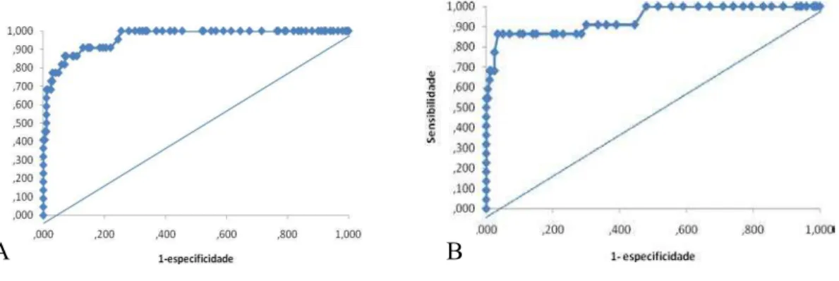
The cut-off point of the ELISA test was defined by the construction of the receiver operator characteristic (ROC) curve, using confidence intervals of 95% and 99%, according to the specifications of Fletcher & Fletcher [26] and Frei et al.. [27]
Accuracy parameters
The accuracy parameters were determined in 22 patients with CPA and in 200 healthy individuals (controls) to determine the sensitivity, specificity, positive predictive value (PPV), negative predictive value (NPV), accuracy (Ac), positive likelihood ratio (PLR) and negative likelihood ratio (NLR), also called verisimilitude ratio, according to the specifications of Fletcher & Fletcher. [26]
PZ Azevedo 22 Cross-reactivity with sera from patients with other granulomatous diseases
The sera of 20 patients with CRC, 23 patients with HST, 28 patients with TB and 50 patients with PCM were used to test the cross-reactivity ratio with diseases that can be considered part of the differential diagnosis of CPA.
Ethics research committee
The project was approved by the Institutional Ethics Research Committee (Process no. 210.781-CEP). The informed consent form was signed by the patients included in the prospective study.
Statistical analysis of results
The likelihoods in dependent populations were compared by Cochran’s Q test, according to the specifications of Curi [29], followed by McNemar’s test, according to Siegel [30]. The degree of agreement between both the two tests was evaluated using the kappa coefficient, according to the specifications of Landis et al. [31]. The kappa coefficient was interpreted as follows: (a) poor: when below 0.00; (b) slight: between 0.00 and 0.20; (c) fair: between 0.21 and 0.40; (d) moderate: between 0.41 and 0.60; (e) substancial: between 0.61 and 0.80; and (d) almost perfect: between 0.81 and 1.00 [31]. For each statistical test, the differences were considered significant when p ≤ 0.05.
Results
PZ Azevedo 23
Case reports
Four cases of AFB were selected to be presented because of their clinical characteristics.
Case 1.
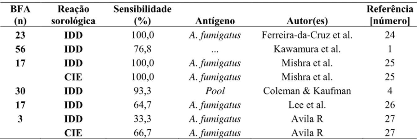
A 80-year-old, white female was hospitalized with a 3-year history of cough with hemoptysis, accompanied by weight loss of 4 kg during this period. She denied previous pulmonary disease and smoking. She reported congestive heart failure and stable angina. The patient weighed 32 kg at admission. Chest CT scan showed cavitary lesions with internal solid content, which were suggestive of aspergilloma (Figure 2). The sputum and bronchoalveolar lavage cytopathology, and lung histopathological examination of tissue obtained by transbronchial biopsy showed the presence of typical Aspergillus spp. structures.
The detection of anti-A. fumigatus antibodies, performed by DID test, was positive (1:128).
PZ Azevedo 24
Figure 2- Chest computed tomography showing areas of parenchymal destruction, decreased vascularization and fibrotic tissue in the right lung apex and left upper lobe, where distortions due to thick-walled cavitary lesions are observed, with internal radiodense content, forming the air crescent sign (arrow), suggestive of fungus ball.
Case 2.
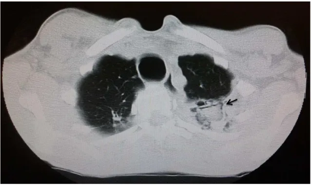
A 44-year-old, mulatto male, farmer, was admitted to the hospital with a 6-month history of productive cough with yellow sputum, dyspnea at rest and weight loss of 13 kg. He also had low evening fever (38°C) for two months and hemoptysis for eight days after admission. He reported two previous episodes of TB, two years before and one year before. A chest CT scan showed areas of consolidation or fibroatelectasis associated with bronchiectasis affecting apical and posterior regions of the left lung, and a cavitary lesion with internal solid content (Figure 3). Seven cytopathological examinations of the sputum and lung histopathology of tissue obtained by transbronchial biopsy showed the presence of typical
Aspergillus spp. structures; and one of the cytopathological examinations also showed the
presence of acid-fast bacilli, Ziehl-Neelsen stained, revealing simultaneous aspergillosis and TB. The detection of anti-A. fumigatus antibodies performed by DID test was negative.
PZ Azevedo 25
months of treatment, the anti-TB regimen was doubled to rifampicin 600 mg and isoniazid 300 mg, which was maintained for 7 months. After discontinuation of these drugs, the daily dose of itraconazole was reduced to 400 mg, which is being maintained. The patient showed clinical cure 10 months after introduction of the antifungal and anti-TB therapy.
Figure 3 - Chest computed tomography showing a consolidation area in the posterior apical segment of the left upper lobe, where a rounded shape lined by a small gas band can be observed, which is characteristic of the air crescent sign (arrow), suggestive of fungus ball. Note also the irregular interlobular septal thickening with signs of architectural distortion.
Case 3.
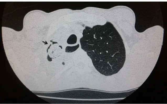
A 51-year-old white male, farmer, was hospitalized with a 20-day history of productive cough and yellowish expectoration, low evening fever and dyspnea on great exertion. A smoker and drinker since he was 15 years old, he reported that two years before he had community bacterial pneumonia caused by Klebsiella pneumoniae. He denied previous
TB. The chest CT scan showed destruction of the right upper lobe and a large apical cavity filled by a fungus ball (Figure 4). The bronchoalveolar lavage cytopathology was negative for fungi and mycobacteria, but the histopathological examination of the lung tissue obtained by transbronchial biopsy, showed the presence of typical Aspergillus spp. structures. The
PZ Azevedo 26
the clinical condition of the patient, drug treatment with itraconazole at a daily dose of 400 mg orally was chosen. The patient progressed with significant clinical improvement and remains under treatment, but still has a cough.
Figure 4 - Chest computed tomography showing atelectasis of the entire right upper lobe associated with bronchiectasis and interposed cavitations. Inside one of the cavities, a rounded image is observed, with a sponge-like aspect, small air spaces inside and the air crescent sign (arrow), suggestive of fungus ball.
Case 4.
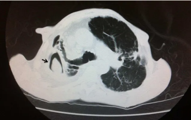
A 72-year-old white male, self-employed, admitted with dyspnea on minimal exertion, associated with productive cough, night sweats and weight loss of 10 kg in two years. A former smoker, he reported two previous episodes of TB, treated 43 and 36 years before. Chest CT scans showed severe destruction of the right upper lobe, with cavitations, one of them filled by a fungus ball (Figure 5). The bronchoalveolar lavage was positive for acid-fast bacilli, Ziehl-Neelsen satained. The lung tissue histopathology, obtained by transbronchial biopsy, showed the presence of typical tissue Aspergillus spp. structures, in addition to Actinomyces sp. accumulation associated with focal calcification. The detection of anti-A. fumigatus antibodies, conducted by DID test, was positive (1:32). Due to severe impairment
PZ Azevedo 27
and itraconazole at a daily dose adjusted to 600 mg orally due to its interaction with rifampicin. To treat the actinomycosis, crystalline penicillin G was administered at a dose of 5,000,000 OU intravenously every 4 hours for 10 days, followed by amoxicillin at a dose of 500 mg orally every eight hours. The patient, still under treatment, has progressed with partial improvement of symptoms.
Figure 5 – Chest computed tomography showing important architectural destruction of the right upper lobe, with deviation of the trachea and mediastinum, presence of bronchiectasis and cavitations, one of which has an oval image bordered by a gaseous contour, characteristic of the air crescent sign (arrow) suggestive of fungus ball.
The procedure most frequently performed in these patients was transbronchial biopsy. The identification of Aspergillus spp. and pulmonary fibrosis were the most common
PZ Azevedo 28 Table 1 – Characterization of the 25 evaluated patients with chronic pulmonary aspergillosis
TB: tuberculosis; PCM: paracoccidioidomycosis; CT: computed tomography; DID: double agar gel immunodiffusion test; TB: transbronchial biopsy ; RSL: resection segment or lobe; ITZ: itraconazole; AMB: amphotericin B; VCZ: voriconazole.
Determination of the cut-off point
The cut-off points were an OD of 0.120 and 0.130 when the A. fumigatus antigen (A)
was used and an OD of 0.090 and 0.100 with the Aspergillus spp. antigen pool (B) and
confidence intervals of 95% and 99%, respectively.
Figure 6 – Receiver operator characteristic curve obtained for determining the cut-off point of the serum level of anti-Aspergillus antibodies, using A. fumigatus antigen (A) and A. fumigatus, A. flavus and A. niger antigen pool
(B), based on 22 patients with chronic pulmonary aspergillosis and 200 healthy blood donors from the same region.
Age Median= 55 (33 - 80)
Sex Male= 20 (80%) Female= 5 (20%)
Underlying diseases TB= 19 (76%) PCM= 2 (8%) Pneummonia= 1 (4%) not specified= 3 (12%)
Clinical manifestations
Weight loss= 72,7% Expectoration= 50,0% Cough= 77,3% Fever= 27,3% Dyspnea= 50,0% Haemoptysis= 63,6% Chest pain= 22,7%
Planigraphy and chest CT BF= 20 (80 %) F=4 (16 %) C= 1 (4 %)
Cytopathological sputum 4/14 = 28,6%
Cytopathological LBA 7/12 = 58,3%
DID admission 13/20 = 65%
Cirurgical procedures BT = 7/ 23 (30,4%) ESL= 4/23 (17,4%)
Antifungal treatment ITZ = 17/19 (89,5%) AMB= 3/19 (15,8%) VCZ= 1/19 (5,3%)
PZ Azevedo 29 Determination of the serum dilution to be used
The serum dilution was chosen by comparing the cut-off values obtained using sera diluted to 1/25, 1/50 and 1/100. The cut-off values, presented as the mean and standard
deviation, were as follows: a) 1/25 dilution: 0.130 ± 0.0237; b) 1/50 dilution: 0.113 ± 0.0215; c) 1/100 dilution: 0.121 ± 0.0313. These values did not differ, so the Aspergillus fumigatus
antigen (p = 0.26) was used. The cut-off values with the A. fumigatus, A. flavus and A. niger
antigen pool were as follows: a) 1/25 dilution: 0.0796 ± 0.0118; b) 1/50 dilution: 0.0874 ± 0.0239; c) 1/100 dilution: 0.0854 ± 0.0159. These values did not differ (p = 0.49), so the serum diluted at 1/100 was chosen because the volume spent in the reactions would be lower.
Accuracy parameters
The use of an antigen pool led to a trend of increasing DID sensitivity of 14%, while
the other accuracy parameters remained unchanged (Table 2). A positive DID test increased by 90.9 to 118.2 times the likelihood of the pre-test diagnosis being aspergillosis, depending on the antigen used (Table 2). The DID sensitivity in patients with AFB (76.5%) was greater than that of patients with CPA, assessed as a whole.
In the ELISA, using a higher cut-off point led to decreased sensitivity for both types of antigens but also to increased PPV and PLR (Table 2). In addition, the use of the antigen pool also led to increased PPV and PLR but decreased sensitivity when the cut-off was higher (Table 2). The ELISA showed higher sensitivity than DID. Regardless of the antigen preparation used, a positive DID test increased much more the chance of pre-test diagnosis than did a positive ELISA test, as indicated by the PLRs (Table 2). Finally, using the higher
cut-off, there was decreased sensitivity, which approached the one presented by DID, but with
PZ Azevedo 30 Table 2 – Accuracy parameters of double agar gel immunodiffusion test - DID and enzyme-linked immunosorbent assay - ELISA, calculated on the evaluation of 22 patients with chronic pulmonary aspergillosis and 200 healthy individuals. Influence of antigen preparations and
cut-offs used in the ELISA test
Serological tests S (%) E (%) PPV (%) NPV (%) PLR (CPRL) NLR
DID 1 45,5 100,0 100,0 93,3 90,9 0,5 DID 2 59,1 100,0 100,0 95,7 118,2 0,4 ELISA 1 (0,120) 81,8 94,0 60,0 97,9 13,6 0,2 ELISA 1 (0,130) 72,7 97,0 76,2 97,0 29,1 0,3 ELISA 2 (0,090) 86,4 96,5 73,1 98,5 24,7 0,1 ELISA 2 (0,100) 59,1 99,5 92,9 95,7 118,2 0,4
DID:double agar gel immunodiffusion; ELISA: enzyme-linked immunosorbent assay; 1: antigen the
Aspergillus fumigatus; 2: pool de antigens the A. fumigatus, A. flavus e A. niger; 0,120, 0,130, 0,090 e 0,100 – cut-off values; S: sensitivity, E: specificity; PPV / NPV: positive and negative predictive values; PLR: positive
likelihood ratio; CPRL: corrected positive likelihood ratio; NLR: negative likelihood ratio.
Cross-reactivity
The detection of anti-Aspergillus antibodies by DID test in sera from patients with
PZ Azevedo 31 Table 3 – Prevalence (percentage) of cross-reactions observed in double agar gel immunodiffusion test- DID and enzyme-linked immunosorbent assay - ELISA. Study of 28 patients with tuberculosis, 23 with histoplasmosis, paracoccidioidomycosis with 50 and 20 with cryptococcosis. Influence of antigen preparation and cut-offs used in the ELISA test.
Disease number Patient DID 1 DID 2 ELISA 1 (0,120) ELISA 1 (0,130) ELISA 2 (0,090) ELISA 2 (0,100) TBC 28 0,0 0,0 21,4 10,7 10,7 0,0
[0,121 - 0,128] [0,138 - 0,156] [0,093 - 0,099]
HST 23 0,0 8,7 30,4 8,7 21,7 13,0
[undiluted - 1/4] [0,120 - 0,128] [0,132 - 0,138] [0,091 - 0,094] [0,103 - 0,115]
PCM 50 2,0 0,0 40,0 36,0 62,0 52,0
[undiluted] [0,120 - 0,128] [0,131 - 0,181] [0,091 - 0,097] [0,101 - 0,140]
CRC 20 0,0 0,0 0,0 0,0 20,0 0,0
[0,091 - 0,098]
DID: double agar gel immunodiffusion test; ELISA: enzyme-linked immunosorbent assay; 1: antigen of
Aspergillus fumigatus; 2: pool of antigens: A. fumigatus, A. flavus e A. niger, ( ) cut-off the test; TBC:
tuberculosis; HST: histoplasmosis; PCM: paracoccidioidomycosis; CRC: cryptococcosis; [ ] range.
Comparison of the sensitivity of the tests
The comparison of the sensitivity of the different diagnostic tests indicated higher frequencies for ELISA using both types of antigen and the lowest frequency for DID1 with A.
fumigatus antigen. GMN and DID2 showed intermediate frequency, which did not differ from
the others (Table 4).
The agreement between the positivity of the GMN test and the detection of antibodies was always small, regardless of the method and the antigen used. The strongest agreement was observed between the ELISA1 and ELISA2. The agreement between the DID
PZ Azevedo 32
b b ab a a
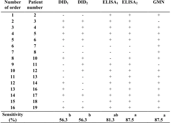
Table 4 - Qualitative results of serological tests performed in 16 patients with chronic aspergillosis. Comparisons carried out among the double agar gel immunodiffusion test (DID) and the enzyme-linked immunosorbent assay (ELISA) using two antigenic preparations, and galactomannan (GMN). Comparisons performed by Cochran Q test, McNemar test, and the bionomial test.
Number
of order number Patient DID1 DID2 ELISA1 ELISA2 GMN
1 2 - - + + +
2 3 + + + + -
3 4 + + + + +
4 5 + + + + +
5 6 + + + + +
6 7 - - - - +
7 8 - - - - +
8 10 + + - + +
9 11 - + + + -
10 12 - + + + +
11 13 - - + + +
12 14 + - + + +
13 16 + - + + +
14 17 + + + + +
15 18 - - + + +
16 19 + + + + +
Sensitivity
(%) 56.3 56.3 81.3 87.5 87.5
- no reagent; +: reagent.
1-Aspergillus fumigatus antigen
2-Pool of antigens from Aspergillus fumigatus, Aspergillus niger and Aspergillus flavus
Q=119.34 (p<0.00001)
Frequencies followed by the same letter do not differ (p>0.05); frequencies followed by different letters are statistically
different (p≤0.05) presented a tendency a statistical difference (0.05 < p ≤ 0.10).
Comparisons, 2x2, of diagnostic method as to sensitivity. Significance represented by p value.
(56.3%) DID1 (56.3%) DID2 (81.3%) ELISA1 (87,5%) ELISA2 (87,5%) GMN DID1 ... 0.36 0.11 0.03 0.06
DID2 ... ... 0.11 0.03 0.09
ELISA1 ... ... ... 0.50 0.50
PZ Azevedo 33
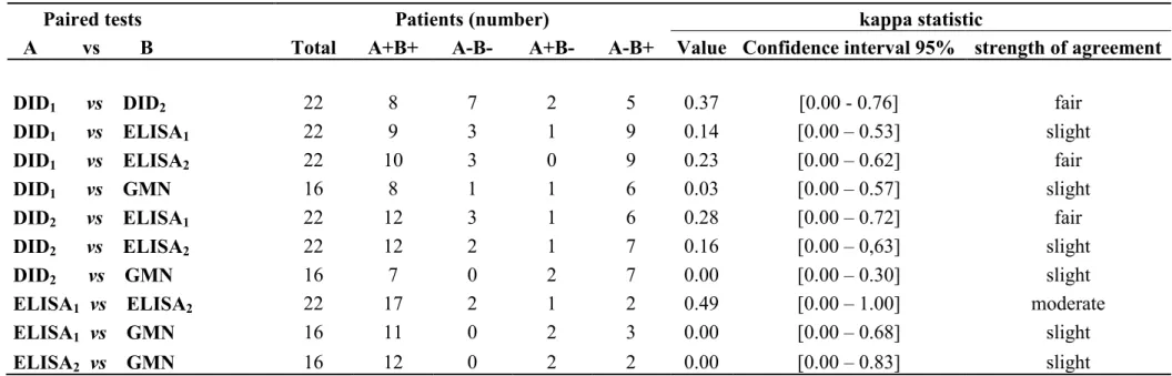
Table 5 - Degree of agreement of diagnosis tests in 22 patients with chonic aspergillosis. Comparison 2x2 using the kappa test.
Paired tests Patients (number) kappa statistic
A vs B Total A+B+ A-B- A+B- A-B+ Value Confidence interval 95% strength of agreement
DID1 vs DID2 22 8 7 2 5 0.37 [0.00 - 0.76] fair
DID1 vs ELISA1 22 9 3 1 9 0.14 [0.00 – 0.53] slight
DID1 vs ELISA2 22 10 3 0 9 0.23 [0.00 – 0.62] fair
DID1 vs GMN 16 8 1 1 6 0.03 [0.00 – 0.57] slight
DID2 vs ELISA1 22 12 3 1 6 0.28 [0.00 – 0.72] fair
DID2 vs ELISA2 22 12 2 1 7 0.16 [0.00 – 0,63] slight
DID2 vs GMN 16 7 0 2 7 0.00 [0.00 – 0.30] slight
ELISA1 vs ELISA2 22 17 2 1 2 0.49 [0.00 – 1.00] moderate
ELISA1 vs GMN 16 11 0 2 3 0.00 [0.00 – 0.68] slight
ELISA2 vs GMN 16 12 0 2 2 0.00 [0.00 – 0.83] slight
DID: Double agar gel immunodiffusion test; ELISA: enzyme-linked immunosorbent assay; GMN: Galactomannan
1- Aspergillus fumigatus antigen
PZ Azevedo 34 Evaluation of the serological follow-up of patients under treatment with itraconazole
The regression curves obtained in the follow-up of patients with positive initial serology according to DID1 and DID2 exhibited the same pattern as a function of treatment
time, with decreased serum levels (p = 0.01). The curves obtained from ELISA were different from those observed with DID because they showed a slight increase in serum antibody levels, more evident when the A. fumigatus antigen was used (p < 0.00001), as shown in
Figure 7.
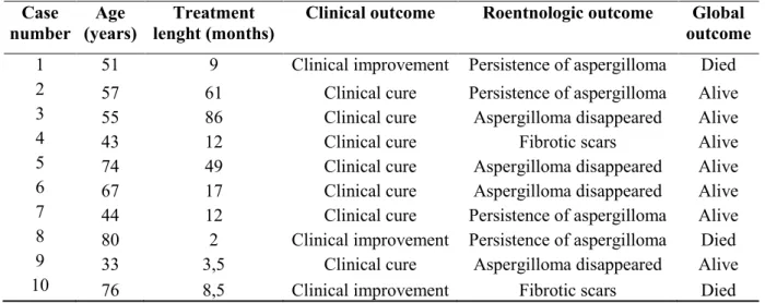
The characterization of the 10 patients evaluated and treated showed clinical cure or improvement in all cases, but with persistence of aspergilloma in three patients (Table 6). The mortality rate was 30%; two patients showed persistence of aspergilloma and the third, fibrotic scars (Table 6).
Table 6 – Progress of 10 patients after treatment, as to age and clinical, roentnologic, and global outcome.
Case
number (years) Age lenght (months) Treatment Clinical outcome Roentnologic outcome outcome Global
PZ Azevedo 35
Figure 7 - Regression analysis representing changes in serum levels of anti-Aspergillus antibodies as a function
of the antifungal treatment period in 10 patients with chronic pulmonary aspergillosis. (A) Curve representing the progression of serum levels determined by DID test with the A. fumigatus antigen and Aspergillus spp.
antigen pool. (B) Curve representing the progression of serum levels determined by ELISA using A. fumigatus
antigen and Aspergillus spp. antigen pool. A
PZ Azevedo 36 Clinical progress. Comparison with double agar gel immunodiffusion test.
Clinical follow-up, evaluated by using complaints scores, showed evident clinical improvement after introduction of antifungal treatment, as it was observed with the decreasing antibodies serum levels, determined by DID test. However, the clinical curve was different from the DID test ones, taken together (Figure 8).
Figure 8 - Regression analyses showing the decreasing antibody serum levels anti-Aspergillus, and evident
clinical improvement after introduction of antifungal treatment. The regression curves are different.
Discussion
The host-parasite interaction between Aspergillus spp. and humans is highly diverse.
In patients with neutropenia, this interaction presents as invasive pulmonary aspergillosis; in some hyperergic patients, as allergic bronchopulmonary aspergillosis; in patients with no obvious cause of immunosuppression, as CPA. The latter has been frequently observed in patients with pulmonary sequelae, such as those observed in pulmonary TB [5] and in chronic obstructive pulmonary disease [32]. In areas where HST is hyperendemic, CPA is widely reported [33]. AIDS patients, whose immune deficiency is linked to the destruction of CD4+ T
PZ Azevedo 37
CPA has several clinical manifestations: aspergilloma, also called AFB, CCPA and CFPA. The pulmonary sequelae caused by TB are an important antecedent of CPA [5,35]. Worldwide, more than 36 million people have been cured of TB from 1995 to 2008, and 9 million new cases per year have been diagnosed. The British Thoracic and Tuberculosis Association noted that 6% of patients with open-scar tuberculous cavities developed aspergillomas in three years, with a mortality of 6% per year [36-38]. In another study, the mortality rate was 31% in 5 years and 56% in 10 years; of the 27 patients who died, 3 of them died due to hemoptysis, 7 due to surgical complications, 6 due to chronic respiratory failure, 6 due to acute pneumonia and 5 due to chronic suppurative pneumonia [36]. In Brazil, in 2003, the incidence of TB was of 44.4 new cases/100,000 inhabitants. In 2013, 71,123 new cases were reported, with an incidence of 35.4 cases/100,000 inhabitants [37]. All these data indicate the importance of CPA for public health and the high number of patients with underlying diseases that favor its appearance. These data also suggest that the prevalence of CPA in our environment must be much higher than the number of cases referred to university hospitals and therefore that many of them have not been diagnosed.
The cases reported presented some special aspects. Although AFB is usually observed in residual parenchymal lesion of the lungs, a case with active TBC was reported, confirming previous reports [39,40]. In addition, the co-morbidity among TBC, AFB and actinomycosis was also reported. It should be considered that Actinomyces sp. can also colonize a pulmonary
cavity, being a differential diagnosis of AFB. The negative inotropic effect caused by itraconazol presented by a patient, as it was previously reported. [41]
However, it should be noted that simple identification in the sputum of fungi that colonize the bronchial tree is not confirmation of the etiologic diagnosis of lung injury, which occurs with Cryptococcus and Aspergillus spp. The solution to this problem depends on
surgical procedures, such as lung biopsy or resection of lung injury and serological tests. Surgical procedures are often aggressive and sometimes contraindicated. Serological tests can be performed using several methods and different antigen preparations. In addition, the presence of GMN and IgG in the serum of these patients can be evaluated.
PZ Azevedo 38
fumigatus, A. flavus and A. niger antigen pool) and qualitative detection of serum GMN in
patients with CPA. Moreover, we evaluated the serological progression of the patients after introduction of the antifungal treatment.
Several serological methods, such as complement fixation reactions, gel precipitation reactions, latex particle agglutination, electrophoretic tests and enzymatic immunoassays have been evaluated for the diagnosis of aspergillosis [9-39]. The immunodiffusion reaction in agar gel is the most used method due to the simplicity of execution and reproducibility [40], which are the reasons that led to its standardization for the detection of anti-Aspergillus antibodies in
most clinical laboratories. However, the antigen concentration required to detect precipitins in the DID is a critical factor of this reaction [41], which can only be read 96 hours after the slides are prepared. With the advent of enzyme immunoassays, the introduction of ELISA test has been encouraged in routine laboratories, given its sensitivity, speed of execution and direct measurement of antibody levels. [25,39-42]
In the present study, the prevalence of patients with AFB among those with CPA, the males, and those with cough, weight loss and hemoptysis as clinical manifestations of pulmonary TB as pre-existing disease confirms the findings of other authors [19,36-38]. The use of serum diluted to 1/100 aimed to better use the serum of each patient, given that the results did not differ from those observed at 1/25 and 1/50 dilutions. The sensitivity of DID test observed in patients with AFB in the present study (76.5%) was lower than that found by other authors (91.0 to 98.0%) [9,43-45]. The finding of high specificity and PPV confirms those same studies [9,43-45]. The PLR or verisimilitude ratio, which was not evaluated by other authors, was very high, which makes the post-test likelihood almost a certainty. This fact justifies defining a confirmed case in patients who have positive DID test with suggestive clinical and radiological manifestations.
PZ Azevedo 39
The antigen preparation used is essential to evaluate the accuracy of serological tests [9]. Because different preparations were used in different studies, comparisons cannot be direct. Nevertheless, the overall set of results can be considered harmonious. The use of an
Aspergillus spp. antigen pool is well indicated because cases of aspergillosis by A. fumigatus, A. flavus, A. niger, A. nidulans and A. terreus have been described, though with a
predominance of the first. [9,43-44,46-47]
The cross-reactivity measured in sera from patients with PCM, HST, TB and CRC were infrequent with DID test, present only with HST and PCM. In contrast, ELISA test showed false-positive results with HST, TB and PCM, and ELISA2 test showed false
positives with the four granulomatous diseases. These findings can be explained by the production of GMN by P. brasiliensis, H. capsulatum and C. neoformans [48-54].
Cross-reactivity with sera from patients with active TB must be carefully evaluated because TB is often the underlying disease of CPA and anti-Mycobacterium tuberculosis antibodies may
persist for a long time.
The serum GMN detection showed the same sensitivity as ELISA test. However, this finding must be carefully interpreted due to the possibility of cross-reactivity with other fungal diseases that also affect the lungs [48-54]. Interestingly, Aspergillus spp. produce
GMN, a complex polysaccharide composed of D-galactose and D-mannose, and C. neoformans also contains a capsular polysaccharide complex, composed of
glucuronoxylomannan (GXM) and GMNs. GXM is found in body fluids, and its identification allows the diagnosis of CRC [55]. Despite the apparent similarity between these polysaccharides, the cross-reactivity in sera from patients with CRC was much less frequent than in patients with HST or PCM.