Review Article
An Historical Perspective on How Advances in Microscopic
Imaging Contributed to Understanding the
Leishmania
Spp.
and
Trypanosoma cruzi
Host-Parasite Relationship
P. T. V. Florentino, F. Real, A. Bonfim-Melo, C. M. Orikaza, E. R. Ferreira, C. C. Pessoa,
B. R. Lima, G. R. S. Sasso, and R. A. Mortara
Departamento de Microbiologia, Imunologia e Parasitologia, Escola Paulista de Medicina, UNIFESP, Rua Botucatu 862, 6th Floor, 04023-062 S˜ao Paulo, SP, Brazil
Correspondence should be addressed to R. A. Mortara; [email protected]
Received 3 December 2013; Accepted 10 January 2014; Published 27 April 2014
Academic Editor: Wanderley de Souza
Copyright © 2014 P. T. V. Florentino et al. his is an open access article distributed under the Creative Commons Attribution License, which permits unrestricted use, distribution, and reproduction in any medium, provided the original work is properly cited.
he literature has identiied complex aspects of intracellular host-parasite relationships, which require systematic, nonreductionist approaches and spatial/temporal information. Increasing and integrating temporal and spatial dimensions in host cell imaging have contributed to elucidating several conceptual gaps in the biology of intracellular parasites. To access and investigate complex and emergent dynamic events, it is mandatory to follow them in the context of living cells and organs, constructing scientiic images with integrated high quality spatiotemporal data. his review discusses examples of how advances in microscopy have challenged established conceptual models of the intracellular life cycles ofLeishmaniaspp. andTrypanosoma cruziprotozoan parasites.
1. Introduction
Leishmaniasis and Chagas disease are tropical diseases caused by protozoan parasites from the Trypanosomatidae
family (Leishmaniaspp. andTrypanosoma cruzi, resp.). hese
protozoans belong to the class Kinetoplastea, a group of lagellated organisms with a peculiar organelle called a
kine-toplast and a single mitochondrion [1]. hese two
trypanoso-matids are responsible for approximately 20 million reported cases of leishmaniasis and Chagas disease and 100,000 deaths per year, primarily in tropical and subtropical areas of the
globe [2]. he negative economic and social impact of these
diseases, especially in Central and South America, is of great
concern [3] and has stimulated scientiic investments into
studying their causative agents. Because the pathogenesis of
Leishmania spp. and T. cruzi involves an intracellular life cycle in human and mammalian hosts, interactions between
the parasite and host cells have been extensively studiedin
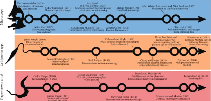
vitro, with particular emphasis on microscopic observations. A timeline showing important historical achievements in
microscope technology andLeishmaniaspp./T. cruzi
knowl-edge is presented inFigure 1.
Remarkable technological advances have increased our ability to sense or experience microscopic agents, buildin-goncepts from scientiic images. Researchers “embody” tech-nology, boosting his/her experience: scientiic images are obtained ater technological mediation between researchers sensorial apparatus (perception) and the object of study
[4]. Increased spatial resolution with the advent of electron
microscopy (EM) enabled access to high quality spatial data for studying the relationship between host cells and pathogens. EM was, and still is, extremely important in determining how viruses, bacteria, fungi, and protozoan
parasites (such asLeishmaniaspp. andT. cruzi) interact with
host cells. However, the singularity of temporal data and lack of integration between high spatial resolution and access to the same individual at diferent time points (due to chemical ixation of samples) led to a fragmented experience of the object and, unfortunately, limitations in a full understanding of how parasites establish and propagate themselves within
their hosts (Box 1).
Factual statements (singular propositions) fragmented in space and time can produce temporal, spatial, and causal gaps
Van Leeuwenhdek (1673)
Visualization of bacteria
and protozoa Oskar Heimstadt (1911) Fluorescence microscope
E. Ruska and M. Knoll (1931) Transmission electron microscopy
Marvin Minsky (1955) Confocal microscope
Alberts Coons (1942)
Immunofluorescence Real-time confocal microscopy using spinning disk
Carlos Chagas (1909) Identifcation ofT. cruzi
Meyer and Barasa (1940) First microcinematographies
of the parasite
Dvorak and Hyde (1973)
Establishment of ive phases of host cell infection by cinematography
Fernandes et al. (2013) Spinning disk Max Knoll
and Fritz Zernike (1935)
Scanning electron microscope and phase contrast microscopy
Julius Ries (1907) Microcinematography
(sea urchins)
John White, Brad Amos and, Mick Fordham (1987) Realization of confocal microscopy
James Wright (1903) Observations of cutaneous lesions
Pulvertaf and Hoyle (1960) Phase contrast microcinematography
(internalization)
Veras, Pouchelet, and Rabinovitch (1994) Microcinematography of parasite cell-to-cell transfers
Samuel Christopher (1904)
Observations of Ruth Lofgren (1950) Transmission electron microscopy
Forestier et al. (2011)
Real and Mortara (2012)
Spinning disk and resonant scanning
Chang and Dwyer (1978) Transmission electron microscopy
of parasitophorous vacuoles
Peters et al. (2008)
Multiphoton intravital imaging
Gaspar Viana (1911)
Conceptualization of parasite’s life cycle
Meyer and Porter (1954)
Transmission electron microscopy
Schenkman and Mortara (1992) Xiao et al. (1988)
infected spleens
Confocal microscopy application
T
ry
pa
n
os
om
a cr
uzi
Leis
h
m
an
ia
sp
p
.
M
icr
os
co
p
y
Figure 1: Timeline showing important historical achievements in microscope technology andLeishmaniaspp./T. cruziknowledge. References from the timeline are shown in the text, and additional references are cited in the igure [10,11], revised in [12,13].
All things must pass; objects are subdued to time and space—these riddling categories have been a matter of intense philosophical and scientiic debate since Aristotle (384-322 BC). A Newtonian perspective assumes that time is an independent entity that passes regardless of physical/chemical changes or an external observer. For Immanuel Kant (1724–1804), time, space, and causality are contained in the experience itself, pertaining essentially to the functioning of the mind [5]. his triad corresponds
to the intrinsic properties of the intellect, which experiences not the reality of the world (conined to experimentally unreachable “things-in-themselves”), but what our senses impose relative to the world we know. To sense time and space as an experimenter is to confer to the external world (and objects of study) a “borrowed human logic, in particular a spatiotemporal pattern which is only human perception in disguise” [5]. his spatiotemporal pattern allows us to put objects of study in a causal logic, explaining past and predicting future events, and interpreting them as goal-directed, or teleological, phenomena [6].
Time and space are problematic categories to the human experience because there is a multiplicity of scales deined by diferent clocks (from subatomic to biological and chronological time) and spatial units in which a plethora of things of human interest are conined,spared from a direct sensorial experience. his is the case of pathogenic microorganisms, hidden from direct human experience and unknown to men until the technological advent of microscopes by Leewenhoek (1632–1723) and the conceptual revolution of the germ theory of disease suggested around the 19th century [7].
Several human pathogens were identiied in the late XIX century ater biomedical institutions had, as a priority, elucidated pathogen life cycles and disease etiology. hen and now, the main scientiic methodological approach to obtain experimental evidence on the life cycles of pathogens has been reductionism, the division of complex systems into smaller intelligible parts. he conceptual framework of a pathogen life cycle has been constructed by a mosaic of separate observations on single factors acquired at deined time points in a deined geographical or physiological location, generally without continuous
observation of the same individual (host or pathogen). Joint analysis of each factor could account for interpretation of the entire system; similarly, single spatiotemporal coordinates accessed before and ater an experimental condition could explain causality. Although it is undeniable that the reductionism paradigm has been responsible for the success of modern science and
technological advances in our society, it “oten disregards the dynamic interaction between parts,” and a complex problem “is oten depicted as a collection of static components” [8]. he notion of space is also dismembered from time in reductionist approaches, and important concepts related to the disambiguation of scientiic images, such as topology and interaction of objects, lack dynamic information and can produce or exacerbate “gaps in experience.” Considering the unpredictability, uniqueness, and structural/dynamic complexity of organisms [6], reducing time and space in disconnected parts in order to understand biological phenomena has led to limitations in scientiic investigation and inadequacy of medical conduct [8].
in experiences, which may be solved by constructing con-ceptual models using solid statistical historical fundamental principles. Due to partial agreement with nature, models have an important predictive power (although to a limited extent) in building an interpretative framework for other researchers until new information (obtained ater technologi-cal improvements) challenges and rebuilds these frameworks
[9]. he life cycles of protozoan parasites, from invasion and
colonization to spreading within the host, are conceptual models based primarily on a reductionist approach that considers nonintegrated time and space observations.
Live recordings of the host-pathogen relationship have been produced as microcinematographic and video technol-ogy has progressed, but the large majority of these stud-ies lack appropriate spatial resolution to observe detailed aspects of the interaction. Integrated or four-dimensional observation of objects approximates our experience to micro-scopic dynamic states, such as oscillatory or chaotic behav-ior, that are unreachable under the conceptual frameworks of static stability and conventional imaging technology, ixed at deined time points or contained in limited
spa-tial/topological regions of the sample [8].
Herein we useLeishmaniaspp. andT. cruzias examples
of how advanced microscopic techniques are circumventing reductionism, integrating or reaching further dimensional scales, and unveiling new aspects of host cell-parasite rela-tionships. Observations of these protozoan parasites will be discussed from a historical point of view considering breakthrough studies and acquisition of new information based on integrated spatiotemporal data.
2. Imaging
Leishmania
spp. and Host Cells
In 1881, Alphonse Laveran (1845–1922) found that a proto-zoan was the etiological agent that caused malaria, which encouraged researchers in the ield of protozoology to describe and investigate protozoan pathogens transmitted to human hosts, especially those carried by insect vectors. his conjuncture led to the investigation of an ancient human malady described in diverse manners in antiquity
and modern times [22, 23]. Discovery of the etiological
agent that causes leishmaniasis, a protozoan parasite from the Trypanosomatidae family, and conceptualization of its life cycle were established from key observations in accor-dance with Koch’s postulates and paradigms of infection and pathogenesis: identify and isolate the microorganisms,
cultivate them in vitro, and establish a causal relationship
with disease. In microscopic observations of Delhi boils, Scottish Surgeon Major David Douglas Cunningham (1843– 1914) found a round-shaped parasite inhabiting cells, and Piotr Borovsky (1898), who observed similar skin lesions (Sart Sore, Turkmenistan), suggested that the intracellular bodies were protozoans. William Leishman (1865–1926) and Charles Donovan (1863–1951) found similar organisms in tissues extracted from the viscera of fatal cases of
kala-azar in India. Attempting to cultivate these organisms in
vitro, Leonard Rogers (1868–1962) and Charles Nicolle (1866– 1936) extracted the round-shaped protozoans from infected
tissues and cultivated them in blood agar culture media. Multiplying lagellated protozoan forms were found in the culture medium, which led to the conclusion that the parasite was a trypanosomatid. Edmond Sergent (1876–1969) and colleagues found that trypanosomatids could be digenetic
parasites, transmitted from insects to mammals [24], and
suggested the same life cycle for those protozoans, which
were then classiied asLeishmania.In vitrocultivation of these
parasites allowed their inoculation into dogs, monkeys, and small rodents, which subsequently developed pathologies similar to the human disease. In 1921, it was experimentally
demonstrated thatPhlebotomus, a tiny sand ly, is the insect
host forLeishmaniaand the transmitter of leishmaniasis [24,
25].
Wright (1869–1928) in 1903 [26] and Christophers (1873–
1978) in 1904 [27] observed that cutaneous lesions or infected
spleens presented massive iniltration of cells containing a large number of oval-shaped parasites. Christophers was the irst to recognize these preferentially infected cells as macrophages, inferring that phagocytosis was responsible for
the uptake of parasites by leucocytes [26, 27]. For decades,
leishmaniasis was considered a disease almost exclusively
of the host macrophage system [28], and phagocytosis is
still considered the primary mechanism ofLeishmaniaspp.
internalization [29].
Pulvertat and Hoyle [30], 56 years ater
Christo-pher’s inferences, recorded the phagocytosis of
Leishma-nia spp. by monocytes/macrophages. Using phase contrast
live microcinematography, the authors described monocyte pseudopodia reaching and taking up leptomonad forms
(now generally called promastigotes) of L. donovani. he
promastigotes display a single lagellum in their anterior poles; Pulvertat and Hoyle demonstrated that promastigote phagocytosis took place from the opposite pole, the posterior, within several minutes. Ater total engulfment, a vacuole is observed around the parasite that may be digested and dis-appear or, alternatively, survives and remains motile within
this compartment. However, Miller and Twohy (1967) [31]
and Akiyama and Haight (1971) [32] found that hamster
macrophage pseudopodia initially internalized promastig-otes by the lagellar anterior pole of the parasite and observed a transient vacuole around it.
Forty years later using 3D and 5D reconstruction images,
Forestier and coworkers (2011) [33] observed thatL. donovani
promastigote uptake by macrophages occurs mainly by the lagellar tip and could also, in exceptional cases, occur through the posterior region and lateral portions of the
body. he authors described four sequential phases of L.
donovanipromastigote establishment in host cells: (i) highly polarized attachment by the lagellar end and internalization in lysosomal compartments; (ii) reorientation; (iii) oscillating movement of the parasite to the periphery of the host cell associated with lysosome exocytosis and minor damage to the host cell; and (iv) loss of motility and inal location of the parasite in parasitophorous vacuoles (PVs) near the host cell nucleus. hese conclusions were only possible due to cutting-edge, high-speed live imaging under modern
micro-scopes [34]. Courret and colleagues (2002) observed similar
macrophages using conventional live imaging techniques of infected samples.
he investigation of Leishmania internalization by
macrophages largely beneited from transmission electron microscopy (TEM). Host cell pseudopodia are formed around entering parasites with concomitant microilament aggregation; sites of close contact between parasite and host cell membranes can be visualized in detail using this
technique [35]. In 1986, Wozencrat and colleagues used
EM to map individual molecules involved in Leishmania
-macrophage interactions. Using immunogold labeling, complement receptors were observed to be associated with the interface between membranes of the macrophage and the
interactingLeishmania,but not with internalized parasites.
hese observations conirmed results published in the same year, demonstrating participation of this receptor in the
direct binding of macrophages toLeishmaniapromastigotes
[36]. It is now recognized that Leishmania internalization
by macrophages is tightly modulated by the irst and third complement receptors (CR1 and CR3) and mannose (MR)
and Fc gamma receptors (Fc�R) [29].
Leishmania internalization by macrophages involves accumulation of actin ilaments at the internalization sites
of the parasite, a feature of phagocytosis [37]. he authors
of the irst studies on the mobilization of host cell com-ponents towards phagocytosed parasites beneited from immunolabeling techniques associated with electron and optical microscopy. he use of antibodies conjugated to luorophores proved to be an easy, accessible technique to
study protein distribution in cell biology [38]. Regarding
Leishmania phagocytosis, luorescence immunolabeling of host GTPases and actin labeling enabled the observation
that these molecules are colocalized during Leishmania
-macrophage interaction [39]. Further, the authors found that
diferent GTPases, Rac1 and RhoA, regulate internalization
of opsonized and nonopsonizedLeishmaniapromastigotes,
respectively. Using the same immunolabeling technique, they also observed that internalization of nonopsonized amastigotes is alternatively regulated by Rac1 but, in this case, the oxidative burst triggered by host phagocytosis
is restrained [40]. hus, diferent receptors (for opsonized
or nonopsonized parasites) trigger diferent GTPases that
modulate host cell responses toLeishmania.
Ater internalization by host cells, Leishmania
para-sites are lodged in PVs, in which they multiply as
oval-shaped amastigotes. Electron micrographs ofLeishmaniaPVs
acquired by Alexander and Vickerman in 1975 and Chang and Dwyer in 1978 demonstrated the phagolysosome-like
nature of the vacuoles developed by this parasite [41, 42].
By loading host cell phagolysosome vesicles with electron-dense compounds, these compounds were observed inside
Leishmania PVs, suggesting that PVs fuse with late endo-somes and secondary lysoendo-somes. In the 1990s, a series of studies from Jean-Claude Antoine demonstrated that PVs acquire early endosome markers such as Rab5 and EEA-1 that are substituted by late endosome markers, such as
Rab7, and glycoproteins associated with lysosomes [43]. he
resulting parasite-containing compartment is a “mature” PV
presenting several phagolysosome features [34,43–46]. PVs
develop diferent morphologies according to Leishmania
species:L. mexicanaandL. amazonensis, for example, present
a spacious PV containing several amastigotes, while most
species (L. major,L. donovani,and others) present a
tight-itting PV in which PV and parasite membranes are in contact
[47, 48]. PV biogenesis is still poorly understood, mainly
because the majority of studies have been performed in ixed cells using endosomal/lysosomal membrane markers.
Spinning disk technology for confocal laser scanning allowed observation of PV biogenesis in live samples from the very early moments of infection at the stage of parasite phagocytosis. Multidimensional images obtained from these techniques allowed for integration of four and even ive
dimensions (x, y, z, time, luorescence) of living cells and
tissues [49]. Lippuner and colleagues [50] were some of
the irst researchers to record PV biogenesis in live samples
using GFP-tagged Rab5 proteins on cells hostingL. mexicana.
he authors demonstrated that the parasite inhabits PVs in which Rab5 GTPases are rapidly excluded from the vacuolar membrane (compared with latex bead phagosomes). hey also documented that a parasite surface component, lipophosphoglycan (LPG), implicated in delaying PV
mat-uration inL. donovani[39] accelerated the exclusion of the
Rab5 marker from PVs.
Beneitting from high resolution and speed, as well as the low photocytotoxicity of the technique, Forestier and
colleagues and Real and Mortara [33, 48] observed the
interaction of PVs with acidiied compartments of host cells. hey dyed vesicles with a lysosomotropic probe (Lysotracker) over time and observed how these labeled vesicles compose PVs. hese acidic vesicles were located around internalized promastigotes minutes ater interaction with host cells, sug-gesting that recently formed PVs promptly fuse with acidic
compartments [33]. he biogenesis of spacious/communal
PVs formed by L. amazonensis versus tight-itting PVs
formed by L. major could also be compared using the
technique. he growth of spacious PVs was accessed in terms of volumetric data in that remodeling restores PV dimensions
ater these large structures fuse together [48]. he ission of
L. majorPVs during parasite intracellular multiplication was also observed for the irst time using GFP-tagged LAMP and Rab7 proteins and multidimensional imaging techniques. hus, the PV membrane could be visualized during
amastig-ote multiplication, unveiling the dynamics of PV ission [48].
However, some aspects of theLeishmanialife cycle, such
as putative host cell collapse due to parasite growth and amastigote spreading to other cells and tissues that must occur in disease persistence, are far from being elucidated and are only hypothetically mentioned in the literature. Laser scanning and/or spinning disk confocal microscopy and intravital imaging techniques are promising tools for investigating these dynamic events. It is diicult to conceive
approaches to evaluateLeishmaniaegress/reinfection when
only taking into account static information from ixed sam-ples.
intracellular environment. If few parasites could be found outside host cells, the question remained as to how they could spread to other cells and tissues and induce skin and organ lesions ater an insect-vector bite.
In 1980, Dennis Snow Ridley, an expert in the pathology
of leprosy, was one of the irst to attempt to studyLeishmania
egress from a host cell [51]. In ixed histological samples from
lesions, he observed “macrophage lysis and the presence of extracellular amastigotes in forms of disease in which parasite numbers were restricted, but not in those in which parasites were freely tolerated.”
In the late 1990s, Rittig et al. [52] used time-lapse
microscopy of infected human peripheral blood monocytes
to properly investigate the dynamic event of Leishmania
majoregress from host cells [52,53]. hey found “numerous host cells simultaneously releasing replicated parasites” in an exocytotic-like process. Also in the 1990s, a series of
unpub-lished cinematographic records of macrophages hosting L.
amazonensiswas made by Michel Rabinovitch and collab-orators at the Institut Pasteur in Paris, France. he record-ings show transference of amastigotes from macrophage-to-macrophage and infected lymphocytes being phagocytosed by macrophages, similar to Trojan horses (supplementary Video 1). hese time-lapse approaches challenged the current
understanding ofLeishmaniaegress based on bacterial and
viral conceptual intracellular cycles, which presume host cell
lysis by multiplication bursts [47].
Although still hypothetical, these egress events are crucial for Leishmania parasites to reach the preferential intracel-lular niche of macrophages ater their inoculation site on the mammalian host skin. From the insect blood meal to
establishment inside macrophages,Leishmaniaparasites are
likely transferred from cell to cell, a process that involves
diversiied host cell lineages. Ater L. major promastigote
forms were inoculated in mice by sand lies, an intense migration of neutrophils was observed at the site of an
insect blood meal 40 minutes post-inoculum [54]. he work
employed multiphoton intravital microscopy (MP-IVM) on mice ear sites where infected sand lies had their blood meal. he technique allowed access to information contained in high depth tissues during transfer of parasites from insects to mice. Neutrophil-depleted mice had a decreased number of
parasites ater one and four weeks ofLeishmaniainoculation
in their ears. his suggests that neutrophils are essential partners in establishment of the parasite in mammalian
hosts in the early stages of infection. Relocation ofL. major
parasites from neutrophil to macrophage populations was inferred ater six days post-inoculum, suggesting a transit of parasites between these two cell types.
Using similar microscopy techniques, dendritic cells were
included asLeishmaniahost cells involved in early
establish-ment of the parasite in mammalian organisms [55]. Injection
ofL. majorpromastigotes into the dermis of mice expressing luorescent-tagged dendritic cells revealed that these cells avidly internalize parasites in the irst three hours post-inoculum.
hus, neutrophils and dendritic cells could participate in
Leishmaniapathogenesis as transient hosts until the parasite reaches its preferential niche, the macrophage. In neutrophils,
L. donovani promastigotes are sheltered in harmless, non-degradative vacuoles until host cell apoptosis. Similar to a Trojan horse, the apoptotic neutrophil is phagocytosed by macrophages that safely transfer the parasites without
exposure to the potentially hostile extracellular milieu [56].
Another interesting tactic ofLeishmaniaegress and transfer
between host cells is mediated by host cell extrusions. As
described by Rittig and Bogdan in 2000 [53], parasites are
extruded from apoptotic host cells and immediately rescued by viable neighbor macrophages (manuscript in preparation).
3. Imaging
Trypanosoma cruzi
and Host Cells
In the early 20th century in Brazil, asLeishmaniawas being
characterized in Europe, Carlos Chagas (1878–1934)
identi-ied the new protozoanTrypanosoma cruzi, its invertebrate
host, and insect vector as well as pathological aspects. In
1909, Chagas named the protozoan Schizotrypanum cruzi
as a tribute to Oswaldo Cruz, his director at Manguinhos
Institute in Rio de Janeiro, Brazil [57]. he parasite showed
morphological features distinct from allTrypanosomaspecies
classiied at that time. he lagellated form of the protozoan,
similar toCrithidia, was found to colonize the posterior gut
of hematophagous triatomines that infested the poorly built dwellings of villagers in Lasance in the northern region of the state of Minas Gerais in Brazil. Ater subjecting experimental
apes to infected triatomines from the genus Corynorhinus
spp., thus applying Koch’s postulates, Chagas was able to identify a lagellated form in the bloodstream of the ape completely diferent from that found in insects. Chagas then associated the presence of the protozoan with the pathology observed in several residents from the region and began to
study three supposedly infected children [57].
Microscopic visualization of the parasite allowed its iden-tiication as a Trypanosomatid based on recognition of the blepharoplast (now called kinetoplast) present in the diferent developmental forms of the parasite. Based on observations and previous knowledge obtained from other protozoan
parasites, such asPlasmodiumspp., Chagas classiied more
than ten diferent evolutionary stages of T. cruzi in ixed
and stained samples [57]. In 1911 with the support of Carlos
Chagas, Gaspar Vianna conducted extensive histological analyses of organs from infected experimental animals, which
led him to simplify the classiication of T. cruzi into two
main evolutionary stages: a round-shaped form without an apparent lagellum (amastigote) and a slim lagellated form
(trypomastigote) [58].
At that time, animals such as monkeys and dogs were
used as experimental models for in vivo infections [57–
60]. Because these were complex models and presented a
challenge for visualizing intracellular parasites, investigation into T. cruzi biology was primarily based on microscopic observations of the peripheral blood from infected animals and patients. Simpliication of experimental models from
whole animals toT. cruzi-infected cell culturesin vitrowas
key to studying theT. cruzilife cycle and its developmental
forms [61,62]. Another important step was establishment of
understanding of the biology of the developmental forms
found in vertebrate host cells and the invertebrate vector [63].
he irst micrographic records of stained cells infected withT. cruziwere published in the 1930s and 1940s [59,61], and the irst microcinematographic record of the intracellular
life cycle of the parasite was presented in the 1940s [64].
he pioneer recordings of Hertha Meyer by directly and continuously accessing parasites within single host
mam-malian cells conirmed the simpliied model of theT. cruzi
intracellular life cycle proposed by Vianna [58]. In
collabo-ration with Keith Porter from Rockefeller University in the USA, Hertha Meyer was the irst investigator to register the
ultrastructure ofT. cruziinvertebrate forms (epimastigotes)
using electron microscopy [65]. Interestingly, T. cruzi was
one of the irst cells observed with this technique [66].
Current detailed knowledge of internal structures of diferent morphological stages of the parasite has been acquired based on comprehensive transmission electron microscopy (TEM) data and gradual improvement of the technique over the
years [66]. hus, based on these early studies, four main
dis-tinct evolutionary stages are assumed inT. cruzi: lagellated
dividing forms (epimastigotes) found in the triatomine gut; infective slim lagellated forms (metacyclic trypomastigotes) at the rectal ampoule that, when released with the feces, may initiate host infection by infecting mammalian host cells; once free in the cytoplasm, they diferentiate into multiplying intracellular round-shaped forms (amastigotes); ater nine
cycles of binary divisions [67], amastigotes diferentiate into
bloodstream trypomastigotes that burst out of infected cells, reach the circulation, and may infect other host cells or a
triatomine in a future blood meal [63].
One of the irst detailed time-lapse studies of the
intra-cellularT. cruzilife cycle was performed in the early 1970s by
Dvorak and Hyde [67]. Using microcinematographic
record-ings, they established a model that involves (i) an invasion (penetration) phase promoted by an infective lagellated form of the parasite; (ii) a irst diferentiation (reorganization) phase in which the lagellated forms turn into oval-shaped amastigote forms; (iii) a multiplication (reproduction) phase in which amastigotes multiply inside host cells; (iv) a second diferentiation phase in which amastigotes diferentiate back into lagellated forms; and (v) the last phase of the intracel-lular cycle (escape) in which the lagellated forms rupture
the host cell and spread to the extracellular milieu [67].
“Continuous observations” by Dvorak and Hyde allowed a better understanding of parasite interactions with the host cell.
Possibly the most extensively studied aspect of theT. cruzi
intracellular cycle is the internalization step, also referred to
as penetration or invasion.T. cruziinfective forms,
includ-ing metacyclic trypomastigotes (MTs), tissue culture trypo-mastigotes (TCTs; analogs to bloodstream trypotrypo-mastigotes), and extracellular amastigotes (EAs), which are obtained by
diferentiating TCTs or bloodstream trypomastigotesin vitro
andin vivo,respectively [68–72], invade host cells through distinct mechanisms that will be discussed in more detail.
In the late 1970s, Zanvil Cohn’s group at Rockefeller University (1926–1993) showed that epimastigotes (noninfec-tive forms) and MTs could be internalized by professional
phagocytes and that only trypomastigotes could enter
non-professional phagocytes via phagocytosis [73]. Additionally,
the group observed that amastigotes released into cell culture supernatants could enter and multiply in all cell types examined. Infectivity of extracellular amastigotes was
con-irmed by others [69,74–76]. Schenkman and colleagues later
observed that MTs and TCTs preferentially entered polar-ized MDCK monolayers at the basolateral regions, whereas nonconluent cell was mostly penetrated by TCTs at their
borders [77]. Using subconluent HeLa cells, Mortara (1991)
[78] observed diferent patterns of parasite internalization
when comparing MTs and EAs. In line with Schenkman’s
(1988) observations [77], MTs preferentially invaded at the
edge of host cells; conversely, EAs initially bound and were then entangled by host cell microvilli at the dorsal surface of HeLa cells before internalization.
As immunoluorescence methodologies became popular in cell biology, they quickly grew to be valuable tools in study-ing T. cruzi-host cell interaction. Additionally, the advent of laser scanning confocal microscopy around the 1990s added signiicant improvements in both lateral and axial resolution on image acquisition compared to conventional wide ield luorescence. Protozoology also largely beneited from these techniques in that one of the irst applications of confocal microscopy in studying the cell biology of parasitic infections was observation of actin redistribution in cells
interacting with trypomastigotes [79]. Additionally, one of
the irst images combining Normarski DIC and confocal luorescence imaging is of a HeLa cell interacting with metacyclic trypomastigotes immunostained with anti-mucin antibody 3F5 (W. Brad Amos, personal communication). he
image shown inFigure 2was that on the cover of a special
issue of Mem´orias do Instituto Oswaldo Cruz [14].
T. cruzi developmental forms and their repertoire of distinct surface proteins trigger diferent signaling pathways that promote invasion. For example, MTs present an 82 kDa surface glycoprotein (GP-82) that is implicated in parasite internalization but does not trigger actin mobilization to
invasion sites [80,81]. So far, the involvement of host cell actin
ilaments in MTs and TCTs invasion remains controversial. Ferreira et al. observed that, during MTs host cell invasion, a surface glycoprotein GP-82 depolymerizes actin micro-ilaments while GP-35/50, another MTs surface molecule,
induces actin recruitment [81]. Proc´opio and colleagues did
not observe inhibitory efect of Cytochalasin D on host cell invasion of G strain MTs, concluding that actin ilaments did
not participate in MTs entry [80]. Regarding TCT invasion,
contradictory results on involvement of host actin have also
been described [79,82–84].
By contrast, it is well established that EAs entry into
host cells is highly dependent on actin mobilization [78].
EA invasion involves actin-rich cup-like structures that
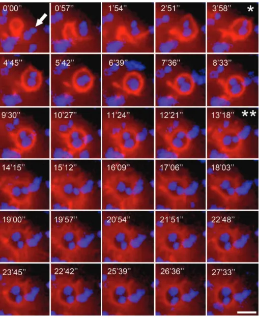
embrace the parasite, called the phagocytic cup (Figure 3
and supplementary Video 2) [85]. Fernandes and colleagues
[86] recently demonstrated that EAs are able to trigger
(a) (b)
Figure 2: Cover of Mem´orias do Instituto Oswaldo Cruz, vol. 86 (1) [14]. Likely the irst DIC image obtained with a confocal microscope (W. Brad Amos, personal communication) showing HeLa cell infection by G strain metacyclic trypomastigotes. On the right, the corresponding image ater immunoluorescence with monoclonal antibody anti-35/50 kDa mucin, suggesting release of the molecule in parts of internalized parasites [15].
other host cell vesicles. EA PVs irst mature into a CD63-, followed by synaptotagmin VII- and then LAMP1-positive structures. hese data show that EAs activate a phagocytic pathway in nonprofessional phagocytes that resembles large
particle uptake by professional phagocytes [86].
Another application of immunoluorescence techniques in this area of research relates to the role of host cell lysosomes inT. cruziinvasion. Tardieux and colleagues [83] observed that lysosomes are recruited to TCT invasion sites, a process dependent on calcium that culminates with the formation
of LAMP-positiveT. cruziPVs [87]. Norma Andrews’ group
(U. Maryland) demonstrated that TCTs induce plasma mem-brane lesions during the invasion process. hese wounds are repaired by lysosomes that secrete sphingomyelinase, an
enzyme that generates ceramide [88]. On the outer lealet of
the plasma membrane, this lipid induces inward budding that could drive parasite internalization. Using live imaging tech-niques, the authors conirmed previous TEM observations, showing the dynamics of lysosome mobilization towards cell
periphery during interaction with trypomastigotes [89].
Based on the observation of PIP-3 recruitment by TCTs at early steps of interaction with mammalian cells, a lysosome-independent pathway for trypomastigote entry has also been
described [90]. Although most of the results in this work
consist of very compelling evidence, it is worth mentioning that Figure 2(related to the attached supplementary video 1) clearly shows moving parasites from as early as 3 min (possibly under the cells). What is then referred to as the “second parasite” also appears moving in the ield (possibly already inside the cell) and the so-called recruitment of Akt-PH-GFP for this parasite, that begins at around 13
minutes, is undoubtedly arising from the protrusion of the
trypomastigote, actively moving from inside the cell. he
implication of this observation is that these trypomastigotes
most likely had invaded the imaged cellbeforethis period.
Considering the theme of this review, this might possibly be regarded as a misinterpretation of a rather compelling live
image ofT. cruzitrypomastigotes interacting with host cells.
Recently, Barrias et al.[91] provided evidence suggesting that
T. cruzitrypomastigotes may also subvert the macropinocytic pathway to enter host cells.
Interestingly, they also reported intracellular trypo-mastigotes protruding from within the host cell ater 15 minutes of infection. Although the authors focused their observations on parasite entry, it appeared that parasites could also attempt to escape or egress from the host cell
[89]. Similar behavior of internalized TCTs pushing out from
infected cells had already been described by Dvorak and
Hyde in their pioneering studies [67]. In 1992, Schenkman
and Mortara [79] observed membrane protrusions and actin
recruitment that were associated with TCT invasion sites in HeLa cells. At that time, ixed samples were visualized by confocal, transmission, and scanning electron microscopy (SEM). Static images were interpreted as depicting events associated with parasite entry. In light of observations made
by Hyde and Dvorak and Fernandes et al. [16,67], formation
of pseudopodia described by Schenkman and Mortara [79]
Figure 3: Actin recruitment by EAs in the phagocytic cup (Supplementary Video 2 available online at http://dx.doi.org/10.1155/2014/565291). HeLa cells transfected with luorescent actin marker were incubated with EAs (arrow) and observed by time-lapse confocal microscopy (Leica SP5 TS) for 30 minutes at one frame per 57 seconds. Total EA internalization occurred within approximately 4 minutes (∗), but actin mobilization difused approximately 9 minutes ater total EA internalization (∗∗) 13 minutes ater recording initiation. Actin is shown in red (Life-actin, ibidi); EA nucleus and kinetoplast are shown in blue (Hoescht 33258). Scale bar, 5�m.
micrographs from ixed samples. In particular, considering
T. cruzitrypomastigotes inside host cells and exposition of
parasite lagella ater host cell membrane damage [67, 89],
static images published years ago could be ambiguously interpreted as both invasion and exit processes.
Ater internalization, a poorly understood aspect of the
T. cruziintracellular life cycle is formation and escape from PVs. Ultrastructural studies demonstrated that, shortly ater
invasion (around 60 minutes),T. cruzitrypomastigotes are
lodged in a vacuole surrounded by a thin membrane, and “at later times, all the parasites were seen free in the cytoplasm”
[73]. his transient PV is able to fuse with host cell lysosomes
in phagocytic and nonphagocytic cells, which is clearly
observed by confocal and electron microscopy [73,89,90,92–
96]. he precise mechanisms by which parasites escape from
PVs into the cell cytoplasm have not been fully disclosed, but
T. cruzitrypomastigotes and amastigotes have been shown to secrete a membrane pore-forming protein, TC-TOX, which is
active at pH 5.5 and could be implicated in PV rupture [97–
99]. he question remains as to whetherT. cruzidiferentiates
into amastigotes inside or outside the PV. de Carvalho and
de Souza [95] suggested that trypomastigotes were able to
disrupt PVs before diferentiation into amastigotes, which is a feature of phagolysosomes in an acidic milieu. Indeed, it is possible to observe small pores in PV membranes that developed ater 1 hour and 30 minutes of trypomastigotes
infection in macrophages using TEM [95]. Using
multidi-mensional live imaging of HeLa cells transfected with
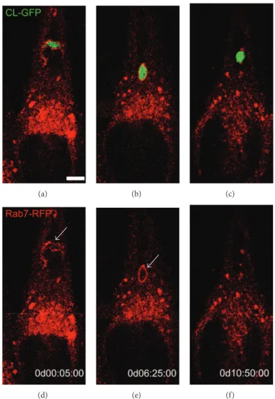
RFP-tagged Rab7 and infected with metacyclic forms ofT. cruzi
expressing GFP, we observed initial morphological changes of MT into round-shaped forms followed by dissolution of
(a) (b) (c)
(d) (e) (f)
Figure 4:T. cruzimetacyclic trypomastigote forms begin to diferentiate into amastigote-like forms inside the parasitophorous vacuole (supplementary Video 3). HeLa cells transfected with Rab7-Red luorescent protein (RFP) were infected with metacyclic trypomastigotes (MTs) from a CL strain transfected with green luorescent protein (GFP). Time-lapse images show the parasite internalized inside the parasitophorous vacuole (PV) labeled with Rab7-RFP (white arrow) ater one hour. MTs diferentiated into round-shaped forms, followed by loss of Rab-7 staining, suggesting parasite escape from the PV. Time-lapse acquisition is displayed as days : hours : minutes : seconds (dd : hh : mm : ss). Scale bar, 5�m. Images acquired with a confocal microscope (Leica SP5 TS).
3). In contrast to previous investigations, the data suggest that MT begins to diferentiate into an amastigote form before escape to the host cell cytosol. Further experiments using
multidimensional images and appropriate markers ofT. cruzi
diferentiation will potentially reveal if diferentiation into amastigotes takes place in PVs or in the cytosol and provide important information for future studies on drug delivery.
Egress from host cells is also poorly understood. Although host cell egress was highlighted in Hertha Meyer’s recordings in the 1940s, there are few studies on the subject. Edgar Rowland’s group was one of the irst to systematically
investigateT. cruziegress using an interesting experimental
approach: culture medium with serum obtained from chron-ically infected mice showed inhibition of parasite egress and
a decrease in intracellular replication in ibroblasts [100,101].
his inhibitory efect was also observed in serum obtained
from chronic chagasic patients [102]. It is possible to
hypothe-size that antibodies (anti-egressins) are reaching intracellular parasites and, according to the authors, promoting
intracel-lular agglutination of T. cruzi forms to block egress. At a
later phase of theT. cruziintracellular life cycle, the plasma
membrane of infected host cells is weakened, leading to
higher permeability to molecules, including antibodies [103].
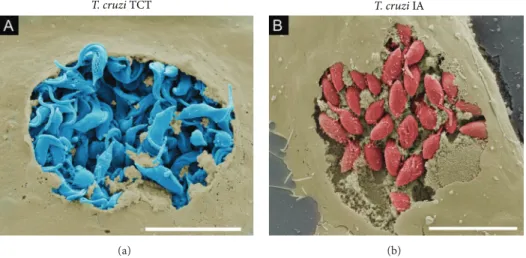
T. cruziegress from host cells has also been investigated by our group. he precise moment of trypomastigote exit from a host cell was captured using ield-emission scanning electron
microscopy (FE-SEM) (Figure 5(a)). FE-SEM is a valuable
microscopy tool to analyze biological surfaces with higher
spatial resolution than SEM [104]. Various morphological
and parasite-host cell interaction-related processes have been highlighted using conventional or FE-SEM, including the
T. cruziTCT
(a)
T. cruziIA
(b)
Figure 5: Visualization of the T. cruzi intracellular life cycle using ield-emission scanning electron microscopy. (a) Tissue cultured trypomastigotes (TCTs) (blue) egress from Vero cells (light brown). (b) Intracellular amastigotes (red) ofT. cruzihosted by Vero cells (light brown). Infected cells were ixed with 4% paraformaldehyde and then subjected to electron scanning processing. Briely, samples were dehydrated in an ethanol series, subjected to critical-point drying from CO2and gold sputtering. In (b), samples processed as in (a) were fractured by adhesive tape and then gold sputtered. Scale bars, 10�m.
(a) (b)
Figure 6: Visualization of bone marrow-derived macrophages infected with L. amazonensis using ield-emission scanning electron microscopy. In (a), intact cell (light grey) and, in (b),L. amazonensisamastigotes (red) within spacious PVs were exposed through fracture by Scotch tape, followed by gold sputtering. he samples were processed as described in the legend ofFigure 5. Scale bar, 10�m.
insect vector and its excretion [106,107], stimuli to
diferenti-ate its life cycle form and invasion [108–110], and cytoskeleton
organization during infection [89, 111]. One of our aims
using this technique was to try and observe intracellular parasites in host cells and entire organs using the ingenious “scotch tape technique,” which fractures the cell monolayer
and tissue samples [112, 113]. his approach allowed us to
observe intracellular amastigotes ofT. cruziin the cytoplasm
of Vero cells (Figure 5(b)) as well as intracellular amastigotes
ofL. amazonensislocated in large vacuoles of macrophages
derived from mouse bone marrow (Figure 6(b)).
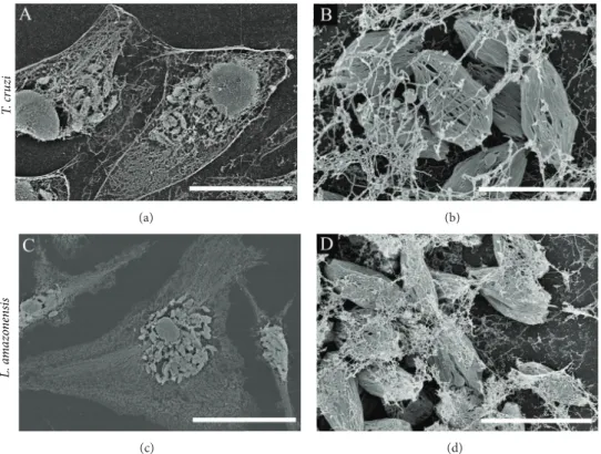
Several protocols have been used to visualize host cytoskeleton interaction with parasites using EM. Fernandes
and colleagues [89] treated infected cells with a membrane
extraction solution containing Triton X-100, taxol, and
phal-loidin to stabilize microtubules and microilaments [17].
his strategy enabled the authors to visualize the initial
invasion proile using TEM (to generate a three-dimensional projection) in which the posterior end of trypomastigotes penetrates underneath HeLa cells, resulting in actin ilament
enrichment at the undulated cell cortex [86]. We used the
same approach to visualize intracellular amastigotes in the
host cell cytoplasm. As shown inFigure 7, we observed
intra-cellular amastigotes ofT. cruzi(Figure 7(a)) andL.
amazo-nensis(Figure 7(b)) hosted by cells in which the cytoskeleton network was preserved. In these images, amastigotes were also subjected to membrane extraction to observe internal structures of the parasites.
Our group has focused eforts on the observation of intracellular parasites in infected hearts of mice at the SEM level. Detailed information from infected cardiac tissue is
relevant for elucidatingT. cruzi pathogenesis due to heart
tissue damage caused by the parasite and/or autoimmune
T
. cr
uzi
(a) (b)
L. a
m
azo
n
ens
is
(c) (d)
Figure 7: Visualization of host cell cytoskeleton networks and intracellular amastigotes ofT. cruzi(a, b) andL. amazonensis(c, d) with host cell cytoskeleton networks. Infected HeLa cells (a, b) and mouse bone marrow macrophages (c, d) were treated with a membrane extraction solution containing Triton X-100, taxol, and phalloidin (to stabilize microtubules and microilaments) [16,17]. Cytoskeletons of infected cells were visualized by ield emission scanning electron microscopy ater processing and gold coating. Scale bars: (a) 20�m; (b) 3�m; (c) 30�m; (d) 5�m.
(a) (b)
Figure 8: Field-emission scanning electron microscopy of mouse hearts infected withT. cruzi.hick parain embedded sections of mouse hearts infected with Y strain metacyclic trypomastigotes were deparainized and processed for ield emission scanning electron microscopy [18–21]. Briely, parain was removed by melting the sections block and then deparainized with xylol and ethanol. Next, heart muscle sections cut with a razor blade were dehydrated in an ethanol series, subjected to critical-point drying, and gold sputtered. (a) Amastigotes (red), scale bar, 4�m; (b) trypomastigotes (blue), scale bar, 10�m.
Pathological investigations on fatal cases of Chagas disease performed by Gaspar Vianna in association with the Ger-man pathologist HerGer-mann D¨urk in 1917 deined acute and chronic phases of the disease, with the latter phase associated
with cardiac involvement [115]. he association between
T. cruzi infection and cardiac failure in chronic patients
was a well-established concept by the 1960s [116]. Common
of research [18–21], but an image of T. cruzi-infected
tis-sue from thick sections (>40�m) has not been produced.
In thick parain histological sections submitted for SEM
processing, we observed T. cruzi amastigote and
trypo-mastigote nests within heart muscle ibers (Figures 8(a)
and 8(b)). his simple, cost-efective, and rapid approach
was applied ater conventional formaldehyde ixation and parain embedding, followed by deparaination with xylol, dehydration with ethanol, critical-point drying, and sputter-coating with gold for SEM. Mice hearts were stored in parain blocks for several years before they were processed using SEM, highlighting the good condition of the tissue and its structures despite a long period of time in storage. A related and relevant issue that deserves more in-depth study is understanding how circulating parasites reach this organ. Intravital imaging techniques of whole animals and multiphoton confocal microscopy of infected tissues should
allow for luorescent-taggedT. cruzitracking in what could
become a challenging and encouraging perspective for future investigations.
4. Concluding Remarks
Innovative techniques consistently improve our interpre-tations of biological processes and their mechanisms in biomedical research. In this review, we presented examples of advances in microscopy that contributed to building concepts regarding host-parasite interactions of the human
kinetoplastid parasites Leishmaniaspp. andT. cruzi. here
are several other cases of conceptual breakthroughs that we did not cover in this review on microscopy, including newly developed techniques that could certainly lead to impor-tant changes in how we conceptualize similar intracellular parasites. Namely, electron tomography in cryopreserved samples allows for 3D reconstruction of infected cells and parasites bypassing cumbersome serial slicing; superreso-lution microscopes (PALM/STORM and STED) increase optical resolution to tens of nanometers and allow for live imaging; bioluminescent parasites could be tracked in whole
organisms using in vivo bioluminescent imaging systems
[117,118]; and use of reporters, probes, or other microcopy
techniques (FRAP, FRET and FLIM) improves microscopic observations regarding biochemical/molecular mechanisms of host/pathogen interactions. We can rely on history to repeat itself in that further studies using these cutting-edge microscopic technologies will change our perception ofLeishmaniaspp. andT. cruziintracellular parasitism and contribute to the development of novel and more eicient strategies of chemotherapy and vaccination.
Disclosure
he authors agree that the irst two authors should be regarded as joint irst authors.
Conflict of Interests
he authors declare that there is no conlict of interests regarding the publication of this paper.
Acknowledgments
he authors would like to thank Maria Cec´ılia Fernandes, Mauro Cortez, and Leonardo Rodrigues de Andrade for
their help with T. cruzi and L. major cytoskeleton
imag-ing (Figure 7); Michel Rabinovith, Patr´ıcia Sampaio Tavares
Veras, and Marcel Pouchelet for the use of the video of
Leishmaniacell-to-cell transfer captured at the Institute Pas-teur; and Andr´e Aguillera for his skillful help with FE-SEM imaging. hey also thank Fundac¸˜ao de Amparo `a Pesquisa do Estado de S˜ao Paulo (FAPESP), Conselho Nacional de Desenvolvimento Cient´ıico e Tecnol´ogico (CNPq), and Coordenac¸˜ao de Aperfeic¸oamento de Pessoal de N´ıvel Supe-rior (Capes) for inancial support through grants and fellow-ships.
References
[1] D. Moreira, P. L´opez-Garc´ıa, and K. Vickerman, “An updated view of kinetoplastid phylogeny using environmental sequences and a closer outgroup: proposal for a new classiication of the class Kinetoplastea,”International Journal of Systematic and Evolutionary Microbiology, vol. 54, no. 5, pp. 1861–1875, 2004. [2] K. Stuart, R. Brun, S. Crot et al., “Kinetoplastids: related
protozoan pathogens, diferent diseases,” Journal of Clinical Investigation, vol. 118, no. 4, pp. 1301–1310, 2008.
[3] W. de Souza, T. M. U. de Carvalho, and E. S. Barrias, “Review on Trypanosoma cruzi: host cell interaction,”International Journal of Cell Biology, vol. 2010, Article ID 295394, 18 pages, 2010. [4] R. Rosenberger, “A case study in the applied philosophy of
imaging: the synaptic vesicle debate,”Science Technology and Human Values, vol. 36, no. 1, pp. 6–32, 2011.
[5] H. Boxenbaum, “Time concepts in physics, biology, and phar-macokinetics,”Journal of Pharmaceutical Sciences, vol. 75, no. 11, pp. 1053–1062, 1986.
[6] E. Mayr, “Cause and efect in biology—kinds of causes, pre-dictability, and teleology are viewed by a practicing biologist,” Science, vol. 134, p. 1501, 1961.
[7] M. Ega˜na Aranguren, K. Wolstencrot, U. Sattler et al., “Using OWL to model biological knowledge,”International Journal of Human Computer Studies, vol. 65, no. 7, pp. 583–594, 2007. [8] A. C. Ahn, M. Tewari, C.-S. Poon, and R. S. Phillips, “he limits
of reductionism in medicine: could systems biology ofer an alternative?”PLoS Medicine, vol. 3, no. 6, pp. 709–713, 2006. [9] R. Sattler, Biophilosophy: Analytic and Holistic Perspectives,
Springer, Berlin, Germany, 1986.
[10] “Milestones in light microscopy,”Nature Cell Biology, vol. 11, pp. 1165–1165, 2009.
[11] G. Q. Xiao, T. R. Corle, and G. S. Kino, “Real-time confocal scanning optical microscope,”Applied Physics Letters, vol. 53, no. 8, pp. 716–718, 1988.
[12] H. Landecker, “Seeing things: from microcinematography to live cell imaging,”Nature Methods, vol. 6, no. 10, pp. 707–709, 2009.
[13] R. Lofgren, “he structure ofLeishmaniatropica as revealed by phase and electron microscopy,”Journal of Bacteriology, vol. 60, pp. 617–625, 1950.
trypomastigotes during invasion of HeLa cells,”Mem´orias do Instituto Oswaldo Cruz, vol. 86, supplement 1, p. 1, 1991. [15] S. Schenkman, M. A. J. Ferguson, N. Heise, M. L. Cardoso de
Almeida, R. A. Mortara, and N. Yoshida, “Mucin-like glycopro-teins linked to the membrane by glycosylphosphatidylinositol anchor are the major acceptors of sialic acid in a reaction cat-alyzed by trans-sialidase in metacyclic forms ofTrypanosoma cruzi,”Molecular and Biochemical Parasitology, vol. 59, no. 2, pp. 293–304, 1993.
[16] M. C. Fernandes, L. R. de Andrade, N. W. Andrews, and R. A. Mortara, “Trypanosoma cruzi trypomastigotes induce cytoskeleton modiications during hela cell invasion,”Mem´orias do Instituto Oswaldo Cruz, vol. 106, no. 8, pp. 1014–1016, 2011. [17] C. Sant’Anna, L. Campanati, C. Gadelha et al., “Improvement
on the visualization of cytoskeletal structures of protozoan parasites using high-resolution ield emission scanning electron microscopy (FESEM),”Histochemistry and Cell Biology, vol. 124, no. 1, pp. 87–95, 2005.
[18] D. A. Gaudet and E. G. Kokko, “Application of scanning electron microscopy to parain-embedded plant tissues to study inva-sive process of plant-pathogenic fungi,”Phytopathology, vol. 74, p. 3, 1984.
[19] H. D. Geissinger, “Correlated light optical and scanning elec-tron microscopy of Gram smears of bacteria and parain sections of cardiac muscle,”Journal of Microscopy, vol. 93, no. 2, pp. 109–117, 1971.
[20] W. P. Wergin, R. W. Yaklich, S. Roy et al., “Imaging thin and thick sections of biological tissue with the secondary electron detector in a ield-emission scanning electron microscope,” Scanning, vol. 19, no. 6, pp. 386–395, 1997.
[21] S. D. Russell and C. P. Daghlian, “Scanning electron microscopic observations on deembedded biological tissue sections: com-parison of diferent ixatives and embedding materials,”Journal of Electron Microscopy Technique, vol. 2, no. 5, pp. 489–495, 1985. [22] R. Lainson and J. J. Shaw,Evolution, Classiication and Geo-graphical Distribution, W. Peters, R. Killick-Kendrick, Eds., Academic Press, San Diego, Calif, USA, 1987.
[23] A. J. Altamirano-Enciso, M. C. A. Marzochi, J. S. Moreira, A. O. Schubach, and K. B. F. Marzochi, “On the origin and spread of cutaneous and mucosal leishmaniasis, based on pre-and post- colombian historical source,”Hist´oria Ciˆencias Sa´ude, Manguinhos,, vol. 10, no. 3, pp. 852–882, 2003.
[24] J.-P. Dedet, “Edmond Sergent’s discoveries on the vectorial transmission of agents of human and animal infectious dis-eases,”Bulletin de la Societ´e de Pathologie Exotique, vol. 100, no. 2, pp. 147–150, 2007.
[25] J. h´eodorid`es, “Historical note on the discovery of the trans-mission of cutaneous leishmaniasis by phlebotomes,”Bulletin de la Societ´e de Pathologie Exotique, vol. 90, no. 3, pp. 177–178, 1997.
[26] J. H. Wright, “Protozoa in a case of tropical ulcer (“Delhi Sore”),” Journal of Medical Research, vol. 10, pp. 472–482, 1903. [27] S. R. Christophers, “On a parasite found in persons sufering
from enlargement of the spleen in India,” Second Report, Oice of the Superintendent of Government Printing, Calcutta, India, 1904.
[28] D. Heyneman, “Immunology of leishmaniasis,”Bulletin of the World Health Organization, vol. 44, no. 4, pp. 499–514, 1971. [29] N. Ueno and M. E. Wilson, “Receptor-mediated phagocytosis
ofLeishmania: implications for intracellular survival,”Trends in Parasitology, vol. 28, pp. 335–344, 2012.
[30] R. J. V. Pulvertat and G. F. Hoyle, “Stages in the life-cycle ofLeishmania donovani,”Transactions of the Royal Society of Tropical Medicine and Hygiene, vol. 54, no. 2, pp. 191–196, 1960. [31] H. C. Miller and D. W. Twohy, “Infection of macrophages in culture by leptomonads ofLeishmania donovani,”he Journal of Protozoology, vol. 14, no. 4, pp. 781–789, 1967.
[32] H. J. Akiyama and R. D. Haight, “Interaction of Leishma-nia donovaniand hamster peritoneal macrophages. A phase-contrast microscopical study,” American Journal of Tropical Medicine and Hygiene, vol. 20, no. 4, pp. 539–545, 1971. [33] C.-L. Forestier, C. MacHu, C. Loussert, P. Pescher, and G.
F. Sp¨ath, “Imaging host cell-Leishmaniainteraction dynamics implicates parasite motility, lysosome recruitment, and host cell wounding in the infection process,”Cell Host and Microbe, vol. 9, no. 4, pp. 319–330, 2011.
[34] N. Courret, C. Fr´ehel, N. Gouhier et al., “Biogenesis of Leish-mania-harbouring parasitophorus vacuoles following phagocy-tosis of the metacyclic promastigote or amastigote stages of the parasites,”Journal of Cell Science, vol. 115, no. 11, pp. 2303–2316, 2002.
[35] M. Aikawa, L. D. Hendricks, Y. Ito, and M. Jagusiak, “Interac-tions between macrophagelike cells andLeishmania braziliensis in vitro,”American Journal of Pathology, vol. 108, no. 1, pp. 50– 59, 1982.
[36] J. M. Blackwell and J. E. Plant, “Expression of the natural resistance gene (Lsh) in wild mice infected experimentally withLeishmania donovaniorSalmonella typhimurium,”Current topics in Microbiology and Immunology, vol. 127, pp. 323–330, 1986.
[37] D. C. Love, M. M. Kane, and D. M. Mosser, “Leishmania amazonensis: the phagocytosis of amastigotes by macrophages,” Experimental Parasitology, vol. 88, no. 3, pp. 161–171, 1998. [38] D. Coling and B. Kachar, “heory and application of
luores-cence microscopy,”Current Protocols in Neuroscience, vol. 2, Unit 2.1, 2001.
[39] R. Lodge and A. Descoteaux, “Leishmania donovani promastig-otes induce periphagosomal F-actin accumulation through retention of the GTPase Cdc42,”Cellular Microbiology, vol. 7, no. 11, pp. 1647–1658, 2005.
[40] R. Lodge and A. Descoteaux, “Phagocytosis of Leishmania donovani amastigotes is Rac1 dependent and occurs in the absence of NADPH oxidase activation,”European Journal of Immunology, vol. 36, no. 10, pp. 2735–2744, 2006.
[41] J. Alexander and K. Vickerman, “Fusion of host cell secondary lysosomes with the parasitophorous vacuoles of Leishmania mexicanainfected macrophages,”he Journal of Protozoology, vol. 22, no. 4, pp. 502–508, 1975.
[42] K. P. Chang and D. M. Dwyer, “Leishmania donovani. Hamster macrophage interactionsin vitro: cell entry, intracellular sur-vival, and multiplication of amastigotes,”Journal of Experimen-tal Medicine, vol. 147, no. 2, pp. 515–530, 1978.
[43] J.-C. Antoine, E. Prina, T. Lang, and N. Courret, “he biogenesis and properties of the parasitophorous vacuoles that harbour Leishmaniain murine macrophages,”Trends in Microbiology, vol. 6, no. 10, pp. 392–401, 1998.
[44] E. Prina and J. Antoine Cl., “Localization and activity of various lysosomal proteases in rat macrophages infected with Leishmania amazonensis,”Pathologie Biologie, vol. 38, no. 10, pp. 1020–1022, 1990.
macrophages maintain an acidic pH,”Infection and Immunity, vol. 58, no. 3, pp. 779–787, 1990.
[46] T. Lang, C. de Chastellier, C. Frehel et al., “Distribution of MHC class I and of MHC class II molecules in macrophages infected withLeishmania amazonensis,”Journal of Cell Science, vol. 107, no. 1, pp. 69–82, 1994.
[47] E. Handman and D. V. Bullen, “Interaction ofLeishmaniawith the host macrophage,”Trends in Parasitology, vol. 18, pp. 332– 334, 2002.
[48] F. Real and R. A. Mortara, “he diverse and dynamic nature of Leishmaniaparasitophorous vacuoles studied by multidimen-sional imaging,”PLoS Neglected Tropical Diseases, vol. 6, no. 2, Article ID e1518, 2012.
[49] T. Lang, H. Lecoeur, and E. Prina, “Imaging Leishmania development in their host cells,”Trends in Parasitology, vol. 25, no. 10, pp. 464–473, 2009.
[50] C. Lippuner, D. Paape, A. Paterou et al., “Real-time imaging ofLeishmania mexicana-infected early phagosomes: a study using primary macrophages generated from green luorescent protein-Rab5 transgenic mice,”FASEB Journal, vol. 23, no. 2, pp. 483–491, 2009.
[51] D. S. Ridley, “A histological classiication of cutaneous leishma-niasis and its geographical expression,”Transactions of the Royal Society of Tropical Medicine and Hygiene, vol. 74, no. 4, pp. 515– 521, 1980.
[52] M. G. Rittig, K. Schr¨oppel, K.-H. Seack et al., “Coiling phago-cytosis of trypanosomatids and fungal cells,” Infection and Immunity, vol. 66, no. 9, pp. 4331–4339, 1998.
[53] M. G. Rittig and C. Bogdan, “Leishmania-host-cell interaction: complexities and alternative views,”Parasitology Today, vol. 16, no. 7, pp. 292–297, 2000.
[54] N. C. Peters, J. G. Egen, N. Secundino et al., “In vivo imaging reveals an essential role for neutrophils in leishmaniasis trans-mitted by sand lies,”Science, vol. 321, no. 5891, pp. 970–974, 2008.
[55] L. G. Ng, A. Hsu, M. A. Mandell et al., “Migratory dermal dendritic cells act as rapid sensors of protozoan parasites,”PLoS Pathogens, vol. 4, no. 11, Article ID e1000222, 2008.
[56] P. Gueirard, A. Laplante, C. Rondeau, G. Milon, and M. Des-jardins, “Traicking ofLeishmania donovanipromastigotes in non-lytic compartments in neutrophils enables the subsequent transfer of parasites to macrophages,”Cellular Microbiology, vol. 10, no. 1, pp. 100–111, 2008.
[57] C. Chagas, “Nova tripanozomiaze humana: estudos sobre a morfolojia e o ciclo evolutivo doSchizotrypanum cruzin. gen., n. sp., ajente etiolojico de nova entidade morbida do homem,” Mem´orias do Instituto Oswaldo Cruz, vol. 1, pp. 159–218, 1909. [58] G. Vianna, “Contribuic¸˜ao para o estudo da anatomia patol´ojica
da “Mol´estia de Carlos Chagas” (Esquizotripanose humana ou tireoidite parasit´aria),”Mem´orias do Instituto Oswaldo Cruz, vol. 3, pp. 276–293, 1911.
[59] E. Dias, “Estudos sobre oSchizotrypanum cruzi,”Mem´orias Do Instituto Oswaldo Cruz, vol. 28, pp. 1–110, 1934.
[60] E. Villela and C. M. Torres, “Histopathology of the central nervous system in experimental paralysis caused by Schizotry-panum cruzi,”Mem´orias do Instituto Oswaldo Cruz, vol. 19, pp. 199–221, 1926.
[61] C. Roma˜na and H. Meyer, “Estudo do ciclo evolutivo do, “Schizotrypanum cruzi” em cultura de tecidos de embri˜ao de galinha,”Mem´orias do Instituto Oswaldo Cruz, vol. 37, pp. 19– 27, 1942.
[62] C. A. Kofoid, F. D. Wood, and E. C. E. McNeil, he Cycle of Trypanosoma Cruzi in Tissue Culture of Embryonic Heart Muscle, University of California Press, Berkeley, Calif, USA, 1935.
[63] Z. Brener, “Biology ofTrypanosoma cruzi,”Annual Review of Microbiology, vol. 27, pp. 347–382, 1973.
[64] H. Meyer and A. Barasa, “Life cycle ofSchizotrypanum cruzi in tissue cultures,”http://www.itarget.com.br/newclients/sbpz.
org.br/2011/extra/download/cruzi1.mpg.
[65] H. Meyer and K. R. Porter, “A study ofTrypanosoma cruziwith the electron microscope,”Parasitology, vol. 44, no. 1-2, pp. 16– 23, 1954.
[66] W. de Souza, “Electron microscopy of trypanosomes—a histor-ical view,”Mem´orias do Instituto Oswaldo Cruz, vol. 103, no. 4, pp. 313–325, 2008.
[67] J. A. Dvorak and T. P. Hyde, “Trypanosoma cruzi: interaction with vertebrate cellsin vitro. I. Individual interactions at the cellular and subcellular levels,”Experimental Parasitology, vol. 34, no. 2, pp. 268–283, 1973.
[68] E. R. Ferreira, A. Bonim-Melo, R. A. Mortara, and D. Bahia, “Trypanosoma cruzi extracellular amastigotes and host cell signaling: more pieces to the puzzle,”Frontiers in Immunology, vol. 3, p. 363, 2012.
[69] V. Ley, N. W. Andrews, E. S. Robbins, and V. Nussenzweig, “Amastigotes ofTrypanosoma cruzisustain an infective cycle in mammalian cells,”Journal of Experimental Medicine, vol. 168, no. 2, pp. 649–659, 1988.
[70] F. M. Lima, P. Oliveira, R. A. Mortara, J. F. Silveira, and D. Bahia, “he challenge of Chagas’ disease: has the human pathogen, Trypanosoma cruzi, learned how to modulate signaling events to subvert host cells?”New Biotechnology, vol. 27, no. 6, pp. 837– 843, 2010.
[71] R. A. Mortara, W. K. Andreoli, N. N. Tantwaki et al., “Mammalian cell invasion and intracellular traicking by Try-panosoma cruziinfective forms,”Anais da Academia Brasileira de Ciencias, vol. 77, no. 1, pp. 77–94, 2005.
[72] S. Tomlinson, F. Vandekerckhove, U. Frevert, and V. Nussen-zweig, “he induction ofTrypanosoma cruzitrypomastigote to amastigote transformation by low pH,”Parasitology, vol. 110, no. 5, pp. 547–554, 1995.
[73] N. Nogueira and Z. Cohn, “Trypanosoma cruzi: mechanism of entry and intracellular fate in mammalian cells,”Journal of Experimental Medicine, vol. 143, no. 6, pp. 1402–1420, 1976. [74] K. Behbehani, “Developmental cycles of Trypanosoma
(Schizotrypanum) cruzi (Chagas, 1909) in mouse peritoneal macrophagesin vitro,”Parasitology, vol. 66, no. 2, pp. 343–353, 1973.
[75] L. Hudson, D. Snary, and S. J. Morgan, “Trypanosoma cruzi: continuous cultivation with murine cell lines,”Parasitology, vol. 88, pp. 283–294, 1984.
[76] S. Chia-Tung Pan, “Trypanosoma cruzi: in vitrointeractions between cultured amastigotes and human skin-muscle cells,” Experimental Parasitology, vol. 45, no. 2, pp. 274–286, 1978. [77] S. Schenkman, N. W. Andrews, V. Nussenzweig, and E. S.
![Figure 2: Cover of Mem´orias do Instituto Oswaldo Cruz, vol. 86 (1) [14]. Likely the irst DIC image obtained with a confocal microscope (W.](https://thumb-eu.123doks.com/thumbv2/123dok_br/17565478.265897/7.900.188.713.107.479/figure-cover-instituto-oswaldo-likely-obtained-confocal-microscope.webp)