535 535 535 535 535 Mem Inst Oswaldo Cruz, Rio de Janeiro, Vol. 95(4): 535-544, Jul./Aug. 2000
Trypanosoma cruzi
The Vector-parasite Paradox
CJ Schofield
London School of Hygiene and Tropical Medicine, London WC1 E7HT, UK
Trypanosoma cruzi and the majority of its insect vectors (Hemiptera, Reduviidae, Triatominae) are confined to the Americas. But while recent molecular studies indicate a relatively ancient origin for the parasite (~65 million years ago) there is increasing evidence that the blood-sucking triatomine vectors have evolved comparatively recently (<5 mya). This review examines the evidence for these ideas, and attempts to reconcile the apparent paradox by suggesting that marsupial opossums (Didelphidae) may have played a role, not just as original reservoir hosts, but also as original vectors of the parasite.
Key words: Trypanosoma cruzi - Triatominae - Didelphidae - evolution
Trypanosoma cruzi, causative agent of Chagas disease, occurs exclusively in the Americas (except for occasional human infections that have travelled elsewhere). Similarly, the majority of its insect vec-tors – blood-sucking Reduviidae of the subfamily Triatominae – also occur primarily in the Americas (except for the aberrant Indian genus Linshcosteus, and the tropicopolitan Triatoma rubrofasciata and its Asian relatives). Parasite and vector are asso-ciated, but a paradox arises from evidence that T. cruzi is a relatively ancient parasite, whereas the Triatominae appear to have evolved comparatively recently (Stevens et al. 2000). Such interpretations may be erroneous, or they may be reconcilable. This review seeks to analyse the available evidence.
EVIDENCE FOR THE ANTIQUITY OF TRYPANOSOMA CRUZI
T. cruzi is widespread in the Americas, from the Great Lakes of the USA to the southern Patagonia of Argentina (roughly 42°N to 46°S). Throughout this region it is a common parasite of small mam-mals, especially nest-building species of rodents and opossums which are commonly associated with silvatic species of Triatominae. Human infections are rare in the USA, but regrettably frequent in Latin America where rural houses are often infested with domestic species of Triatominae. It is clear that most transmission of T. cruzi to humans is via fae-cal deposits from infected Triatominae, although other routes of transmission are also possible, no-tably by blood transfusion from infected donors,
transplacental transmission from infected mothers, and oral route transmission by eating infected ma-terial (for review see Pipkin 1969). But even for transmission via infected triatomine faeces, there is still doubt about the frequency of parasite pas-sage across intact human skin. Many authorities consider that transmission most commonly occurs when the infected faecal deposits are inadvertently passed to the mucosa of eye, nose or mouth, across which the parasite can pass quite readily. Similarly, transmission to small mammals would seem most likely to occur when the mammal eats an infected bug, or licks triatomine faecal deposits while groom-ing the haircoat (Diotaiut et al. 1995). Oral route transmission would seem to be the primitive state. If the first line of evidence for the relative an-tiquity of T. cruzi comes from its wide distribution in the Americas, the second comes from compara-tive clinical studies. Human infections are often virulent, with some mortality during the acute parasitaemic phase of infection, and severe tissue lesions developing in up to 30% of chronic human infections. Similar pathology can be seen in do-mestic animals such as cats and dogs, and also in some strains of sinanthropic murid rodents (espe-cially laboratory rats and mice). In contrast, severe pathology has not been recorded in commonly in-fected wild hosts such as opossums, armadillos, and cricetid rodents. This comparison suggests that those mammals of greater antiquity in the Americas have had a longer association with T. cruzi, compared to those mammals imported more recently from the Old World, allowing time for some degree of co-evolution leading to attenuated viru-lence.
The third line of evidence comes from genetic studies. Phylogenetic analysis based on ssu rRNA sequence data indicate that the genus Trypano-soma is monophyletic (Stevens & Gibson 1999, Stevens et al. 1999a,b, 2000) and this conclusion is This work has benefitted from international
collabora-tion through the ECLAT network.
Fax: +33.450.20.63-77. E-mail: [email protected] Received 13 April 2000
536 536 536 536
536 T. cruzi The Vector-parasite Paradox CJ Schofield
endorsed by studies based on the GAPDH gene (Wiemer et al. 1995, Alvarez et al. 1996, Hannaert et al. 1998), and on 9S and 12S mitochondrial rRNA genes (Lake et al. 1988), elongation factor 1α (Hashimoto et al. 1995), trypanothione reductase and α-tubulin (Alvarez et al. 1996) and phospho-glycerate kinase (Adjé et al. 1998). All trypano-somes are parasitic, so the idea of of a monophyl-etic origin suggests an ancestral parasitic form that gave rise to the mammalian trypanosomes of Af-rica, AmeAf-rica, and Australasia, as well as the vari-ous forms that parasitise fish. The implied connec-tion between these groups is offered by plate tec-tonic theory, suggesting that the original parasitic forms developed prior to splitting up the continents during the mezozoic era (~230 mya). However, the ssu rRNA data also indicate monophyly for the ‘cruzi clade’, where this group includes the various forms of T. cruzi, together with T. rangeli, various trypanosomes of bats, and an unamed species iso-lated from an australian kangaroo (Stevens et al. 2000). Development of the cruzi clade is thus sug-gested to have initiated prior to splitting up the southern supercontinent (Gondwanaland) in the cenozoic era. The earliest forms of cruzi itself are deduced to have been associated with marsupial opossums at the time of separation of South America from Gondwanaland about 40 mya.
Within T. cruzi, there is strong evidence for at least two main lineages, originally denoted as Z1 and Z2 on the basis of their isoenzyme profiles (Miles 1979), subsequently denoted as lineage 2 and lineage 1, repectively, on the basis of molecu-lar markers: RAPDs (Tibayrenc et al. 1993, Souto et al. 1996, Brisse et al. 1998), miniexon gene se-quences and 24S lsu rRNA sese-quences (Souto et al. 1996, Zingales et al. 1998, Fernandes et al. 1998), cytochrome-b sequences (Brisse 1997) and the topoisomerase locus (Dos Santos & Buck 1999). These two lineages are now denoted as cruzi 1 (= Z1of Miles 1979, and lineage 2 of Souto et al. 1996) and cruzi 2 (= Z2 of Miles 1979, and lineage 1 of Souto et al. 1996) (Memórias 1999). Of these, cruzi1 seems the more homogenous and, from studies throughout the Americas, seems primitively asso-ciated with opossums (Didelphis spp.). By con-trast, cruzi 2 shows a number of well-characterised natural clones found mainly in southern cone
coun-tries, and seems primitively associated with rodents. T. cruzi 2 in human infections is also associated with chronic intestinal lesions (eg. megaoesopha-gus, megacolon) as well as the cardiopathy that characterises cruzi 1 infections. T. cruzi Z3 of Miles (1979) is also heterogeneous, and seems by analy-sis of miniexon gene sequences to have affinity with cruzi 1, although it can generally be recognised as a sub-clade characterised by a ~50bp insertion in the non-transcribed spacer region of the miniexon (Fernandes et al. 1998). It is almost invari-ably silvatic, with current evidence suggesting a primitive association with armadillos. T. cruzi marinkellei (sometimes referred to as T. cruzi Z4) is strongly associated with bats.
Other parasites grouped within the cruzi clade of Stevens et al. (2000) are T. dionisii and T. vespertilionis of bats, T. conorhini of rats, T. rangeli which can transiently infect a wide range of mammals but seems primarily associated with Rhodnius species, T. leeuwenhoeki and T. minasense which seem similar to T. rangeli, and the unamed trypanosome species from an Austra-lian kangaroo. Their data incompletely resolve the position of the flea-transmitted rodent trypano-somes T. lewisi, T. musculi, and T. microtis, but place them close to the cruzi clade.
EVIDENCE FOR THE ANTIQUITY OF TRIATOMINAE
The Triatominae are defined as Reduviidae (Hemiptera, Heteroptera) that suck vertebrate blood (Jeannel 1919, Lent & Wygodzinsky 1979) in con-trast to the other 30 or so reduviid subfamilies1 that prey on invertebrates. The Reduviidae them-selves are clearly an ancient family, with the fossil record suggesting that the earliest predatory forms may have derived from phytophagous Hemiptera during the Permian/Triassic periods some 230 mya (Evans 1956, Wootton 1981). Such forms would clearly predate haematophagy since the earliest mammals and birds seem to have arisen during the Jurassic period some 50 million years later. Preda-tory reduviids are now of worldwide distribution, with well over 6,000 described species (Maldonado Capriles 1990). All reduviid subfamilies now seem to be represented in the Americas, and at least three subfamilies of predatory reduviid are represented in Oligocene and Eocene amber (25-65 mya) from Mexico and the Caribbean – Apiomerinae (Capriles et al. 1993a), Emesinae (Thomas 1992, Capriles et al. 1993b) and Holoptilinae (Poinar 1991). In contrast, there appears to be no evidence of fossilised haematophagous forms.
The evolution of feeding habits within the Heteroptera has been subject to considerable dis-cussion (see: Cobben 1978, 1979, Sweet 1979, Schuh 1986,). Blood-sucking most probably derived from 1 At the time of writing, there is no universal agreement
537 537537 537 537 Mem Inst Oswaldo Cruz, Rio de Janeiro, Vol. 95(4), Jul./Aug. 2000
a predaceous habit, with the intermediate stages perhaps being predation on guilds of nest-dwell-ing invertebrates followed by facultative blood feeding from the vertebrates occupying the nests (Schofield 1988, Schofield & Dolling 1993). Within the heteropteran Hemiptera, there are many spe-cies that suck vertebrate blood to a greater or lesser degree (Table I). Even some phytophagous spe-cies will probe vertebrates and may imbibe blood on occasion. The frequency of haematophagous behaviour suggests that facultative blood-sucking is a relatively simple step within the Heteroptera, and seems to be particularly frequent amongst predatory groups of Reduviidae and Lygaeidae, and also amongst the Anthocoridae which are believed to have given rise to the obligate haematophagous families of Cimicidae and Polyctenidae (see: Southwood & Leston 1959, Schuh & Slater 1995). In the converse sense, the predatory state is re-flected in many species of Triatominae. Examples include T. rubrofasciata, which was considered beneficial in some huts in SE Asia because of prey-ing on caterpillars that would otherwise damage the palm thatch roofs (Kalshoven 1970), T. rubrovaria which can be fed on spiders and silk-worm larvae in the laboratory (Abalos & Wygodzinsky 1951, Lent & Wygodzinsky 1979) and, along with T. circummaculata, has been shown to complete its entire life cycle on a diet of either ver-tebrate blood or cockroach haemolymph (Lorosa et al. 2000). Young nymphs of Eratyrus mucronatus seem preferentially to feed on invertebrates, while older nymphs and adults preferentially feed on ver-tebrate blood (Miles et al. 1981). A further transi-tional stage between predator and blood-sucker is indicated by cannibalistic behaviour (‘cleptohemo-deipnonism’ of Ryckman 1951) where, under crowded laboratory conditions, triatomine nymphs that cannot reach the vertebrate host will penetrate feeding nymphs and take blood through them. Some triatomine species seem to have predomi-nantly cleptohaematophagic behaviour, for example Belminus herreri that preferentially takes bloodmeals by feeding from recently engorged Rhodnius (Sandoval et al. 2000). These observa-tions not only suggest that Triatominae have evolved the blood-sucking habit relatively recently, but could be interpreted to indicate that some are still in the process of doing so.
Morphological features used to distinguish Triatominae from other Reduviidae, such as the straight rostrum adpressed to the gula, and the ability of the third rostral segment to flex upwards, seem to have been derived in association with adap-tations for feeding on vertebrate hosts (Cobben 1978, Schofield & Dolling 1993), and more strin-gent differences between the haematophagous and
predatory forms of Reduviidae are not apparent. Even characters that can be helpful in determina-tion, such as the laterally inserted antennae, ab-sence of dorsal abdominal scent glands, and fea-tures of the external genitalia and wing venation, are all shared with one or more of the predatory reduviid groups. Indeed, the morphological simi-larities between some predatory Reduviidae and blood-sucking Triatominae are so striking that at least one predator has been erroneously described as a new species of Triatominae (see Lent 1982). Carcavallo et al. (1999) offer several other examples of these similarities – predatory reduviids that are morphologically almost indistinguishable from one or other group of blood-sucking Triatominae, and often occur in the same type of habitat. We must conclude that the blood-sucking Triatominae are generally poorly differentiated from predatory redu-viids, both in body form and habitat, which is again suggestive that they have evolved the blood-suck-ing habit relatively recently.
In contrast to the Triatominae however, both the Cimicidae and Polyctenidae are highly evolved as blood-suckers, with an almost ectoparasitic habit and highly specialised morphology (see Schofield & Dolling 1993). Most of the 93 species of Cimicidae occur in the Middle East and Africa (Table II) and those that occur in the Americas are generally species associated with humans and/or domesticated or sinanthropic animals – suggest-ing that they have been exported to the Americas in recent, postcolombian times. In Africa, the niche occupation of Cimicidae is very similar to that of the Triatominae in the Americas, suggesting that both have followed a similar evolutionary route. But the complete absence of Triatominae from Af-rica [except for T. rubrofasciata exported in sailing ships to some African ports (see Gorla et al. 1997)]
TABLE I Blood-sucking Hemiptera
Lineage Facultative Obligate
blood-suckers blood-suckers
Lygaeidae Cleradini
Reduviidae Emesinae Harpactorinae Peiratinae Reduviinae
Triatominae Triatominae
Anthocoridae Doufouriellini Xylocorini
538 538 538 538
538 T. cruzi The Vector-parasite Paradox CJ Schofield
tempts the idea that evolution of blood-sucking reduviids in Africa was inhibited by prior evolu-tion of those blood-sucking anthocorids, now known as Cimicids, that had already occupied the niches available for that type of evolutionary pro-gression. The high degree of morphological specialisation of the Cimicidae suggests that they evolved earlier than the Triatominae, so that it would appear that the Triatominae evolved independently in the Americas, after separation of the Atlantic divide, rather than having arisen also in Africa but subsequently becoming locally extinct. Blood-sucking reduviids have also evolved independently in the Indian subcontinent (a biogeographic island for most of its existence) to give the aberrant triatomine genus Linshcosteus. However, neither Cimicidae nor Triatominae appear to have arisen elsewhere in Asia, which may relate to the prolif-eration of the Polyctenidae in the Asia-Pacific re-gion (Table II) and may also explain why T. rubrofasciata was able to differentiate quite rap-idly on arrival in east Asia during the 17-18th cen-turies (Gorla et al. 1997).
host and becoming physiologically capable of in-gesting and diin-gesting vertebrate blood. To avoid host predation requires behavioural adaptations – cryptic behaviour and inverse activity patterns, feeding when the host is asleep – and also physi-ological adaptions of biting and salivary function to avoid undue host disturbance when feeding. For mammals, the bite of predatory reduviids tends to be extremely painful, and at least one species of Apiomerinae is known (Apiomerus lanipes) that will readily feed on laboratory mice, but generally kills them by anaphylactic shock within a few min-utes of beginning to feed (MH Pereira & L Diotaiuti, pers. commun.). But the bite of many blood-suck-ing Triatominae is also very painful (Ryckman & Bentley 1979). Bites of Panstrongylus geniculatus on pigs and humans in the Amazon region leave painful lesions that resemble cutaneous leishma-niasis (Valente et al. 1998) and there is at least one record of a person succumbing to anaphylactic shock after being bitten by T. rubrofasciata (Teo & Cheah 1973). We must conclude that many Triatominae remain imperfectly adapted for feed-ing on vertebrates – another indication of recent evolution as blood-suckers.
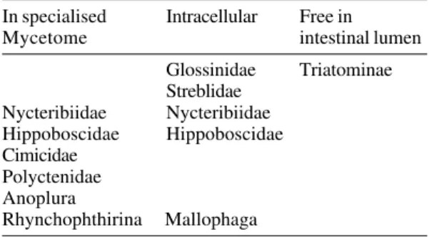
At the physiological level also, there is strong evidence for imperfect adaptation to a diet of verte-brate blood. All obligate blood-sucking insects seem to require symbionts, which are believed to provide vitamins such as folate that may be scarce in vertebrate blood (see Nyirady 1973). But whereas all other obligate blood-suckers carefully store spe-cific symbionts either intracellularly or in a special organ, the mycetome, the Triatominae invariably have a wide range of bacteria (not all of which may be important) living freely in the gut lumen (Table III). This would suggest them to be at an early stage in adaptation to obligate haematophagy. TABLE II
Biogeographical diversity of blood-sucking Hemiptera. Number of genera and species in Africa,
Asia and the Americas
Africa Asia Americas
Cimicidae
Genera 11 8 14
Species 76 11 20
Polyctenidae
Genera 3 4 1
Species 7 10 16
Triatominae
Genera 1 2 14
Species 1 13 116
Within the Americas, it is clear that the Triatominae could not have followed the proposed evolutionary route from predator to nest-dwelling blood-sucker prior to extensive development of nest-dwelling vertebrates. Adaptation to vertebrate nests would only be warranted if these represented a reasonably abundant resource, but would offer important advantages in terms of protection from climatic extremes (allowing reproduction to proceed with less dependence on seasonal climate) and more abundant proteinaceous food sources. However, exploitation of vertebrate blood requires important physiological and behavioural changes, especially in terms of avoiding predation by the vertebrate
TABLE III
Location of intestinal symbionts of obligate blood-sucking insects (excluding facultative blood-blood-sucking groups, and those that make use of other fluids as well
as vertebrate blood)
In specialised Intracellular Free in
Mycetome intestinal lumen
Glossinidae Triatominae Streblidae
Nycteribiidae Nycteribiidae Hippoboscidae Hippoboscidae Cimicidae
Polyctenidae Anoplura
539 539539 539 539 Mem Inst Oswaldo Cruz, Rio de Janeiro, Vol. 95(4), Jul./Aug. 2000
There is also evidence for rapid evolutionary change within the Triatominae. For example, com-parison of derived populations of Rhodnius prolixus in Central America, with their putative ancestral populations in Venezuela and Colombia, shows them to morphometrically distinguishable and also to have a reduced genome as indicated by far fewer RAPD bands. And yet historical recon-struction indicates separation of the Central Ameri-can and South AmeriAmeri-can forms to be due to acci-dental human intervention a mere 85 years ago (Dujardin et al. 1998a). Similarly, comparison of domestic T. infestans from Uruguay, with their origi-nal silvatic populations in Bolivia, again shows clear morphometric and genetic differentiation over a time scale that appears to be little more than 100 years (Dujardin et al. 1998a,b). A further well-studied example is that of T. rubrofasciata, exported from a New World origin on sailing ships to port areas throughout the tropics, but speciating in eastern Asia – to give seven species defined by morpho-logical characteristics – over a period that cannot have been much more than about 200 years (Gorla et al. 1997, Patterson 1999). Even in the space of a few generations in laboratory colony, morphologi-cal changes leading to apparent differentiation can be demonstrated (eg. Dujardin et al. 1999, Galindez-Girón et al. 2000).
All the evidence presented above points in the same direction. As blood-suckers, Triatominae seem capable of evolving rapidly, and appear to have done so quite recently. And their current geo-graphical distribution accords fully with this idea, since the distribution of species and species groups matches modern American geography, rather than ancient scenarios. In almost all cases so far exam-ined, species groups, complexes, and the smaller genera, occupy discrete geographic areas con-strained by post-pleistocene geographic features (Schofield 1988, Schofield & Dujardin 1999). The only exceptions are discontinuities in the distribu-tion of R. prolixus, T. infestans, and T. rubrofasciata, which are consistent with human intervention, and in the distribution of T. maculata (Venezuela) and T. pseudomaculata (Northeastern Brazil) which is consistent with their association with migrating birds. However, there is strong evidence to sug-gest that the Triatominae represent a polyphyletic grouping, with the smaller genera and many of the species groups of Triatoma assumed to have arisen from different predatory ancestors (Schofield & Dujardin 1999, Dujardin et al. 2000). Thus, although the evidence presented here suggests recent evo-lution of haematophagy in the Reduviidae to give the various forms now grouped as the Triatominae, there is no reason to suggest that such adapta-tions were simultaneous, nor that some form of
haematophagy could not have arisen in some ear-lier groups that have since become extinct.
RECONCILING THE PARADOX
In contrast to infection of a tsetse fly with T. brucei, which leads to a very complex system of defence and counter-defence mechanisms by fly and parasite (see Welburn & Maudlin 1999), infec-tion of a triatomine with T. cruzi leads to an ex-tremely modest interaction (see Brener 1979). This is reflected in the resulting infection rates, which tend to be extremely low in Glossinidae, but can often exceed 50% in Triatominae – simply mirroring the cumulative probability of taking an infected blood meal (Schofield 1994). Indeed, T. cruzi seems a relatively non-specific parasite, able to develop, at least partially, in a wide range of invertebrates including leeches, ticks, bedbugs, and even experi-mentally-infected caterpillars (eg. Brumpt 1912, Mazzotti & Osorio 1943, Goldman 1950, Marsden & Pettit 1969) (none of these acts as vector how-ever, since there is no adaptation for subsequent transmission). We can infer that association be-tween glossinids and T. brucei seems to be rela-tively ancient, while the association between Triatominae and T. cruzi may be be relatively re-cent. But as we have seen, there is evidence that T. cruzi itself may be relatively ancient, arising in as-sociation with marsupials when South America was still connected to Antartica and Australia over 65 mya. Here is the apparent paradox – ancient para-site and recent vector.
540 540 540 540
540 T. cruzi The Vector-parasite Paradox CJ Schofield
known to develop in insects, and it may be that freitasi is transmitted directly between opossums, for example via their anal gland secretions. More-over, subsequent studies of naturally infected opos-sums appeared to reveal not just T. cruzi and T. freitasi but also other flagellates in the anal glands (MP Deane, pers. commun. 1985, Deane & Jansen 1988, Jansen et al. 1988). Opossums are omnivo-rous, readily feeding on a wide range of vegetable and animal tissues, so the finding of trypanosomes and other flagellates in their anal glands suggests that these organisms were able to penetrate the oral mucosa and pass in the bloodstream to the glands. In other words, the capacity for mucosal penetration and subsequent survival in the blood would seem to be shared innately by a number of protozoan flagellates. The monophyletic origin of extant Trypanosomatidae may have been a flagel-late of plants or primitive insects, that first entered mammals by being eaten by an omnivorous marsu-pial and migrating to the anal glands. Subsequent adaptation to a further bloodstream form would then make the adapting parasite available for transmis-sion by blood-sucking insects.
So far in this hypothesis, we have a primitive trypanosomatid associated with didelphids in the southern supercontinent (Gondwanaland) during the early tertiary period about 65 mya. Then, around 40 mya, South America becomes separated from Antartica, and the fore-runners of modern Ameri-can didelphids commence their northerly spread. We can propose that during this time, the trypanosomatid is being transmitted directly be-tween opossums via their anal gland secretions (as demonstrated by Jansen & Deane 1985) and/or urine (as demonstrated by McKeever et al. 1958, and Olsen et al. 1964). But by the late tertiary or early pleistocene, some 2-5 mya, opossums would be common throughout South America, along with other nest-building vertebrates such as furnariid and psittacid birds, armadillos, and various forms of cricetid rodent. We can imagine that the habi-tats offered, and their associated guilds of nest-dwelling invertebrates, would be attractive to a wide range of reduviid predators – many of which would develop as facultative blood-suckers, ca-pable of imbibing opossum blood infected with the primitive trypanosome.
But a feature of opossum lodges is that they also offer refuges to other vertebrates, just as opos-sums themselves may utilise abandonned nests of birds or rodents, or even armadillo burrows. So the advent of blood-sucking Reduviidae, today classi-fied as Triatominae, would have provided the pre-cise vehicle for spreading the trypanosome to new hosts. In birds it would not survive – killed by a form of complement-mediated lysis in bird blood
(Kierszenbaum et al. 1976). But in rodents, armadil-los, and bats, it would not only survive, but en-counter profound selection pressures leading to the development of new forms distinguishable to-day by a range of genetic markers.
develop-541 541541 541 541 Mem Inst Oswaldo Cruz, Rio de Janeiro, Vol. 95(4), Jul./Aug. 2000
ment [observations that heavily infected salivary glands of Rhodnius tend to loose their characteris-tic red colouration (eg. Añez 1983) suggest that the parasites may be utilising the red nitrophorins, which could explain both their predilection for Rhodnius species, as well as contributing to vec-tor mortality]. And perhaps the evolutionary cost of this adaptation is mirrored by reduced survivor-ship in the mammalian hosts, possibly due to loss of membrane neuraminidase which seems to be in-volved in passage through the vector haemolymph by rangeli, and in penetrating vertebrate cells by cruzi (D’Alessandro & Saravia 1992).
This theory reconciles the apparent disparity between the antiquity of T. cruzi, and the relatively recent development of blood-sucking Triatominae. It is consistent with the relative homogeneity of cruzi 1, compared to the multiple clones of cruzi 2 and other forms proposed as derivatives from the cruzi stem. But the theory implies that the putative derivatives from the original cruzi stem have radi-ated in comparatively recent times in response to being vectored into new hosts by blood-sucking insects, which is not at all consistent with current attempts to date the divergence of cruzi using mo-lecular clocks. On the basis of 18S rDNA se-quences, for example, the divergence between cruzi 1 and cruzi 2 has been dated at 88 mya (before the arrival of opossums and rodents in South America2) and divergence between T. cruzi and T. rangeli was put at 475 mya (Briones et al. 1999). This is difficult to reconcile with the geological record, since 475 mya was the time of ammonites and early fish. Early insects arose during the upper Paleozoic about 330 mya, with early mammals and birds arising in the Jurassic period about 200 mya, so there is no obvious clue as to how cruzi and rangeli could have diverged, or even existed as such, at 475 mya. Perhaps part of the problem is
the assumption of strict homology between 18S sequences, whereas at least two divergent 18S types have been demonstrated in the cruzi genome (see Stothard 2000), and the assumption of a relatively low rate of sequence divergence (Briones et al. 1999) together with the idea that molecular clocks would run at a constant rate, whereas it may be that the intense selection pressure associated with ad-aptation to a completely new host – as in the pro-posed step from cruzi 1 to cruzi 2 – may distort the clock speed even of apparently ‘neutral’ genes such as 18S.
ACKNOWLEDGEMENTS
To several authorities for their contributions to the discussions that formed the basis of this review, particu-larly Jamie Stevens, Harry Noyes, Octavio Fernandes, Lileia Diotaiuti, Jean-Pierre Dujardin, Michel Tibayrenc, and Fernando Dias de Avila Pires. Errors of fact or interpretation however, are entirely my own.
REFERENCES
Abalos JW, Wygodzinsky P 1951. Las Triatominae Argentinas (Reduviidae, Hemiptera). An Inst Med
Regional, Tucuman, Monografia 2: 1-179
Adjé CA, Opperdoes FR, Michels PAM 1998. Molecu-lar analysis of phosphoglycerate kinase in
Trypanoplasma borreli and the evolution of this en-zyme in Kinetoplastida. Gene217: 91-99. Alvarez F, Cortinas MN, Musto H 1996. The analysis
of protein coding genes suggests monophyly of
Try-panosoma. Mol Phylog Evol5: 333-343.
Añez N 1983. Studies on Trypanosoma rangeli Tejera, 1920. VI. Developmental pattern in the haemolymph of Rhodnius prolixus. Mem Inst Oswaldo Cruz78: 413-419.
Brener Z 1979. O parasito: relações hospedeiro-parasito. In Z Brener & ZA Andrade (eds), Trypanosoma cruzi
e Doença de Chagas, Guanabara Koogan, Rio de Janeiro, p. 1-41.
Briones RSM, Souto RP, Stolf BS, Zingales B 1999. The evolution of two Trypanosoma cruzi subgroups inferred from rRNA genes can be correlated with the interchange of American mammalian faunas in the Cenozoic and has implications to pathogenicity and host specificity. Mol Bioch Pathog104: 219-232. Brisse S 1997. Phylogénie Moléculaire des Clones
Naturels de Trypanosoma cruzi, Agent de la Maladie de Chagas: Évolution Clonale, Recombinaison Génétique, et Relations Phylogénétiques avec d’Autres Espèces du Sous-genre Schizotrypanum, PhD The-sis, Université de Montpellier II, Montpellier, France.
Brisse S, Barnabé C, Tibayrenc M 1998. Trypanosoma
cruzi: how many relevant phylogenetic subdivisions are there? ParasitolToday14: 178-179.
Brumpt E 1912. Le Trypanosoma cruzi evolue chez
Conorhinus megistus, Cimex lecularius, Cimex boueti
et Ornithodorus moubata. Cycle evolutif de ce para-site. Bull Soc Pathol Exotique5: 360-367.
Capriles JM, Santiagoblay JA, Poinar GO 1993a. 2 The earliest marsupial fossils, in Asia-America, have
542 542 542 542
542 T. cruzi The Vector-parasite Paradox CJ Schofield
Apicrenus fossilis gen and sp-n (Heteroptera, Redu-viidae, Apiomerinae) from Dominican amber (lower oligocene-upper eocene). Entomol Scandinavica24: 139-142.
Capriles JM, Santiagoblay JA, Poinar GO 1993b.
Paleoploiariola venosa, a new fossil Emesinae (Heteroptera, Reduviidae) genus and species from Dominican amber. J Agric Univ Puerto Rico77: 95-100.
Carcavallo RU, Jurberg J, Lent H 1999. Phylogeny of the Triatominae. In Atlas of Chagas Disease
Vec-tors in the Americas, Vol. 3, Fiocruz, Rio de Janeiro, p. 925-969.
Clemens WA 1966. Origin and early evolution of marsu-pials. Evolution22: 18.
Cobben RH 1978. Evolutionary trends in Heteroptera. Part II. Mouthpart-structures and feeding strategies.
Mededlingen Landbouwhogeschool, Wageningen78: 1-407.
Cobben RH 1979. On the original feeding habits of the Hemiptera (Insecta): a reply to Merrill Sweet. An
Entomol Soc America72: 711-715.
D’Alessandro A, Saravia NG 1992. Trypanosoma
rangeli. In JP Kreier & JR Baker (eds), Parasitic
Protozoa, 2nd ed., Vol. 2, Academic Press, San Di-ego, p. 1-54.
Davis NT 1969. Contribution to the morphology and phylogeny of the Reduvioidea. Part IV. The Harpactaroid complex. An Entomol Soc Am62: 74-94.
Deane MP, Jansen AM 1986. Another Trypanosoma distinct from Trypanosoma cruzi multiplies in the lumen of the anal gland of the opossum Didelphis
marsupialis. Mem Inst Oswaldo Cruz81: 131-132. Deane MP, Jansen AM 1988. From a mono to a dige-netic life-cycle: how was the jump for flagellates of the family Trypanosomatidae. Mem Inst Oswaldo
Cruz83: 273-275.
Deane MP, Jansen AM 1990. Developmental stages of
Trypanosoma (Megatrypanum) freitasi Rego, Magalhães & Siqueira, 1957 in the opossum
Didel-phis marsupialis (Marsupialia, Didelphidae). J
Protozool37: 44-47.
Deane MP, Lenzi HL, Jansen AM 1984. Trypanosoma
cruzi: vertebrate and invertebrate cycles in the same mammal host, the opossum Didelphis marsupialis.
Mem Inst Oswaldo Cruz79: 513-515.
Deane MP, Lenzi HL, Jansen AM 1986. Double devel-opment cycle of Trypanosoma cruzi in the opos-sum. Parasitol Today2: 146-147.
Diotaiut L, Pereira AS, Loiola CF, Fernandes AJ, Schofield CJ, Dujardin JP, Dias JCP, Chiari E 1995. Inter-relation of sylvatic and domestic transmission of
Trypansoma cruzi in areas with and without domes-tic vectorial transmission in Minas Gerais, Brazil.
Mem Inst Oswaldo Cruz 90: 443-448.
Dos Santos WG, Buck GA 1999. Polymorphisms at the topoisomerase II gene locus provides more evidence for the partition of Trypanosoma cruzi into two major groups. J Eukar Microbiol46: 17-23.
Dujardin JP, Muñoz M, Chavez T, Ponce C, Moreno J, Schofield CJ 1998a. The origin of Rhodnius prolixus
in Central America. Med Vet Entomol12: 113-115. Dujardin JP, Schofield CJ, Tibayrenc M 1998b.
Popula-tion structure of Andean Triatoma infestans: allozyme frequencies and their epidemiological relevance. Med
Vet Entomol12: 20-29.
Dujardin JP, Schofield CJ, Panzera F 2000. Les Vecteurs
de la Maladie de Chagas. Recherches Taxonomiques, Biologiques et Génétiques, Académie Royale des Sci-ences d’Outre Mer, Brussels, 162 pp.
Dujardin JP, Steindel M, Chavez T, Machane M, Schofield CJ 1999. Changes in the sexual dimorphism of Triatominae in the transition from natural to arti-ficial habitats. Mem Inst Oswaldo Cruz94: 565-569. Evans JW 1956. Palaeozoic and mesozoic Hemiptera
(Insecta). Austral J Zool 4: 165-258.
Fernandes AJ 1989. Importancia de Didelphis albiventris
e Panstrongylus megistus na Interacão dos Ciclos de Transmissão do Trypanosoma cruzi no Municipio de Bambuí, Minas Gerais, Thesis, UFMG, Belo Horizonte, 116 pp.
Fernandes AJ, Chiari E, Rodrigues RR, Dias JCP, Romanha AJ 1991. The importance of the opossum (Didelphis albiventris) as a reservoir for
Trypano-soma cruzi in Bambuí, Minas Gerais state. Mem
Instit Oswaldo Cruz 86: 81-85.
Fernandes AJ, Diotaiuti L, Chiari E, Dias JCP 1987. Natural infection of Didelphis albiventris by
Trypa-nosoma cruzi and Trypanosoma freitasi. Mem Instit
Oswaldo Cruz82 (Suppl.): 65.
Fernandes AJ, Diotaiuti L, Dias JCP, Rnmanha AJ, Chiari E 1989. Infecção natural das glandulas anais de gambas (Didelphis albiventris) pelo Trypanosoma cruzi no município de Bambui – MG. Mem Inst Oswaldo
Cruz 84: 87-93.
Fernandes O, Souto RP, Castro JA, Pereira JB, Fernandes NC, Junqueira ACV, Naiff RD, Barrett TV, Degrave W, Zingales B, Campbell DA, Coura JR 1998. Bra-zilian isolates of Trypanosoma cruzi from humans and triatomines classified into two lineages using mini-exon and ribosomal RNA sequences. Am J Trop Med
Hyg58: 807-811.
Galindez-Girón I, Torres E, Galvão C, Magalhães dos Santos C, Lizano E, Barazarte R, Marquez J 1999. Influencia da manutenção em laboratorio sobre o fenotipo de Rhodnius robustus Larrousse, 1927 (Hemiptera, Reduviidae, Triatominae). Entomol Vect
6: 677-703.
Goldman M 1950. The experimental infection of pupae of Philosamia cynthia Drury (Lepidoptera: Saturniidae) with Trypanosoma cruzi. J Parasitol
36: 1-8.
Gorla DE, Schofield CJ, Dujardin JP 1997. Biosystem-atics of Old World Triatominae. Acta Trop63: 127-140.
Hannaert V, Opperdoes FR, Michels PAM 1998. Com-parison and evolutionary analysis of the glycosomal glyceraldehyde-3-phosphate dehydrogenase from different Kinetoplastida. J Mol Evol47: 728-738. Hashimoto T, Nakamura Y, Kamaishi T, Adachi J,
543 543543 543 543 Mem Inst Oswaldo Cruz, Rio de Janeiro, Vol. 95(4), Jul./Aug. 2000
Mol Bioch Parasitol70: 181-185.
Hoare CA 1972. TheTtrypanosomes of Mammals, Blackwell Scientific Publications, Oxford.
Jansen AM, Deane MP 1985. Trypanosoma cruzi infec-tion of mice by ingesinfec-tion of food contamined with material of the anal glands of the opossum Didelphis
marsupialis. XII Reunião Anual sobre Pesquisa Básica em Doença de Chagas, Caxambu, BI-09. Jansen AM, Carreira JC, Deane MP 1988. Infection of a
mammal by monogenetic insect trypanosomatids (Kinetoplastida, Trypanosomatidae). Mem Inst
Oswaldo Cruz83: 271-272.
Jeannel R 1919. Henicocephalidae et Reduviidae. In
Voy-age de Ch. Alluaud et R. Jeannel en Afrique orientale (1911-1912). Resultats scientifiques – Hemiptera, vol. 3, Paris, p.131-314.
Kalshoven LGE 1970. Observations of the blood-suck-ing reduviid Triatoma rubrofasciata (De Geer) in Java. Entomologische Berichten, Amsterdam30: 41-47.
Kierszenbaum F, Ivanyi J, Budzko DB 1976. Mecha-nism of natural resistance to trypanosomal infec-tion. Role of complement in avian resistance to
Try-panosoma cruzi infection. Immunology30: 1-6. Lake JA, De La Cruz VF, Ferreira PCG, Morel C,
Simpson L 1988. Evolution of parasitism: Kinetoplastid protozoan history reconstructed from mitochondrial rRNA gene sequences. Proc Nat Acad
Sci USA85: 4779-4783.
Lent H 1982. Microtriatoma pratai Sherlock & Guitton, 1982 e sinonimo do hemiptero predador
Ara-domorpha championi Lent & Wygodzinsky, 1944 (Reduviidae, Reduviinae). Mem Inst Oswaldo Cruz
77: 449-451.
Lent H, Wygodzinsky P 1979. Revision of the Triatominae (Hemiptera, Reduviidae), and their sig-nificance as vectors of Chagas disease. Bul Am Mus
Nat Histo163: 123-520.
Lenzi HL, Jansen AM, Deane MP 1984. The recent discovery of what might be a primordial escape mechanism for Trypanosoma cruzi. Mem Inst
Oswaldo Cruz79: 13-18.
Lorosa ES, Jurberg J, Souza ALA, Vinhaes MC, Nunes IM 2000. Hemolinfa de blatideos na manutenção do ciclo biológico silvestre de Triatoma rubrovaria (Blanchard 1843) e Triatoma circummaculata (Stal, 1859). Rev Soc Bras Med Trop 35 (Supl.1): 172. Maldonado Capriles J 1990. Systematic catalague of the
Reduviidae of the world (Insecta: Heteroptera).
Car-ibbean J Sci (Special edition) x + 1-694.
Marsden PD, Pettitt LE 1969. The survival of
Trypano-soma cruzi in the medicinal leech (Hirudo
medicinalis). Trans R Soc Trop Med Hyg63: 414-415.
Mazzotti L, Osorio MT 1943. Experimentos de transmisión de Trypanosoma cruzi en cuatro especies de Ornithodorus. Rev Inst Salub Enferm Trop, Mexico
4: 163-165.
McKeever S, Gorman GW, Norman L 1958. Occurrence of a Trypanosoma cruzi-like organism in some mam-mals from southwestern Georgia and northwestern Florida. J Parasitol44: 583-587.
Memórias 1999. Recommendations from a satellite meet-ing. Mem Inst Oswaldo Cruz94 (Suppl.1): 429-432. Miles MA 1979. Transmission cycles and the heteroge-neity of Trypanosoma cruzi. In WHR Lumsden & DA Evans (eds), Biology of the Kinetoplastida, Vol. 2, London, Academic Press, p. 117-196.
Miles MA, Souza AA de, Povoa M 1981. Chagas dis-ease in the Amazon basin. III. Ecotopes of ten triatomine bug species (Hemiptera: Reduviidae) from the vicinity of Belém, Pará State, Brazil. J Med
Entomol18: 266-278.
Naiff RD, Barrett TV, Arias AR 1987. Trypanosoma
cruzi nas galndulas anais de Didelphis marsupialis: primero registro de infecções naturais. X Congresso da Sociedade Brasileira de Parasitologia, Salvador, BA, abstract 165.
Nyirady SA 1973. The germfree culture of three species of Triatominae: Triatoma protracta (Uhler),
Tri-atoma rubida (Uhler) and Rhodnius prolixus Stal. J
Med Entomol10: 417-448.
Olsen PF, Shoemaker JP, Turner HF, Hays KL 1964. Incidence of Trypanosoma cruzi (Chagas) in wild vectors and reservoirs in east-central Alabama. J
Parasitol 50: 599-603.
Patterson J 1999. A Morphometric Investigation of the
Relationships between Triatoma rubrofasciata (Hemi-ptera: Reduviidae: Triatominae), Old World Tri-atoma and Representatives of the New World Spe-cies, Thesis, LSHTM, London, 44 pp.
Pipkin AC 1969. Transmision of Trypanosoma cruzi by arthropod vectors: anterior versus posterior route infection. Internl Rev Trop Med3: 1-47.
Poinar GO 1991. Praecoris dominicana gen n, sp n (Hemiptera, Reduviidae, Holoptilinae) from Domini-can amber, with an interpretation of past behavior based on functional morphology. Entomol
Scandinavica22: 193-199.
Putshkov VG, Putshkov PV 1985. A catalogue of As-sassin-bug genera of the world (Heteroptera, Redu-viidae). Vestnik Zoologica, 112 pp.
Ryckman RE 1951. Recent observations of cannibalism in Triatoma (Hemiptera: Reduviidae). J Parasitol37: 433-434.
Ryckman RE, Bentley DG 1979. Host reactions to bug bites (Hemiptera, Homoptera): a literature review and annotated bibliography. Calif Vect Views26: 1-49. Sandoval CM, Joya MI, Gutierez R, Angullo VM 2000.
Cleptohaematophagy of the triatomine bug Belminus
herreri. Med Vet Entomol14: 100-101.
Schofield CJ 1988. Biosystematics of the Triatominae. In M Service, Biosystematics of Haematophagous
Insects, Clarendon Press, Oxford, p. 284-312. Schofield CJ 1994. Triatominae - Biology & Control,
Eurocommunica Publications, West Sussex, UK, 80 pp.
Schofield CJ, Dolling WR 1993. Bedbugs and kissing-bugs (bloodsucking Hemiptera). In RP Lane & RWCrosskey (eds), Medical Insects and Arachnids,
Chapman & Hall, London, UK, p. 483-516. Schofield CJ, Dujardin JP 2000. Theories on the evolution
544 544 544 544
544 T. cruzi The Vector-parasite Paradox CJ Schofield
Heteropteran classification. Ann Rev Entomol31: 67-93.
Schuh RT, Slater JA 1995. True Bugs of the World
(Hemi-ptera: Heteroptera) Classification and Natural His-tory, Cornell University Press, New York, 336 pp. Souto RP, Fernandes O, Macedo AM, Campbell DA,
Zingales B 1996. DNA markers define two major phylogenetic lineages of Trypanosoma cruzi. Mol
Bioch Parasitol 83: 141-152.
Southwood TRE, Leston D 1959. Land and Water Bugs
of the British Isles, Frederick Warne, London, xi + 436 pp.
Steindel M, Pinto CJC 1988. Trypanosoma cruzi devel-opment in the anal glands of experimentally infected
Lutreolina crassicaudata (Marsupialia, Didelphidae).
Mem Inst Oswaldo Cruz83: 387.
Steindel M, Scholz AF, Toma HK, Schlemper Junior BR 1988. Presence of Trypanosoma cruzi in the anal glands of naturally infected opossums (Didelphis
marsupialis) in the State of Santa Catarina, Brazil.
Mem Inst Oswaldo Cruz83: 135-137.
Stevens J, Gibson W 1999. The evolution of salivarian trypanosomes – Second Internet Conference on Salivarian Trypanosomes. Mem Inst Oswaldo Cruz
94: 225-228.
Stevens JR, Noyes HA, Dover GA, Gibson WC 1999a. The ancient and divergent origins of the human patho-genic trypanosomes, Trypanosoma brucei and T.
cruzi. Parasitology118: 107-116.
Stevens JR, Noyes HA, Schofield CJ, Gibson W 2000. The molecular evolution of Trypanosomatidae. Adv
Parasitol48: 1-53.
Stevens JR, Teixeira MMG, Bingle LEH, Gibson WC 1999b. The taxonomic position and evolutionary relationships of Trypanosoma rangeli. Internl J
Parasitol29: 749-757.
Stothard JR 2000. Trypanosome trees and homologies.
Parasitol Today16: 173.
Sweet MH 1979. On the original feeding habits of the
Hemiptera (Insecta). Ann Entomol Soc America72: 575-579.
Teo SK, Cheah JS 1973. Severe reaction to the bite of the triatomine bug (Triatoma rubrofasciata) in Singapore. J Trop Med Hyg76: 161-162.
Thomas DB 1992. A fossil Empicoris Wolff (Reduvi-idae, Heteroptera) from Mexican amber with remarks on the phylogenetic status of the fossil genus
Alumeda Popov. J NY Entomol Soc100: 535-539. Tibayrenc M 1995. Population genetics of parasitic
pro-tozoa and other microorganisms. Adv Parasitol36: 47-115.
Tibayrenc M, Neubauer K, Barnabe C, Guerrini F, Skarecky D, Ayala FJ 1993. Genetic characteriza-tion of six parasitic protozoa: parity between ran-dom-primer DNA typing and multilocus enzyme electrophoresis. Proc Natl Acad Sci USA 90: 1335-1339.
Valente VC, Valente SAS, Noireau F, Carrasco HJ, Miles MA 1998. Chagas disease in the Amazon Basin: association of Panstrongylus geniculatus (Hemiptera: Reduviidae) with domestic pigs. J Med Entomol35: 99-103.
Welburn SC, Maudlin I 1999. Tsetse-trypanosome in-teractions: rites of passage. Parasitol Today 15: 399-403.
Wiemer EAC, Hannaert V, Van Den Ijssel PRLA, Van Roy J, Opperdoes FR, Michels PAM 1995. Mo-lecular analysis of glyceraldehy3-phosphate de-hydrogenase in Trypanoplasma borreli: an evolu-tionary scenario of subcellular compartmentation in Kinetoplastida. J Mol Evol 40: 443-454.
Wootton RJ 1981. Palaeozoic insects. Ann Rev Entomol
26: 319-344.