Relato de Caso / Case Report
IJD
ISSN:1806-146XThe use of a magnetic attachment in a removable partial denture of
a patient with periodontal tissue loss
O uso dos magnetos em prótese parcial removível de pacientes com perda de tecido periodontal
Adriana da Fonte Porto Carreiro 1
Cátia Maria Fonseca Guerra 2
Sandra Lúcia Dantas de Moraes 3
Arcelino Farias Neto 4
Érica Miranda de Torres 5
Erika Oliveira de Almeida 6
1 - Associate Professor, Department of Dentistry, Rio Grande do Norte Federal University.
2- Associate Professor, Department of Prosthodontics and Buco Facial Surgery, Pernambuco Federal University. 3 - Assistant Professor, Department of Restorative Dentistry, Pernambuco University.
4 - Master Degree in Dentistry,
Department of Dentistry, Rio Grande do Norte Federal University.
7 - Associate Professor, Department of Stomatology Science, Goias Federal University.
6 - Doctorate Student in Dental Prosthesis, Araçatuba School of Dentistry, Sao Paulo State University.
Correspondência:
Erika Oliveira de Almeida – Departamento de Materiais Odontológico e Prótese. Rua José Bonifácio, 1193, Araçatuba-SP, Brazil, 16015-050.
FAX: 5518- 36363332. Email: [email protected]
ABSTRACT
The use of magnets in removable partial dentures is a valid alternative for eliminating prosthetic structures that may interfere in esthetics without losing retention and stability. Magnets can also be recommended for patients with periodontal tissue loss, since they can decrease transmission of forces to remaining teeth. The aim of this clinical report was to present an alternative prosthetic rehabilitation treatment for periodontally-compromised abutment teeth, and to point out the advantages and disadvantages of using magnets in removable partial dentures (overdentures) from the functional, biological, and esthetic point of view.
Key-words: overdenture, denture partial removable, esthetics, denture retention
RESUMO
O uso dos magnetos em prótese parcial removível é uma alternativa viável para eliminar a estrutura metálica que pode interferir na estética sem perder retenção e estabilidade. Os magnetos podem ser recomendados para pacientes com perda de tecido periodontal, desde que eles diminuam a transmissão de forças ao dente remanescente. O objetivo deste relato de caso clínico foi apresentar uma alternativa de tratamento protético para dentes pilares comprometidos periodontalmente e descrever as vantagens e desvantagens do uso dos magnetos em prótese parcial removível sobre os pontos de vista funcionais, biológicos e estéticos.
Palavras-chave: : revestimento da dentadura, prótese parcial removível, estética, retenção em dentadura.
INTRODUCTION
When teeth present periodontal tissue loss, but tissues remain healthy, oral rehabilitation of the patient is possible with overdentures. Overdentures cover one or more teeth or dental implants, restoring the entire dentition. They help preserve natural teeth or roots that are often indicated for extraction because of periodontal tissue loss. Maintaining these teeth or roots enables a delay in alveolar bone resorption and preserves periodontal proprioception and masticating efficiency1, improving the
quality of life and oral health of older people.
A range of attachment systems are used to retain overdentures. The most common devices are the bar-clip, O-ring, the ERA system, and magnets. The bar-clip, ERA
system, and the O-ring offer greater retaining power for overdentures than magnets2; however, magnets transmit lower force levels to the teeth than other types of attachments 3. Surface abrasion due to wear
caused by the eccentric movements in the mouth does not affect their retentive force significantly4. Laboratory studies show that magnets have more constant retentive properties and are less susceptible to fatigue than stud attachments5,6. The use of magnets in removable partial dentures promotes adequate retention and stability, eliminates prosthetic structures that may compromise esthetics, and improves the distribution of forces to the dental support structures, mainly by dissipating lateral forces7.
them to stabilize the prosthesis in a jaw with advanced alveolar bone resorption. For some time the magnet of choice for retaining dental and maxillofacial prostheses was samarium-cobalt (Sm-Co); however, this magnet was considered fragile and was later substituted8,9. The most commonly
magnet used in prosthetic dentistry today is neodymium-iron-boron. It combines high magnetic saturation with good resistance to demagnetization and produces higher amounts of energy than any other known magnetic material. Since one of its limitations is low resistance to corrosion by oral fluids, it must be coated by another material, generally titanium10.
Magnets that combine cobalt and platinum are also used. An example is Dyna® (Dental Engineering, Bergen op
Zoom, The Netherlands), which is a system that has variable retention of between 300 and 500 grams/force11. A new alloy of iron and platinum has recently been investigated for use in prosthetic dentistry, owing to its strong force of attraction and high magnetic saturation. Since this alloy contains a high percentage of platinum, it is expected to offer excellent resistance to corrosion and may be used as a substitute for Nd-Ir-B12.
There are several reports of the growing use of magnets in prosthetic dentistry13–20. Matsumura and Kawasaki13 reported the use
of magnets in sectional prostheses for patients with maxillofacial defects. According to these authors, magnets may substantially improve prosthodontic treatment for these patients. Thean et al14 reported the use of
magnetic retainers in removable partial dentures. These magnets exhibited no deterioration after 3 years in function; however, long-term prospective studies on implant-retained overdentures are limited. Interestingly, Tan and Walmsley15 reported a case where the patient successfully wore a mandibular implant-retained overdenture with magnets for 12 years. Barker and Cooper16 described a unilateral removable partial denture with a hinged arm with magnetic retention and a prosthetic flange component that was provided for a patient with localized periodontal tissue loss. Finally, Rockman et al17 presented a case report demonstrating a technique using magnets to enhance the retention of maxillary and mandibular prostheses in a 9-year-old boy with Ectodermal Dysplasia.
The aim of this clinical report is to show an alternative prosthetic treatment for patients with periodontal tissue loss, and to
emphasize the advantages and disadvantages of using magnets in removable partial dentures (overdentures) from the functional, biological, and esthetic point of view.
CLINICAL REPORT
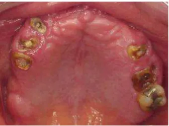
A 67-year-old man came to our clinic for prosthodontic treatment. He wore an interim fixed upper denture and a lower removable partial denture (RPD). In the upper arch, the remaining teeth (maxillary right second molar, maxillary right first molar, maxillary right first pre-molar, maxillary left canine, maxillary left first pre-molar and maxillary left second pre-molar) had fractures, caries, and periodontal tissue loss (Fig. 1). During extra-oral clinical examination, the patient showed signs of shortening in the lower 1/3 of the face, pronounced facial sulcus, inverted lips, and angular cheilitis.
Figure 1.Initial aspect of the patient’s
upper arch
After obtaining radiographs and study casts mounted on a semi-adjustable articulator, a treatment plan of an upper RPD retained by magnets and a new lower conventional RPD was proposed. The
patient’s consent for the treatment was
impression was taken using silicone on the original lower RPD of the patient.
An upper wax-up trial base was prepared and adjusted to guide the mounting of the upper model onto the articulator, using the facial arch. To mount the lower model, a registration of the maxillomandibular relation was taken. The upper artificial teeth were arranged according to esthetic references and were clinically evaluated. A space was observed between the upper trial denture and the lower RPD, indicating the need to correct the posterior occlusion. The upper denture was fabricated, and the occlusion surface of the original lower RPD was adapted. This adjustment was initially made by putting tracing compound (Impression Compound, Kerr, Orange, CA) on the lower RPD posterior surfaces in order to restore the occlusion. Care was taken to isolate the upper teeth. The compound was removed from one side, and the opposite side was adjusted for direct application of chemically activated acrylic resin (Dencôr, Clássico, São Paulo, Brazil). After polymerization, this process was repeated on the other side. The lower RPD surfaces were polished.
The provisional upper denture was installed and recoated with resilient resin (Coe-soft, GC America, Alsip, IL) for greater comfort. The occlusion was checked. After a period of 60 days, the patient felt comfortable and presented occlusal stability. In the next stage, due to the endodontic and periodontal conditions, the roots of the maxillary first right molar and maxillary left canine were removed, while the maxillary first right molar, maxillary first left pre-molar, and maxillary second left pre-molar were endodontically re-treated, and the filling in the maxillary second right molar was replaced. The top of the roots was prepared to obtain the most horizontal surface possible and to avoid the influence of lateral loads (Fig. 2).
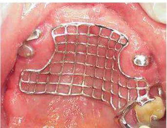
An impression of the prepared root canals was made using Duralay resin (Reliance, Worth, IL), which left a retentive area at the cervical level. This allowed impression and transfer with an addition silicone (Aquasil Ultra, Dentsply, Milford, DE) (Fig. 3). The cores were cast in special metal for Dyna®, and tested in the mouth. All adjustments were made, and conventional cementing was performed using zinc phosphate cement (Zinc Cement, S.S. White, Gloucester, England) (Fig. 4).
Figure 2. Upper remaining tooth after root
canal preparation.
Figure 3. Upper arch impression. The root
canals were directly modeled in Duralay resin.
Figure 4. Fitting of upper framework. No
metal was used in the core areas to avoid interference with the magnetic attachments.
preparations, such as occlusal support niches and guideplanes. Final upper and lower impressions were made with custom trays. Due to the large edentulous area in the upper arch, the impression was made with a combination of tracing compound and Impregum® (3M ESPE, Seefeld, Germany).
In the lower arch, the impression was made with alginate (Jeltrate, Dentsply, Milford, DE). The master models obtained were checked again in the surveyor and used to fabricate the metallic structures. Full palate coverage was proposed for the upper arch, excluding the metallic structure design in the core areas (Fig. 4).
The metallic structures were tested and adjusted in the oral cavity, observing the correct relationship between their elements and abutment teeth and residual ridge. A functional lower impression was taken using
McCracken’s altered model21. The final
models were mounted on the semi-adjustable articulator. The facial arch was used to mount the upper model, and wax rims were used to take the maxillomandibular ratio.
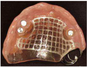
Artificial teeth were mounted and tested in the oral cavity to check occlusion and esthetic results. The dentures were manufactured and installed (Fig. 5) and the magnets were positioned onto the cores before directly fixing on the upper denture base with colorless acrylic resin (Jet, Clássico, São Paulo, Brazil) (Fig. 6). The patient was then instructed with regard to correct insertion and removal procedures, adequate cleaning, and the need for future adjustments.
Figure 5. Clinical view of the new dentures
in position
DISCUSSION
Even when remaining teeth have periodontal tissue loss, they can support a denture and transmit masticatory pressures to the periodontal ligament receptors. This
improves the patient’s oral perception,
causing an increase of roughly 20% in masticatory efficiency. In such cases, prosthetic planning must be carried out to provide an adequate distribution of forces to the abutment teeth. An overdenture retained by magnets may provide an excellent option, since it offers adequate retention and decreases the transmission of excessive forces to the remaining teeth3,7.
Figure 6. Internal view of the overdenture
with the magnets fixed
According to Gillings and Samant18, magnetic retention presents very little risk of trauma to the root that supports the overdenture, and the lateral forces imposed on the root are very small. Additionally, the magnetic overdentures are more stable and retentive than conventional partial dentures, and they are easily removed and seated without the patient having to grapple with clasps and complex paths of insertion, thereby improving esthetics, function, and comfort14,17. As such, a
magnetically-retained overdenture, as made in this study, may represent a good alternative for rehabilitating patients with reduced periodontium. This type of retention is effective, as it dissipates lateral forces to these structures and provides esthetic rehabilitation to a partially edentulous patient.
fluids, which requires them to be enclosed in an inert metal alloy of stainless steel or titanium11.
REFERENCES
1. Highton R, Caputo AA, Kinni M, Matyas J. The interaction of a magnetically retained denture with osseointegrated implants. J Prosthet Dent. 1988;60:486-490.
2. Naert I, Gizani MV, Steenberghe DV. A 5-year prospective randomized clinical trial on the influence of splinted and unsplinted oral implants retaining a mandibular overdenture: prosthetic aspects and patient satisfaction. J Oral Rehabil. 1999;26:195-202.
3. Fujimoto T, Niimi A, Murakami I, Ueda M. Use of new magnetic attachments for implant-supported overdentures. J Oral Implantol. 1998;24:147-151. 4. Huang Y, Tawada Y, Hata Y, Watanabe F. The change in retentive force of magnetic attachment by abrasion. Odontology 2008;96(1):65-8.
5. Rutkunas V, Mizutani H, Takahashi H. Evaluation of stable retentive properties of overdenture attachments. Stomatologija 2005;7(4):115-20.
6. Rutkunas V, Mizutani H, Takahashi H. Influence of attachment wear on retention of mandibular overdenture. J Oral Rehabil. 2007;34(1):41-51. 7. Chung KH, Chung CY, Cagna DR, Cronin RJ. Retention characteristics of attachment systems for implant overdentures. J Prosthodont. 2004;13:221-226.
8. Walmsley AD. Magnetic retention in prosthetic dentistry. Dent Update. 2002;29:428-433.
9. Gonda T, Ikebe K, Ono T, Nokubi T. Effect of magnetic attachment with stress breaker on lateral stress to abutment tooth under overdenture. J Oral Rehabil. 2004;3:1001-1006.
10.Yiu EYL, Fang DTS, Chu FCS, Chow TW. Corrosion resistance of iron-platinum magnets. J Dent. 2004;32:423-429.
11.Lemon JC, Brigoni RA, Collard SM, Martin JW, Powers JM, Chambers MS. In vitro effect of microwave irradiation on the retentive force of magnets. J Prosthet Dent. 2004;91:368-373.
12.Riley MA, Walmsley AD, Harris IR. Magnets in prosthetic dentistry. J Prosthet Dent. 2001;86:137-142. 13.Matsumura H, Kawasaki K. Magnetically connected removable sectional denture for a maxillary defect with severe undercut: A clinical report. J Prosthet Dent. 2000;84:22-26.
14.Thean HP, Khor SK, Loh PL. Viability of magnetic denture retainers: a 3-year case report. Quintessence Int. 2001;32:517-520.
15.Tan AS, Walmsley AD. Mandibular implant-retained overdenture with magnets: a case report. Dent Update 2004;31(2):104-8.
16.Barker D, Cooper A. A novel use of a unilateral hinged partial denture. Br Dent J. 2006;201:571-573. 17.Rockman RA, Hall KB, Fiebige M. Magnetic retention of dental prostheses in a child with ectodermal dysplasia. J Am Dent Assoc. 2007;138:610-615. 18.Gillings BRD, Samant A. Overdenture with magnetic attachments. Dent Clin North Am. 1990;34:683-709. 19.Sasaki H, Kinouchi Y, Tsutsui H, Yoshida Y, Ushita T. A magnetic attachment for overdentures. J Prosthet Dent. 1984;51:450-455.