The ATM Signaling Cascade Promotes
Recombination-Dependent Pachytene Arrest
in Mouse Spermatocytes
Sarai Pacheco1,2☯, Marina Marcet-Ortega1,2☯, Julian Lange3, Maria Jasin4,
Scott Keeney3,5*, Ignasi Roig1,2*
1Genome Integrity and Instability Group, Institut de Biotecnologia i Biomedicina, Universitat Autònoma de Barcelona, Cerdanyola del Vallès, Spain,2Cytology and Histology Unit, Department of Cell Biology, Physiology and Immunology, Universitat Autònoma de Barcelona, Cerdanyola del Vallès, Spain,3Molecular Biology Program, Memorial Sloan Kettering Cancer Center, New York, New York, United States of America,
4Developmental Biology Program, Memorial Sloan Kettering Cancer Center, New York, New York, United States of America,5Howard Hughes Medical Institute, Memorial Sloan Kettering Cancer Center, New York, New York, United States of America
☯These authors contributed equally to this work.
*[email protected](SK);[email protected](IR)
Abstract
Most mutations that compromise meiotic recombination or synapsis in mouse spermato-cytes result in arrest and apoptosis at the pachytene stage of the first meiotic prophase. Two main mechanisms are thought to trigger arrest: one independent of the double-strand breaks (DSBs) that initiate meiotic recombination, and another activated by persistent combination intermediates. Mechanisms underlying the recombination-dependent arrest re-sponse are not well understood, so we sought to identify factors involved by examining mutants deficient for TRIP13, a conserved AAA+ ATPase required for the completion of meiotic DSB repair. We find that spermatocytes with a hypomorphicTrip13mutation (Trip13mod/mod) arrest with features characteristic of early pachynema in wild type, namely, fully synapsed chromosomes without incorporation of the histone variant H1t into chroma-tin. These cells then undergo apoptosis, possibly in response to the arrest or in response to a defect in sex body formation. However, TRIP13-deficient cells that additionally lack the DSB-responsive kinase ATM progress further, reaching an H1t-positive stage (i.e., similar to mid/late pachynema in wild type) despite the presence of unrepaired DSBs. TRIP13-defi-cient spermatocytes also progress to an H1t-positive stage if ATM activity is attenuated by hypomorphic mutations inMre11orNbs1or by elimination of the ATM-effector kinase CHK2. These mutant backgrounds nonetheless experience an apoptotic block to further spermatogenic progression, most likely caused by failure to form a sex body. DSB numbers are elevated inMre11andNbs1hypomorphs but notChk2mutants, thus delineating genet-ic requirements for the ATM-dependent negative feedback loop that regulates DSB num-bers. The findings demonstrate for the first time that ATM-dependent signaling enforces the normal pachytene response to persistent recombination intermediates. Our work supports the conclusion that recombination defects trigger spermatocyte arrest via pathways than are genetically distinct from sex body failure-promoted apoptosis and confirm that the latter
OPEN ACCESS
Citation:Pacheco S, Marcet-Ortega M, Lange J, Jasin M, Keeney S, Roig I (2015) The ATM Signaling Cascade Promotes Recombination-Dependent Pachytene Arrest in Mouse Spermatocytes. PLoS Genet 11(3): e1005017. doi:10.1371/journal. pgen.1005017
Editor:Michael Lichten, National Cancer Institute, UNITED STATES
Received:March 11, 2014
Accepted:January 22, 2015
Published:March 13, 2015
Copyright:© 2015 Pacheco et al. This is an open access article distributed under the terms of the
Creative Commons Attribution License, which permits unrestricted use, distribution, and reproduction in any medium, provided the original author and source are credited.
Data Availability Statement:All relevant data are within the paper and its Supporting Information files.
can function even when recombination-dependent arrest is inoperative. Implications of these findings for understanding the complex relationships between spermatocyte arrest and apoptosis are discussed.
Author Summary
Meiosis is the specialized cell division by which haploid cells are produced. As germ cells enter the first meiotic prophase, programmed double-stranded breaks (DSBs) are formed throughout the genome. Repair of these DSBs by homologous recombination is crucial for proper segregation of homologous chromosomes at the end of the first meiotic division, and thus, for the production of haploid gametes. Moreover, failure to correctly repair these DSBs can have deleterious effects on the genomic integrity of offspring. To ensure that meiocytes that fail to repair meiotic DSBs do not complete meiosis, recombination is tight-ly controlled. However, the signaling pathway(s) tying meiotic recombination to meiotic progression in mouse spermatocytes is not known. We report here that the ATM-signaling pathway, composed of the MRE11 complex, ATM and CHK2, is responsible for activation of the recombination-dependent arrest that occurs inTrip13mutant mouse spermato-cytes, which accumulate unrepaired DSBs during meiotic prophase.
Introduction
Meiosis generates haploid cells from a diploid progenitor by coupling one round of genome replication to two rounds of chromosome segregation. During prophase of the first division, SPO11 protein forms double-strand breaks (DSBs), whose repair enables homologous chromo-somes to pair, synapse and recombine [1]. DSB repair failure has deleterious effects, so meiotic recombination is monitored to ensure its completion [2–8]. When recombination intermedi-ates persist, a system often referred to as the pachytene checkpoint is activated to delay or stop meiotic progression or, in some cases, to initiate programmed cell death [9]. In this paper, we use the term“checkpoint”in the well-established sense of a signaling mechanism that creates a relationship (dependency) between two otherwise independent meiotic processes [8]. In this context, checkpoint responses to a cellular defect could have any of several non-exclusive downstream consequences including arrested differentiation, cell cycle arrest, and/or pro-grammed cell death.
Mouse spermatocytes unable to complete recombination (e.g., if they lack the strand-ex-change protein DMC1 [10,11]) arrest differentiation without expressing the testis-specific his-tone variant H1t [2], which is a marker of mid-pachynema and later stages of spermatogenesis [12,13]. In contrast, spermatocytes unable to make DSBs at all (i.e., in the absence of SPO11 [14,15]) are able to progress to a state where H1t is expressed [2] (SeeS1 Tablefor a more de-tailed summary of the phenotypes of these and other mouse mutants used in this study). Thus, male mammalian meiocytes respond differently to the inability to complete recombination once it has begun as opposed to the absence of recombination initiation. In both types of mu-tant, however, spermatocytes undergo apoptosis during pachynema [16]. It is now clear that one DSB-independent trigger of spermatocyte apoptosis involves failure to form a sex body [16], the transcriptionally repressed, heterochromatic domain encompassing the nonhomolo-gous portions of the X and Y chromosomes. Sex body failure enables expression of sex chromo-some genes that for unknown reasons are deleterious for pachytene spermatocytes [17]. This
hhmi.org/). This work was supported by the Ministerio de Ciencia e Innovación (BFU2010-18965,http:// www.mineco.gob.es/, IR) and by the NIH (R01 GM105421,http://www.nih.gov/, MJ and SK). The funders had no role in study design, data collection and analysis, decision to publish, or preparation of the manuscript.
cell elimination mechanism depends on the checkpoint kinase ATR [18]. We will refer to sper-matocyte arrest in response to DSB repair defects, measured as a block to H1t expression, as re-combination-dependent arrestto distinguish it from the DSB-independentsex body-deficient arrest.
It has been suggested that both types of arrest are sufficient to trigger apoptosis [10,14,15], although this remains unproven for recombination-dependent arrest. Interestingly, however, even though spermatocytes arrest with physiological states characteristic of different points in prophase depending on the type of recombination defect, they undergo apoptosis at an equiva-lent stage when evaluated in the context of development of the seminiferous epithelium [2,19]. Spermatogenesis occurs within the seminiferous tubules, which can be classified among twelve epithelial stages (I to XII) according to the cell types found in each cross-section [20]. Despite showing different arrest points based on H1t staining,Dmc1−/−andSpo11−/−spermatocytes
undergo apoptosis at the same epithelial stage (stage IV), which is the equivalent of mid-pachy-nema for wild-type spermatocytes [2,19], and which is the time when H1t incorporation would normally have occurred [13] (described in more detail below). The inability of epithelial staging alone to discriminate between different physiological arrest points emphasizes the importance of molecular cytological methods for characterizing mutant spermatocyte behavior [2]. A straightforward interpretation is that recombination-dependent arrest blocks spermatogenic differentiation at a stage equivalent to early pachynema, with cells remaining in this arrested state until apoptosis is triggered in epithelial stage IV, equivalent to the time unarrested cells would have transitioned into mid-pachynema.
Exploring mechanisms unique to recombinant-dependent arrest is challenging: meiotic re-combination drives pairing and synapsis of homologous chromosomes in mouse, so most mu-tants that fail to complete recombination also show widespread chromosome asynapsis, which indirectly blocks sex body formation [16]. Thus, mutants likeDmc1that retain numerous unre-paired DSBs at early pachynema also display rampant chromosomal asynapsis, which impedes sex body formation [2,16]. Interpreting this arrest phenotype is further complicated if asynap-sisper secan trigger a response separate from its effects on sex body formation [21]. Since these mutants have such a complex array of severe defects, it is difficult to use them to explore mechanisms unique to recombination-dependent arrest. Thus, we wished to use mutants that cannot complete DSB repair but that do complete synapsis.
Two mutants are known to enter pachynema with unrepaired DSBs yet with substantially normal autosomal synapsis and apparently normal sex body formation, providing an opportu-nity to investigate factors that specifically regulate recombination-dependent arrest during meiosis. One mutant isTrip13mod/mod, which contains a hypomorphic gene trap allele of Trip13(Trip13mod, for“moderately defective”; also termedTrip13Gt) [22,23]. This allele sub-stantially reduces expression of TRIP13, a AAA+ ATPase required for meiotic recombination. MostTrip13mod/modspermatocytes arrest at pachynema with unrepaired DSBs and complete autosomal synapsis, then subsequently undergo apoptosis [22,23]. We show here that arrest oc-curs prior to histone H1t incorporation, similar toDmc1−/−mutants. A small fraction of cells
manage to evade arrest and finish meiosis (“escapers”), presumably because they complete mei-otic recombination [22,23]. Nonetheless, the great majority of TRIP13-deficient spermatocytes undergo what has been inferred to be a recombination-dependent arrest in pachynema since autosomal synapsis is completed and spermatocytes display apparently normal looking sex bodies [22,23].
The second mutant is theSpo11+/−Atm−/−mouse [24,25]. In somatic cells, the ATM kinase
spermatocytes experience greatly elevated DSB levels [27]. As a result, cells are unable to repair DSBs sufficiently and thus fail to complete synapsis or form sex bodies, arresting at pachynema [28]. However, introduction ofSpo11heterozygosity reduces SPO11 activity, partially decreas-ing DSB numbers, and so suppresses some of the phenotype ofAtm−/−spermatocytes [27].
Al-thoughSpo11+/−Atm−/−spermatocytes are unable to repair all DSBs at pachynema, they
complete autosome synapsis and sex body formation [24,25].
Remarkably, unlikeTrip13mod/modspermatocytes,Spo11+/−Atm−/−spermatocytes do not
ar-rest or undergo apoptosis at pachynema despite the presence of multiple unrepaired DSBs, in-stead progressing to metaphase I before initiating programmed cell death [24,25]. To explain this difference in arrest behavior, we hypothesized that ATM is a critical component of the re-combination-dependent arrest mechanism. Consistent with this idea, the canonical ATM-ef-fector kinase CHK2 is required to arrestTrip13mod/modoocytes [29]. Here we test this
hypothesis by performing epistasis experiments combiningTrip13mod/modwith mutations that eliminate ATM entirely, attenuate ATM activity, or eliminate a downstream effector kinase. Our findings reveal that recombination-dependent arrest in meiosis prior to the H1t-positive substage of pachynema depends on the MRE11 complex, ATM and CHK2.
Results
Different arrest and apoptosis responses in pachynema in
Trip13
mod/modand
Spo11
+/−Atm
−/−spermatocytes despite persistence of unrepaired
DSBs in both genotypes
It has been proposed that the arrest and subsequent apoptosis ofTrip13mod/modspermatocytes at pachynema are both consequences of unrepaired DSBs activating a recombination-dependent checkpoint arrest mechanism [16,22,23]. To test this hypothesis, we asked ifTrip13mod/modcells with unrepaired DSBs were indeed undergoing apoptosis while retaining developmental charac-teristics of early pachynema. We detected apoptotic cells by performing TUNEL staining on spermatocyte spreads previously immunostained for three markers:γH2AX, a phosphorylated form of histone variant H2AX that is a marker of DSBs and sex body formation [30]; SYCP3, a component of the synaptonemal complex [31]; and H1t. ForSpo11−/−spermatocytes, in which
sex body failure triggers arrest and apoptosis after H1t incorporation has begun [2], all TUNEL-positive cells were also TUNEL-positive for H1t staining (n = 59,S1A Fig.). By contrast, forDmc1−/−
sper-matocytes, all TUNEL-positive cells were H1t-negative (n = 70, P0.0001, Fisher’s exact test, S1B Fig.). These results further validate use of H1t as a molecular marker to distinguish between different arrest points in meiotic prophase.
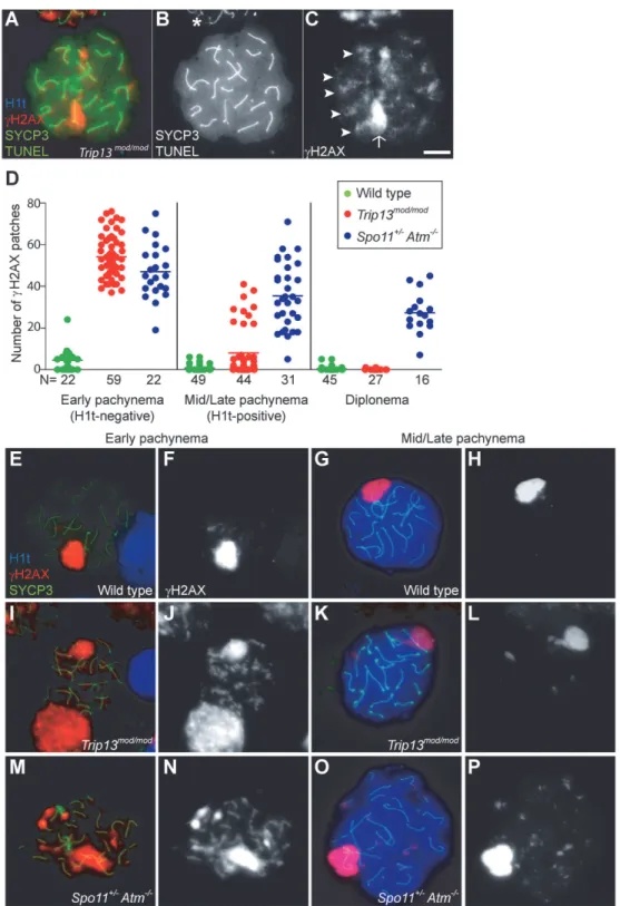
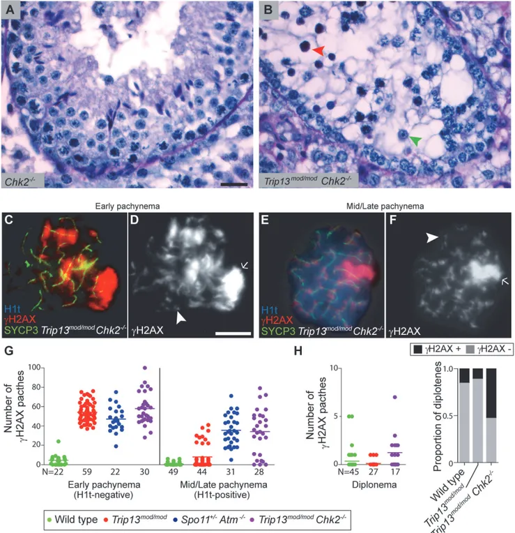
Nearly allTrip13mod/modTUNEL-positive cells (97.2%, n = 106) were H1t-negative (Fig. 1A-B), implying that most apoptotic cells had arrested in a developmental state with characteristics of early pachynema, as predicted. AllTrip13mod/modTUNEL-positive spermatocytes displayed multipleγH2AX patches (Fig. 1C), confirming the presence of unrepaired DSBs. A grossly nor-mal sex body was also observed, agreeing with prior results [22,23], although further analysis described below revealed subtle sex body defects.
Fig 1.Trip13mod/modspermatocytes activate a recombination-dependent arrest at pachynema.(A) ApoptoticTrip13mod/modspermatocyte showing the axial element protein SYCP3 (green), TUNEL (green), mid/late pachytene stage marker H1t (blue) andγH2AX staining (red). Absence of H1t staining (blue) in the TUNEL-positive cell indicates that this is an early pachytene stage spermatocyte. (B) TUNEL staining appears as pan-nuclear labelling that overlaps all chromatin, which is observed in only a fraction of the analysed cells (asterisk shows an adjacent TUNEL-negative cell). (C)γH2AX localizes to the sex body (arrow) and unrepaired breaks (arrowheads). (D) Number of autosomalγH2AX patches (similar to
Fig. 1K-L), but still significantly more than wild-type cells (0.8 ± 1.5; P = 0.016;Fig. 1D, G-H). The remainder (22.7%) had moreγH2AX patches than the first population (29.4 ± 6.7; P0.0001), but significantly less than H1t-negative cells (P0.0001). For the fewTrip13mod/mod cells that reached diplonema, however, the number ofγH2AX patches was indistinguishable from wild type (0.1 ± 0.3 vs. 0.4 ± 1.1, respectively; P = 0.183, negative binomial regression; Fig. 1D). A straightforward interpretation is that stochastic differences between TRIP13-deficient cells in the number of unrepaired DSBs translate into different arrest responses: cells with many unrepaired DSBs experience recombination-dependent arrest at early pachynema (H1t-nega-tive), cells with an intermediate number progress to an H1t-positive stage but then undergo apoptosis (possibly because of the DSBs, sex body defects, or both; see below), and the most repair-proficient subset of cells progress still further to diplonema. Additional implications of these patterns are addressed in Discussion.
Spermatocytes fromSpo11+/−Atm−/−mice also displayγH2AX patches late in prophase I
yet progress to the first division; this difference from ATM-proficientTrip13mod/modmice is consistent with the hypothesis that cells respond differently to persistent DSBs in the absence of ATM. Using H1t staining to more precisely define prophase stages, we observed that H1t-positiveSpo11+/−Atm−/−spermatocytes had numerousγH2AX patches, only
*20% fewer
than H1t-negativeSpo11+/−Atm−/−spermatocytes (Fig. 1D, M-P). This is in marked contrast
toTrip13mod/modmice, where most cells that progressed to an H1t-positive state had much fewerγH2AX patches than the earlier H1t-negative cells (see above). Moreover, H1t-positive Spo11+/−Atm−/−spermatocytes had significantly moreγH2AX patches than total H1t-positive
Trip13mod/modcells (35.5 ± 15.6, P0.0001;Fig. 1D). Thus, in the absence of ATM, cells with numerous unrepaired DSBs can progress to mid/late pachynema (and beyond).
Trip13
mod/modSpo11
+/−Atm
−/−spermatocytes progress to an
H1t-positive state in greater numbers despite a high level of persistent DSBs
The above results are consistent with our hypothesis that unrepaired DSBs at early pachynema inTrip13mod/modspermatocytes trigger a recombination-dependent checkpoint arrest that is mediated by ATM. To test this idea, we wished to ask if removing ATM activity allows
TRIP13-deficient cells to progress further into prophase. It is uninformative for this purpose to queryTrip13mod/modAtm−/−double mutants because theAtm−/−mutation by itself causes an
early block at an H1t-negative stage as a consequence of the enormous (>10-fold) increase in
DSB numbers (seeDiscussion) [2,27]. Instead, sinceSpo11heterozygosity ameliorates this cat-astrophic effect of ATM deficiency, we tested the epistasis relationship between theTrip13mod/
modphenotype (arrest in an H1t-negative state) andSpo11+/−Atm−/−phenotype (no pachytene
arrest) by analyzingTrip13mod/modSpo11+/−Atm−/−mice (hereafter referred to as
“TSA triple mutant”). As detailed below, we indeed find that absence of ATM allows TRIP13-deficient spermatocytes to progress to an H1t-positive state, but that additional defects that cause effi-cient mid/late-pachytene arrest and apoptosis of TSA triple mutant spermatocytes become ap-parent. (Note that DSBs in theSpo11+/−Atm−/−background are still substantially elevated
relative to wild type (*6-fold) [27], so no aspect of the triple mutant phenotype can be
as-cribed to a reduction in DSBs below wild-type levels.)
of cells counted per each stage and genotype. Primary data are provided inS1 Dataset. (E-P) Representative cells from the indicated genotypes stained for H1t (blue),γH2AX (red) and SYCP3 (green). InTrip13mod/mod
, mid/late pachytene cells have substantially fewerγH2AX patches than early pachytene spermatocytes. Bar in (C) represents 10μm and applies to all panels.
First, we measured testis size as a readout of spermatogenetic progression since mice with spermatogenic failure have small testes [14,15]. Sizes ofTrip13mod/modand TSA triple mutant testes were indistinguishable from one another (P = 0.6, t test), both being smaller than in Spo11+/−Atm−/−mice (P0.05,Table 1) and approximately one third the size of wild-type
tes-tes (P0.0001,Table 1). These results indicate that TSA triple mutant mice experience spermatogenic failure.
Second, we assessed the timing of apoptosis by histological staging of seminiferous tubules. ManyTrip13mod/modspermatocytes underwent apoptosis in tubules at epithelial stage IV, cor-responding to mid pachynema (Fig. 2A,B), as previously shown [23]. In contrast, and consis-tent with absence of pachytene arrest,Spo11+/−Atm−/−spermatocytes apoptosed in tubules at
epithelial stage XII (Fig. 2C), corresponding to metaphase I. Arrest ofSpo11+/−Atm−/−
sper-matocytes at this point is thought to be caused largely by a spindle checkpoint response to achiasmate (unconnected) chromosomes, particularly the X-Y pair [32]. Consistent with the small testis sizes, TSA triple mutant animals showed spermatocyte apoptosis at stage IV (Fig. 2D). Furthermore, whereas few wild-type tubules had>5 TUNEL-positive cells (1.0%,
Table 1), bothTrip13mod/modsingle mutant and TSA triple mutant testes displayed many such apoptotic tubules (21.6% and 26.0% respectively, P0.0001 compared to wild type, Fisher’s exact test,Table 1). Surprisingly, no TSA triple mutant cells escaping stage IV apoptosis were observed (Fig. 2B-D), in contrast toTrip13mod/modorSpo11+/−Atm−/−testes [22–24].
Although these results indicate that ATM deficiency (in theSpo11+/−Atm−/−background)
does not suppress the pachytene apoptosis ofTrip13mod/modmutant spermatocytes, they do not precisely define the stage of meiotic arrest. To address this question, we examined progression of chromosome synapsis in spermatocyte spreads stained with anti-SYCP3 antibodies. Out of total SYCP3-positive primary spermatocytes, 3.3% had progressed beyond pachynema in Trip13mod/mod, but none in TSA triple mutants (P0.0001, Fisher’s exact test,Table 1). This lack of escapers here and in the analysis of tubule sections indicates that overall spermatogenic failure is more penetrant in TSA triple mutants. Moreover, all TSA triple mutant spermatocytes displayed substantial autosome asynapsis (n = 703,Fig. 2E, H-I), unlike either theTrip13mod/
modorSpo11+/–Atm−/−mutants [23,24]. Because failure to complete synapsis has been
Table 1. Testicular and meiotic phenotypes of wild-type and mutant mice.
Wild type
Trip13mod/ mod
Spo11+/− Atm–/–
Trip13mod/ modSpo11+/
−Atm–/–
Mre11ATLD/ ATLD
Trip13mod/mod Mre11ATLD/ATLD
Nbs1ΔB/
ΔB
Trip13mod/ mod
Nbs1ΔB/ΔB
Chk2–/ –
Trip13mod/ modChk2–/–
N.T.W. (mean ±SEM)
0.66± 0.03
0.20±0.01 0.36± 0.04
0.21±0.05 0.65±0.05 0.19±0.02 0.51a 0.21±0.02 0.59± 0.1
0.19±0.02
Epithelial Arrest No arrest IV (escapers) XII (escapers) IV (no escapers)
No arrest IV (no escapers) No arrest IV (no escapers) No arrest IV (escapers) % Tubules with
˃5 TUNEL-positive cells (N) 1.0a (100) 21.6a (102)
11.9 (101) 26.0a(100) 7.0a(100) 16.0a(100) 10.0a (100)
25.0a(100) 3.0a (100)
16.0a(100)
% cells Post-pachynema (N)
31.6 (973)
3.3 (705) 15.6 (700) 0.0 (703) 25.2 (309) 0.0 (348) 49.8 (211)
0.3 (350) 18.1 (299)
1.9 (360)
% cells H1t positive (N)
50.3*
(350)
20.8 (567) 45.8*
(500)
47.7*(532) 61.06*
(208)
73.3*(221) 64.0*
(100)
43.4*(219) 57.0*
(100)
46.0*(237)
N.T.W.: Normalized Testis Weight expressed as a percentage of body weight.
a: one mouse analysed.
*: Significantly different from what is found inTrip13mod/modmice, P˂0.05, Fisher’s exact test.
associated with the inability to properly repair meiotic DSBs, these results could suggest that TRIP13-deficient spermatocytes are unable to cope with the increased DSB numbers produced in theSpo11+/−Atm−/−background [27], leading to synaptic catastrophe.
One consequence of synaptic failure is the inhibition of sex body formation [16]. Accordingly, we did not observe any TSA triple mutant cells with theγH2AX immunostaining pattern diag-nostic of sex body formation (Fig. 2E, G). Instead,γH2AX signal was mostly localized as patches on both unsynapsed and synapsed portions of chromosomes and did not accumulate in any par-ticular region of the nucleus. The complete failure to form sex bodies in TSA triple mutants likely accounts for the highly penetrant arrest in pachynema and apoptosis at stage IV.
To determine whether ATM imposes a recombination-dependent arrest early in pachynema that is relieved in TSA triple mutant spermatocytes, we examined H1t incorporation. In wild type, 50.3% of SYCP3-positive cells were also positive for H1t (Table 1), whereas only 20.8% of SYCP3-positive cells fromTrip13mod/modtestes were H1t-positive (Table 1). Presumably, most of these H1t-positive cells were escapers that would complete meiosis (see above). Notably, 47.7% of SYCP3-positive cells from TSA triple mutants were H1t-positive (see representative image inS2 Fig.), atwo-fold increase compared toTrip13mod/modsamples but similar to that in wild-type and inSpo11+/−Atm−/−animals (45.8%,Table 1). Thus, theSpo11+/−Atm−/−
com-bination is epistatic toTrip13mod/modmutation for the ability of spermatocytes to incorporate H1t.
We note that the relatively normal H1t pattern inSpo11+/−Atm−/−animals (S1 Table) and
the strong block to H1t incorporation inAtm−/−single mutants [2] argue against the possibility
that ATM is required to prevent premature H1t expression in early pachynema. To confirm this conclusion, we compared timing of H1t expression in testis sections from wild type and TSA triple mutants. In wild type, H1t was first detected in mid-pachytene spermatocytes in stage IV tubules, and was absent from early pachytene spermatocytes in stage I-III tubules, as previously reported [13] (seeS3A,B Fig.and its legend). Staging is less precise for immuno-fluorescently stained tubules of mutants that lack post-meiotic cell types [19]. Nonetheless, we observed that leptotene, zygotene, and most if not all early pachytene spermatocytes were H1t-negative, and that H1t-positive cells did not appear until approximately mid pachynema in TSA triple mutants (i.e., in tubule sections that were likely in stage IV) (seeS3C Fig.and its leg-end). Thus, there is no evidence for substantially premature expression of H1t in ATM-defective spermatocytes.
The increased number of H1t-positive cells implies that the TSA triple mutant spermato-cytes are not being restrained by the recombination-dependent arrest mechanism. This further reinforces the conclusion above that the apoptosis occurring in these cells is attributable to the absence of a sex body. If our interpretation is correct, we would expect that absence of the re-combination-dependent checkpoint allows spermatocytes to progress to an H1t-positive state even if they have many unrepaired DSBs. Indeed, H1t-positive TSA triple mutant spermato-cytes displayed significantly moreγH2AX patches thanTrip13mod/modcells (61.0 ± 18.8, P0.0001, t test;Fig. 2J). The large number ofγH2AX patches also supports the conclusion that the TSA triple mutants do not experience a reduction in DSBs relative toTrip13mod/mod alone. The finding that absence of ATM allows cells to progress to an H1t-positive state even if they contain numerous unrepaired DSBs indicates that ATM is required for implementation of recombination-dependent arrest at early pachynema.
The MRE11 complex promotes recombination-dependent arrest
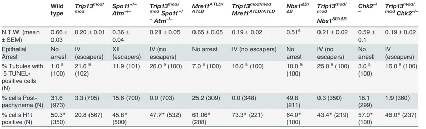
Fig 2.Trip13mod/modSpo11+/−Atm−/−spermatocytes arrest at epithelial stage IV, but present autosomal asynapsis, multiple unrepaired DSBs and fail to form a sex body at mid/late pachynema.(A-D) Testis cross-sections showing individual seminiferous tubules from the indicated genotypes, stained with PAS-Haematoxylin. (A) In wild type, spermatogonia, spermatocytes and spermatids are present. (B) Stage IV tubule inTrip13mod/modwith apoptotic
development, but mice bearing hypomorphicNbs1orMre11mutations that attenuate ATM activation and/or diminish phosphorylation of subsets of ATM targets are viable (Nbs1ΔBor Mre11atld) [34,35].Nbs1ΔB/ΔBandMre11ATLD/ATLDmice are subfertile, with spermatocytes dis-playing some synaptic defects and accumulating early recombination markers at pachynema, suggesting that the MRE11 complex plays a role in meiotic recombination in mammals [36], as in budding yeast and other organisms [37]. Testis samples from both mutants showed a small elevation in the fraction of spermatocytes that were H1t-positive (Table 1). This could be a con-sequence of previously documented differences in the overall distribution of meiotic cell types [36], but the important conclusion for purposes of this study is that the mutant spermatocytes are readily able to progress through pachynema and beyond in roughly normal numbers.
To further define the ATM-dependent pathway leading to recombination-dependent arrest, we asked whether these MRE11-complex mutations mimic ATM deficiency with respect to the meiotic progression phenotype ofTrip13mod/modspermatocytes. Indeed, similar to the TSA tri-ple mutant,Trip13mod/modNbs1ΔB/ΔBandTrip13mod/modMre11ATLD/ATLDmutants had approxi-mately two-fold (43.4%) and three-fold (73.3%) more H1t-positive spermatocytes, respectively, thanTrip13mod/modsingle mutants (Table 1). Importantly, these mutants progressed despite having high numbers of unrepaired DSBs: H1t-positive spermatocytes inTrip13mod/mod Nbs1ΔB/ΔBandTrip13mod/modMre11ATLD/ATLDmutants had significantly moreγH2AX patches than theTrip13mod/modmutant (59.7 ± 12.4 and 54.1 ± 10.8, respectively;Fig 2J), but similar numbers to the TSA triple mutant (P>0.05). These hypomorphic MRE11 complex mutations
are thus epistatic toTrip13mod/modwith respect to the ability of cells to progress to an H1t-posi-tive stage. These results support the hypothesis that the MRE11 complex activates the ATM-mediated recombination-dependent arrest that halts development of mostTrip13mod/modsingle mutant spermatocytes at early pachynema.
Also akin to the TSA triple mutant, these double mutants experienced a more penetrant spermatogenic failure (i.e., few or no escapers) than in theTrip13mod/modsingle mutant despite better progression beyond early pachynema.Trip13mod/modNbs1ΔB/ΔBandTrip13mod/mod Mre11ATLD/ATLDmice had small testes (Table 1), and histological analysis revealed many simi-larities betweenTrip13mod/modNbs1ΔB/ΔBandTrip13mod/modMre11ATLD/ATLDdouble mutants and the TSA triple mutant: apoptosis at epithelial stage IV (Figs.3andS4), a high number of apoptotic cells per tubule (Table 1), and no evidence of cells escaping and completing meiosis (Figs3. andS4). Likewise, cytological analysis showed that noTrip13mod/modMre11ATLD/ATLD spermatocytes and only a negligible fraction ofTrip13mod/modNbs1ΔB/ΔBspermatocytes (0.3%) progressed beyond pachynema, similar to the TSA triple mutant (P˃0.05, Fisher’s exact test, Table 1) but significantly different from theTrip13mod/modsingle mutant (P0.0001 and P = 0.0014, respectively). Also similar to TSA triple mutants, most double mutant spermatocytes had substantial synaptic defects, with only 0.5% ofTrip13mod/modMre11ATLD/ATLD(n = 348) and 5.4% ofTrip13mod/modNbs1ΔB/ΔB(n = 350) SYCP3-positive spermatocytes showing
Atm−/−tubule at stage XII, which is characterized by spermatocytes at metaphase I (red arrowhead) [24]. The presence of lagging chromosomes in some
metaphase I spermatocytes is thought to be the cause of apoptosis. Green arrowhead denotes a spermatid that has escaped meiotic arrest. (D)Trip13mod/ mod
Spo11+/−Atm−/−tubule at stage IV, presenting multiple apoptotic spermatocytes at pachytene stage (red arrowhead). No spermatids were observed.
(E-H) TwoTrip13mod/modSpo11+/−Atm−/−spermatocytes, one at preleptonema (left) and the other at mid/late pachynema (right), stained for H1t (blue, F), γH2AX (red, G) and SYCP3 (green, H). A high degree of asynapsis occurs in the H1t-positive cell, where long stretches of axial elements are visible (arrowhead). (I) Enlarged image of a bivalent that has successfully synapsed only a portion of the homologous chromosome pair (arrow). (J) Number of
γH2AX patches in mid/late pachytene spermatocytes of the indicated genotypes. Triple and double mutants that generate more DSBs and fail to complete synapsis (highlighted in dark grey) display moreγH2AX patches at mid/late pachynema thanTrip13mod/modChk2−/−cells (highlighted in light grey). Data for
wild type,Trip13mod/mod
andSpo11+/−Atm−/−reproduced fromFig. 1Dfor comparison. N shows the total number of cells counted per each genotype.
Asterisk marks statistically significant differences compared toTrip13mod/mod
, P0.0001, t test. Primary data are provided inS1 Dataset. Bar in (A) represents 20μm and applies to panels (A-D). Bar in (H) represents 10μm and applies to panels (E-H).
complete autosomal synapsis. These values are much lower thanTrip13mod/modSYCP3-positive spermatocytes (40.7%, n = 705, P0.0001, Fisher’s exact test). As expected from the substantial asynapsis, the double mutants showed no evidence of sex body formation as assessed by γH2AX staining (Figs.3andS4). Taken together, we infer from these data that the more pene-trant spermatogenic failure observed inTrip13mod/modNbs1ΔB/ΔBandTrip13mod/mod
Mre11ATLD/ATLDtestes is most probably due to activation of the sex body-deficient arrest, as in TSA triple mutants. Further, our results confirm that the sex body-deficient arrest mechanism can operate even when recombination-dependent arrest is compromised. This was also indicat-ed by the arrest ofSpo11−/−mutants as well as repair-proficient mutants with defects in meiotic
sex chromosome inactivation (MSCI) [2,16].
The MRE11 complex controls DSB numbers
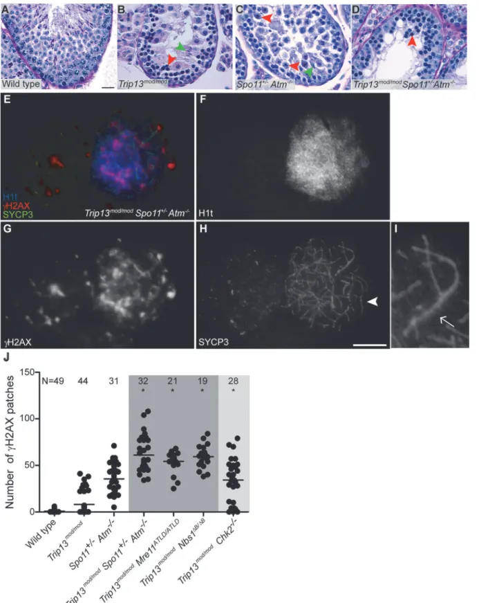
We reasoned that the attenuated ATM signaling inMre11andNbs1hypomorphic mutants might lead to elevated DSB numbers. To test this, we examined SPO11-oligonucleotide com-plexes, which provide a measure of whole-testis DSB levels, inMre11ATLD/ATLDandNbs1ΔB/ΔB single mutant mice [27]. (Note that this class of hypomorphic MRE11-complex mutation is un-like theRad50Stype of mutation, which in yeast blocks endonucleolytic release of SPO11 from Fig 3.Trip13mod/modNbs1ΔB/ΔB
spermatocytes arrest in mid/late pachynema with unrepaired DSBs at epithelial stage IV.(A-B) Cross sections of Nbs1ΔB/ΔB
andTrip13mod/modNbs1ΔB/ΔB
stage IV tubules stained with PAS-Haematoxylin. (C-D) Early (C) and mid/late (D) pachyteneTrip13mod/modNbs1ΔB/ΔB
spermatocytes stained for H1t,γH2AX, and SYCP3. Both cells present multiple unrepaired DSBs and incomplete synapsis. Bar in (A) represents 20μm and applies to panels (A-B). Bar in (D) represents 10μm and applies to panels (C-D).
DSB ends [33]). Indeed, we found thatMre11ATLD/ATLDandNbs1ΔB/ΔBmice displayed an*
2-fold increase compared to wild-type littermates (Fig. 4A-B). This elevation is less than what is seen inAtm–/–(
*12-fold) orSpo11+/–Atm−/−testes (*6-fold), presumably because theMre11
andNbs1hypomorphs attenuate but do not eliminate ATM activity [34,35]. (Note that the over-all fertility phenotype of theMre11andNbs1single mutants is also significantly milder than for mice lacking ATM (S1 Table)). These results provide strong evidence that ATM-mediated feed-back control of DSBs involves an MRE11 complex-dependent ATM activation pathway similar to the response to ionizing radiation. In contrast,Trip13mod/modanimals had roughly normal lev-els of SPO11-oligonucleotide complexes (*70% of wild type,Fig. 4C). BecauseTrip13mod/mod
single mutants have defects in completing DSB repair, we speculate that the autosomal synaptic failure inTrip13mod/modMre11ATLD/ATLDorTrip13mod/modNbs1ΔB/ΔBmice is a synthetic defect caused by an inability of TRIP13-deficient cells to tolerate increased DSB numbers resulting from ATM activation defects. An alternative but not mutually exclusive possibility is that the MRE11 complex and TRIP13 synergistically promote synapsis separately from their effects on recombination.
CHK2 is involved in recombination-dependent arrest but does not
regulate DSB levels
The CHK2 kinase is an effector of the ATM-signaling cascade in response to ionizing radiation [38]. In meiosis in several species, CHK2 function is required to efficiently activate the recom-bination-dependent checkpoint [9]. In mouse, CHK2 is required to arrest oocytes with unre-paired DSBs, although it has been suggested based on histological analysis that CHK2 does not play a similar role in spermatocytes [29]. To test for CHK2 involvement in recombination-de-pendent arrest in mouse spermatocytes, we analyzedTrip13mod/modChk2−/−mice.
The testicular phenotype ofTrip13mod/modChk2−/−mice was similar to theTrip13mod/mod
single mutant for testis size and histological tubule classification (apoptosis of spermatocytes at epithelial stage IV, but with presence of some spermatids), but also, notably, for the percentage of SYCP3-expressing cells that had progressed beyond pachynema (1.9%, P = 0.2462, Fisher’s exact test;Table 1andFig. 5A-B). Thus, CHK2 deficiency does not cause a more penetrant spermatogenic failure in the context of theTrip13mod/modmutation, unlike MRE11 complex hypomorphs or the absence of ATM itself. Moreover, CHK2-deficient testes displayed similar levels of SPO11-oligonucleotide complexes as wild-type littermates (Fig. 4D). Thus, CHK2 is not required for proper control of meiotic DSB formation, clearly separating CHK2 from both the MRE11 complex and ATM in the regulation of SPO11 activity. These results further sup-port the conclusion above that the synaptic defects and more penetrant block to spermatogene-sis observed in TSA triple mutants and inTrip13mod/modMre11ATLD/ATLDandTrip13mod/mod Nbs1ΔB/ΔBdouble mutants are attributable to the increased DSB numbers.
Notably, however,Trip13mod/modChk2−/−spermatocytes showed evidence of a defect in
re-combination-dependent arrest, because two-fold more H1t-positive spermatocytes were ob-served than with theTrip13mod/modsingle mutant (Table 1). Similarly to TSA triple mutants and the other double mutants described above, these H1t-positive spermatocytes presented moreγH2AX patches (34.1 ± 23.3; P0.0001, t test;Fig. 5E-G). Also, whereas 77.3% of the H1t-positiveTrip13mod/modspermatocytes (i.e., those inferred to be relatively recombination-proficient escapers) had very fewγH2AX patches (<8 per cell), only 21.4% of cells from
Trip13mod/modChk2−/−mice were in this category (P0.0001, Fisher’s exact test), with the
Fig 4. The MRE11 complex, but not TRIP13 or CHK2, modulates SPO11-oligonucleotide complex levels.
SPO11-oligonucleotide complexes were immunoprecipitated from extracts of testes of the indicated genotypes, labelled with terminal transferase and32P-nucleotide, and resolved by SDS-PAGE. Top image in each panel,
autoradiograph; bottom image, SPO11 Western blot where the two major SPO11 isoforms,αandβ, are indicated. The vertical lines next to the autoradiographs indicate the signal from SPO11-oligonucleotide complexes. Asterisk indicates non-specific signal from the labelling reaction. Short arrow designates the migration position of the heavy chain of the antibody used to immunoprecipitate SPO11. (A-B) BothMre11and Nbs1mutants have increased levels of SPO11-oligonucleotide complexes compared to littermate controls (1.9± 0.4 fold, mean±SD of the relative signal intensity compared to a wild-type control, n = 3 mice, and 1.8±0.2 fold, n = 3, respectively). (C) SPO11-oligonucleotide complex levels are slightly reduced inTrip13mod/mod
samples (0.7 ±0.2,, n = 3), similar to other recombination-deficient mutants, such asDmc1–/–[27]. The SPO11αisoform is not detected inTrip13mod/modtestes as observed in other mutants that arrest at pachynema [27]. (D)Chk2−/−testes
have similar levels of SPO11-oligonucleotide complex as controls (1.0±0.2 fold, n = 3).
Fig 5. CHK2 is required to maintain early pachytene arrest caused by TRIP13 deficiency.(A-B) Cross-sections ofChk2−/−andTrip13mod/modChk2−/−
testes stained with PAS-Haematoxylin.Chk2−/−testes contained all spermatogenic cell types, whereasTrip13mod/modChk2−/−spermatocytes arrested in
tubules at epithelial stage IV (red arrowhead). Note the presence of spermatids (green arrowhead) indicating that someTrip13mod/modChk2−/−cells manage
to complete meiosis. (C-F) Early and mid/late pachyteneTrip13mod/modChk2−/−spermatocytes stained for H1t,γH2AX, and SYCP3. Both early and mid/late
pachytene cells exhibit multipleγH2AX patches (arrowheads) corresponding to unrepaired DSBs, as well as accumulation ofγH2AX on the chromatin of the sex chromosomes (arrows). (G) Quantification of the number ofγH2AX patches inTrip13mod/modChk2−/−spermatocytes. Data for wild type,Trip13mod/mod
andSpo11+/−Atm−/−reproduced fromFig. 1Dfor comparison, N shows the total number of cells counted per each stage and genotype. (H)γH2AX patches in
diplotene cells. The plot on the left shows the number ofγH2AX patches per cell. Note that overplotting of zero values obscures information about proportions of cells that lack aγH2AX patch, so the plot on the right displays proportions of cells with or without aγH2AX patch. Primary data for panels G and H are provided inS1 Dataset. N shows the total number of cells counted per each stage and genotype. Bar in (A) represents 20μm and applies to panels (A-B). Bar in (D) represents 10μm and applies to panels (C-F).
CHK2 allows spermatocytes with numerous unrepaired DSBs to bypass the recombination-de-pendent arrest during pachynema, reminiscent of what occurs in oocytes [29]. If this is correct, we would also expect to find evidence of unrepaired DSBs in those spermatocytes that had es-caped pachytene arrest entirely. Indeed,γH2AX patches were substantially more prevalent at diplonema inTrip13mod/modChk2−/−spermatocytes: only 11.1% of diploteneTrip13mod/mod
cells hadγH2AX patches, but 52.9% of theTrip13mod/modChk2−/−spermatocytes had one or
more of these markers of unrepaired DSBs (P = 0.0045, Fisher’s exact test) (Fig. 5H). (Mean ± SD of 0.1 ± 0.3 forTrip13mod/mod(Fig. 1D) vs. 1.2 ± 1.8 (n = 17) forTrip13mod/modChk2–/–; P =
0.0005, negative binomial regression;S1 Dataset.) We conclude that CHK2, like the MRE11 complex and ATM, is required to activate the recombination-dependent arrest occurring in Trip13mod/modspermatocytes.
Trip13
mod/modChk2
−/−spermatocytes have sex body defects
Although CHK2 deficiency allowed more efficient progression of TRIP13-deficient cells to an H1t-positive stage, the overall spermatogenic failure was not alleviated, such that most
Trip13-mod/modChk2−/−spermatocytes still underwent apoptosis in tubules at epithelial stage IV
(Fig. 5B). We therefore hypothesized that the sex bodies ofTrip13mod/modChk2−/−
spermato-cytes may have subtle defects that impede their function. Indeed, mostTrip13mod/modChk2−/−
spermatocytes showed an abnormally elongatedγsH2AX-positive sex body chromatin domain (57.8%, N = 45,Fig. 5E-F), unlike the condensed, rounded structure seen in most wild-type H1t-positive spermatocytes (Fig. 1F,H). In addition, the intensity of the sex-bodyγH2AX sig-nal was reduced to about half the wild-type level on average in H1t-positive pachytene
Trip13-mod/modChk2−/−spermatocytes (S5 Fig.; P0.0001, t test). Although most H1t-positive
pachyteneTrip13mod/modcells had sex bodies with normal morphology, a significant minority also showed the abnormal elongated form (11.3%, n = 53, P0.0001, Fisher’s exact test) and sex bodyγH2AX intensity was reduced on average (P = 0.033; seeS5 Fig.).
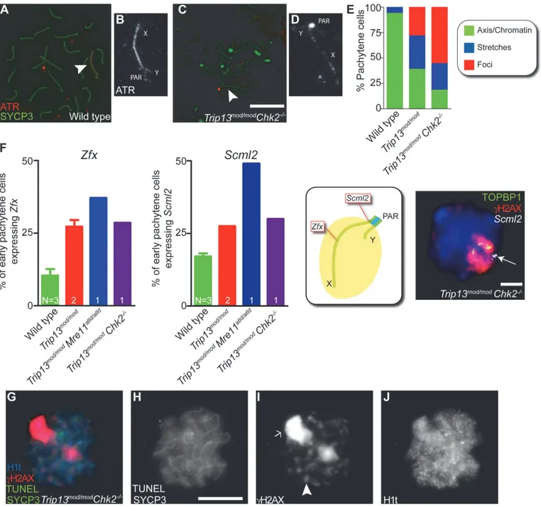
ATR is the kinase responsible for the bulk of H2AX phosphorylation in sex bodies [18,24,25]. In wild type, most pachytene spermatocytes showed ATR staining either as a con-tinuous signal on the sex chromosome axes or spread to the XY chromatin (93.6%, n = 47; Fig. 6A-B), as previously reported [39]. The remainder of the cells displayed stretches of ATR partially covering the X and Y axes. By contrast, only 18.0% ofTrip13mod/modChk2−/−
pachy-tene cells had the axial or chromatin-associated ATR staining most commonly found in wild-type cells, 26.2% had short stretches of continuous ATR signal partly covering sex chromosome axes, and most cells had only focal ATR staining (55.7%, n = 61;Fig. 6C-E).Trip13mod/mod sper-matocytes had an intermediate phenotype: 38.8% displayed ATR completely covering the XY axes or chromatin, 32.8% showed ATR stretches over the XY axes, and 28.4% had only discrete ATR foci along the X and Y (n = 67,Fig. 6E). These results suggest that CHK2 deficiency may exacerbate a defect in ATR loading on the sex chromosomes in TRIP13-deficient cells.
To further explore ATR-dependent processes, we examined SUMO-1, which is loaded onto sex chromosomes at pachynema in an ATR-dependent manner [18]. In all wild-type cells (n = 52) and 86.0% ofTrip13mod/modcells (n = 50), we detected SUMO-1 covering the chromatin of the sex body. In contrast, only 72.3% ofTrip13mod/modChk2−/−pachytene spermatocytes
con-tained SUMO-1 in their sex bodies, and of these, SUMO-1 signal had failed to spread over the entire XY chromatin in 60.6% of cells (n = 45,S6 Fig.).
Fig 6. Sex body deficiency inTrip13mutants.(A-D) Wild-type andTrip13mod/modChk2−/−pachytene spermatocytes stained for ATR and SYCP3. Arrowheads
indicate the sex chromosomes. Note the relatively continuous ATR staining on the X and Y axes in wild type compared with the focal ATR staining in the mutant. (E) Quantification of the different ATR staining patterns found in the genotypes presented. (F) Left panels, percent of early pachytene-stage spermatocytes expressingZfxandScml2in wild-type,Trip13mod/mod
,Trip13mod/modMre11ATLD/ATLD
andTrip13mod/modChk2−/−mice. The N value in each bar represents the
number of mice analyzed per each gene and genotype. Middle panel, cartoon of a sex body displaying the relative position ofZfxandScml2within the X chromosome. Right image, representativeTrip13mod/modChk2−/−spermatocyte displaying TOPBP1 (green) andγH2AX (red) immunostaining andScml2
RNA-FISH signal (white, arrow). Just 10.9%±3.8 (mean±SD) of wild-type spermatocytes (N = 225 cells) expressZfxand 17.1%±1.9 expressScml2(N = 200). Mouse that fail to synapse the X and Y chromosomes, likeTrip13mod/modMre11ATLD/ATLD
, present more spermatocytes expressing these genes than wild-type mice (forZfx: 37.1%, N = 35 and forSmcl2: 49.1%, N = 53, P0.0001 and P = 0.0002 respectively, Fisher’s exact test).Trip13mod/modmice present more cells expressing these genes than wild-type mice (forZfx: 27.2%±3.2, N = 158; forSmcl2: 27.5%±0.0, N = 160, P<0.005, 1 way Anova). Similarly,Trip13mod/mod Chk2−/−spermatocytes are more likely to express these X-linked genes than wild-type cells (forZfx: 28.6%, N = 70; forSmcl2: 30.0%, N = 80, P = 0.0217 and P =
0.0009 respectively, Fisher’s exact test). (G-J) Mid/late pachytene-stage, TUNEL-positiveTrip13mod/modChk2−/−spermatocyte immunostained for SYCP3 (green,
H),γH2AX (red, I) and H1t (blue, J). Note the presence of an elongated sex body (arrow), multipleγH2AX patches (arrowhead) and H1t in the chromatin of the apoptotic cell. Bars in (C) and (H) represent 10μm and apply to panels (A,C) and (G-J), respectively. Bar in (F) represents 5μm.
X-linked genes:Zfx, located in an interstitial region; andScml2, located near the pseudoautoso-mal boundary at the centromere-distal end of the chromosome (Figs.6FandS7). As expected, only a minority of wild-type cells expressedZfxorSmcl2(10.9% ± 3.8 and 17.1% ± 1.9, mean ± SD, respectively,Fig. 6F), whereas a significantly larger fraction of cells expressed these genes in mutants that fail to form a sex body at pachynema, likeTrip13mod/modMre11ATLD/ATLD sper-matocytes (forZfx: 37.1% and forSmcl2: 49.1%, P0.0001 and P = 0.0002 respectively, Fisher’s exact test). As predicted from the altered sex body morphology, we found thatTrip13mod/mod andTrip13mod/modChk2−/−spermatocytes also showed increased expression of these X-linked
genes at early pachynema (Trip13mod/mod:for Zfx: 27.2% ± 3.2 and forSmcl2: 27.5% ± 0.0, P<0.005, 1 way Anova;Trip13mod/modChk2–/–:for Zfx: 28.6% and forSmcl2: 30.0%, P = 0.0217
and P = 0.0009 respectively, Fisher’s exact test). We conclude that sex body function is altered inTrip13mutants.
The levels of sex chromosome gene misexpression are similar to those reported to be suffi-cient to arrest spermatocytes at pachynema in other mutants [21]. Indeed, when we assessed whetherTrip13mod/modChk2−/−pachytene spermatocytes undergo apoptosis at an H1t-positive
stage, as occurs in mutants that fail to form a sex body (S1 Fig.) [2], we found that 98.5% of TUNEL-positiveTrip13mod/modChk2−/−spermatocytes analyzed were also H1t-positive,
con-trasting what occurs inTrip13mod/modmutants (N = 67, 1 mouse, P0.0001, Fisher’s exact test, Fig. 6G-J) but mimicking what is seen inSpo11−/−mutants (P = 1, Fisher
’s exact test). In sum-mary, the mild sex body defect inTrip13mod/modtestes was more pronounced inTrip13mod/mod Chk2−/−spermatocytes, where it resulted in the failure to silence the X and Y chromosomes at
pachynema, suggesting that the stage IV apoptosis observed in these double mutant mice is triggered by the sex body-deficient arrest mechanism.
Discussion
Recombination-dependent arrest prior to H1t incorporation depends on
the MRE11 complex, ATM and CHK2 in mouse
Two major mechanisms are postulated to mediate arrest of recombination-defective mouse spermatocytes during the pachytene stage [2]. One occurs when sex body formation is impeded by massive synaptic failure, irrespective of the presence of uncompleted recombination events, such as when no DSBs are generated at all [16]. Sex body defects allow expression of X and Y chromosome genes that are sufficient to induce apoptosis [17]. In this scenario, the arrest and later cell death might be caused by misregulation of gene expression, not activation of a check-pointper se.
The other arrest mechanism is proposed to occur when recombination intermediates persist at pachynema [2,16], similar to what is seen in many other organisms [9]. In most species ana-lyzed, ATR (Mec1 in budding yeast) activates this checkpoint, most likely due to the presence of single-stranded DNA [6]. However, recent observations in yeast suggest that Tel1 (ATM) might also be involved [40] and our findings clearly indicate that the ATM signaling cascade is crucial for recombination-dependent arrest in mammalian spermatocytes.
A recent study found that the previously described recombination-dependent arrest of Atm−/−oocytes [4] is mediated by CHK2 [29]. It was further suggested that a protein kinase
other than ATM, possibly ATR, is involved in recombination-dependent arrest in oocytes [29]. While our results suggest that ATM is the principal kinase mediating this arrest in spermato-cytes, we do not exclude the possibility that ATR also participates in this mechanism, either in wild type or specifically inAtm−/−cells, in the latter case by replacing some ATM functions (e.
In somatic cells, the MRE11 complex participates in ATM activation, and mutations that impair the MRE11 complex lead to an inefficient G2/M checkpoint [26]. NBS1 also specifically promotes ATM phosphorylation of some of its substrates [33]. Our findings show that recom-bination-dependent arrest at pachynema has similarities to the G2/M checkpoint in somatic cells, as previously speculated [42]. These similarities may be useful for identifying other key members of the meiotic pathway.
Involvement of the effector kinase CHK2 in mammalian recombination-dependent arrest, further supported by observations in oocytes [29], underlines differences between organisms [9]. In budding yeast, for instance, the checkpoint depends on the meiosis-specific CHK2 ho-molog, Mek1. In contrast,D.melanogasteruses the non-meiosis-specific CHK2 homolog, Mnk, and inC.elegansCHK1, but not CHK2, is required for arrest. This variety emphasizes the importance of studying mouse meiosis to characterize this pathway in mammals.
This and previous studies establish H1t expression as a molecular marker of a response to recombination defects in spermatocytes, and our current findings establish that this response occurs via an ATM signaling cascade. Although available data rule out substantially premature H1t expression, it is possible that H1t is expressed slightly earlier than normal in the absence of DSBs or in the absence of the ATM response; such an effect might contribute to the high ob-served fraction of spermatocytes that are H1t-positive in the relevant mutants. However, if so, we note that this would be consistent with our conclusion that regulation of H1t expression is a checkpoint response since there are many instances where cell cycle regulated events occur pre-maturely when either the monitored event or the monitoring mechanism are missing (e.g., yeast Ndt80 expression when DSBs or Mec1 signaling are absent [43] or anaphase onset when the spindle assembly checkpoint is deactivated [44]). It is formally possible that this response has little or no other consequence than the control of H1t expression, and that spermatocytes do not otherwise“care”that they have sensed the presence of unrepaired DSBs. We consider this hypothesis unlikely since it would mean that the male germline in mouse, unlike mouse oocytes and meiotic cells in all other species studied to date, ignores molecular signals that re-port directly on the progression of one of the most central aspects of meiotic chromosome dy-namics (recombination), and ignores signals that have pronounced consequences in other mouse cell types. Instead, we favor the interpretation that the ATM-dependent response de-scribed here is an integral part of meiotic quality control surveillance during spermatogenesis.
The recombination-dependent checkpoint is tolerant of a certain level of
unrepaired DSBs
TRIP13, MRE11 complex and ATM interplay to promote homologous
chromosome synapsis
As previously stated, homologous chromosome synapsis depends on meiotic recombination [48]. Thus, failure to complete synapsis might be interpreted as a consequence of defective ho-mologous recombination. Unlike the relevant single mutants, spermatocytes from TSA triple mutants and fromTrip13mod/modMre11ATLD/ATLDandTrip13mod/modNbs1ΔB/ΔBdouble mu-tants fail to complete synapsis in all, or almost all, cells [22–25,36]. The MRE11 complex, ATM and TRIP13 have been reported to promote meiotic recombination [22–25,36]. Thus, the fact that double and triple mutants present problems with completing synapsis suggests that the MRE11 complex and ATM could synergize with TRIP13 to promote proper DSB repair during meiotic prophase. However, we provide evidence that proteins required to activate ATM (MRE11 and NBS1) are also involved in the regulation of DSB formation. Thus, sinceSpo11+/−
Atm−/−spermatocytes as well as cells fromMre11ATLD/ATLDandNbs1ΔB/ΔBsingle mutants
incur more DSBs than wild-type cells and because TRIP13-deficient cells are unable to com-plete meiotic recombination [22,23], this opens the possibility that TRIP13-defective cells can-not deal with the additional DSBs formed in the absence, or reduction, of ATM activity. Alternatively or in addition, the synapsis phenotype observed in TSA triple mutants or
Trip13-mod/modMre11ATLD/ATLDandTrip13mod/modNbs1ΔB/ΔBdouble mutants might be a
manifesta-tion of a funcmanifesta-tion of ATM and/or the MRE11 complex in promoting synapsis more directly and synergistically with TRIP13.
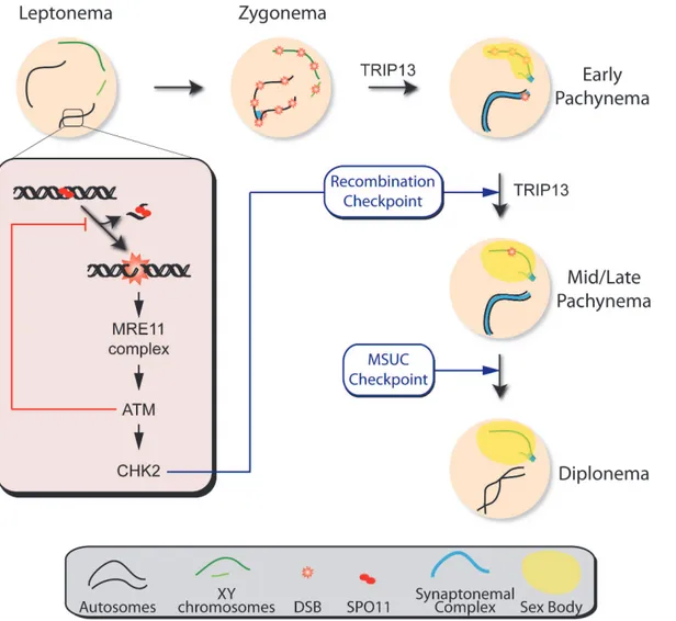
The array of phenotypes in the mutants analyzed here suggests that ATM participates in dif-ferent aspects of meiotic prophase (Fig. 7). Our results further demonstrate that, while meiotic progression depends on the MRE11 complex, ATM and CHK2, the regulation of SPO11 activi-ty requires the MRE11 complex to activate ATM but is independent of CHK2. This finding may indicate that direct ATM phosphorylation targets exert control of DSB formation, as has been argued in yeast [49]. Interestingly, yeast Mek1 promotes inter-homolog bias in the repair of DSBs [50,51]. The fact that homologous chromosome synapsis is not altered in the absence of mouse CHK2 suggests that this protein is not involved in recombination partner choice in mammals, although we cannot exclude that compensation by related kinases (e.g., CHK1) could be responsible for the absence of an obvious phenotype inChk2−/−spermatocytes.
TRIP13 participates in proper sex body formation
The apoptosis of spermatocytes in tubules at epithelial stage IV observed inTrip13mod/mod Chk2−/−testis can be explained if TRIP13 also contributes to the formation of a functional sex
body (Fig. 7). In support of this hypothesis, we observed defective loading of ATR onto XY chromatin in pachytene spermatocytes fromTrip13mod/modChk2−/−mice and in a subset of
pachytene cells fromTrip13mod/modsingle mutants. This defect leads to inappropriate H2AX phosphorylation and SUMO-1 incorporation into the sex body, resulting in inefficient sex chromosome silencing and apoptosis at mid/late pachynema. TRIP13 is required for normal chromosomal association of HORMA (“Hop1, Rev7 and Mad2”)-domain proteins HOR-MAD1 and HORMAD2 during meiotic prophase [52]. HORMAD1 and HORMAD2 localize to the chromosome axes at leptonema and disappear from synapsed regions during zygonema as synapsis progresses. At pachynema, HORMAD1 and HORMAD2 accumulate at the unsy-napsed regions of the X and Y chromosomes where they attract the machinery required to si-lence these chromosomes [21,53]. InTrip13mod/modspermatocytes, HORMAD1 and
Chk2−/−pachytene spermatocytes. It is worth noting thatTrip13mod/modChk2−/−cells do
as-semble ATR foci, presumably at resected DSB sites containing single-stranded DNA. This focal ATR localization has been reported previously to be HORMAD2-independent [21]. However, loading of ATR along the entire length of the unsynapsed chromosome axis is HORMAD2-de-pendent [21,53]. Furthermore,ZfxandScml2expression levels in early pachytene-stageTrip13 mutant spermatocytes are similar to those reported inHormad2−/−cells [21]. These findings
lead us to propose that proper HORMAD1 and HORMAD2 localization may underlie the function of TRIP13 in sex body formation.
The unexpected failure to properly form a sex body found inTrip13mutants opens the pos-sibility that, although this and other studies have clearly shown that there are at least two dis-tinct arrest mechanisms to respond to recombination or sex body defects [2,16], the apoptosis occurring after the activation of these two arrest mechanisms may be a response only to the common MSCI failure. Nonetheless, we favor the hypothesis that recombination-dependent arrest is sufficient to trigger apoptosis because unrepaired DSBs provoke programmed cell Fig 7. Functions of the ATM signaling pathway during mouse meiotic prophase.We propose that DSBs induced by SPO11 at the beginning of meiotic prophase are sensed by the MRE11 complex, which activates ATM. ATM inhibits further DSB formation via a feedback loop and promotes DSB repair. ATM activates CHK2, which controls cell cycle progression but is not involved in regulating DSB formation. Recombination progression leads to homologous chromosome synapsis and sex body formation. Besides participating in recombination, TRIP13 is also needed for effective sex body formation. Proper recombination and sex body formation are required to permit meiotic progression.
death in oocytes (where MSCI defects are not an issue) via the conventional DNA damage re-sponse effector molecules, p53 and p63 [4,22,29]; and because MRE11- and ATM-dependent signaling can directly promote apoptosis in somatic cells [26]. Addressing this issue will require novel meiotic mutants that display recombination defects in the context of an intact ATM sig-naling cascade and without MSCI defects.
Materials and Methods
Mutant mice
Trip13,Spo11,Atm,Dmc1,Mre11,Nbs1andChk2mutations were generated previously [10,14,23,28,34,35,54]. All lines were maintained on a C57BL6–129Sv mixed background. Ex-periments were performed using at least two animals in comparison with littermate controls (either homozygous or heterozygous for the wild-type alleles) or, when appropriate littermates were unavailable, control animals obtained from litters of the same matings. Genotyping was performed by PCR analysis of tail-tip DNA as previously described [23].
Cytology and histology
Testes were harvested from two- to five-month-old animals and processed for histology or cy-tology, as previously described [23,55]. Immunofluorescence was performed using standard methods [47] and antibodies [23]. Additional primary antibodies used were: guinea pig anti-H1t (kindly donated by M.A. Handel, Jackson Lab) at 1:500 dilution [12] and a mouse anti-SUMO-1 (clone 21C7, Invitrogen) at 1:100 dilution. TUNEL-staining on IF-stained slides was performed using anin situcell death detection kit (Roche Diagnostics) according to the manu-facturer’s instructions, as reported previously [56].
RNA FISH and immunofluorescence
RNA FISH was carried out with digoxigenin-labeled probes as previously described [57]. BAC DNA probes used were:Zfx, bMQ-372M23 (from Mouse bMQ BAC library) andScml2, RP24-204O18 (from CHORI BACPAC library). Briefly, BAC-containing bacteria were grown in an overnight LB-Chloramphenicol culture at 37°C and BAC DNA was isolated using a standard miniprep method. Approximately 2μg of BAC DNA was labelled using DIG-Nick Translation Mix (Roche) and precipitated with Cot-1 DNA (Invitrogen) and salmon sperm DNA (Strata-gene). Mouse testes were minced and cells were permeabilized with CSK buffer (100 mM NaCl, 300 mM sucrose, 3 mM MgCl2, 10 mM PIPES, 0.5% Triton X-100, 2 mM vanadyl ribo-nucleoside (New England Biolabs)), fixed with 4% paraformaldehyde and dehydrated through an ice-cold ethanol series. DNA-BAC probes were denatured for 10 min at 80°C, pre-hybrid-ized for 30 min at 37°C, and added to the slides for an overnight incubation at 37°C. Stringency washes were performed and digoxigenin was detected using anti-digoxigenin-FITC (1:10, Millipore). RNA FISH was then followed by TOPBP1 (1:50, Abcam) andγH2AX (1:100, Milli-pore) immunostaining. Cells were examined on an Olympus IX70 inverted microscope. Images were captured using a computer-assisted (DeltaVision) CCD camera (Photometrics), and pro-cessed for publication using ImageJ and Photoshop. Early pachytene cells were defined based on a continuous TOPBP1 staining along the X and Y chromosome axes.
SPO11-oligonucleotide complex detection and Western blotting
(Spo11–180) on Protein A-agarose beads (Roche). SPO11-oligonucleotide complexes were la-beled with [α-32P] dCTP using terminal deoxynucleotidyl transferase (Fermentas), released from the beads by boiling in Laemmli buffer, and fractionated by SDS-PAGE. The electropho-resed products were transferred onto polyvinylidene fluoride (PVDF) membrane. Radiolabeled species were detected using Fuji phosphor screens and quantified with ImageGauge software. The same PVDF membrane was then subjected to Western analysis using the SPO11 monoclonal antibody.
Statistical analysis
One-way ANOVA, Student's t tests and Fisher's exact tests were performed using GraphPad Prism software and/or GraphPad QuickCalcs online resource (http://www.graphpad.com/ quickcalcs/). For comparing counts ofγH2AX foci, we used t tests for simplicity to compare be-tween genotypes at pachynema because the count data were not highly skewed (i.e., were rea-sonably approximated by a normal distribution). However, for diplotene cells we instead used negative binomial regression because the count distributions at this stage were highly skewed for theTrip13mod/modChk2−/−sample and contained many zero values for all samples.
Regres-sion was carried out using the glm.nb function from the MASS package (verRegres-sion 7.3–33) in R (version 3.1.1).
Ethics statement
All experiments performed in this study comply with US and EU regulations and were ap-proved by the MSKCC Institutional Animal Care and Use Committee and the UAB Ethics Committee and the Catalan Government.
Supporting Information
S1 Fig.Dmc1−/−spermatocytes arrest at early pachynema andSpo11−/−spermatocytes ar-rest at mid/late pachynema.Spo11–/–(A,C,E,G) andDmc1–/–(B,D,F,H) spermatocyte spreads were immunostained for H1t,γH2AX and SYCP3, and subjected to TUNEL staining as well. (H) The cell on the right side of the panel presents pan-nuclear staining characteristic of the TUNEL reaction (also seen in G). One mouse analyzed per genotype. Bar in (G) represents 10μm and applies to all panels.
(TIF)
S2 Fig. Triple mutant spermatocytes accumulate H1t.Representative wild-type (A-D), Trip13mod/mod(E-H) andTrip13mod/modSpo11+/−Atm–/–(I-L) testis sections immunostained
for H1t (green) andγH2AX (red). Note the significant increase in the number of spermatocytes positive for H1t in the triple mutant section compared to theTrip13mod/modsample. Bar in (A) represents 10μm and applies to panels A, E and I.
(TIF)
S3 Fig. Timing of H1t incorporation into the chromatin of TSA triple mutant spermato-cytes is not detectably altered compared to wild type.Individual panels in (A) and (C) show representative seminiferous tubule sections of the indicated epithelial stages from wild-type andTrip13mod/modSpo11+/−Atm−/−testis sections immunostained for H1t (green) andγH2AX
and diplotene (D-Sp)); Sd, spermatid; Se, Sertoli cell. (A, B) H1t in wild type. Seminiferous tu-bule staging is based on the array of different cell types contained in a tutu-bule section, with mor-phological differences regarding particular organelles used as markers to more precisely distinguish between cellular subtypes [19]. Tubule staging is more challenging using only the information obtained by immunofluorescence analysis of chromatin proteins, as compared to the histological staining methods traditionally used. Nonetheless, unambiguous staging can be performed in wild type testis, which contains all spermatogenic cell types. Leptotene spermato-cytes make up the outermost layer of cells in stage IX and X tubules, with a layer of late pachy-tene cells and a layer of spermatids beginning to elongate their nuclei located more centrally toward the tubule lumen. Zygotene cells are in the outer layer of stage XI-XII tubules, which are further characterized for the presence of spermatids with a clearly elongated head. The outer layer of stage I tubules contains early pachytene cells that show the first manifestation of a stretched sex body; these tubules also contain two kinds of spermatids, round and elongated. From stage II-III onwards, spermatogonia for the next wave of spermatogenesis have prolifer-ated to sufficient numbers to constitute a distinct basal layer of cells, so spermatocytes are now located in the second layer of cells. In stage II-III tubules, the outer layer of spermatogonia is followed by a layer of H1t-negative pachytene spermatocytes with a more condensed sex body, then a layer of H1t-positive round spermatids, and finally elongated spermatids. Stage IV tu-bules are similar, with the important exception that the pachytene spermatocytes are now clearly H1t positive. Stage IV can be clearly distinguished from other stages by examining the round spermatids: this stage is characterized by the presence of the acrosomal granules that begin to form the acrosomal vesicle on top of the nuclear envelope of round spermatids. This creates a small, rounded depression (angle<40°) in the nuclei of these cells that clearly marks
this stage (see (B) for a magnified image of the spermatids boxed in yellow in the stage IV tu-bule section). These results demonstrate that H1t protein expression first becomes detectable in mid-pachytene spermatocytes in stage IV tubules, consistent with prior studies [13]. (C) H1t expression timing is not grossly altered in the absence of ATM. Accurate classification of indi-vidual tubule stages is more difficult in mutants (including the TSA triple mutant) that experi-ence spermatogenic arrest, because these mutants lack the post-meiotic cell types that facilitate a clear differentiation between stages [19]. Nonetheless, we can conclude that H1t expression does not begin until after the earliest part of pachynema in the TSA triple mutant, because spermatocytes were H1t-negative in all analyzed sections for which spermatocytes made up the outermost layer (i.e., corresponding to leptotene, zygotene or early pachytene spermatocytes from tubule stages IX-XII or stage I). In tubules in which spermatocytes made up the second layer of cells (i.e., stages II-IV), we observed that these pachytene spermatocytes were H1t-neg-ative in some tubules and H1t-positive in others. In tubules with H1t-positive pachytene cells, we also frequently observed apoptotic figures (arrowheads in bottom-most panel), so we infer these are stage IV tubules. We cannot distinguish with absolute certainty between these tubule stages, so we cannot exclude the possibility that H1t expression begins slightly earlier than in wild type (e.g., in stage III, or earlier in stage IV than would normally occur), but the data are fully consistent with the hypothesis that H1t expression occurs with similar timing in the TSA triple mutant as in wild type.
(TIF)
S4 Fig.Trip13mod/modMre11ATLD/ATLDspermatocytes arrest in mid/late pachynema with unrepaired DSBs at epithelial stage IV.(A-B) Cross-sections ofMre11ATLD/ATLDand
Trip13-mod/modMre11ATLD/ATLDstage IV tubules stained with PAS-Haematoxylin. (C-D) Early (C)