RESEARCH ARTICLE
Srs2 and Mus81
–
Mms4 Prevent
Accumulation of Toxic Inter-Homolog
Recombination Intermediates
Kenji Keyamura, Kota Arai, Takashi Hishida*
Department of Life Science, Graduate School of Science, Gakushuin University, Tokyo, Japan
Abstract
Homologous recombination is an evolutionally conserved mechanism that promotes genome stability through the faithful repair of double-strand breaks and single-strand gaps in DNA, and the recovery of stalled or collapsed replication forks.Saccharomyces cerevi-siaeATP-dependent DNA helicase Srs2 (a member of the highly conserved UvrD family of helicases) has multiple roles in regulating homologous recombination. A mutation
(srs2K41A) resulting in a helicase-dead mutant of Srs2 was found to be lethal in diploid, but not in haploid, cells. In diploid cells, Srs2K41Acaused the accumulation of inter-homolog joint molecule intermediates, increased the levels of spontaneous Rad52 foci, and induced gross chromosomal rearrangements. Srs2K41Alethality and accumulation of joint molecules were suppressed by inactivating Rad51 or deleting the Rad51-interaction domain of Srs2, whereas phosphorylation and sumoylation of Srs2 and its interaction with sumoylated prolif-erating cell nuclear antigen (PCNA) were not required for lethality. The structure-specific complex of crossover junction endonucleases Mus81 and Mms4 was also required for via-bility of diploid, but not haploid,SRS2deletion mutants (srs2Δ), and diploidsrs2Δmus81Δ mutants accumulated joint molecule intermediates. Our data suggest that Srs2 and Mus81–
Mms4 have critical roles in preventing the formation of (or in resolving) toxic inter-homolog joint molecules, which could otherwise interfere with chromosome segregation and lead to genetic instability.
Author Summary
Homologous recombination (HR) is a DNA-repair mechanism that is generally consid-ered error free because it uses an intact sister chromatid as a template. However, in diploid cells, HR can also occur between homologous chromosomes, which can lead to genomic instability through loss of heterozygosity. This alteration is often detected in genetic disor-ders and cancer, suggesting that tight control of this process is required to ensure genome stability. Yeast Srs2, conserved from bacteria to humans, plays multiple roles in the regula-tion of HR. We show here that a helicase-dead mutant of Srs2,srs2K41A, is lethal in diploid cells but not in haploid cells. Expression of Srs2K41Ain diploid cells causes inter-homolog
a11111
OPEN ACCESS
Citation:Keyamura K, Arai K, Hishida T (2016) Srs2 and Mus81–Mms4 Prevent Accumulation of Toxic Inter-Homolog Recombination Intermediates. PLoS Genet 12(7): e1006136. doi:10.1371/journal. pgen.1006136
Editor:Michael Lisby, University of Copenhagen, DENMARK
Received:January 4, 2016
Accepted:May 31, 2016
Published:July 7, 2016
Copyright:© 2016 Keyamura et al. This is an open access article distributed under the terms of the Creative Commons Attribution License, which permits unrestricted use, distribution, and reproduction in any medium, provided the original author and source are credited.
Data Availability Statement:All relevant data are within the paper and its Supporting Information files.
Funding:This work was funded by grants-in-aid for Scientific Research (23114007) from the Ministry of Education, Culture, Sports, Science and Technology (MEXT), Japan to TH. The funders had no role in study design, data collection and analysis, decision to publish, or preparation of the manuscript.
joint molecule intermediates to accumulate, and leads to gross chromosomal rearrange-ments. Moreover,srs2Δmus81Δdouble mutants have a severe diploid-specific growth
defect with accumulation of inter-homolog joint molecules. These data demonstrate that Srs2 and Mus81-Mms4 participate in essential pathways preventing accumulation of inter-homolog recombination intermediates, thereby reducing the risk of genome instability.
Introduction
Genomes are constantly challenged by endogenous metabolic products or exogenous physical or chemical agents that can generate DNA lesions. When they go unrepaired, these DNA lesions cause stalled replication forks and/or replication-fork collapse, leading to the accumula-tion of single-stranded DNA (ssDNA) gaps or DNA double-strand breaks (DSBs). Homolo-gous recombination (HR) is a highly conserved DNA-repair mechanism that is essential for the faithful repair of DSBs and has an important role in the repair of post-replicative ssDNA gaps [1–3]. Therefore, dysregulated or incomplete repair by HR can lead to genomic instability, which is a hallmark of cancer.
Rad51 is a central factor in DSB repair by HR. Rad51 forms nucleoprotein filaments on ssDNA tracts generated by 5’to 3’ssDNA resection from DSBs. Rad51 filaments mediate strand invasion into homologous DNA duplexes, leading to the formation of D-loops [4,5]. HR intermediates, including D-loops, can enter one of two HR sub-pathways: the synthesis-dependent strand-annealing (SDSA) pathway, which generates non-crossover products, and the canonical DSB repair (DSBR) pathway, which generates crossover or non-crossover prod-ucts [6,7]. In the SDSA pathway, a newly synthesized ssDNA strand is displaced from the D-loop to anneal to the complementary strand in the original duplex, resulting in a non-crossover outcome with no change to the template DNA [1]. The DSBR pathway involves D-loop exten-sion and annealing of the displaced strand to a second ssDNA tail of the broken duplex, form-ing a DNA intermediate termed the double Holliday junction. InSaccharomyces cerevisiae, several helicases function in crossover control. Srs2 and Mph1 act independently to promote SDSA by processing the HR intermediates downstream of D-loop formation [8–11]. Sgs1, together with Top3 and Rmi1, can dissociate double Holliday junctions to generate non-cross-over products, thus preventing crossnon-cross-overs in the DSBR pathway [8,12–14]. Alternatively, dou-ble Holliday junctions can be resolved to produce crossover or non-crossover products by structure-specific endonucleases, such as the Mus81–Mms4 complex, the Slx1–Slx4 complex, and Yen1 [15–17].
Moreover, Srs2 promotes the SDSA pathway during mitotic DSB repair by removing the Rad51 filament from the second end of the DSB, and/or by facilitating the dissociation of the invading strand from the D-loop [30–32]. Phosphorylation of Srs2 by cyclin-dependent kinase 1 (Cdk1) stimulates the SDSA pathway [33]. Taken together, these observations suggest that Srs2 has two distinct functions in HR; it prevents unscheduled recombination by inhibiting Rad51-dependent formation of joint molecules and it promotes efficient DSB repair by the SDSA pathway.
During HR in diploid cells, sister chromatids are the preferred templates for HR-mediated repair (inter-sister HR), but homologous chromosomes can also be used to restore the broken DNA (inter-homolog HR), although much less efficiently. Because sister chromatids are identi-cal, inter-sister HR is genetically silent. By contrast, the use of homologous chromosomes as repair templates has important consequences for genetic stability, and loss of heterozygosity is a frequent outcome [34]. The frequency of loss of heterozygosity is high in cancerous and aged cells, which has raised interest in dissecting the mechanisms of HR [35]. The HR process has to be tightly controlled to protect against genetic instability, but little is known about the relative contributions of each HR pathway to the processing of the two classes of recombination inter-mediate, involving either sister chromatids or homologs.
Our experiments were designed to explore the role of Srs2 in haploid and diploid cells by phenotypic characterization of a number ofsrs2mutants as a function of cell ploidy. The Srs2 helicase-deficient mutant (srs2K41A) caused diploid-specific lethality. This lethality was sup-pressed by deletion ofRAD51, but was independent of the phosphorylation and sumoylation of Srs2 and of its interaction with sumoylated PCNA. Expression of Srs2K41Ain diploid cells led to a specific increase in G2/M-arrested cells, more abundant inter-homolog joint molecules and
increased gross chromosomal rearrangements, such as chromosome loss and translocations.
srs2Δmus81Δdouble mutants also demonstrated a severe, diploid-specific growth defect, with
the concomitant accumulation of joint molecules. These results suggest that the mechanisms of processing inter-sister and inter-homolog joint molecules differ significantly. We propose that Srs2 and Mus81–Mms4 have critical roles in processing inter-homolog joint molecules, which could otherwise interfere with chromosome segregation and lead to genetic instability.
Results
Helicase-dead
srs2
K41Ais lethal in diploid yeast
A previous study showed thatsrs2Δdiploid cells are more sensitive to methyl methanesulfonate
(MMS) thansrs2Δhaploid cells [21,36]. This ploidy-specific sensitivity to MMS is thought to
reflect lethal outcomes of inter-homolog HR events in the absence of wild-type Srs2. To under-stand the role of Srs2 in inter-homolog HR, we constructed four mutants ofsrs2:srs2K41Alacks helicase activity [37],srs27AVcannot undergo Cdk1-dependent phosphorylation [38,39],
srs23KRcannot undergo sumoylation [40], andsrs2ΔSIMlacks the protein motif that mediates interaction with sumoylated PCNA [26]. Thesesrs2mutants and wild-typeSRS2were expressed in yeast from low-copy centromeric (pRS415_LEU2) plasmids under control of the
SRS2promoter. The plasmids were introduced intosrs2Δhaploid or diploid cells and selected
on SC+Glucose medium lacking leucine (SC+Glu-Leu). In this initial screen, no diploid colo-nies expressing Srs2K41Awere detected (Table 1), suggesting thatsrs2K41Acould be lethal or could block growth ofsrs2Δdiploid cells. To test this possibility, ansrs2K41Aallele was
inte-grated at theSRS2genomic locus of haploid yeast. The integrating cassette included down-streamHIS3orLEU2selectable markers (srs2K41A_HIS3orsrs2K41A_LEU2). The endogenous
SRS2allele in a haploid strain was also linked toHIS3orLEU2selectable markers as a control (SRS2_HIS3orSRS2_LEU2). AMATαstrain carryingsrs2K41A_LEU2was crossed toMATa
strains bearingsrs2K41A_HIS3,SRS2_HIS3, orsrs2Δ::HIS3. Diploids from these crosses were
selected for growth on SC+Glu medium lacking histidine and leucine. As shown inFig 1A, the
srs2K41A/srs2Δheterozygotes andsrs2K41A/srs2K41Ahomozygotes did not grow on the selection
medium, whereas heterozygoussrs2K41A/SRS2diploids exhibited normal growth. This demon-strates thatsrs2K41Amutants are lethal in diploids.
Srs2
K41Adifferentially inhibits growth of haploid and diploid yeast cells
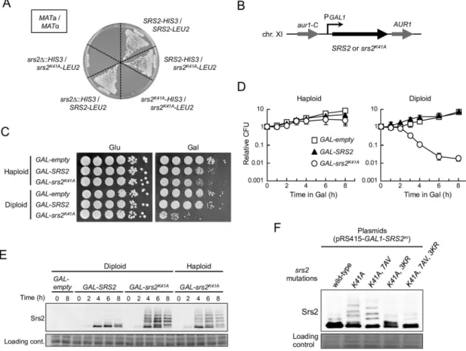
To investigate whysrs2K41Ais lethal in diploid cells, Srs2K41Aand wild-type Srs2 were expressed under the control of the inducibleGAL1promoter from a single-copy integrated allele at the chro-mosomalAUR1locus ofsrs2Δdiploid and haploid cells (Fig 1B). Hereafter, these strains are
referred to asGAL-srs2K41AandGAL-SRS2, respectively. AGAL-emptystrain (essentially the same as ansrs2Δstrain) was constructed in a similar manner, as an additional control. The resul-tant haploid and diploid strains grew normally in 2% glucose-containing medium (YPD) (Fig 1C
andS1A Fig), enabling the effect of conditional expression of Srs2K41Aand Srs2 to be investigated. To determine the level of expression of Srs2 in this experimental system,GAL-SRS2diploid cells were grown for 6 h in the presence of 2% raffinose medium (YPR) and various concentra-tions of galactose, and whole-cell extracts were prepared and analyzed by immunoblotting with an antibody to Srs2. The results revealed that Srs2 protein was absent in cells grown in YPD or YPR, and that the abundance of Srs2 increased with increasing galactose concentration (S1B Fig). Control experiments established thatGAL-SRS2diploid cells grew normally in the pres-ence of 0.02% galactose, but poorly in the prespres-ence of 0.2% galactose, because of high overex-pression of Srs2 (Fig 1CandS1C Fig), as previously reported [36]. In addition, expression of Srs2K41A, but not wild-type Srs2, inhibited growth (despite the presence of the chromosomal
SRS2+allele) when moderately expressed in the presence of 0.05% galactose, whereas similar growth defects were not observed in the presence of 0.02% galactose (S1D Fig). Thus,srs2K41Ais essentially a dominant-negative allele, and its dominancy is dependent on the ratio of wild-type Srs2 to Srs2K41A. We conclude that expression of Srs2 from theGAL1promoter in the presence of 0.02% galactose generates a physiologically-relevant protein level, and, for the remainder of this study, cells carryingGAL1promoter-driven expression strains were grown in YPD or YPR to repress Srs2 expression, and in YPR medium containing 0.02% galactose to induce Srs2.
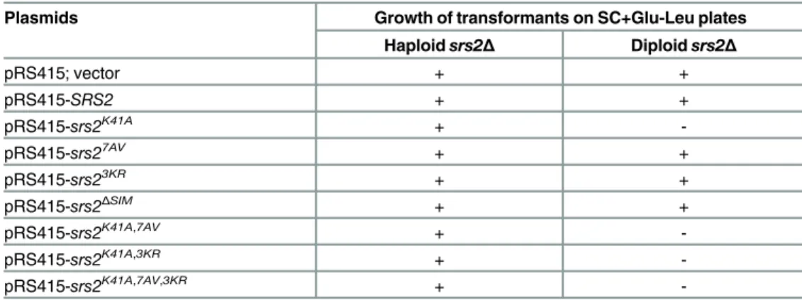
Table 1. srs2K41A
is lethal in diploids, but not in haploids.
Plasmids Growth of transformants on SC+Glu-Leu plates
Haploidsrs2Δ Diploidsrs2Δ
pRS415; vector + +
pRS415-SRS2 + +
pRS415-srs2K41A +
-pRS415-srs27AV + +
pRS415-srs23KR + +
pRS415-srs2ΔSIM + +
pRS415-srs2K41A,7AV +
-pRS415-srs2K41A,3KR +
-pRS415-srs2K41A,7AV,3KR +
-Thesrs2Δhaploid and diploid strains transformed with each of the pRS415 derivatives were incubated at 30°C for 3 days on SC+Glu-Leu plates. The pRS415-based vectors contain theSRS2alleles under the control of endogenousSRS2promoter. +, viable colonies were detected at similar levels to those with an empty-vector control; -, no colonies were detected.
Expression of Srs2
K41Areduces viability of the
srs2
Δ
diploid strain
To examine whetherGAL-srs2K41Adiploid cells could recover from growth arrest in galactose-containing medium, cells transiently grown in the presence of 0.02% galactose were transferred back to glucose-containing medium to determine the plating efficiency. The plating efficiency ofGAL-srs2K41Adiploids decreased rapidly with>3 h incubation in the presence of galactose,
whereas no significant effect on growth was observed forGAL-srs2K41Ahaploid cells, or GAL-emptyandGAL-SRS2haploid or diploid cells, even after incubation for 8 h in 0.02% galactose
Fig 1. Physiological expression of Srs2K41Acauses diploid-specific lethality.(A)MATa haploid cells were mated withMATα cells on YPD plates, generatingMATa/MATαdiploid cells. The indicated diploid cells were then selected at 30°C for 3 days on
SC+Glu plates lacking histidine and leucine (SC+Glu-His-Leu). (B) A DNA fragment with a galactose-inducible promoter and wild-typeSRS2(GAL-SRS2),srs2K41A(GAL-srs2K41A), or no insertion (GAL-empty) was integrated into theAUR1locus ofsrs2Δcells. In allGAL-promoter-integrated haploid and diploid strains, the endogenous copy ofSRS2was deleted to eliminate the expression of wild-type Srs2 from its own locus. (C) Cells grown in YPD medium were diluted and spotted onto YPD plates and YPR + 0.02% galactose plates. These plates were incubated at 30°C for 3 days. (D) For quantitative assays, cells grown to early logarithmic phase in YPD were transferred to YPR containing 0.02% galactose for further incubation, and then plated on YPD to determine the plating efficiency. Cell viability is represented as relative colony-forming units (CFU), such that CFU = 1 at 0 h. Data were obtained from at least three independent experiments. Error bars indicate the standard error for each data point.GAL-empty(open squares);
GAL-SRS2(filled triangles);GAL-srs2K41A(open circles). (E) The indicated haploid and diploid strains were grown at 30°C in YPR + galactose (0.02%) medium, and cells were harvested at the indicated time points. Protein extracts were prepared and separated by 6% SDS-PAGE, followed by western blotting with anti-Srs2 antibodies. (F) Thesrs2Δdiploid strains carrying the indicated plasmids were grown in SC+Glu-Leu and then transferred into SC-Leu (2% raffinose + 0.2% galactose) medium for 6 h to induce Srs2. Protein extracts were prepared and separated by 6% SDS-PAGE, followed by western blotting with anti-Srs2 antibodies.
doi:10.1371/journal.pgen.1006136.g001
(Fig 1D). These data show that a physiological level of Srs2K41Areduces viability of diploid cells, but not haploid cells.
The lethality of
GAL-srs2
K41Adiploids does not depend on
post-translational modification
In the course of these studies, Srs2K41Aisolated from haploid and diploid cells was observed as multiple slow-migrating protein species on SDS-PAGE when cells were grown in the presence of 0.02% galactose (Fig 1E). Because Srs2 is phosphorylated and sumoylated in response to DNA damage [33,38,39], we postulated that the slower-migrating forms of Srs2K41Aprotein are phos-phorylated and/or sumoylated isoforms of the protein. To test this hypothesis, plasmids that expressed Srs2K41A, Srs2K41A,7AV, Srs2K41A,3KR, and Srs2K41A,7AV,3KRfrom theGAL1promoter were introduced intosrs2Δdiploid cells. Each strain was grown to early logarithmic phase in
glu-cose medium and transferred to galactose medium, and protein extracts were prepared and ana-lyzed by western blot with an antibody to Srs2. This analysis revealed that Srs2K41A,7AV, which lacked phosphorylation sites, existed as three sumoylated isoforms that moved slightly faster than modified isoforms of Srs2K41Aon electrophoresis (Fig 1F). Srs2K41A,3KR, which lacked sumoylation sites, existed as phosphorylated isoforms (Fig 1F). As expected,srs2K41A,7AV,3KR, in which all phosphorylation and sumoylation sites had been mutated, resulted in a considerable reduction in expression of modified isoforms of Srs2 (Fig 1F). These results indicate that Srs2K41Acan be sumoylated and phosphorylated in the absence of DNA damage. To determine whether these modifications of Srs2K41Aaffected diploid-specific lethality, yeastCEN/ARS plas-mids (in whichsrs2K41A,srs2K41A,7AV,srs2K41A,3KR, andsrs2K41A,7AV,3KRwere under the control of the endogenousSRS2promoter) were constructed and transformed into thesrs2Δdiploid strain.
The result showed that nosrs2Δtransformants expressing Srs2K41Aor its derivatives were viable
(no colonies were detected) (Table 1), indicating that neither phosphorylation nor sumoylation is required for the lethal effects of Srs2K41Ain diploid yeast.
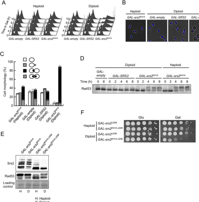
GAL-srs2
K41Adiploids arrest at G
2/M
To learn more about howsrs2K41Akills diploid yeast cells, the cell-cycle distribution and cell morphology ofGAL-srs2K41Acells were investigated in haploid and diploid cells. Cells were grown to early logarithmic phase in the presence of glucose, transferred to YPR containing 0.02% galactose, and then analyzed by flow cytometry. InGAL-emptyandGAL-SRS2haploids and diploids, cell-cycle progression was not significantly altered by galactose induction (Fig 2A). However,GAL-srs2K41Adiploids, but not haploids, showed apparent cell-cycle arrest at G2/M after induction of Srs2K41A. The 4C peak appeared to broaden with prolonged incubation
of cells in the presence of 0.02% galactose (Fig 2A). Similar effects have been observed after extended treatment with nocodazole, a microtubule-depolymerizing drug that causes G2/M
arrest [41]. Consistent with this interpretation, approximately 80% ofGAL-srs2K41Adiploids assumed the characteristic morphology of G2/M arrest, which involves large-budded cells with
one nucleus at the bud neck and a short spindle (Fig 2B and 2CandS2 Fig). These results sug-gest that, in diploids, Srs2K41Acauses cell-cycle arrest after bulk DNA synthesis is complete.
The lethality of
GAL-srs2
K41Adiploids is not dependent on its interaction
with sumoylated PCNA
Fig 2. Expression of Srs2K41Acauses G
2/M arrest in diploids but not in haploids.(A) Asynchronous cells were grown at 30°C in YPR + galactose (0.02%), and samples were collected at the indicated time points. DNA content was measured by FACS. (B and C) Cells grown in YPR + galactose (0.02%) medium for 8 h were fixed with ethanol and stained with 4,6-diamidino-2-phenylindole (DAPI) to visualize the DNA. Representative morphology observed after transfer to the YPR + galactose (0.02%) medium for 8 h is shown in (B). Cells with no bud (G1phase), cells with small bud (S phase), and large-budded cells with one or two nuclei at the bud neck (G2/M phase) were scored (C). The results represent the averages of at least three independent measurements. Error bars indicate the standard error for each data point. (D) The DNA-damage checkpoint is activated insrs2K41Ahaploid and diploid cells. The indicated haploid and diploid strains were grown in YPD medium. Cells were transferred to YPR + 0.02% galactose to induce Srs2 expression and then cultured at 30°C for the indicated times. Protein extracts were prepared and separated by 6% SDS-PAGE, followed by western blotting with anti-Rad53 antibody. (E)GAL-srs2K41AandGAL-srs2K41A,ΔSIMhaploid and diploid cells were grown
in YPD medium. Cells were transferred to YPR + 0.02% galactose to induce Srs2 expression and then cultured at 30°C for 6 h. Protein extracts were prepared and separated by 6% SDS-PAGE, followed by western blotting with anti-Srs2 or Rad53 antibodies. (F) Cells grown in YPD were diluted and spotted onto YPD plates (Glu) and YPR + 0.02% galactose plates (Gal). These plates were incubated at 30°C for 3 days.
doi:10.1371/journal.pgen.1006136.g002
studies showed that the protein product ofsrs2ΔSIM, which cannot interact with sumoylated PCNA, undergoes dramatically less sumoylationin vivo[40], andsrs2ΔSIMmutation suppresses the replication defects associated with overexpression of Srs2 in haploid cells [42]. In our study, the phenotypes ofGAL-srs2K41A,ΔSIMdiploid and haploid cells were examined. Rad53 phosphorylation and Srs2 sumoylation (and phosphorylation) were significantly reduced at 6 h afterGAL-srs2K41A,ΔSIMhaploid cells were transferred to 0.02% galactose, compared with levels inGAL-srs2K41Ahaploid cells (Fig 2E). By contrast, substantial Rad53 phosphorylation was still observed inGAL-srs2K41A,ΔSIMdiploid cells, although Srs2 phosphorylation and sumoylation were strongly reduced compared with levels inGAL-srs2K41Adiploid cells (Fig 2E). In addition,
GAL-srs2K41A,ΔSIMdiploids, but not haploids, had severe growth defects (Fig 2F). These results indicate that thesrs2K41Alethality in diploid cells is not associated with activation of the DNA damage checkpoint through its interaction with sumoylated PCNA.
The
srs2
K41Alethality in diploids is dependent on homologous
recombination
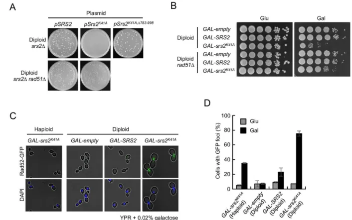
A well-characterized role of Srs2 is that of anti-recombinase, and in this context Srs2 disman-tles Rad51 nucleofilaments on ssDNA [27,28]. Toxic HR intermediates might, therefore, accu-mulate insrs2K41Adiploid cells, which could explain the ploidy-specific lethality of this allele. Consistent with this hypothesis,rad51Δsrs2Δdiploid strains expressing Srs2K41Afrom a
plas-mid vector were viable (Fig 3A). Similarly, the growth inhibition ofGAL-srs2K41Adiploids in the presence of 0.02% galactose was suppressed by therad51Δmutation (Fig 3B). Moreover,
srs2Δdiploid cells expressing Srs2K41A,Δ783–998, which lacks the Rad51 interaction domain in
Srs2 [28], were also viable (Fig 3A). Taken together, these results indicate that the lethality of
srs2K41Ain diploids is associated with Rad51-dependent HR in diploids.
Srs2
K41Acauses Rad52-GFP foci to accumulate in diploid cells
Rad52 nuclear focus formation is an indication of HRin vivo, and many mutants with
genome-maintenance defects have increased numbers of Rad52 foci compared with their wild-type counterparts [43]. The frequency of spontaneous Rad52 foci was, therefore, quantified in
GAL-srs2K41Acells and appropriate control cells expressing GFP-tagged Rad52 from the endog-enousRAD52genomic locus. Few Rad52-GFP foci were observed when cells were grown in glucose-containing medium (Fig 3C and 3D). However, after 8 h incubation in 0.02% galactose, Rad52-GFP foci were markedly increased inGAL-srs2K41Adiploids compared withGAL-SRS2
diploid andGAL-srs2K41Ahaploid cells, and most of the foci occurred in large-budded cells with a single nucleus (Fig 3C and 3D). These findings suggest thatGAL-srs2K41Adiploid cells accumulate HR intermediates at a much higher frequency thanGAL-srs2K41Ahaploid cells.
Inter-homolog joint molecules accumulate in
GAL-srs2
K41Adiploid cells
To test directly whether Srs2K41Acaused joint molecules to accumulate insrs2Δdiploids, diploid
suggest that Rad51 and Srs2K41Acollaborate in diploid cells to generate DNA structures that are not able to migrate out of the well during PFGE. In this context, it should be noted that the
rad51Δmutation did not suppress Rad53 activation inGAL-srs2K41AandGAL-srs2K41A,ΔSIM dip-loid cells under the same conditions (S3B Fig), suggesting that joint moleculesper seare not direct signals for Rad53 activation.
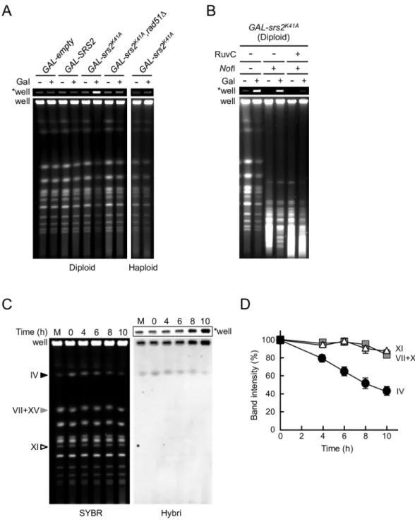
To characterize the chromosomal structures that accumulated inGAL-srs2K41Adiploid cells, chromosomal DNA samples were digested with the rare-cutter restriction endonucleaseNotI prior to PFGE. AlthoughNotI digests yeast chromosomes into multiple large and small frag-ments, the intensity of the DNA signal in the wells did not change significantly after digestion withNotI (Fig 4B). This observation suggested thatGAL-srs2K41Adiploid cells accumulated branched DNA structures, which were enriched even after digestion withNotI. To test this pos-sibility,NotI-digested or non-digested chromosomal DNA samples were digested with purified RuvC fromEscherichia coli. RuvC is a highly specific endonuclease that resolves Holliday junc-tions, although it also cleaves three-stranded junctions and nicked Holliday junctions [44,45]. The results showed that the action of RuvC releasedNotI-digested chromosomal fragments into the PFGE gel (Fig 4B), whereas non-migratory chromosomal DNA withoutNotI treat-ment was hardly resolved by RuvC (S3C Fig).NotI digestion could conceivably facilitate the
Fig 3. Expression of Srs2K41Acauses toxic recombination intermediates to accumulate in diploids.(A)srs2Δandsrs2Δ rad51Δdiploid cells were transformed with each of the pRS415 derivatives bearingSRS2,srs2K41A, orsrs2K41A,Δ783–998, and the cells were incubated at 30°C for 3 days on SC+Glu-Leu plates. (B) Cells grown in YPD were diluted and spotted onto YPD plates (Glu) and YPR + 0.02% galactose plates (Gal). These plates were incubated at 30°C for 3 days. (C) Images of cells with Rad52-GFP foci in Srs2K41A-expressing diploid cells. The indicated strains were grown at 30°C for 8 h in YPD (Glu) or YPR + 0.02% galactose (Gal). Cells were collected, stained with 4,6-diamidino-2-phenylindole (DAPI), and examined by fluorescence microscopy. (D) Quantitation of cells with Rad52-GFP foci in Srs2K41A-expressing diploid cells. Error bars indicate the standard error for each data point. Representative images of Rad52-GFP foci and DAPI staining are shown in (C).
doi:10.1371/journal.pgen.1006136.g003
Fig 4. Inter-homolog joint molecules containing Holliday junction-like structures accumulate in GAL-srs2K41A
diploid cells.(A–D) PFGE analysis of chromosomal DNA from Srs2K41A-expressing diploid cells. (A) The indicated haploid and diploid strains were grown in YPR or YPR + 0.02% galactose for 8 h. Chromosomal DNA was separated by PFGE and detected by staining with SYBR green. (B)GAL-srs2K41Adiploid cells were grown at 30°C
for 4 h in YPR or YPR + 0.02% galactose medium. DNA was isolated using agarose-gel blocks, digested withNotI orNotI + RuvC, and subjected to PFGE. (C) TheGAL-srs2K41Adisome IV strain was transferred to
SC+Raffinose-His+G418 medium containing 0.5% galactose to induce the expression of Srs2K41A. Chromosomal DNA was analyzed by PFGE, followed by hybridization with a chr. IV probe.“SYBR”indicates a SYBR-green-stained gel,
“Hybri”indicates a Southern analysis with the chr. IV probe, and“M”indicates haploid DNA as a size marker. (D) The band intensities of chromosomes IV (circle), VII+XV (square), and XI (triangle) detected by staining the gel were quantified and are shown relative to 100% at time 0. Error bars indicate the standard error for each data point.
“*well”indicates an image at the well of the gel shown at low exposure.
formation of catalytically competent joint molecule configurations for RuvC cleavage, since junction incision by RuvC is dependent on configuration [46,47]. These results suggest that RuvC-cleavable joint molecules accumulate inGAL-srs2K14Adiploid cells.
To further examine whether the DNA structures inGAL-srs2K14Adiploid cells were prod-ucts of inter-homolog HR, ansrs2Δhaploid strain that carried an additional copy of
chromo-some IV (henceforth known as thesrs2Δdisome) was constructed. Notably, no colonies were
obtained when Srs2K41Awas expressed from a plasmid vector insrs2Δdisomes, but the growth
defect was rescued by deletion ofRAD51(S4A Fig). These suggest that the additional copy of a donor sequence (homologous chromosome) is a cause of the lethality ofsrs2Δdisomes
express-ing Srs2K41A, and that the growth defect ofGAL-srs2K14Adisomes is the result of Rad51-depen-dent HR. Similar experiments were performed in ansrs2Δdisome in whichGAL-srs2K41Awas
integrated at the chromosomalAUR1locus (henceforth known as theGAL-srs2K41Adisome). TheGAL-srs2K41Adisome strain failed to grow in the presence of galactose, whereas the hap-loid control strain grew normally under same conditions (S4B Fig). In PFGE analysis, the
GAL-srs2K41Adisome strain, but notGAL-emptyandGAL-SRS2disome strains, showed a spe-cific loss of signal corresponding to chromosome IV in galactose-induced cells, whereas no other chromosomes were similarly affected (Fig 4C and 4DandS4C Fig). This conclusion was confirmed by Southern blotting with a probe for chromosome IV, which showed a reduction in hybridization signal in the gel and augmentation of the hybridization signal in the well dur-ing Srs2K41Aexpression (Fig 4C). These results suggest that inter-homolog joint molecules accumulate inGAL-srs2K41Adiploid and disome cells.
Srs2
K41Ainduces chromosomal instability in diploid cells
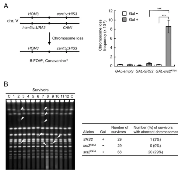
Our results led to the hypothesis that unresolved joint molecules form insrs2K41Adiploid cells, leading to chromosomal instability and cell death. To test this hypothesis, the frequency of loss of a pair of chromosome V homologs marked withCAN1on the right arm andURA3on the left arm was calculated and compared inGAL-SRS2andGAL-srs2K41Adiploid cells (Fig 5A, left panel) [48]. The observed frequency of chromosome loss in galactose-inducedGAL-srs2K41A
diploid cells was 15-fold higher than in galactose-inducedGAL-SRS2diploid cells (Fig 5A, right panel), suggesting thatGAL-srs2K41Adiploid cells have a defect in chromosome segrega-tion, which leads to a high rate of aneuploidy. Indeed, this result probably underestimated the chromosome-loss frequencies in galactose-inducedGAL-srs2K41Adiploid cells because it only detected aneuploid cells that remained viable after re-plating on glucose-containing medium. To directly investigate genomic integrity, chromosomal DNA was isolated from surviving cells and analyzed by PFGE. Chromosomal abnormalities were observed in 3% (1 of 29) of galac-tose-inducedGAL-SRS2and 0% (0 of 29) glucose-repressedGAL-srs2K41Adiploid cells (Fig 5B). By contrast, 20 of 68 survivors (29%) obtained from galactose-inducedGAL-srs2K41A dip-loid cells showed abnormal chromosome compositions; both aneupdip-loidy and chromosomal translocations were detected (Fig 5B). Thus, the expression of Srs2K41Ain diploids dramatically increases the rates of gross chromosomal rearrangements.
Srs2 and Mus81
–
Mms4 are essential for growth of diploid cells
It has been reported that sensitivity to MMS is enhanced insrs2Δdiploid cells relative to their
haploid counterparts [21]. To gain insight into inter-homolog HR, a genome-wide screen for diploid-specific sensitivity to MMS was conducted using a library (n4,800) of viable haploid
and diploid deletion mutants, directly testing for a ploidy-specific phenotype in the presence of MMS. The complete results of the screen will be described elsewhere. Three genes were identi-fied that function in the processing of HR intermediates (SRS2,MUS81, andMMS4). Our
investigation focused on a subset of HR genes includingSGS1,MPH1,MUS81,MMS4,RAD1,
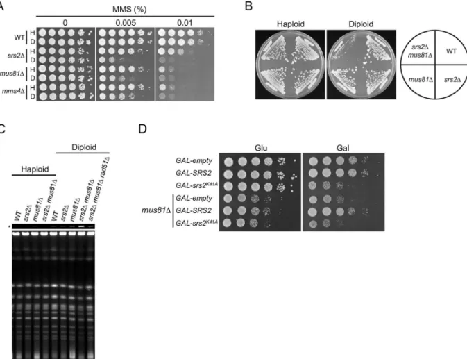
RAD10,YEN1,SLX1, andSLX4, which are involved in the processing of D-loops, Holliday junc-tions, and similar structures [11,17]. We reconfirmed thatmus81Δandmms4Δstrains were
more sensitive to MMS as diploids than as haploids, whereas other HR-deficient diploid strains had similar MMS sensitivity to their haploid counterparts (Fig 6AandS5 Fig). Mus81 interacts with Mms4 to form a structure-specific nuclease, which cleaves a variety of branched structures, including 3' flaps, D-loops, and nicked Holliday junctions [49–51]. These results suggest that Mus81–Mms4 has an important role in the resolution of inter-homolog joint molecules.
The genetic relationship between Srs2 and Mus81–Mms4 was investigated by comparing the growth and viability of haploid and diploidsrs2Δandmus81Δsingle-mutant and Fig 5. Expression of Srs2K41Aincreases genome instability in diploids.(A) Schematic representation of the chromosome V homolog used to monitor chromosome loss (left panel). Clones resistant to both 5-fluoroorotic acid (5-FOA) and canavanine (5-FOARCanR) arise because of loss of the entire chromosome with the markers.
GAL-empty,GAL-SRS2, andGAL-srs2K41Astrains were cultured in glucose medium, after which they were placed on YPR or YPR + 0.02% galactose medium for 8 h to induce Srs2 or Srs2K41A. The frequency of loss of chr. V was calculated as described in Materials and Methods (right panel). Error bars indicate the standard error for each data point.***indicates a p value<0.005, calculated using a Student’s two-tailed t test. (B) The indicated cells were grown for 8 h in YPR or YPR + 0.02% galactose medium. The cultures were diluted appropriately and spread onto YPD plates. Chromosomal DNA from the obtained colonies (the survivors) was analyzed using PFGE. The percentage of survivors with aberrant chromosomes is indicated. A representative gel image is shown in the left panel, and abnormal chromosome bands are indicated with arrowheads.
double-mutant strains. Thesrs2Δmus81Δhaploid double mutant grew just as well as either
single mutant, whereassrs2Δmus81Δdiploid cells grew very poorly (Fig 6B). A similar
effect was seen insrs2Δmms4Δcells (S6A Fig). Poor growth ofsrs2Δmus81Δdiploids was
rescued by expression of plasmid-bornesrs27AV,srs23KR, orsrs2ΔSIM, but not by the plasmid vector alone (S6B Fig, upper panel), which indicated that Srs2 rescued the growth defect of the double-mutant strain in the absence of phosphorylation, sumoylation, or interaction with sumoylated PCNA. However, plasmid-bornesrs2Δ783–998, which lacks a
Rad51-interac-tion domain, did not complement the severe growth defect of srs2Δmus81Δdiploid cells
(S6B Fig, upper panel), and deletion ofRAD51orRAD52rescued the growth defect (S6C Fig). These results suggest that Mus81–Mms4 and Srs2 have essential roles in inter-homolog HR.
Biochemical and two-hybrid studies have shown that, in addition to Srs2Δ783–998, Srs2Δ875–902and Srs2L844Aare deficient in Rad51 interaction [52,53]. These results suggest that the amino acid residues of Srs2 that are critical for binding to Rad51 are localized in sep-arate regions within Srs2 residues 783–998. In our study, plasmids were constructed in which
srs2L844A,srs2Δ875–902, andsrs2L844A,Δ875–902were under the control of the endogenousSRS2
promoter. Poor growth ofsrs2Δmus81Δdiploids was rescued by expression of plasmid-borneSRS2,srs2L844A,srs2Δ875–902, orsrs2L844A,Δ875–902, but notsrs2Δ783–998(S6B Fig,
lower panel). Similarly,srs2Δdiploid cells expressingsrs2K41A,L844A,srs2K41A,Δ875–902, or
srs2K41A,L844A,Δ875–902from pRS415 were not able to form viable colonies (S6D Fig). These
results suggest that Srs2L844Aand Srs2Δ875–902retain some ability to interact with Rad51in vivo, consistent with the results of a previous study that the phenotype ofsrs2Δ875–902cells is
similar to wild-type [52].
srs2
Δ
mus81
Δ
double mutants accumulate joint molecules in diploid
cells
Mostsrs2Δmus81Δdiploids arrested with 4C DNA content and were large-budded cells with
a single nucleus, suggesting a significant delay of entry into mitosis (S7 Fig). The simplest explanation for the synergistic growth defect insrs2Δmus81Δdiploids is that the double
mutants had unresolved inter-homolog joint molecules, which resulted in G2/M arrest, as
observed insrs2K41Adiploids. PFGE analysis consistently revealed that chromosomal DNA fromsrs2Δmus81Δdiploid cells, but not from their haploid counterparts, formed structures
that failed to enter the gel (Fig 6C). Moreover, these DNA accumulations insrs2Δmus81Δ diploid cells were suppressed by therad51Δmutation (Fig 6C). This ploidy-specific behavior is consistent with our other results, and probably reflects the accumulation of inter-homolog joint molecules.
The phenotypic similarity betweensrs2K41Aandsrs2Δmus81Δsuggested the possible functional interaction between Srs2K41Aand Mus81. To address this possibility, GAL-srs2K41Amus81Δ,GAL-SRS2 mus81Δ, andGAL-empty mus81Δdiploid strains were con-structed, and the effect of expressing Srs2K41Aor Srs2 insrs2Δmus81Δdiploids in the pres-ence of 0.02% galactose was examined. In a control experiment, expression of wild-type Srs2 complemented the growth defect of theGAL-SRS2 mus81Δmutant (Fig 6D). Notably, whereas Srs2K41Aexpression aggravated the growth of theGAL-srs2K41Adiploid strain, it had no effect on the growth of theGAL-srs2K41Amus81Δdiploid strain (Fig 6D). These results suggest that thesrs2K41Amutant behaves similarly to thesrs2Δmus81Δdouble mutant. It should be noted thatsrs2K41Awas lethal in diploid yeasts, butsrs2Δmus81Δdiploid cells were viable, albeit with poor growth, suggesting that the growth defect insrs2K41Adiploids is more toxic than insrs2Δmus81Δdiploids.
Discussion
Inter-homolog recombination intermediates accumulate in Srs2
K41A-expressing diploid cells
Srs2 has a dual function in HR, preventing unscheduled recombination and promoting the SDSA pathway during DSB repair. Our results showed thatsrs2K41Adiploids, but not haploids, had a severe defect in growth.GAL-srs2K41Adiploid cells showed an elevated number of Rad52 foci and an increase in the rate of gross chromosomal rearrangements, suggesting a high rate of spontaneous HR-associated DNA damage. Indeed, these growth defects were suppressed by inactivation of Rad51 and also by deletion of the Rad51 interaction domain of Srs2K41A. These results imply that DSBs are not responsible for the toxic effects ofsrs2K41Ain diploid yeast, since the repair of DSBs is essential for cell survival and requires functional HR. Indeed, in PFGE analysis, fragmented chromosomes were not detected, but joint molecule accumulations
Fig 6.srs2Δmus81Δdiploid cells exhibit a severe growth defect.(A) MMS sensitivity of haploid and diploid mutant cells. Cells were grown and spotted on YPD plates containing the indicated concentration of MMS at 10-fold serial dilutions, and incubated at 30°C for 3 days. (B) The indicated haploid and diploid strains were grown on YPD plates at 30°C for 3 days. (C) Accumulation of joint molecules insrs2Δmus81Δdiploid cells, but not in haploid cells. DNA isolated from asynchronous cultures of wild-type (WT),srs2Δ,
mus81Δ,srs2Δmus81Δ, andsrs2Δmus81Δrad51Δcells was subjected to PFGE and detected by staining with SYBR green. An asterisk (*) indicates well images obtained at low exposure. (D) The effect of Srs2K41Aexpression on the growth ofsrs2Δmus81Δ diploid cells. The indicated diploid strains grown in YPD were diluted and spotted onto YPD (Glu) and YPR + 0.02% galactose (Gal) plates. These plates were incubated at 30°C for 2 days.
were observed in Srs2K41A-expressing diploid cells. To repair ssDNA gaps by HR, homologous sequences located on the same or different chromosomes can serve as templates. Especially, inter-homolog HR occurs only in diploids, whereas inter-sister HR can occur in both haploid and diploid yeasts. Our results suggest, therefore, that the diploid-specific lethality ofsrs2K41A
is the result of a failure to resolve joint molecules formed during inter-homolog HR.
Post-translational modification of Srs2
K41Ais not required for
diploid-specific lethality
Srs2 is phosphorylated by Cdk1 and sumoylated in response to DNA damage [33,38]. Cdk1-dependent phosphorylation of Srs2 is required to promote the SDSA pathway for DSB repair. Srs2 sumoylation may have a role in the assembly and/or stabilization of protein com-plexes involved in DNA repair, although its biological significance remains obscure [40]. In this study, low-abundance Srs2K41Aunderwent both phosphorylation and sumoylation at mul-tiple sites in haploid and diploid cells, even in the absence of DNA damage. Mutational analysis revealed that sumoylation and phosphorylation of Srs2K41Awere largely independent events, which was consistent with the results of a previous study [40]. Moreover, our data demon-strated that Srs2K41Asumoylation and phosphorylation were dispensable forsrs2K41Alethality in diploids. By contrast, the lethality ofsrs2K41Ain diploids required its interaction with Rad51. These results suggest that the removal of toxic Rad51 filaments by the Srs2 translocase activity may be essential for the viability of diploid cells.
Our results showed that the DNA damage checkpoint, as monitored by Rad53 phosphoryla-tion, was constitutively activated in haploid and diploid cells expressing Srs2K41A. A previous study showed that overexpression of wild-type Srs2 in haploid cells activates the DNA damage checkpoint in a manner that requires the Srs2 SIM domain [42]. Similar observations were made in our experiments inGAL-srs2K41Ahaploid cells, suggesting that activation of the DNA damage checkpoint insrs2K41Ahaploids depends both on DNA replication and the interaction between Srs2 and sumoylated PCNA. By contrast, checkpoint activation and growth inhibition were still observed inGAL-srs2K41A,ΔSIMdiploid cells. Thus,GAL-srs2K41A,ΔSIMdiploids might accumulate more (or a different type of) DNA lesions than haploid cells of the same genotype, triggering the DNA damage checkpoint. In addition, therad51Δmutation did not suppress diploid-specific (unrelated to sumoylated PCNA) Rad53 activation ofsrs2K41A,ΔSIMdiploids, suggesting that this checkpoint activation is unlikely to be associated with the lethality of
srs2K41Ain diploid cells.
Mus81 and Srs2 have critical roles in the processing of inter-homolog
joint molecules
Inter-homolog recombination intermediates form infrequently during HR in mitotic yeast cells. However, if they form, efficient resolution is required to prevent interference with proper chromosome segregation. Our data suggest that Srs2K41Ais recruited to inter-homolog recom-bination intermediates through its interaction with Rad51, and, further, that Srs2K41Ainhibits processing of these intermediates, possibly because it lacks a functional helicase/translocase activity. Thus, a possible explanation for Srs2K41Alethality is that in addition to impeding Srs2-dependent HR, it actively blocks a second repair pathway that resolves inter-homolog joint molecules by other helicases or endonucleases. Srs2 has been shown to physically interact with Mus81–Mms4, and to remove Rad51 from DNA, enabling Mus81–Mms4 to access and cleave DNA [54]. In addition, Srs2 and Mus81 co-localize after DNA damage, although Mus81 is fully proficient in focus formation in the absence of Srs2 [54]. A plausible mechanism for a second repair pathway is the resolution of inter-homolog joint molecules by Mus81–Mms4
endonuclease. In this context, it is notable that each of thesrs2Δ,mus81Δ, andmms4Δ
muta-tions resulted in greater sensitivity to MMS in diploids than in haploids, which was not true of
sgs1Δmutations. Moreover,srs2Δmus81Δdiploid cells exhibited a severe growth defect and a
marked accumulation of joint molecule intermediates, which were also observed in Srs2K41A -expressing diploid cells. It remains unclear whether the non-migratory DNA complexes observed during PFGE are direct substrates for Mus81–Mms4. However, our genetic and phys-ical studies showed that therad51Δmutation could suppress both lethality and joint molecule accumulation insrs2K41Aandsrs2Δmus81Δdiploids. Moreover, expression of Srs2K41A
aggra-vated the growth ofsrs2Δdiploid cells, whereas it did not affect growth insrs2Δmus81Δdiploid
cells. Taken together, these findings suggest that the lethality ofsrs2K41Aandsrs2Δmus81Δ dip-loid cells was likely to be associated with joint molecule accumulation, and that Srs2K41A actively blocks at least in part the Mus81–Mms4 pathway.
These diploid-specific phenotypes ofsrs2K41Aandsrs2Δmus81Δimply that inter-homolog and inter-sister HR are somewhat mechanistically different in the processing of HR intermedi-ates. Previous studies in haploid yeast reported that thesgs1Δsrs2Δandsgs1Δmus81Δdouble
mutants, but notsrs2Δmus81Δ, are lethal in haploid yeast [55–57]. Sgs1–Top3–Rmi1 (STR) is
required to prevent mitotic crossovers by dissolving double Holliday junction structures through the formation of hemicatenanes [8,13,14]. These results indicate that Sgs1 has an important role in dissociating joint molecule intermediates that arise during HR. A possible explanation for the differential results in haploid and diploid yeasts is that some of the inter-homolog joint molecules that accumulate insrs2K41Aandsrs2Δmus81Δdiploid cells cannot be
resolved by the STR complex. Cohesion complexes are recruited to sites of DNA damage inde-pendently of DNA replication and have a role in suppressing inter-homolog HR by holding sis-ter chromosomes together [58–60]. We speculate that the STR complex might have limited ability to dissociate inter-homolog joint molecules that contain sister-chromatid cohesin rings because cohesin complexes sterically block the formation of hemicatenanes by restricting the decatenation of topologically linked DNA structures between homologous chromosomes. Alternatively, inter-homolog joint molecules might include specific substrates for Mus81– Mms4, such as a single Holliday junction that cannot be resolved by the STR complex. Indeed, it has been reported that joint molecules formed in themus81Δmutant contain single Holliday junctions [11].
Our results demonstrate that Srs2 and Mus81–Mms4 participate in essential pathways to prevent the accumulation of toxic inter-homolog joint molecules. In this context, Srs2 may pre-vent formation of joint molecule structures resulting from inter-homolog HR, whereas
Mus81–Mms4 might have a downstream role in promoting their resolution. Unprocessed inter-homolog joint molecules result in chromosome nondisjunction, leading to genetic insta-bility and a high likelihood of cell death. Uncontrolled inter-homolog HR in human cells is associated with genomic instability, such as loss of heterozygosity and gross chromosomal rear-rangements, which are hallmarks of cancer cells. Hence, elucidation of the mechanisms con-trolling inter-homolog HR in diploid yeast could provide new insights into the mechanisms of cancer and aging in humans.
Materials and Methods
Strains and plasmids
Media and growth conditions
Cells were grown in yeast extract–peptone–dextrose medium containing 0.01% adenine sulfate (YPD) at 30°C. Cells transformed with pRS415 derivatives were selected on Synthetic Complete (SC)+Glucose medium lacking leucine (SC+Glu-Leu). For Srs2 expression from theAUR1
locus, cells grown exponentially in YPD or YP-2% raffinose (YPR) medium were further incu-bated at 30°C for various times in YPR medium containing galactose. In a mating assay to pro-duce diploid cells, the resulting diploid cells were selected on SC+Glu medium lacking both histidine and leucine. Disome cells transformed with pRS415 derivatives were selected on SC +Glu medium lacking both leucine and histidine and containing 300μg/mL G418
(Sigma-Aldrich). Cells resistant to both canavanine and 5-fluoroorotic acid (5-FOA) were selected on SC+Glu medium lacking arginine and containing 60μg/mL canavanine and 1 mg/mL 5-FOA.
For Srs2 expression from p415GAL1 derivatives, cells grown in SC+Glu-Leu medium were washed with water, and the cells (1×10⁷cells/mL) were further incubated at 30°C for 6 h in SC-Leu medium containing 2% raffinose and 0.2% galactose. For Srs2 expression from the
AUR1locus of disome strains, cells grown in SC+Glu-His+G418 medium containing 0.05μg/
mL aureobasidin A were washed with water, and the cells (2×10⁶cells/mL) were further incu-bated at 30°C for 3 h in SC-His+G418 medium containing 2% raffinose in place of glucose and then transferred to 0.5% galactose-containing medium.
Preparation of yeast extracts and western blotting
Protein extracts were prepared from 1×10⁸cells using the trichloroacetic acid (TCA) method, as described previously [61]. Proteins were separated by SDS-PAGE, transferred to PVDF membrane, and probed with anti-Srs2 or anti-Rad53 polyclonal antibodies (Santa Cruz).
PFGE analysis
Yeast chromosomes were separated with CHEF-Mapper XA (Bio-Rad) in 0.8% agarose with 0.5×TBE buffer and stained using ethidium bromide or SYBR Green I (Life Technologies). Gel images were acquired with an LAS4000 mini system (GE Healthcare). The intensity of chromo-some bands was quantified using Image J software (NIH). For samples digested withNotI and RuvC, the plugs prepared for PFGE were washed twice more with H buffer (Takara) containing 0.01% Triton X-100 and then washed twice with the same buffer containing 1.3 mM phenyl-methylsulfonyl fluoride (PMSF). The plugs were treated with 300 units/mLNotI at 37°C for 16 h in the same buffer. TheNotI treated plugs were washed twice with a buffer containing 20 mM Tris-HCl (pH7.5), 10 mM Mg(OAc)2, and 1 mM DTT, and then washed twice with the
same buffer containing 1.3 mM PMSF. The plugs were further incubated at 37°C for 16 h in the same buffer containing 8μg/mL RuvC. After incubation, the plugs were treated with
pro-teinase K and washed twice with 0.5×TBE for PFGE analysis. Southern blotting was performed essentially as described previously [62]. Chromosomes were transferred to a charged nylon membrane (Hybond-N+; GE Healthcare) and hybridized with alkaline phosphatase-labeled probes, which were prepared from PCR products (chromosome IV; 463,680–463,707). After hybridization, the membrane was treated with CDP-Star (GE Healthcare), and chromosomes were detected with the LAS4000 mini imaging system.
Chromosome-loss frequency
The frequency of loss of a pair of chromosome V homologs marked withCAN1on the right arm andURA3on the left arm was determined. Briefly, cells were grown in SC+Glu medium lacking histidine and uracil, and further incubated at 30°C for 3 h in YPR medium. After
incubation, galactose (0.02%) was added to the cultures, followed by incubation at 30°C for 8 h. Cells from each culture were washed and spread onto plates at an appropriate dilution to deter-mine the total cell number (on YPD plates) and the cell number with allelic loss of chromo-some V (on SC+Glu plates containing canavanine and 5-FOA). Colonies arising on YPD and SC+Glu plates containing canavanine and 5-FOA were counted after 3 or 4 days of growth at 30°C. The chromosome-loss frequency was determined from the number of colonies with both CanRand 5-FOARper mL divided by the number of viable cells per mL, and the average from three independent experiments was calculated. p values were calculated using a Student’s two-tailed t test.
Other methods
Fluorescence-activated cell sorting (FACS) analysis, microscopy, and spot assays for MMS sen-sitivity were performed as described previously [63].
Supporting Information
S1 Fig. Characterization of theGAL-SRS2andGAL-srs2K41Astrains.(A) Cells grown in
YPD medium for 8 h were stained with DAPI to evaluate nuclear and cellular morphology under a microscope. The results show the averages of three independent measurements. Error bars indicate the standard error for each data point. (B)GAL-SRS2diploid cells were grown at 30°C in YPR medium containing various concentrations of galactose, and cells were harvested at 6 h. Protein extracts were prepared and separated by 6% SDS-PAGE, followed by western blotting with anti-Srs2 antibodies. (C) Wild-type,srs2Δ, andGAL-SRS2diploid cells grown in
YPD medium were diluted and spotted onto YPD plates and YPR plates containing 0.02% or 0.2% galactose. These plates were incubated at 30°C for 2 days. (D) The indicated diploid strains grown in YPD medium were diluted and spotted onto YPD plates and YPR plates con-taining 0.02% or 0.05% galactose. These plates were incubated at 30°C for 2 days.
(PDF)
S2 Fig. Analysis of GFP-fused alpha-tubulin foci inGAL-srs2K41Adiploid cells.
GAL-srs2K41Adiploid cells were grown at 30°C for 8 h in YPD or YPR + 0.02% galactose medium. Cells were collected, stained with DAPI, and examined by fluorescence microscopy. Represen-tative images of Tub1-GFP foci and DAPI staining are shown.
(PDF)
S3 Fig. PFGE analysis and Rad53 phosphorylation ofGAL-srs2K41Adiploid cells.(A)
GAL-srs2K41Adiploid cells grown in YPR + 0.02% galactose medium were collected at the indicated time points. Chromosomal DNA was separated by PFGE and detected by staining with SYBR green.“well”indicates images taken at low exposure. (B) The indicated diploid strains were
grown in YPD medium. Cells were transferred to YPR + 0.02% galactose medium to induce Srs2 expression and then cultured at 30°C for 6 h. Protein extracts were prepared and separated by 6% SDS-PAGE, followed by western blotting with an anti-Rad53 antibody. (C) GAL-srs2K41Adiploid cells were grown at 30°C for 4 h in YPR or YPR + 0.02% galactose. Chromo-somal DNA was isolated in agarose-gel blocks, digested with RuvC at 37°C for 16 h, and sub-jected to PFGE as described above.“well”indicates images taken at low exposure.
(PDF)
S4 Fig. Analysis ofGAL-srs2K41Adisome IV cells.(A)srs2Δandsrs2Δrad51Δhaploid cells or
srs2Δandsrs2Δrad51Δdisome IV cells were transformed with pRS415 vector derivatives
and disome IV strains grown in SC+Glu-His+G418 were diluted and spotted onto SC-His +G418 containing 2% glucose or 2% raffinose + 0.5% galactose. These plates were incubated at 30°C for 3 days. (C) TheGAL-emptydisome IV and theGAL-SRS2disome IV strains were transferred to SC-His+G418 containing 2% raffinose + 0.5% galactose, and incubated for the indicated times. Chromosomal DNA was separated by PFGE and stained with SYBR green. “M”indicates haploid DNA as a size marker. The band intensities of chromosomes IV (circle), VII+XV (square), and XI (triangle) detected by staining the gel were quantified and are shown relative to 100% at time 0.
(PDF)
S5 Fig. Screening for diploid-specific methyl methanesulfonate (MMS)-sensitive mutants.
The indicated haploid deletion mutants (H) and their diploid counterparts (D) grown in YPD medium were diluted and spotted onto YPD plates containing MMS (0%, 0.005%, 0.01%, and 0.02%). These plates were incubated at 30°C for 3 days.
(PDF)
S6 Fig. Analysis ofsrs2Δmms4Δandsrs2Δmus81Δdiploid cells.(A) The indicated haploid
and diploid strains were grown on YPD plates at 30°C for 3 days. (B)srs2Δmus81Δdiploid
cells carrying the indicated plasmids were streaked onto SC+Glu-Leu plates. The plates were incubated at 30°C for 3 days. (C) Therad51Δorrad52Δmutations suppress the severe growth
defect ofsrs2Δmus81Δdiploid cells. Cells were streaked onto YPD plates, and the plates were
incubated at 30°C for 3days. (D) Thesrs2Δhaploid or diploid strains were transformed with
pRS415 derivatives carryingSRS2,srs2K41A,srs2K41A,L844A,srs2K41A,Δ875–902, andsrs2K41A,L844A,
Δ875–902, and the plates were incubated at 30°C for 3 days.
(PDF)
S7 Fig. Fluorescence-activated cell sorting (FACS) analysis ofsrs2Δmus81Δdiploid cells.
Asynchronous diploid cells were grown at 30°C in YPD medium, and samples were collected. DNA content was measured by FACS. The same samples were stained with DAPI to visualize the DNA, and then observed by microscopy.
(PDF)
S1 Table.S.cerevisiaestrains used in this study.
(DOCX)
S1 File. Construction of strains and plasmids.
(DOCX)
Acknowledgments
We thank A. Amon and G. Liberi for strains and plasmids. We thank Ms. M. Matsuda for tech-nical assistance.
Author Contributions
Conceived and designed the experiments: TH. Performed the experiments: KK KA TH. Ana-lyzed the data: KK TH. Contributed reagents/materials/analysis tools: KK KA TH. Wrote the paper: KK TH.
References
1. Krogh BO, Symington LS (2004) Recombination proteins in yeast. Annu Rev Genet 38: 233–271. PMID:15568977
2. Branzei D, Foiani M (2008) Regulation of DNA repair throughout the cell cycle. Nat Rev Mol Cell Biol 9: 297–308. doi:10.1038/nrm2351PMID:18285803
3. Ciccia A, Elledge SJ (2010) The DNA damage response: making it safe to play with knives. Mol Cell 40: 179–204. doi:10.1016/j.molcel.2010.09.019PMID:20965415
4. Sung P, Klein H (2006) Mechanism of homologous recombination: mediators and helicases take on regulatory functions. Nat Rev Mol Cell Biol 7: 739–750. PMID:16926856
5. San Filippo J, Sung P, Klein H (2008) Mechanism of eukaryotic homologous recombination. Annu Rev Biochem 77: 229–257. doi:10.1146/annurev.biochem.77.061306.125255PMID:18275380
6. Krejci L, Altmannova V, Spirek M, Zhao X (2012) Homologous recombination and its regulation. Nucleic Acids Res 40: 5795–5818. doi:10.1093/nar/gks270PMID:22467216
7. Mathiasen DP, Lisby M (2014) Cell cycle regulation of homologous recombination in Saccharomyces cerevisiae. FEMS Microbiol Rev 38: 172–184. doi:10.1111/1574-6976.12066PMID:24483249 8. Ira G, Malkova A, Liberi G, Foiani M, Haber JE (2003) Srs2 and Sgs1-Top3 suppress crossovers during
double-strand break repair in yeast. Cell 115: 401–411. PMID:14622595
9. Prakash R, Satory D, Dray E, Papusha A, Scheller J, et al. (2009) Yeast Mph1 helicase dissociates Rad51-made D-loops: implications for crossover control in mitotic recombination. Genes Dev 23: 67–
79. doi:10.1101/gad.1737809PMID:19136626
10. Sebesta M, Burkovics P, Haracska L, Krejci L (2011) Reconstitution of DNA repair synthesis in vitro and the role of polymerase and helicase activities. DNA Repair (Amst) 10: 567–576.
11. Mazon G, Symington LS (2013) Mph1 and Mus81-Mms4 prevent aberrant processing of mitotic recom-bination intermediates. Mol Cell 52: 63–74. doi:10.1016/j.molcel.2013.09.007PMID:24119400 12. Wu L, Hickson ID (2003) The Bloom's syndrome helicase suppresses crossing over during homologous
recombination. Nature 426: 870–874. PMID:14685245
13. Liberi G, Maffioletti G, Lucca C, Chiolo I, Baryshnikova A, et al. (2005) Rad51-dependent DNA struc-tures accumulate at damaged replication forks in sgs1 mutants defective in the yeast ortholog of BLM RecQ helicase. Genes Dev 19: 339–350. PMID:15687257
14. Bzymek M, Thayer NH, Oh SD, Kleckner N, Hunter N (2010) Double Holliday junctions are intermedi-ates of DNA break repair. Nature 464: 937–941. doi:10.1038/nature08868PMID:20348905 15. Ip SC, Rass U, Blanco MG, Flynn HR, Skehel JM, et al. (2008) Identification of Holliday junction
resol-vases from humans and yeast. Nature 456: 357–361. doi:10.1038/nature07470PMID:19020614 16. Agmon N, Yovel M, Harari Y, Liefshitz B, Kupiec M (2011) The role of Holliday junction resolvases in
the repair of spontaneous and induced DNA damage. Nucleic Acids Res 39: 7009–7019. doi:10.1093/ nar/gkr277PMID:21609961
17. Sarbajna S, West SC (2014) Holliday junction processing enzymes as guardians of genome stability. Trends Biochem Sci 39: 409–419. doi:10.1016/j.tibs.2014.07.003PMID:25131815
18. Rong L, Klein HL (1993) Purification and characterization of the SRS2 DNA helicase of the yeast Sac-charomyces cerevisiae. J Biol Chem 268: 1252–1259. PMID:8419328
19. Marini V, Krejci L (2010) Srs2: the "Odd-Job Man" in DNA repair. DNA Repair (Amst) 9: 268–275.
20. Lawrence CW, Christensen RB (1979) Metabolic suppressors of trimethoprim and ultraviolet light sen-sitivities of Saccharomyces cerevisiae rad6 mutants. J Bacteriol 139: 866–887. PMID:383698 21. Aboussekhra A, Chanet R, Z Z Cassier CC, Heude MFF (1989) RADH, a gene of Saccharomyces
cere-visiae encoding a putative DNA helicase involved in DNA repair. Characteristics of radH mutants and sequence of the gene. Nucleic Acids Res 17: 7211–7219. PMID:2552405
22. Schiestl RH, Prakash S, Prakash L (1990) The SRS2 suppressor of rad6 mutations of Saccharomyces cerevisiae acts by channeling DNA lesions into the RAD52 DNA repair pathway. Genetics 124: 817–
831. PMID:2182387
23. Aguilera A, Klein HL (1988) Genetic control of intrachromosomal recombination in Saccharomyces cer-evisiae. I. Isolation and genetic characterization of hyper-recombination mutations. Genetics 119: 779–790. PMID:3044923
24. Miura T, Shibata T, Kusano K (2013) Putative antirecombinase Srs2 DNA helicase promotes noncross-over homologous recombination avoiding loss of heterozygosity. Proc Natl Acad Sci U S A 110: 16067–16072. doi:10.1073/pnas.1303111110PMID:24043837
25. Papouli E, Chen S, Davies AA, Huttner D, Krejci L, et al. (2005) Crosstalk between SUMO and Ubiquitin on PCNA Is Mediated by Recruitment of the Helicase Srs2p. Mol Cell 19: 123–133. PMID:15989970 26. Pfander B, Moldovan GL, Sacher M, Hoege C, Jentsch S (2005) SUMO-modified PCNA recruits Srs2
27. Veaute X, Jeusset J, Soustelle C, Kowalczykowski SC, Le Cam E, et al. (2003) The Srs2 helicase pre-vents recombination by disrupting Rad51 nucleoprotein filaments. Nature 423: 309–312. PMID:
12748645
28. Krejci L, Van Komen S, Li Y, Villemain J, Reddy MS, et al. (2003) DNA helicase Srs2 disrupts the Rad51 presynaptic filament. Nature 423: 305–309. PMID:12748644
29. Burkovics P, Sebesta M, Sisakova A, Plault N, Szukacsov V, et al. (2013) Srs2 mediates PCNA-SUMO-dependent inhibition of DNA repair synthesis. EMBO J 32: 742–755. doi:10.1038/emboj.2013. 9PMID:23395907
30. Aylon Y, Liefshitz B, Bitan-Banin G, Kupiec M (2003) Molecular dissection of mitotic recombination in the yeast Saccharomyces cerevisiae. Mol Cell Biol 23: 1403–1417. PMID:12556499
31. Dupaigne P, Le Breton C, Fabre F, Gangloff S, Le Cam E, et al. (2008) The Srs2 helicase activity is stimulated by Rad51 filaments on dsDNA: implications for crossover incidence during mitotic recombi-nation. Mol Cell 29: 243–254. doi:10.1016/j.molcel.2007.11.033PMID:18243118
32. Robert T, Dervins D, Fabre F, Gangloff S (2006) Mrc1 and Srs2 are major actors in the regulation of spontaneous crossover. EMBO J 25: 2837–2846. PMID:16724109
33. Saponaro M, Callahan D, Zheng X, Krejci L, Haber JE, et al. (2010) Cdk1 targets Srs2 to complete syn-thesis-dependent strand annealing and to promote recombinational repair. PLoS Genet 6: e1000858. doi:10.1371/journal.pgen.1000858PMID:20195513
34. Moynahan ME, Jasin M (2010) Mitotic homologous recombination maintains genomic stability and sup-presses tumorigenesis. Nat Rev Mol Cell Biol 11: 196–207. doi:10.1038/nrm2851PMID:20177395 35. Henning W, Sturzbecher HW (2003) Homologous recombination and cell cycle checkpoints: Rad51 in
tumour progression and therapy resistance. Toxicology 193: 91–109. PMID:14599770
36. Mankouri HW, Craig TJ, Morgan A (2002) SGS1 is a multicopy suppressor of srs2: functional overlap between DNA helicases. Nucleic Acids Res 30: 1103–1113. PMID:11861900
37. Krejci L, Macris M, Li Y, Van Komen S, Villemain J, et al. (2004) Role of ATP hydrolysis in the antire-combinase function of Saccharomyces cerevisiae Srs2 protein. J Biol Chem 279: 23193–23199. PMID:15047689
38. Liberi G, Chiolo I, Pellicioli A, Lopes M, Plevani P, et al. (2000) Srs2 DNA helicase is involved in check-point response and its regulation requires a functional Mec1-dependent pathway and Cdk1 activity. Embo J 19: 5027–5038. PMID:10990466
39. Chiolo I, Carotenuto W, Maffioletti G, Petrini JH, Foiani M, et al. (2005) Srs2 and Sgs1 DNA helicases associate with Mre11 in different subcomplexes following checkpoint activation and CDK1-mediated Srs2 phosphorylation. Mol Cell Biol 25: 5738–5751. PMID:15964827
40. Kolesar P, Sarangi P, Altmannova V, Zhao X, Krejci L (2012) Dual roles of the SUMO-interacting motif in the regulation of Srs2 sumoylation. Nucleic Acids Res 40: 7831–7843. doi:10.1093/nar/gks484
PMID:22705796
41. Endo K, Mizuguchi M, Harata A, Itoh G, Tanaka K (2010) Nocodazole induces mitotic cell death with apoptotic-like features in Saccharomyces cerevisiae. FEBS Lett 584: 2387–2392. doi:10.1016/j. febslet.2010.04.029PMID:20399776
42. Leon Ortiz AM, Reid RJ, Dittmar JC, Rothstein R, Nicolas A (2011) Srs2 overexpression reveals a heli-case-independent role at replication forks that requires diverse cell functions. DNA Repair (Amst) 10: 506–517.
43. Alvaro D, Lisby M, Rothstein R (2007) Genome-wide analysis of Rad52 foci reveals diverse mecha-nisms impacting recombination. PLoS Genet 3: e228. PMID:18085829
44. Benson FE, West SC (1994) Substrate specificity of the Escherichia coli RuvC protein. Resolution of three- and four-stranded recombination intermediates. J Biol Chem 269: 5195–5201. PMID:8106501 45. Fogg JM, Lilley DM (2000) Ensuring productive resolution by the junction-resolving enzyme RuvC:
large enhancement of the second-strand cleavage rate. Biochemistry 39: 16125–16134. PMID:
11123941
46. Shida T, Iwasaki H, Saito A, Kyogoku Y, Shinagawa H (1996) Analysis of substrate specificity of the RuvC holliday junction resolvase with synthetic Holliday junctions. J Biol Chem 271: 26105–26109. PMID:8824253
47. Shah R, Cosstick R, West SC (1997) The RuvC protein dimer resolves Holliday junctions by a dual inci-sion mechanism that involves base-specific contacts. EMBO J 16: 1464–1472. PMID:9135161 48. Klein HL (2001) Spontaneous chromosome loss in Saccharomyces cerevisiae is suppressed by DNA
damage checkpoint functions. Genetics 159: 1501–1509. PMID:11779792
49. Boddy MN, Gaillard PH, McDonald WH, Shanahan P, Yates JR 3rd, et al. (2001) Mus81-Eme1 are essential components of a Holliday junction resolvase. Cell 107: 537–548. PMID:11719193
50. Kaliraman V, Mullen JR, Fricke WM, Bastin-Shanower SA, Brill SJ (2001) Functional overlap between Sgs1-Top3 and the Mms4-Mus81 endonuclease. Genes Dev 15: 2730–2740. PMID:11641278 51. Fricke WM, Bastin-Shanower SA, Brill SJ (2005) Substrate specificity of the Saccharomyces cerevisiae
Mus81-Mms4 endonuclease. DNA Repair (Amst) 4: 243–251.
52. Colavito S, Macris-Kiss M, Seong C, Gleeson O, Greene EC, et al. (2009) Functional significance of the Rad51-Srs2 complex in Rad51 presynaptic filament disruption. Nucleic Acids Res.
53. Islam MN, Paquet N, Fox D 3rd, Dray E, Zheng XF, et al. (2012) A variant of the breast cancer type 2 susceptibility protein (BRC) repeat is essential for the RECQL5 helicase to interact with RAD51 recom-binase for genome stabilization. J Biol Chem 287: 23808–23818. doi:10.1074/jbc.M112.375014
PMID:22645136
54. Chavdarova M, Marini V, Sisakova A, Sedlackova H, Vigasova D, et al. (2015) Srs2 promotes Mus81-Mms4-mediated resolution of recombination intermediates. Nucleic Acids Res 43: 3626–3642. doi:10. 1093/nar/gkv198PMID:25765656
55. Gangloff S, Soustelle C, Fabre F (2000) Homologous recombination is responsible for cell death in the absence of the Sgs1 and Srs2 helicases. Nat Genet 25: 192–194. PMID:10835635
56. Fabre F, Chan A, Heyer WD, Gangloff S (2002) Alternate pathways involving Sgs1/Top3, Mus81/ Mms4, and Srs2 prevent formation of toxic recombination intermediates from single-stranded gaps cre-ated by DNA replication. Proc Natl Acad Sci U S A 99: 16887–16892. PMID:12475932
57. Bastin-Shanower SA, Fricke WM, Mullen JR, Brill SJ (2003) The mechanism of Mus81-Mms4 cleavage site selection distinguishes it from the homologous endonuclease Rad1-Rad10. Mol Cell Biol 23: 3487–3496. PMID:12724407
58. Unal E, Heidinger-Pauli JM, Koshland D (2007) DNA double-strand breaks trigger genome-wide sister-chromatid cohesion through Eco1 (Ctf7). Science 317: 245–248. PMID:17626885
59. Strom L, Karlsson C, Lindroos HB, Wedahl S, Katou Y, et al. (2007) Postreplicative formation of cohe-sion is required for repair and induced by a single DNA break. Science 317: 242–245. PMID:
17626884
60. Covo S, Westmoreland JW, Gordenin DA, Resnick MA (2010) Cohesin Is limiting for the suppression of DNA damage-induced recombination between homologous chromosomes. PLoS Genet 6: e1001006. doi:10.1371/journal.pgen.1001006PMID:20617204
61. Hishida T, Ohya T, Kubota Y, Kamada Y, Shinagawa H (2006) Functional and physical interaction of yeast Mgs1 with PCNA: impact onRAD6-dependent DNA damage tolerance. Mol Cell Biol 26: 5509–
5517. PMID:16809783
62. Keyamura K, Sakaguchi C, Kubota Y, Niki H, Hishida T (2013) RecA protein recruits structural mainte-nance of chromosomes (SMC)-like RecN protein to DNA double-strand breaks. J Biol Chem 288: 29229–29237. doi:10.1074/jbc.M113.485474PMID:23974212