and Simulated Dissociative Identity States
A. A. T. Simone Reinders1,2*, Antoon T. M. Willemsen3, Herry P. J. Vos4, Johan A. den Boer5, Ellert R. S. Nijenhuis6
1King’s College London, Institute of Psychiatry, Department of Psychosis Studies, London, United Kingdom,2Department of Neuroscience, University Medical Center Groningen, and BCN Neuroimaging Center, University of Groningen, Groningen, The Netherlands,3Department of Nuclear Medicine & Molecular Imaging, University Medical Center Groningen, University of Groningen, The Netherlands,4Outpatient Department Addiction Clinic Groningen/Drenthe, The Netherlands,5University Center of Psychiatry, University Medical Center Groningen, University of Groningen, The Netherlands,6Top Referent Trauma Center Mental Health Care Drenthe, Assen, The Netherlands
Abstract
Background: Dissociative identity disorder (DID) is a disputed psychiatric disorder. Research findings and clinical
observations suggest that DID involves an authentic mental disorder related to factors such as traumatization and disrupted attachment. A competing view indicates that DID is due to fantasy proneness, suggestibility, suggestion, and role-playing. Here we examine whether dissociative identity state-dependent psychobiological features in DID can be induced in high or low fantasy prone individuals by instructed and motivated role-playing, and suggestion.
Methodology/Principal Findings:DID patients, high fantasy prone and low fantasy prone controls were studied in two
different types of identity states (neutral and trauma-related) in an autobiographical memory script-driven (neutral or trauma-related) imagery paradigm. The controls were instructed to enact the two DID identity states. Twenty-nine subjects participated in the study: 11 patients with DID, 10 high fantasy prone DID simulating controls, and 8 low fantasy prone DID simulating controls. Autonomic and subjective reactions were obtained. Differences in psychophysiological and neural activation patterns were found between the DID patients and both high and low fantasy prone controls. That is, the identity states in DID were not convincingly enacted by DID simulating controls. Thus, important differences regarding regional cerebral bloodflow and psychophysiological responses for different types of identity states in patients with DID were upheld after controlling for DID simulation.
Conclusions/Significance:The findings are at odds with the idea that differences among different types of dissociative
identity states in DID can be explained by high fantasy proneness, motivated role-enactment, and suggestion. They indicate that DID does not have a sociocultural (e.g., iatrogenic) origin.
Citation:Reinders AATS, Willemsen ATM, Vos HPJ, den Boer JA, Nijenhuis ERS (2012) Fact or Factitious? A Psychobiological Study of Authentic and Simulated Dissociative Identity States. PLoS ONE 7(6): e39279. doi:10.1371/journal.pone.0039279
Editor:Jerson Laks, Federal University of Rio de Janeiro, Brazil
ReceivedMarch 5, 2012;AcceptedMay 16, 2012;PublishedJune 29, 2012
Copyright:ß2012 Reinders et al. This is an open-access article distributed under the terms of the Creative Commons Attribution License, which permits unrestricted use, distribution, and reproduction in any medium, provided the original author and source are credited.
Funding:AATSR is supported by the Netherlands Organization for Scientific Research (www.nwo.nl), NWO-VENI grant no. 451-07-009. The funders had no role in study design, data collection and analysis, decision to publish, or preparation of the manuscript.
Competing Interests:The authors have declared that no competing interests exist.
* E-mail: [email protected]
Introduction
Despite its inclusion in the Diagnostic Manual for Mental Disorders [1], the genuineness of dissociative identity disorder (DID) continues to be disputed. Supporters of the diametrically opposed trauma-related and non-trauma-related views have been engaged since decades in a passionate debate regarding its validity as a mental disorder, and whether it is related to traumatization or to fantasy proneness, suggestibility, suggestion, and simulation [2–10].
The non-trauma-related position [2,3,7,11–13], also referred to as the sociocognitive model of DID [14–16], holds that DID is a simulation caused by high suggestibility and/or fantasy proneness [17–21], suggestive psychotherapy and other suggestive sociocul-tural influences (e.g., the media and/or the church). According to this model, ‘‘[t]he rules for enacting the [DID] role […] are as follows: (a) Behave as if you are two (or more) separate people who
inhabit the same body. (b) Act as if the you I have been addressing thus far is one of those people and as if the you I have been talking to is unaware of the other coinhabitants. (c) When I provide a signal for contacting another coinhabitant, act as though you are another person. To the extent that patients behave in terms of these rules, the ‘‘classic’’ symptoms [of DID] follow by implication and do not have to be taught through direct instruction or further suggestion’’, Spanos (p.239 [15]). Fantasy proneness and suggest-ibility are highly correlated [18,22–25], and dissociative symptoms were found to be correlated with fantasy proneness, heightened suggestibility, and susceptibility to pseudomemories [11,26].
Despite this lack of empirical support, the sociocognitive and fantasy based model of DID is influential in contemporary psychiatry and there have been proposals to prevent the inclusion of DID in the DSM-V [31].
The trauma-related perspective entails that DID is related to a combination of factors that include chronic emotional neglect and emotional, physical, and/or sexual abuse from early childhood, insufficient integrative capacity, attachment disorder, and lack of affect-regulation by caretakers [27,32–35]. In this view DID is thought to be at the far end of the spectrum of trauma-related psychiatric disorders, i.e. being a severe form of post-traumatic stress disorder (PTSD) [33,36].
Holders of the trauma-related view acknowledge that: some features of dissociative identity states can be influenced by sociocultural factors [33], that false positive cases of DID have evolved in a treatment setting, and that some psychiatric patients imitate DID [37]. However, they also note that there are differences between authentic and imitated DID and that there is no evidence that DID can (sub-)consciously be created by sociocultural factors [27]. Furthermore, even if DID symptoms can be created iatrogenically or enacted [14] this does not mean that genuine trauma-related DID does not exist [38].
According to the DSM-IV [1], DID is characterized by, among others, the presence of two or more distinct `identities’ or
`personality states’. Different proposed labels include `different emotional states’,`alters’,`dissociative parts of the personality’ [33], and ` dissociative identity states.’ Following previously used descriptions and terminology [39,40] different types of dissociative identity states are indicated here as neutral identity states (NIS) and trauma-related identity states (TIS). These indicators are derived from the terms ‘apparently normal part of the personality (ANP)’ and ‘emotional part of the personality (EP)’ respectively, which are used in the theory of structural dissociation [33,41]. This theory defines dissociation as a division of personality into different types of subsystems, each with their own first-person perspective, that is, their own point of view as to who they are, what the world is like, and how they relate to that world [42]. As NIS DID patients concentrate on functioning in daily life, commonly try to hide their pathology, and have not sufficiently integrated (e.g., have partial or complete amnesia) traumatic memories. That is, NIS fails to relate the trauma-related nature to its self [39]. In contrast, TIS does have conscious access to these memories, recalls them as personal experiences and is bodily and emotionally affected by them. That is, as TIS the patients are fixated in traumatic memories and engage in defensive actions such as freeze and flight, when they are or feel threatened [41,43], thereby activating fast subcortical response routes in the brain [40,44]. TIS who engage in active kinds of physical defence (e.g., freeze, flight, fight) would involve dominance of the sympathetic nervous system, whereas those who engage in total submission (i.e.,playing dead) would be primarily mediated by the dorsal vagal branch of the parasympathetic nervous system [45].
Proponents of the sociocognitive view have argued that the different patterns of subjective, psychophysiological, and neural activity for NIS and TIS in response to a trauma-memory script that Reinders et al. [39,40] documented, might be due to fantasy proneness, suggestion and role-playing, and that they do not prove a traumagenic origin of DID. Obtaining independent proof of childhood traumatization in adulthood is most difficult. However, the claim that the previously reported results constitute effects of fantasy proneness, suggestion, and role-playing is open to test. Thus, the present study involves a psychobiological comparison between NIS and TIS engaging in active kinds of physical defence in DID patients (i.e., the DID identity states from Reinders et al.
[39,40]), and simulated NIS and TIS in high and low fantasy prone mentally healthy women who do not report a trauma history and who are instructed and motivated to role-play these different identity states (i.e., simulated identity states).
Thea priorihypotheses of the current study were: (i) important previously found psychophysiological and neurobiological differ-ences between NIS and TIS engaging in active kinds of physical defence in DID patients [39,40] are upheld when controlling for fantasy proneness, suggestion, and instructed and motivated role-playing, and (ii) the upheld psychophysiological and neurobiolog-ical differences for NIS and TIS in DID patients include higher sympathetic nervous system activation (e.g. higher heart rate and systolic bloodpressure) and subcortical activity (e.g. the amygdala and caudate nucleus) for TIS in DID, and (iii) hyperactivation of the cortical multimodal posterior association areas (e.g. the intraparietal sulcus and (pre-)cuneus) for NIS in DID when listening to personal trauma scripts.
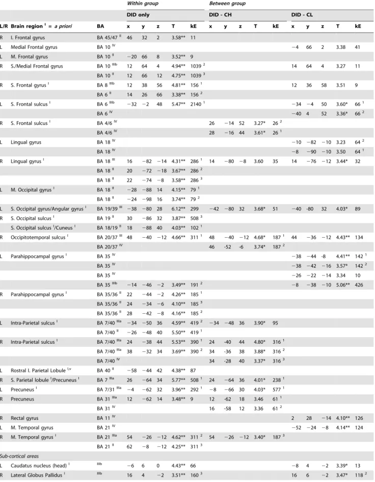
Results
Twenty-nine subjects participated in the brain imaging study: 11 patients with dissociative identity disorder (DID), 10 high fantasy prone DID simulating controls, and 8 low fantasy prone DID simulating controls. The controls were instructed to enact the two DID identity states: a neutral identity state (NIS) and a trauma-related identity state (TIS). Brain imaging data, autonomic (systolic and diastolic blood pressure, discrete heart rate and heart rate variability (HRV)) and subjective (controls’ subjective sensorimotor and emotional experiences) reactions were obtained. DID patients, as well as high fantasy prone and low fantasy prone controls were studied in the two different types of identity states during a memory script (MS) driven (neutral or trauma-related autobiographical texts) imagery paradigm. The brain imaging data of the three groups was statistically analyzed in SPM5 in a three-by-two-by-two factorial design which allows for the assessment of various effects, e.g., main effects and simple subtraction analyses (within and between identity state) within and between the three groups.
Autonomic and Subjective Reactions
Statistical results of the autonomic and subjective reactions analyses between the three groups are presented in Table 1. Mean values and the direction of the responses are depicted in Figure 1. Significant differences were found for most of the measured variables between the DID patients and both control groups (see for details Table 1) for dissociative identity state (DIS), DIS*group, MS, MS*group, DIS*MS, and DIS*MS*group.
Regional Cerebral Blood Flow Changes
Covariate data. T-tests were used to test if a significant (p,0.05) difference in regional cerebral bloodflow (rCBF) variance between the DID and control groups was explained by the subjective or objective covariates (i.e. the principal components (PC), see below). No brain areas for which a significant difference was present between the DID patients and the high or low fantasy prone controls respectively were found.
findings are shown in Figure 2. Commonalities in brain activation between patients and controls were found (data not shown).
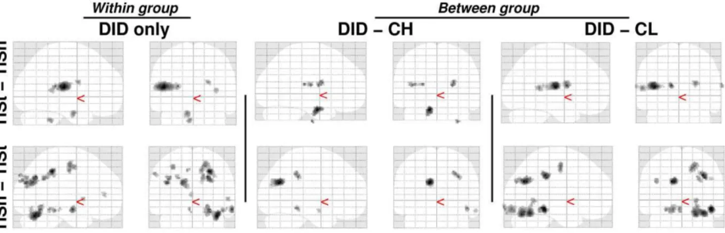
Trauma-related MS effects within identity
state. Trauma-related MS effects within both TIS and NIS are given in Table 3. TIS showed significant regionally specific increases and decreases in cerebral blood flow, when processing the trauma-related MS as compared to the neutral MS, between the DID and both the high and low fantasy prone control groups. These findings are depicted in Figure 3 and 4.
Trauma-related MS effects between identity
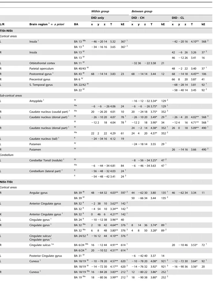
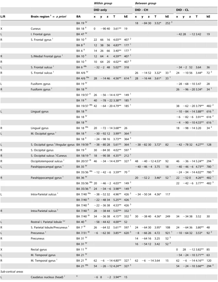
state. Trauma-related MS effects between DIS are given in Table 4. Different rCBF patterns were found for NIS and TIS, when processing the trauma-related MS, between the DID and both the high and low fantasy prone control groups. These differential rCBF patterns are shown in Figure 5 and 6. The results indicate that, for some areas (e.g. the parahippocampal gyrus in the comparison NISt-TISt or the caudate nucleus in the comparison TISt-NISt), the difference in blood flow between patients and controls is larger than the difference between the DID identity states.
Discussion
The present study was performed to examine whether earlier reported results [39,40] for DID hold after correcting for potential iatrogenic and other sociogenic effects. To this end, we tested whether these findings can be simulated by motivated role-enactment and/or is facilitated by a high level of fantasy proneness [18] by re-investigating the patient population from Reinders et al. [39,40]. Results of a sub-study (see Methods and Supporting Information S1) show that DID patients have a fantasy proneness score of 9.83 (SD 5.25), which approximates the normal population, indicating that fantasy proneness might not play a
major role in DID. This finding is consistent with the current psychobiological results. Neither high nor low fantasy prone healthy controls, instructed and motivated to simulate two different types of dissociative identity states in DID (i.e., NIS and TIS), mimicked previously observed psychophysiological and neural reactions that are associated with these identity states in DID [39,40], which is supportive of our firsta priorihypothesis.
From results shown in Figures 2, 3, and 5 a general feel of the effects can be obtained. Figures 2 and 5 and the top row of Figure 3 show that in the high fantasy prone control group more of the original DID rCBF patterns are apparent, while the low fantasy prone control group show less similarities with the original DID rCBF patterns, for example the disappearance of the left amygdala activation. Less similarities between patients only and patients versus controls means more overlap in rCBF patterns. In other words the less differences in the rCBF patterns between patients only and patients versus controls, the better the controls simulate DID. Thus, relatively speaking, low fantasy prone controls simulated the performance of DID patients better than high fantasy prone controls. This result is the opposite from the direction indicated by holders of the sociocognitive and fantasy based model of DID [17,19–21,46]. As patients and controls were scanned in a highly similar experimental setting and because controls were highly motivated to simulate DID, commonalities in brain activation between patients and controls were expected. Despite the overlap in brain activation between patients and controls important previously found psychophysiological and neurobiological differences between NIS and TIS in DID patients were upheld when controlling for fantasy proneness, suggestion, and instructed and motivated role-playing, which is supportive of our firsta priorihypothesis.
The activated areas seem to be subdivided in two distinct neural networks, where the NIS activates areas in the cerebral cortex, Table 1.Between group: Subjective and autonomic reactions.
Between group: DID versus CH Between group: DID versus CL
DIS
DIS* Group MS
MS* Group
DIS * MS
DIS*MS* Group DIS
DIS* Group MS
MS* Group
DIS * MS
DIS*MS* Group
Subjective ratings
sensory rating ,0.001** ,0.001** ,0.001** ,0.001** ,0.001** ,0.001** ,0.001** ,0.007** ,0.001** ,0.001** ,0.001** 0.001**
emotional rating ,0.001** n.s. ,0.001** 0.001** ,0.001** 0.031* ,0.001** 0.076 ,0.001** ,0.001** ,0.001** 0.030*
Autonomic reactions
heart rate frequency 0.011* 0.002** ,0.001** ,0.001** 0.009* 0.036* 0.018* 0.010* ,0.001** ,0.001** 0.021* 0.023*
systolic blood pressure 0.058 0.015* 0.006** 0.005** 0.044* 0.018* 0.080 0.034* 0.006** 0.025* 0.017* n.s.
diastolic blood pressure 0.043* n.s. 0.001** 0.002** n.s. n.s. n.s. n.s. 0.001** 0.004** n.s. n.s.
HRV-AVG n.s. 0.078 0.006** 0.003** 0.017* 0.036* n.s. n.s. 0.009* 0.015* 0.054 0.033*
Factorial statistical analyses of the between group (DID versus high or low fantasy prone DID simulating healthy controls, respectively) subjective reactions (emotional and sensori-motor ratings) and autonomic (discrete heart rate, systolic and diastolic blood pressure and heart rate variability) measurements. The statistical analyses consist of the two main effects and the accompanying interaction effect. Statistical values are reported inpvalues.
** =p,0.0083 (i.e., corrected for multiple comparisons). * = p,0.05 (i.e., uncorrected for multiple comparisons). DID = dissociative identity disorder.
CH = high fantasy prone DID simulating controls. CL = low fantasy prone DID simulating controls. DIS = dissociative identity state.
DIS*Group = interaction effect. MS = memory script. MS*Group = interaction effect. DIS * MS = interaction effect. DIS*MS*Group = interaction effect.
while the TIS mainly activates subcortical areas (e.g., see Table 2 and Figure 2). The Tables show a detailed listing of all the brain areas involved. The brain areas marked with aIIin the Tables are brain areas non-specific to DID as they disappear after comparing to a control group, i.e. these areas share commonalities between patients and controls. The brain areas marked with aIIIandIVin the Tables are brain areas specific to DID. The areas in the latter group are areas that were not reported earlier as they were ‘‘subtracted out’’ in the within group comparisons.
Our findings support the cortico-limbic inhibition model of trauma-related dissociative disorders [41,47]. Results of both the NISt-TISt comparison and the main effect of NIS show significant overlap with the activated network of brain regions during emotional memory suppression of unwanted memories in mentally healthy individuals [48], for example in frontal areas (BA 4/6/8/ 10/47), cingulate cortex (BA 32), and intraparietal sulcus (BA 7/ 40). Anderson et al. [48] did not find all of these brain areas. There is significant overlap between our study and their study, but the brain areas involved in the modulation of access to trauma-related memory in our patient population are of larger number. This might be an indication that, when functioning as NIS, in DID patients different cortical processes are involved that modulate conscious and subconscious perception of trauma-related infor-mation. These areas, e.g. (pre-)cuneus (BA 7/39, 18/19), fusiform gyrus (BA 18/19/37), lingual gyrus (BA 18), occipital gyrus (BA 18/19/37), and the parahippocampal gyrus (BA 35/36), are located in the posterior association areas (PAA) and have been indicated to be involved in multimodal [49,50] somato-sensory
integration [51,52] of information, especially in relation to attention and perceptual awareness [49]. Hyperactivation of cortical multimodal association areas for NIS in DID when listening to personal trauma scripts constituted our thirda priori hypothesis. We thus propose that for emotional memory suppression, or NIS’ mental avoidance [41], of unwanted memories in DID the PAA fulfils a pivotal role.
There are notable similarities in the patterns of brain activation for DID patients, as revealed in the main effect TIS and the TISt-NISt comparison, and mentally healthy individuals unsuppressed memory retrieval [48]. Both groups had increased activation of the insula (BA 13) and parietal operculum (BA 40/43). We did not find the hippocampus to play a role in memory retrieval in DID patients, despite the fact that this area has been indicated in memory processing in mentally healthy individuals [48]. Instead we found that the caudate nucleus was activated when DID patients listened to the trauma-memory scripts as TIS. Acute stress can be associated with a shift from hippocampal involvement to caudate nucleus involvement [53,54]. Thus, acute stress is linked with caudate nucleus-dependent stimulus-response type reactivity at the expense of hippocampal dependent spatial learning and memory [53]. According to the theory of structural dissociation [33,41] listening to a description of a personal traumatic memory in an experimental setting constitutes a consciously experienced acute stressor for TIS, because as this dissociative identity state DID patients do not manage to mentally avoid the relevant memory. When DID patients as TIS are confronted with reminders of traumatic memories, they may initiate a caudate
Figure 1. Graphical representation of averages and direction of subjective emotional experiences, subjective sensori-motor
experiences, and cardiovascular responses.The dashed line depicts the response of the neutral identity state (NIS) when listening to the
neutral or trauma-related memory script (MS). The solid line depicts the response of the traumatic identity state (TIS) when listening to the neutral or trauma related MS. All three groups are presented per variable: the dissociative identity disorder patients (DID) in pink, the high fantasy prone DID simulating controls (CH) in cyan and the low fantasy prone DID simulating controls (CL) in blue. See Table 1 for the statistical values.
Table 2.Main effects.
Within group Between group
DID only DID - CH DID - CL
L/R Brain regionI =a priori BA x y z T kE x y z T kE x y z T kE
Main Effect TIS
Cortical areas
L Insula BA 13IIIb
246 220 14 4.76 2581
246 224 18 4.41** 3021
R Insula BA 13IIIb 32
224 18 3.87 422 46
22 22 3.98** 12013
L Orbitofrontal cortex BA 11IV
230 34 222 3.95 96
BA 11IV
224 56 220 3.73 61
L Parietal operculumI BA 40IIIb
254 214 16 4.02 2582
254 214 16 3.45* 3022
R Parietal operculumI BA 40/43II 52 2 20 3.97 641
BA 40/43II 48
26 26 3.52 642
R Postcentral gyrusI BA 43III 68
214 14 4.60 1771 68 -10 16 4.25* 941 68
214 18 4.65** 1461
R Precentral gyrusI BA 6IIIb 66 6 20 4.06 1772 66 8 20 3.57** 1462
L S. Temporal gyrus BA 22/42IV
268 228 18 3.63** 641
BA 22/42IV
260 234 16 3.20 642
R S. Temporal gyrus BA 22/42IV 70
222 16 3.27 942
BA 22/42IV 72
240 10 3.52 17
Subcortical areas
L AmygdalaI IIIa
26 26 226 4.30 29 26 26 226 3.73* 35
L Caudate nucleus (anterior part) IV
224 14 18 3.53** 3383
R Caudate nucleus (caudal part)I II 20
226 20 4.56 421
L Caudate nucleus (dorsal part)I III
226 210 20 4.72 2032
224 24 20 3.88* 1442
222 26 20 4.90** 3381 III
218 24 20 4.98 2031
212 2 18 3.96* 1441
214 4 18 4.76** 3382
R Caudate nucleus (dorsal part)I III 22 2 22 4.69 2291 24 4 20 4.84* 6251 26
24 16 5.73** 12011
IV 24
24 18 4.71* 6252
R Caudate nucleus (lateral-dorsal part)I
IIIb 32
22 22 4.05 2292 32 6 20 4.74** 12012
L Caudate nucleus (tail)I II
226 236 12 4.45 43
R PutamenI IV 26 216 16 3.90* 6253
Cerebellum
L Cerebellar Tonsil (nodule)I IIIa
26 244 234 4.73 88 26 246 234 3.60* 29
R Nucleus DentatusI IV 12
248 230 3.48* 10
Main Effect NIS
Cortical areas
R Angular gyrus BA 39IIIa 42
272 34 4.16** 5082 44
262 30 3.66 2382
BA 39IV 50
266 34 3.41 2383
R Anterior Cingulate gyrusI BA 32II 2 46 6 4.94** 10391
R Cingulate gyrusI BA 32IIIa 2 18 42 4.93** 21403 4 8 50 3.24 10
BA 32IV 8 16 34 3.44* 18
L Cingulate sulcus BA 31II
214 240 40 3.91** 4193
R Cingulate sulcusI,u BA 6/24IIIb 16
212 44 5.38** 21402 18
212 46 3.33 10
L CuneusI BA 18/19IIIa
216 -88 32 3.82** 2923
216 -88 32 3.79* 5772
BA 18/19IIIa
210 278 20 3.91** 2922
210 278 20 3.61* 5773
R CuneusI BA 18/19IIIa 14
278 34 3.87** 1022 12
280 22 3.75* 1841
BA 18/19IV 16
290 38 3.62* 1842
X Cuneus BA 18II 0
290 40 3.77** 13
R Fusiform gyrusI BA 19II 42
264 220 5.00** 2591
BA 19/37II 28
258 214 3.60** 2592
L I. Frontal gyrus BA 47IV
Table 2.Cont.
Within group Between group
DID only DID - CH DID - CL
L/R Brain regionI =a priori BA x y z T kE x y z T kE x y z T kE
R I. Frontal gyrus BA 45/47II 46 32 2 3.58** 11
L Medial Frontal gyrus BA 10IV 24 66 2 3.38 41
L M. Frontal gyrus BA 10II
220 66 8 3.52** 9
R S./Medial Frontal gyrus BA 10IIIb 12 64 4 4.94** 10392 14 64 4 3.27 11
BA 10II 12 66 12 4.75** 10393
R S. Frontal gyrusI BA 8IIIb 12 38 56 4.81** 1561 12 36 58 3.51 9
BA 6II 14 26 66 3.38** 1562
L S. Frontal sulcusI BA 6IIIb
232 22 48 5.47** 21401
234 24 50 3.60* 661
BA 6IV
240 4 52 3.36* 662
R S. Frontal sulcusI BA 4/6IV 26
214 52 3.27* 262
BA 4/6IV 28
216 44 3.61* 261
L Lingual gyrus BA 18IV
210 282 210 3.23 642
BA 18IV
28 290 210 3.50 641
R Lingual gyrusI BA 18III 16
282 214 4.31** 2861 14
280 28 3.60 35 14 276 212 3.44* 32
BA 18II 20
272 218 3.67** 2862
BA 18II 22
274 28 3.58** 2863
L M. Occipital gyrusI BA 18II
228 288 14 4.15** 791
BA 18II
224 298 16 3.74** 792
L S. Occipital gyrus/Angular gyrusI BA 19/39III
238 280 28 6.12** 299 242 280 32 3.68* 51 240 -80 32 4.03* 89
R S. Occipital sulcusI BA 19II 30
286 32 3.87** 5083
S. Occipital sulcusI/CuneusI BA 18/19II 18
288 40 4.03** 1021
R Occipitotemporal sulcusI BA 20/37III 48
240 212 4.66** 3111 48
240 212 4.68* 1871 44
236 212 4.43** 134
BA 20/37IV 46 -52 -6 3.74* 1872
L Parahippocampal gyrusI BA 35IV
238 244 -8 4.41** 1421
BA 35IV
238 242 216 3.57* 1422
BA 35IV
226 222 214 3.34 10
BA 35IIIb
214 246 22 3.49** 1912
28 238 210 5.06** 426
R Parahippocampal gyrusI BA 35/36II 22
244 22 4.26** 1851
BA 35/36II 24
234 26 4.10** 1853
BA 35/36II 28
242 28 4.16** 1852
L Intra-Parietal sulcusI BA 7/40IIIa
234 250 36 4.59** 4192
234 248 36 3.90* 95
BA 7/40II
226 248 40 5.50** 4191
R Intra-Parietal sulcusI BA 7/40IIIa 24
238 44 5.53** 3901 24 -40 44 4.80* 3161
BA 7/40IIIa 38
232 34 3.69** 3902 34 -36 38 3.88* 3162
BA 7/40IV 34 -28 40 3.37* 3163
L Rostral I. Parietal LobuleI,v BA 40II
258 244 42 4.38** 87
R S. Parietal lobuleI/PrecuneusI BA 7IIIa 26
264 34 5.77** 5081 24
264 36 4.01* 2381
L PrecuneusI BA 7/31IIIa
24 262 32 3.96** 2921
28 266 30 4.03* 5771
R Precuneus BA 31IIIa 12
262 14 3.48** 9 12 -62 18 3.46 611
BA 31IV 16 -58 12 3.36 612
R Rectal gyrus BA 11IV 2 28
214 4.10** 126
L M. Temporal gyrus BA 21IV
252 224 28 4.14** 124
R M. Temporal gyrusI BA 21IIIa 54
226 212 4.62** 3112 54
226 212 3.40* 1873
BA 21II 62
28 212 4.25** 3113
Sub-cortical areas
L Caudatus nucleus (head)I IIIb
26 6 0 4.43** 66 28 4 22 3.39* 13
R Lateral Globus PallidusI IIIb 16 4
22 3.51** 1603 16 6
mediated reflex-like flight-fright-freeze response [55,56] which reaction is also supported by an accompanying amygdala activation [44,57]. Another, but compatible, explanation for increased caudate and amygdala activation in DID patients as compared to controls is a heightened memory sensitivity for
negative valanced information [58]. These findings for TIS are supportive of our seconda priorihypothesis.
To date, experimental research of inter-identity amnesia in DID has produced mixed results. One study [59] demonstrated evidence for inter-identity amnesia, which is in line with the Table 2.Cont.
Within group Between group
DID only DID - CH DID - CL
L/R Brain regionI =a priori BA x y z T kE x y z T kE x y z T kE
IV 20 -6
26 3.57* 1181 IIIb
24 0 22 4.25** 1601 24 0 22 3.36* 1183
R Substania Nigra IV 8
218 210 3.45 16
R ThalamusI II 8
28 0 4.19** 1602
Cerebellum
L Cerebellum (anterior lobe)I II
24 240 212 4.84** 1911
Overview of brain areas with statistically significant cerebral blood flow changes when comparing DID patients to high or low DID simulating controls (CH and CL respectively) for the dissociative identity state main effects.
DID = dissociative identity disorder patient group. CH = high fantasy prone DID simulating control group. CL = low fantasy prone DID simulating control group. I=A prioribrain areas based on Reinders et al. (2006) [40]. II= Brain areas found only in the DID within group analysis.
III= Brain areas found in the DID within group analysis, in the DID versus CH between group analysisandin the DID versus CL between group analysis. IIIa= Brain areas found in the DID within group analysisandin the DID versus CH between group analysis.
IIIb= Brain areas found in the DID within group analysisandin the DID versus CL between group analysis.
IV= Brain areas not found in the DID within group analysisbutappears in the between group analysis DID versus CHorDID versus CL. 1= first peak voxel in the cluster of the specified size.
2= second peak voxel in the cluster of the specified size. 3
= third peak voxel in the cluster of the specified size.
** = whole brain multiple comparison correction (p,0.05) using false discovery rate statistics [85]. * = region of interest multiple comparison correction (p,0.05) using false discovery rate statistics [85]. u= Callosomarginal sulcus (SCM) ( = Cingulate sulcus).
v= Supramarginal gyrus (Rostral I. Parietal Lobule).
(x, y, z) = MNI coordinates in mm. L/R = Left/Right.
kE = clustersize in voxels (one voxel is 26262 mm). NIS = neutral identity state.
TIS = trauma-related identity state. BA = Brodmann area.
I. = inferior; M. = middle; S. = superior. doi:10.1371/journal.pone.0039279.t002
Figure 2. ‘‘Glass brain’’ renderings showing the dissociative identity state main effects, both for the trauma-related identity state (TIS) and for the neutral identity state (NIS), for the dissociative identity disorder (DID) group (left) and the comparison of this
group to the high (middle) and low (right) fantasy prone DID simulating controls (CH and CL respectively).See Table 2 for the specific
areas.
Table 3.Memory script effects within dissociative identity state.
Within group Between group
DID only DID - CH DID - CL
L/R Brain RegionI= a priori BA x y z T kE x y z T kE x y z T kE
TISt - TISn
Cortical areas
L InsulaI BA 13II
246220 14 5.32 5691
BA 13III
238214 14 4.36 5692
238214 14 3.61* 481
238214 14 4.61* 3271
L Parietal operculumI BA 40IIIb
248230 12 4.02 5693
250222 12 3.39* 3272
R Postcentral gyrus BA 43IV 68
214 18 3.58 19
R I. Temporal gyrus BA 20IV 32
212244 3.57 9
L S. Temporal gyrus BA 42IV
268228 14 3.51 952
BA 22IV
258240 12 3.73 951
Sub-cortical areas
L AmygdalaI IIIa
21026 224 4.18 63 21224 226 4.05* 132
L Caudate nucleus (dorsal part)I IV
212 4 16 4.13* 39
R Caudate nucleus (dorsal part)I III 22 0 22 4.05 28 24
22 14 3.64* 531 26 2 20 3.76* 56
R Caudate nucleus
(lateral-dorsal part)I
IV 28 6 14 3.18* 532
L Caudate nucleus (tail)I IIIa
224234 16 4.36 20 222224 16 3.60* 13
R Caudate nucleus (tail) II 36
238 10 3.80 24
L Putamen IV
224218 14 3.30 482
224218 14 3.22 3273
Cerebellum
L Cerebellar Tonsil (nodule)I II
26 242234 4.07 34
TISn - TISt
Cortical areas
R Angular gyrus BA 39IV 48
274 32 3.30 11
L Anterior Cingulate gyrus BA 32II
22 44 8 3.61 14
L Posterior Cingulate gyrusI BA 31II
28 238 46 3.78 11
R Cingulate sulcusI,u BA 6/24IIIb 20
210 52 4.02 1161 20
210 52 3.76* 101
BA 6/24II 14 212 46 3.90 1162
L CuneusI BA 18/19IIIa
210278 24 3.81 1772
210278 24 3.39* 2102
R Cuneus BA 18/19II 16
284 28 3.41 643
R I. Frontal gyrus BA 44II 56 10 2 3.69 18
R Fusiform gyrusI BA 19/37IIIb 26
256214 4.11 3452 26
258 218 3.77* 4783
BA 18IV 26
296 220 3.48 382
BA 19/37IIIb 36
256220 3.80 3453 34
258 220 4.11 4781
BA 19/37II 38
266224 4.65 3451
L Lingual gyrus BA 18IV
214288 214 3.73 1391
BA 18IV
210282 210 3.23 1392
R Lingual gyrus BA 18IV 22
272 222 4.09 4782
BA 18IV 24
2100210 3.65 381
L S. Occipital gyrusI/Angular gyrus BA 19/39IIIb
240280 26 4.19 39 242280 28 3.59 29
R S. Occipital gyrusI BA 19II 30
284 30 3.96 26
R S. Occipital sulcusI/CuneusI BA 18/19II 18
290 38 3.86 641
R Occipitotemporal sulcusI BA 20/37III 48
234216 3.96 341 48
238212 3.65 23 46 236 214 4.31* 89
L Parahippocampal gyrus BA 35/36IV
224234 214 3.61 23
L Intra-Parietal sulcusI BA 7/40II
238252 32 3.99 27
BA 7/40II
222248 34 3.81 14
R Intra-Parietal sulcusI BA 7/40II 28
238 44 4.54 1101
BA 7/40III 38
236 36 3.37 1102 34
current findings. Other studies [60–65] found inter-identity transfer of newly learned non-autobiographical stimuli, even though the ‘‘amnestic’’ identity reported subjective amnesia for these stimuli. Several principles might explain the inconsistent findings: (i) Inter-identity amnesia may only exist for stimuli that have personal relevance for the ‘‘amnestic’’ identity. In the cited studies [59–65], it was not assessed if or to what degree the applied stimuli had autobiographical meaning for the tested ‘‘amnestic’’ and ‘‘mnestic’’ dissociative identities. Our study included trau-matic memories that were subjectively autobiographical for TIS but not for NIS, and found that NIS and TIS had different subjective, psychophysiological, and neural reactions to a descrip-tion of the involved traumatic memories. We also found that as a NIS, DID patients did not relate these traumatic memories to themselves [39]. These results indicate the importance of using autobiographical information when investigating inter-identity amnesia in DID. (ii) Inter-identity amnesia may predominantly exist between different types of dissociative identities, particularly between neural and trauma-related identity states.This has been
clinically observed, theoretically proposed [33,41] and is in line with our results. Unfortunately, in most studies [59–66] it was not assessed what types of dissociative identities participated, e.g. NIS or TIS. Therefore, we strongly recommend that in future research in DID the types of dissociative identities are verified and reported and that test material is used that is subjectively autobiographical for one dissociative identity, but not for another.
The sociocognitive view of DID entails the idea that this disorder can be easily and readily created in motivated suggestible individuals and that few suggestions would suffice to generate the symptoms of DID [15] (see Supporting Information S2). Still, one might argue that the current brief practice of DID simulation is insufficient to simulate the psychobiological profiles of NIS and TIS. Even if years of practice could generate these profiles, our findings suggest that fantasy proneness is not the driving factor because low fantasy prone controls simulated the performance of DID patients better than high fantasy prone controls. This result is the opposite from the direction indicated by holders of the sociocognitive and fantasy based view. Therefore we feel that our Table 3.Cont.
Within group Between group
DID only DID - CH DID - CL
L/R Brain RegionI= a priori BA x y z T kE x y z T kE x y z T kE
L Rostral I. Parietal LobuleI,v BA 40II
260244 40 3.77 19
R S. Parietal Lobule/PrecuneusI BA 7IIIb 24 264 36 4.32 65 28 266 32 3.49* 34
L Precentral sulcusI BA 4/6II
23028 52 3.85 43
L PrecuneusI BA 7/31III
216268 28 4.22 1771
212266 26 4.25* 2101
216268 28 4.26* 181
BA 7/31II
210264 32 3.74 1773
R (Pre-)Cuneus/Parieto-occipital sulcusBA 7/19II 18
278 34 3.79 642
R M. Temporal gyrusI BA 21II 54
226212 3.58 342
BA 21IIIa 62
26 214 4.24 48 60 22 216 3.33 11
Cerebellum
L Cerebellum (anterior lobe) IIIb
24 242214 3.79 11 24 244 214 3.62 481
IV
24 236 212 3.43 482
Overview of brain areas with statistically significant cerebral blood flow changes when comparing DID patients to high or low DID simulating controls (CH and CL respectively) for the trauma-related memory script effects within dissociative identity state.
DID = dissociative identity disorder patient group. CH = high fantasy prone DID simulating control group. CL = low fantasy prone DID simulating control group. I=A prioribrain areas based on Reinders et al. (2006) [40]. II= Brain areas found only in the DID within group analysis.
III= Brain areas found in the DID within group analysis, in the DID versus CH between group analysisandin the DID versus CL between group analysis. IIIa= Brain areas found in the DID within group analysisandin the DID versus CH between group analysis.
IIIb= Brain areas found in the DID within group analysisandin the DID versus CL between group analysis.
IV= Brain areas not found in the DID within group analysisbutappear in the between group analysis DID versus CHorDID versus CL. 1= first peak voxel in the cluster of the specified size.
2= second peak voxel in the cluster of the specified size. 3= third peak voxel in the cluster of the specified size.
** = whole brain multiple comparison correction (p,0.05) using false discovery rate statistics [85]. * = region of interest multiple comparison correction (p,0.05) using false discovery rate statistics [85]. u= Callosomarginal sulcus (SCM) ( = Cingulate sulcus).
v= Supramarginal gyrus (Rostral I. Parietal Lobule). (x, y, z) = MNI coordinates in mm.
L/R = Left/Right.
kE = clustersize in voxels (one voxel is 26262 mm).
NISn = neutral identity state exposed to the neutral memory script. NISt = neutral identity state exposed to the trauma-related memory script. TISn = trauma-related identity state exposed to the neutral memory script. TISt = trauma-related identity state exposed to the trauma-related memory script. BA = Brodmann area.
study provides an important contribution to the etiology discus-sion.
For the first time, it is shown using brain imaging that neither high nor low fantasy prone healthy women, who enacted two different types of dissociative identity states, were able to substantially simulate these identity states in psychobiological terms. These results do not support the idea of a sociogenic origin for DID.
Methods
Participants
Controls. Mentally healthy females were recruited by local newspaper advertisements. Respondents were sent a letter in
which the study was explained and in which they were invited to complete three questionnaires: (i) the Traumatic Experiences Checklist (TEC) [67], a self-report questionnaire assessing potentially traumatizing events such as physical abuse and emotional neglect, (ii) the Somatoform Dissociation Questionnaire (SDQ-20 [68–70], a self-report questionnaire evaluating the severity of somatoform dissociative symptoms, e.g., analgesia, anesthesia, motor inhibitions), and (iii) the Creative Experiences Questionnaire (CEQ) [18] which measures fantasy proneness. Exclusion criteria were the presence of medical, neurological or psychiatric problems in the past or the present, the use of psychotropic medication 15 days prior to examination, participa-tion in a positron emission tomography (PET) or other study that involved administration of radiation in the year prior to this study,
Figure 3. ‘‘Glass brain’’ renderings show differences in the processing of the trauma-related text (indicated with a small ‘t’) and the
neutral text (indicated with a small ‘n’) within the trauma-related identity state (TIS).Differences in regional cerebral blood flow patterns
for the dissociative identity disorder (DID) group (left) and the comparison of this group to the high (middle) and low (right) fantasy prone DID simulating controls (CH and CL respectively) are depicted. See Table 3 for the specific areas.
doi:10.1371/journal.pone.0039279.g003
Figure 4. The brain areas indicated with the blue cross (i.e. the peak voxel) are (from top left to bottom right): the left amygdala,
the left insula, the left precuneus, and the right occipitotemporal sulcus.These areas have the most significant rCBF differences between
the dissociative identity disorder patients and high and low fantasy prone DID simulating controls (CH and CL respectively) and is shown both in directionality, i.e. the bar graphs, and location, i.e. shown on a coronal overlay (left in the picture is left in the brain). Results show the differential processing of the trauma-related text versus the neutral text within the TIS, when comparing the DID groups to the high fantasy prone control group (left) and low fantasy prone control group (right).
Table 4.Memory script effects between dissociative identity states.
Within group Between group
DID only DID - CH DID - CL
L/R Brain regionI =a priori BA x y z T kE x y z T kE x y z T kE
TISt-NISt
Cortical areas
L InsulaI BA 13IIIb
246220 14 5.32 3671
242220 16 4.10** 5682
BA 13II
234216 16 3.65 3672
R Insula BA 13IV 42
26 26 3.26 372
BA 13IV 46
212 26 3.41 16
L Orbitofrontal cortex BA 11IV
232 36 222 3.58 21
R Parietal operculum BA 40/43IV 48
22 22 3.40 371
R Postcentral gyrusI BA 43III 68
214 14 3.83 23 68 214 14 3.44 12 68 214 18 4.45** 106
R Precentral gyrus BA 6IV 66 8 20 3.87 41
L S. Temporal gyrus BA 22/42IV
268228 14 3.81 921
BA 22IV
258240 14 3.45 922
Sub-cortical areas
L AmygdalaI IV
216212232 3.34* 1292 IIIa
26 26 226 4.06 24 26 26 226 3.75* 1291
R Caudate nucleus (caudal part)I IIIa 20
226 20 4.01 10 20 224 18 3.75* 3523
L Caudate nucleus (dorsal part)I III
226210 20 4.07 781
226210 20 3.49* 292
22624 20 4.02** 5683 III
212 2 18 4.04 782
212 2 18 3.90* 34 212 4 16 4.71** 5681
R Caudate nucleus (dorsal part)I IV 24
22 14 4.38* 3521 26 0 18 5.09** 4901 IIIa 22 2 22 4.29 61 24 4 20 4.37* 3522
L Caudate nucleus (tail)I II
224234 16 4.12 19
L Putamen IV
224218 14 3.55 291
R Putamen IV 26
214 16 3.66 4902
Cerebellum
L Cerebellar Tonsil (nodule)I IV
28 256234 3.25* 472 IIIa
26 244234 4.81 84 26 246234 3.63 471
L Cerebellum (lateral part)I II
256248232 4.03 241 II
254248242 3.41 242
NISt-TISt
Cortical areas
R Angular gyrus BA 39III 48
264 32 4.03** 5972 44
262 30 3.80 1351 46
262 34 3.34 11
BA 39IV 50
266 34 3.44 1352
L Anterior Cingulate gyrus BA 32II 22 38 10 3.62** 1422
BA 32II
24 54 10 3.34** 1423
X Anterior Cingulate gyrusI BA 32II 0 46 6 4.21** 1421
L Cingulate gyrusI BA 24II
210212 38 3.96** 40
R Cingulate gyrusI BA 32IIIa 2 18 42 4.64** 3761 8 14 36 3.74* 891
BA 32IIIa 6 8 48 3.80** 3763 4 8 50 3.28* 892
L Cingulate sulcus/
Cingulate gyrusI BA 24/32 II
216 12 44 4.10** 3762
R Cingulate sulcusI,u BA 6/24IIIb 16
212 44 4.91** 6141 20
210 46 3.53* 721
BA 6/24II 20
210 52 4.51** 6143
L Posterior Cingulate gyrus BA 31IV
26 242 40 3.37 14
L CuneusI BA 18/19III
210278 20 4.57** 6201
210278 20 4.08* 9212
212272 30 3.64* 921
BA 18/19III
214272 30 4.17** 6202
214276 32 3.92* 9213
216290 36 3.56* 20
R CuneusI BA 18/19IIIa 16
284 28 3.83** 2123 12
280 22 3.86* 2521
BA 19IIIa 18
280 36 3.99** 2122 18
Table 4.Cont.
Within group Between group
DID only DID - CH DID - CL
L/R Brain regionI =a priori BA x y z T kE x y z T kE x y z T kE
BA 19IV 18
284 30 3.52* 2523
X Cuneus BA 18II 0 290 40 3.61** 19
L I. Frontal gyrus BA 47IV
242 28 212 3.42 19
R S. Frontal gyrusI BA 10II 22 66 16 4.03** 4072
BA 8II 12 38 56 4.83** 1771
BA 6II 14 26 66 3.40** 1772
R S./Medial Frontal gyrusI BA 10II 12 64 4 4.59** 4071
R BA 10II 10 64 20 4.02** 4073
L S. Frontal sulcusI BA 6IIIb
23222 48 5.02** 318 23422 52 3.64 36
R S. Frontal sulcusI BA 4/6IV 26
214 52 3.32* 332 24
210 56 3.44* 722
BA 4/6IIIa 28
214 46 4.56** 6142 28
216 44 3.61* 331
L Fusiform gyrus BA 19IV
228268210 3.47 28
R Fusiform gyrusI BA 18IV 26
296220 3.34* 341
BA 19/37II 26
256214 4.10** 1491
BA 19II 40
278222 3.38** 1852
BA 19/37IIIb42
264220 4.79** 1851 38
262220 3.79** 4822
L Lingual gyrus BA 18IV
210284214 3.88** 6163
BA 18IV
26 28226 3.91** 6162
BA 18IV
24 290210 4.33** 6161
R Lingual gyrus BA 18IIIb 20
272214 3.68** 26 18 298214 3.20 342
L M. Occipital gyrusI BA 18II
230292 12 3.99** 3642
BA 18II
224298 16 3.73** 3643
L S. Occipital gyrusI/Angular gyrus BA 19/39III
238280 28 5.61** 3641
238282 30 3.72* 82 242278 32 4.27** 128
R S. Occipital gyrus BA 19II 30
284 30 4.02** 5973
R S. Occipital sulcusI/Cuneus BA 18/19II 18
290 38 4.35** 2121
R Occipitotemporal sulcusI BA 20/37III 46
234214 4.39** 3272 48
240212 4.53* 92 46 236214 5.24** 2941
L Parahippocampal gyrusI BA 35IV
24024624 3.75 18 24024626 4.73** 7801
BA 35/36IIIb
21224226 3.59** 702
224234214 4.02** 7803
R Parahippocampal gyrusI BA 36IV 20
252 2 3.46* 521 22
252 0 4.26** 4821
BA 35/36IIIb20
24622 4.03** 1492 22
24226 3.77** 4823
BA 35/36II 24
23426 3.98** 1493
L Intra-Parietal sulcusI BA 7/40IIIa
238252 32 4.96** 4262
234250 34 4.36* 117
BA 7/40II
222248 34 5.25** 4261
BA 7/40II
222236 38 4.53** 4263
R Intra-Parietal sulcusI BA 7/40II 28
238 44 5.07** 3321
BA 7/40III 34
236 38 4.15** 3322 30
238 40 4.36* 249 34 234 38 3.52 30
L Rostral I. Parietal lobuleI,v BA 40II
258244 42 4.00** 52
R S. Parietal lobule/PrecuneusI BA 7III 26
264 32 5.61** 5971 24
264 30 3.95* 108 24 264 36 3.80** 48
L PrecuneusI BA 7/31III
26 262 30 3.85** 6203
28 266 26 4.72 9211
210264 32 3.53* 922
R Precuneus BA 31IV 14
264 16 3.25 523
BA 31IV 16
254 12 3.42 522
X Rectal gyrus BA 11IV 0 28
212 3.82** 85
L M. Temporal gyrus BA 21IV
254224210 3.71** 63
R M. Temporal gyrusI BA 21III 62
26 214 4.80** 3271 62
26 214 3.64 15 62 26 214 4.16** 120
BA 21IIIb 54
226212 4.24** 3273 54
224210 3.66** 2942
Sub-cortical areas
L Caudatus nucleus (head)I II
and pregnancy. A total of 18 healthy controls participated in the study, which was approved by the Medical Ethical Committee of the University Medical Center Groningen.
After inclusion, written and oral information on dissociative identity states (i.e. NIS and TIS) in DID and instructions on how to simulate these dissociative identity states was given to the controls. It was checked whether the controls understood this Table 4.Cont.
Within group Between group
DID only DID - CH DID - CL
L/R Brain regionI =a priori BA x y z T kE x y z T kE x y z T kE
R Lateral Globus PallidusI IV 24
28 28 3.77** 851 IIIb
24 0 22 4.13** 701 24 0 22 3.34* 852
R Medial Globus Pallidus II 14
26 22 3.44** 702
Cerebellum
L Cerebellum (anterior lobe) IIIb
26 242212 4.23** 701
24 242212 4.53** 7802
Overview of brain areas with statistically significant cerebral blood flow changes when comparing DID patients to high or low DID simulating controls (CH and CL respectively) for the trauma-related memory script effects between dissociative identity state.
DID = dissociative identity disorder patient group. CH = high fantasy prone DID simulating control group. CL = low fantasy prone DID simulating control group. I=A prioribrain areas based on Reinders et al. (2006) [40]. II= Brain areas found only in the DID within group analysis.
III= Brain areas found in the DID within group analysis, in the DID versus CH between group analysisandin the DID versus CL between group analysis. IIIa= Brain areas found in the DID within group analysisandin the DID versus CH between group analysis.
IIIb= Brain areas found in the DID within group analysisandin the DID versus CL between group analysis.
IV= Brain areas not found in the DID within group analysisbutappear in the between group analysis DID versus CHorDID versus CL. 1= first peak voxel in the cluster of the specified size.
2= second peak voxel in the cluster of the specified size. 3= third peak voxel in the cluster of the specified size.
** = whole brain multiple comparison correction (p,0.05) using false discovery rate statistics [85]. * = region of interest multiple comparison correction (p,0.05) using false discovery rate statistics [85]. u= Callosomarginal sulcus (SCM) ( = Cingulate sulcus).
v= Supramarginal gyrus (Rostral I. Parietal Lobule). (x, y, z) = MNI coordinates in mm.
L/R = Left/Right.
kE = clustersize in voxels (one voxel is 26262 mm).
NISn = neutral identity state exposed to the neutral memory script. NISt = neutral identity state exposed to the trauma-related memory script. TISn = trauma-related identity state exposed to the neutral memory script. TISt = trauma-related identity state exposed to the trauma-related memory script. BA = Brodmann area.
I. = inferior; M. = middle; S. = superior. doi:10.1371/journal.pone.0039279.t004
Figure 5. ‘‘Glass brain’’ renderings show differences in the processing of the trauma-related text (indicated with a small ‘t’)
between the trauma-related identity state (TIS) and the neutral identity state (NIS).Differences in regional cerebral bloodflow patterns for
the dissociative identity disorder (DID) group (left) and the comparison of this group to the high (middle) and low (right) fantasy prone DID simulating controls (CH and CL respectively) are depicted. See Table 4 for the specific areas.
information. A template for training themselves in switching between the simulated identity states was provided. Controls were then questioned about how they constructed the two identity states, whether they encountered difficulties and if so, they were given support to improve their roles as NIS and TIS. To help the controls simulate NIS and TIS, they were asked to recall two experiences they had had earlier in their life, an emotionally neutral experience and an emotionally painful experience. Controls were asked to provide their most painful memory to serve as an analogue for the patients’ personal trauma memories, as well as a neutral personal episodic memory. Controls were subsequently instructed how to write the autobiographical analogue neutral and ‘‘trauma’’ memory scripts. For the experiment they had to train themselves in being in a neutral state, the NIS who is unresponsive or under-responsive to the painful experience, and in being in a state in which they re-experience the painful memory, the TIS. The consecutive and final check on the capability to simulate the two different dissociative identity states consisted in checking whether their description of their neutral and painful experiences (that was to be casted in an audiotape recording) met the instructions on how to enact a DID patient.
In the two or more weeks preceding the PET scans, candidate control subjects practiced simulating NIS and TIS, as well as alternating between NIS and TIS using detailed role instructions. One of the investigators (H.V.) contacted the candidates per telephone during this preparatory phase to ensure that they followed the instructions and to offer further suggestions for optimizing their role performance. One candidate felt unable to simulate the roles satisfactorily, and was therefore excluded. Prior to the actual PET scanning, H.V. checked if the candidates experienced and judged that they were able to simulate the roles of NIS and TIS. During the actual scanning, he checked if they engaged in the requested simulations, and immediately after the role performances, he checked if the controls generally felt they had simulated the roles of NIS and TIS effectively. All controls
passed these various checks. In addition, immediately after each text condition, H.V. administered a detailed questionnaire that inquired after the controls’ subjective sensorimotor and emotional experiences during their role performance. This questionnaire was identical to the one in the patient study [39,40], which was administered by the patients’ therapist, and debriefed six subjective emotional experiences (fear, sorrow, sadness, anger, shame and disgust) and ten sensorimotor experiences (visual, kinesthetic, auditory, olfactory+gustatory reactions, pain, physical numbness, body stiffening, paralysis and restlessness) were debriefed. In addition, the presence of the identity state under investigation and the interference among identity states were also debriefed. Using this questionnaire, H.V. or the patients’ therapist could structurally evaluate if the intended NIS or TIS had been present during the experimental condition. Statistical analyses of the simulation performance in terms of their subjective experi-ences, i.e. the subjective sensorimotor perception and emotional response, during the scanning by the two control groups are provided in Supporting Information S2.
As we did not have CEQ values for the patients (see also Supporting Information S1) we could not control for fantasy proneness by including a covariate. Therefore, the controls were divided into two groups based on their CEQ scores resulting in a high fantasy prone group (n = 10, age 38.2 (SD 10.9), TEC 0.7 (SD 1.3), SDQ-20 22 (SD 2.4)) with CEQ 13.7 (SD 3.2) and a low fantasy prone group (n = 8, age 42.5 (SD 10.1), TEC 0.4 (SD 0.5), SDQ-20 20.9 (SD 1.5)), with CEQ 3.9 (SD 1.6). A CEQ cut-off for high fantasy proneness of 10 was used, which the developers of the CEQ recommended for the current sample [71].
Patients
A detailed description of the DID patients can be found elsewhere [39,40]. In short: Eleven patients (all female, age 41.0, SD 6.1) participated: (i) whose treatment had progressed to Phase II [72], which involves therapeutic exposure to trauma-related
Figure 6. The brain areas indicated with the blue cross (i.e. the peak voxel) are (from top left to bottom right): the right caudate
nucleus (dorsal part) (2x), the left precuneus, and the right occipitotemporal sulcus. These areas involve the most significant rCBF
difference between the dissociative identity disorder patients and high and low fantasy prone DID simulating controls (CH and CL respectively) and is shown in both directionality, i.e. the bar graphs, and location, i.e. shown on a coronal overlay (left in the picture is left in the brain). Results show the differential processing of the trauma-related text between the TIS and the NIS, when comparing the DID groups to the high fantasy prone control group (left) and low fantasy prone control group (right).
memories, (ii) who met criteria for DID, as operationalized in the Structured Clinical Interview for DSM-IV Dissociative Disorders (SCID-D [73]), and (iii) had at least one TIS and one NIS that they could activate on demand [33] and (iv) the involved TIS had displayed signs of sympathetic nervous system dominance under perceived threat in clinical situations.
To establish the CEQ values in DID patients an independent and representative sample of DID patients (n = 42) completed the CEQ. Details regarding this substudy can be found in the Supporting Information S1.
Stimulus Scripts
During scanning, patients and controls listened to descriptions of the neutral episodic memories and memories of traumatizing or most painful events that only TIS experienced as a personal memory [74]. These memories were cast, prior to the PET session, by the therapist or one of the principal investigators (H.V.) in terms of stimulus descriptions, and were subsequently audio-taped in a neutral tone of voice as 120 second scripts for playback during the PET investigation.
PET Procedure
The PET (Siemens/CTI ECAT HR+) procedure for the controls was close to identical to the patients [39,40]. In contrast to patients the controls did not habituate to the PET environment prior to the investigation as anxiety levels were expected to be low. Approximately two hours prior to the PET investigation the continuous ECG registration was started, obtaining the five frequency and time domain variables [75,76]. No urine samples were obtained for the control groups, both medication and drugs use were verbally debriefed according to standard control research practice.
For the controls one extra set of the four conditions was added to increase statistical power. The scanning sequence was therefore NISn, NISt, TISn, TISt, TISn, TISt, NISn, NISt, TISn, TISt, NISn and NISt. The last minor character (n or t) denotes the content of the memory script (MS: neutral or trauma-related). For patient comfort considerations, i.e. minimizing the number of identity state switches, a fixed condition order was used, which was also used for the controls to minimize methodological differences. Immediately following the end of each script, blood pressure (systolic and diastolic) and discrete heart rate frequency were measured and the six subjective emotional and ten sensorimotor experiences were debriefed. Finally, the presence of the identity state under investigation and the interference among identity states were also debriefed.
Image Acquisition and Data Processing
Data acquisition, reconstruction, attenuation correction, spatial transformation, spatial smoothing (isotropic Gaussian kernel of 12 mm) and global normalization were performed as usual [39,40,77]. SPM5 (www.fil.ion.ucl.ac.uk/spm) was used for spatial transformation to the MNI template (using heavy regularization) [78,79] and statistical analysis [80] of both patient and control data.
Data Analysis: Autonomic and Subjective Reactions Statistical analysis, missing value analysis and principal compo-nents (PC) analysis were performed with SPSS-PC 15.0 (2006) in an identical manner as was done for the patient data [39,40]. Results withp,0.05 are reported as significant. Within SPSS two two-by-two-by-two factorial design were defined with the first factor Group, consisting of the levels DID and the high fantasy prone controls or
the low fantasy prone controls, a second factor identity state, consisting of the levels NIS and TIS, and the third factor was MS, consisting of the levels neutral and trauma-related. For one high fantasy prone and one low fantasy prone subject heart rate variability (HRV) data could not be obtained. In addition, the data, including the PET data, from two NISt conditions was removed as the control subjects reported not to be able to maintain as a NIS. One TISn condition was removed from the low fantasy prone data as the subject reported not to be able to maintain a TIS. Bonferoni correction to correct for multiple testing was applied.
Data Analysis: PET-data
The patient PET data included in the current study is identical to the data as included and described in our previous publications [39,40]. This study assessed various effects, e.g., main effects and simple subtraction analyses (within and between identity state) within the DID group using SPM99. This data was re-analyzed in SPM5 and is referred to as the ‘‘within DID only’’ analyses.
From the 10 high fantasy prone healthy controls the PET data of one subject was lost due to storage failure at the PET center. The data of the three groups was statistically analyzed in SPM5 in a three-by-two-by-two factorial design [81–84]. The general linear model (GLM) consisted of the three factor main effects, the four conditions and a group by condition interaction.
In addition, the subjective reactions and the autonomic reactions were included as group specific covariates of interest after PC analysis [39,40]. The variance in the subjective ratings could be described with the first two, six, and five PC for the DID, high and low fantasy prone groups respectively, explaining 64%, 68%, and 72% of the variance. The variance in the autonomic reactions could be described with the first three PC for each of the DID, high and low fantasy prone groups, explaining 85%, 82%, and 87% of the variance respectively. Finally, the global cerebral blood flow (CBF) was included as a nuisance covariate (AnCova by subject).
Hypothesis Testing
Previously reported significant findings were tested using a between group subtraction of the within group results (e.g. DID(TISt-NISt)-Control(TISt-NISt)). Commonalities in brain activation between patients and controls were tested using global null conjunction analyses [83].
Statistical Inference and Reporting