Histological and ultrastructural
aspects of the tongue in
undernourished rats
Summary
Marilda Aparecida Milanez Morgado de Abreu1,Luc Louis Maurice Weckx2, Cleonice Hitomi
Watashi Hirata3
1 Master’s degree, graduate studies at the Stomatology Sector, Otorhinolaryngology and Disturbances of Human Communication Department and the Dermatology
Department; Sao Paulo Federal University/Paulista Medical School.
2 Lecturer, Full Professor of the Pediatric Otorhinolaryngology and Disturbances of Human Communication Department; Hea of the Otorhinolaryngology and Head &
Neck Surgery Department, Sao Paulo Federal University/Paulista Medical School.
3 Master’s, Doctor, Head of the Stomatology Sector, Otorhinolaryngology and Disturbances of Human Communication Department and the Dermatology Department;
Sao Paulo Federal University/Paulista Medical School. Sao Paulo Federal University/Paulista Medical School (UNIFESP/EPM).
Address for correspondence: Marilda Aparecida Milanez Morgado de Abreu - Rua Brasil 1599 Centro Dracena SP 17900-000 Telephone: (0xx18) 3821-4630 - Fax: (0xx18) 3821-2276 - E-mail: lfmabreu@uol.com.br
This paper was submitted to the ABORL-SGP (Publication Management System) of the RBORL on 17/10/2005 and approved on 5/5/2006. Paper submitted to the ABORL-CCF SGP (Management Publications System) on October 17th, 2005 and accepted for publication on May 5th, 2006.
T
here are few published studies on the effects of protein-caloric undernourishment on the oral mucosa. Objectives: The objective of this study is to verify the histological and ultrastructural aspects of the tongue mucosa in protein-caloric undernourished adult rats. Material and Methods: A clinical experimental study was done in thirty Wistar rats, 15 controls and 15 with protein-caloric undernourishment. The last group received ration in small amounts, with a reduced casein content, during 45 days. Rats were weighed every 3 days, from the first (90 days of life) to the last day of a 45-day dietary period, when they were sacrificed. Plasma was used for protein electrophoresis and their tongues were prepared for light and scanning electron microscopy. Analysus of variance and Student’s t-test were used for statistical analysis. Results: A significant decrease in weight and in plasma proteins was found in protein-caloric undernourished rats compared to the control group. Histological findings revealed no differences between the two groups and there were no statistically significant differences in the filiform papilla count under the scanning electron microscopy. Conclusion: Protein-caloric undernourishment does not cause alterations in the tongue mucosa of adult rats.Keywords: protein-energy malnutrition, histology, tongue, scanning electron microscopy.
ORIGINAL ARTICLE
INTRODUCTION
Although clinical findings in the mouth such as tongue mucosa atrophy, loss of papillae, and angular cheilitis, are frequently related to nutrition, there are few studies on the effect of protein-energy malnutrition over
the oral mucosa and the tongue1-4.
Winkler and Nakamoto in 19825 studied the effects
of prenatal malnutrition on tissue development in the mouth of newborn rats. They described histological alte-rations on the tongue such as reduced cell numbers and the increased size of these cells. Malnutrition also affected incisor and molar dental bacteria. Results suggested that adequate protein intake was extremely important for the healthy development of teeth and the tongue.
Aldred et al. in 19896 reported an increase in yeasts
in the mouth of malnourished children, but found no other clear clinical signs.
The aim of this paper is to study the effect of pro-tein-energy malnutrition on the mucosa of the tongue of adult rats, using optical and scanning electron microsco-py.
MATERIAL AND METHODS
This is an experimental study made on thirty “WISTAR” rats aged 90 days and weighing between 180 and 220 grams. Animals were placed in individual cages measuring 40 x 20 x 13 cm each, containing sawdust and water with no restrictions.
Animals were randomly divided into two groups each containing 15 rats. The first group (control) received ration composed of casein (18%), corn (4%), salt (4%), multivitamins (1%), benzoate (0.1%), oil (8%), sugar (10%), corn starch excipient qs (100%). The second group (with protein-energy malnutrition) received a similar ration but with changes in the relative components of casein (1%), given as ration weighing 6 grams/day.
Rats were weighed every 3 days from the first day (age 90 days) until the final day of the 45-day diet period. At this point, having been weighed, the animals were guillotined. Blood was collected and plasma was obtained by centrifugation for protein electrophoresis.
The tongue was sectioned and completely removed. The anterior region of the tongue was sectioned for light and electron microscopy.
Fragments were included in paraffin for microtome sectioning and hematoxylin-eosin (HE) stained. Slides were studied under a common optical microscopy at 50 and 400 times amplification. The following parameters were assessed: epithelial layer uniformity or lack thereof, the presence or absence of an inflammatory infiltrate in the lamina propria, and areas with or without filiform and fungiform papillae in the anterior region of the tongue. Uniformity was considered as the regular distribution of
layers with similar cell maturity.
Seven cases in each group (control and malnouri-shed) were assessed by electron microscopy. The number of filiform papillae was one of the study parameters; counts were obtained from electron microscopy pictures taken using VP 120 black-and-white film. Inclusion criteria were: the number of whole filiform papillae (which included a base and a conical projection) counted on pictures
magni-fied 100 times, measuring 9 x 9 cm, using a 2.3 cm2 area,
in each case counting 5 to 10 regions. Tangential papillae in the left and lower borders were included; tangential papillae in the right and upper borders were excluded;
regions with fungiform papillae were excluded7.
Statistical analysis included the analysis of variance to compare average rat weight, protein electrophoresis and the papillary count. Student’s T-test was used to study the
difference between two independent samples8 with a 0.05
or 0.5% significance level.
RESULTS
Weight increased in the control group and decrea-sed in the protein-energy malnutrition group after 45 days. Statistical analysis revealed a significant difference between both samples on the 45th day, as shown on Chart 1.
The plasma protein electrophoresis profile disclosed a significant reduction in total plasma proteins in the pro-tein-energy malnutrition group on the 45th day compared to controls (Tables 1 and 2).
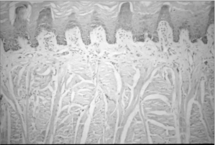
Common light microscopy histology revealed no differences between the control group and the protein-energy malnutrition group for the parameters we studied (Figures 1 and 2).
Common light microscopy histology of the tongues of rats in both groups showed stratified epithelium with uniform layers in 15 cases. Neither group had an inflam-matory infiltrate or areas of papillary loss in the tongue. Keratinization of the ventral surface of the tongue was seen in 2 cases in the control group (with thickening of the keratin layer in 1 control group case) and in no case of the protein-energy malnutrition group.
Chart 1. Histogram of rat body weight averages in the control group
Table 1. Results of plasma protein electrophoresis in rats for the control group and the malnutrition group on the 45th day (metrics are total
proteins in densitometric units).
Case Control group Protein-energy malnutrition group
1 280 210
2 310 200
3 310 215
4 270 200
5 300 230
6 270 205
7 320 200
8 288 215
9 276 220
10 337 230
11 327 200
12 300 220
13 317 259
14 280 260
15 326 250
Average 301 221 Standard deviation 22 20
Table 2. Analysis of variance of densitometry results on total
elec-trophoresis in the control group and the protein-energy malnutrition group rats.
Sources of variation Degrees of freedom
Test T calc T crit. Protein-energy
mal-nutrition group rats 14 10,04 (*) 2,05 Control group rats 14
28
Figure 1. Photomicrograph of a sagittal section of a filiform papilla.
Stratified epithelium of the normal tongue mucosa in the control group - Magnification - 100X.
Figure 2. Photomicrograph of a sagittal section of a filiform papilla.
Stratified epithelium of the normal tongue mucosa of rats with protein-energy malnutrition - Magnification - 100X.
There were no statistically significant differences in the filiform papillae count by electron microscopy on the tongue in both the control group (7 cases) and the protein-energy malnutrition group (7 cases), as seen on Tables 3, 4 and 5 (Figures 3 and 4).
DISCUSSION
Malnutrition is a significant public health problem in the world; protein-energy malnutrition is one of the most severe forms of this condition. It affects mostly children in poorer coutries, where the prevalence is estimated as 1/3
of children between birth and five years of age9.
The World Health Organization defines protein-energy malnutrition as “the range of pathological con-ditions arising from a deficiency of cell intake, transport or use of nutrients, commonly associated with infection, occurring with greater frequency in infants and pre-school
children”10.
Protein-energy malnutrition causes a variety of clinical conditions, the most apparent being effects on body measurements and weight as a whole or for specific organs. Malnutrition develops in a sequence starting from exclusive protein deficiency (kwashiorkor) to
protein-ener-gy deficiency (marasmus)11. In man, nutritional deficiency
is never an exclusively caloric or protein deficiency; both are usually associated.
Figure 3. Photomicrograph of the anterior dorsal surface of the adult rat tongue. Filiform and fungiform papillae over which are taste buds. Control group. Original Magnification - 200X.
Figure 4. Photomicrograph of the anterior dorsal surface of the adult
rat tongue showing a papilla inclined antero-posteriorly. Protein-energy malnutrition group. Original Magnification - 200X.
Table 3. Count of filiform papillae on the anterior region of the tongue
by unit area (2.3 cm2). Magnification - 100x; control group (electron microscopy).
Control
group Number of papillae per area
C1 11 13 9 11 9 11
C2 15 14 17 15 16
C3 18 17 17 17 17
C4 17 20 20 17 17
C5 10 10 8 9 10 11
C6 11 13 11 13
C7 12 11 13 12 12
Average 13,44 Standard
deviation 3,3 Sum 484 Sum of
squares 6900
Table 4. Count of filiform papillae on the anterior region of the tongue
by unit area (2.3 cm2). Magnification - 100x; protein-energy malnutrition group (electron microscopy).
Protein-energy
malnu-trition group Number of papillae per area
D1 17 18 15 19 15
D2 16 14 10 12
D3 11 12 14 11
D4 14 14 15
D5 13 12 10 11 15 10
D6 11 11 11 14
D7 11 11 11 12
Average 13 Standard deviation 2,41
Sum 390
Sum of squares 5244
Table 5. Analysis of variance of the number of filiform papillae in the
anterior region of the tongue by unit area.
Degrees of free-dom
Test Tcalc Tcrit No of papillae in rats with
protein-energy malnutrition 30 0.60 2.00 No of papillae in control
group rats 36
Total 66
glossities and stomatitis1-4. In out study the 45-day
protein-energy deficient diet effectively produced malnutrition in rats aged 90 days, as shown in the statistically significant differences between the control group and the protein-energy malnutrition group; there was both weight loss and a reduction in total proteins. However, the histological study of the tongue of protein-energy malnutrition rats, both under common optical microscopy and electron mi-croscopy did not reveal changes such as glossitis or loss or filiform papillae.
There are no published papers on the effects of pro-tein-energy malnutrition specifically on the mucosa of the mouth or tongue. There is one paper describing changes in
prenatal malnutrition5. In contrast, there are many reports
on the clinical, histological and ultra-structural changes of the tongue mucosa in iron deficiency, particularly
deficiency, in particular vitamin B deficiency12,13, both of
which describe tongue mucosa alterations such as lobu-lation, fissures, edema and loss of papillae. These studies corroborate our daily clinical findings, where glossitis and loss of papillae are seen in patients with iron deficiency, whereas no evident tongue alterations are seen in patients with protein-energy malnutrition.
Methodological limitations do not allow us to state with certainty that there are no alterations of the tongue mucosa in animals subjected to protein-energy malnutri-tion, as we studied only adult animals. If our study had included animals in the growing or prenatal phases, which are more sensitive to malnutrition, we might have found some evident change. A further point is the duration of malnutrition. Chronic malnutrition could possibly lead to
such changes, as described by some authors11,14. Therefore,
our investigation opens the door for further studies and new methods.
Using other animal species to understand what ha-ppens on the tongue in protein-energy malnutrition leads to a further issue, which is to extrapolate such results to human beings. Although we found no published papers comparing the tongue mucosa between these species, it is known that rats have biological similarities to human beings, which is why it is used so frequently as an
expe-rimantal animal15.
Regardless of our belief that glossitis is connected to malnutrition and hypovitaminosis in general, we do not exclude the possibility of further reviews on this subject, as other causes are certainly a matter for research. Studies involving prolonged or chronic protein-energy malnutri-tion, or conducted in prenatal or growing animals, could add further elements to clarify such issues.
CONCLUSION
Histology and ultra-structural studies of the anterior region of the tongue in 90-day rats subjected to protein-energy malnutrition for 45 days reveal that:
• Protein-energy malnutrition does not alter the tongue mucosa in adult rats.
REFERENCES
1. Steele B, Sofaer JA, Southam JC. Short communication lingual epi-thelial thickness in mice with inherited iron-deficiency anaemia (sla). Archs Oral Biol 1981;26:343-4.
2. Rennie JS, MacDonald DG. Cell kinectics of hamster ventral tongue epithelium in iron deficiency. Archs Oral Biol 1984;29(3):195-9. 3. Rennie JS, MacDonald DG. Quantitative histological analysis of the
epithelium of the ventral surface of hamster tongue in experimental iron deficiency. Archs Oral Biol 1982;27:393-7.
4. Scott J, Valentine JA, Hill CA et al. Morphometric analysis of atrophic changes in human lingual epithelium in iron deficiency anaemia. J Clin Path 1985;38:1025-9.
5. Winkler MM, Nakamoto T. Effects of prenatal malnutrition on deve-lopment of oral tissue in neonatal rats. Ann Nutr Metab 1982;26:248-53.
6. Aldred MJ, Arendorf TM, Wade WG et al.. Frequency and density of yeasts in the mouths of malnourished children. Comm Dent Oral Epid 1989;17(3):136-8.
7. Loud AV, Anversa P. Biology of disease. Morphometric analysis of biologic processes. Lab Invest 1984;50:250-61.
8. Sieger S. Estatística no paramétrica aplicada a las ciencias de la con-duta. 2ª ed., Editorial Trillas: México, 1975; p.346.
9. Onis M, Habicht J. Anthropometric reference data for international use: recommendations from a World Health Organization Expert Committee. Am J Clin Nutr 1996;64(4):650-8.
10. SISVAN. Sistema de Vigilância Alimentar e Nutricional. Ministério da Saúde. Instituto Nacional de Alimentação e Nutrição. Brasília: SISVAN-INAN 1991: [Boletim Nacional nº1].
11. Alves DC. Desnutrição. In: Augusto ALP, Alves DC, Mannarino IC, Gerude M, organizadores. Terapia nutricional. São Paulo: Atheneu; 1995. p.220.
12. Hjorting-Hansen E, Bertram V. Oral aspect of pernicious anaemia. Brit Dent J 1968;125(6):266-70.
13. Pindborg JJ. Atlas of diseases of the oral mucosa, 4th., Saunders: Copenhagen, 1985; p.130-49.
14. Batista Filho M. Prevalência e estágios da desnutrição protéico-calórica em crianças da cidade de São Paulo [tese]. São Paulo: Faculdade de Saúde Pública da Universidade de São Paulo; 1976.